女性生殖器は「腹膜のかぶり方」で理解する——卵管と子宮は広間膜という1枚の膜のヒダに包まれ、卵巣だけがその膜を破って腹腔に露出している。
会陰は内臓学の最後の項目で、2つの三角と2枚の隔膜という枠を先に立ててから筋を当てはめる。
この範囲の芯は「女性の腹膜腔は外界と繋がっている」ことである。卵管の腹腔口が唯一の開口で、そこから上行感染も、卵子の受け渡しも、子宮外妊娠も起こる。男性で完全に閉鎖しているのと対をなす。
- 卵巣と卵管
- 卵巣の内部 — 白膜・皮質・髄質
- 子宮 — 部位・傾き・支持
- 腟と外陰
- 腟の血管と神経 — 上は子宮と、下は会陰と同じ
- 会陰 — 2つの三角と2枚の隔膜
- 尿生殖三角の層 — 深会陰隙・会陰膜・浅会陰隙・会陰筋膜
- 骨盤隔膜と骨盤底 — 2枚の筋膜と、5つの仕事
- 坐骨肛門窩 — 6方向の壁
- 卵巣
- 卵管端・子宮端
- 固有卵巣索
- 卵巣提索
- 卵管
- 卵管漏斗
- 卵管膨大部
- 卵管峡部
- 子宮底
- 子宮体
- 子宮腔
- 子宮頸(腟部・腟上部)
- 外子宮口
- 子宮の膀胱面・腸面
- 子宮広間膜
- 子宮円索
- 腟・腟ヒダ
- 腟円蓋(後部・前部・側部)
- 大陰唇
- 小陰唇
- 腟前庭
- 外尿道口
- 腟口
- 陰核(脚・体・亀頭)
- 発生 — Müller 管が合わさって子宮になる
- 卵巣と卵管Ovarium et tuba uterina
- 卵巣の内部 — 白膜・皮質・髄質Cortex et medulla ovarii
- 子宮 — 部位・傾き・支持Uterus
- 腟と外陰Vagina et organa genitalia externa
- 腟の血管と神経 — 上は子宮と、下は会陰と同じVasa vaginae
- 会陰 — 2つの三角と2枚の隔膜Perineum
- 尿生殖三角の層 — 深会陰隙・会陰膜・浅会陰隙・会陰筋膜Spatium perinei profundum et superficiale
- 骨盤隔膜と骨盤底 — 2枚の筋膜と、5つの仕事Diaphragma pelvis
- 坐骨肛門窩 — 6方向の壁Fossa ischioanalis
- 卵巣Ovarium
- 卵管端・子宮端Extremitas tubaria, uterina
- 固有卵巣索Ligamentum ovarii proprium
- 卵巣提索Ligamentum suspensorium ovarii
- 卵管Tuba uterina
- 卵管漏斗Infundibulum tubae uterinae
- 卵管膨大部Ampulla tubae uterinae
- 卵管峡部Isthmus tubae uterinae
- 子宮底Fundus uteri
- 子宮体Corpus uteri
- 子宮腔Cavitas uteri
- 子宮頸(腟部・腟上部)Cervix uteri
- 外子宮口Ostium uteri
- 子宮の膀胱面・腸面Facies vesicalis, intestinalis uteri
- 子宮広間膜Ligamentum latum uteri
- 子宮円索Ligamentum teres uteri
- 腟・腟ヒダVagina, rugae vaginales
- 腟円蓋(後部・前部・側部)Fornix vaginae, pars posterior, anterior, lateralis
- 大陰唇Labia majora pudendi
- 小陰唇Labia minora pudendi
- 腟前庭Vestibulum vaginae
- 外尿道口Ostium urethrae externum
- 腟口Ostium vaginae
- 陰核(脚・体・亀頭)Clitoris
- 発生 — Müller 管が合わさって子宮になるDuctus paramesonephricus
- まとめ
卵巣と卵管Ovarium et tuba uterina
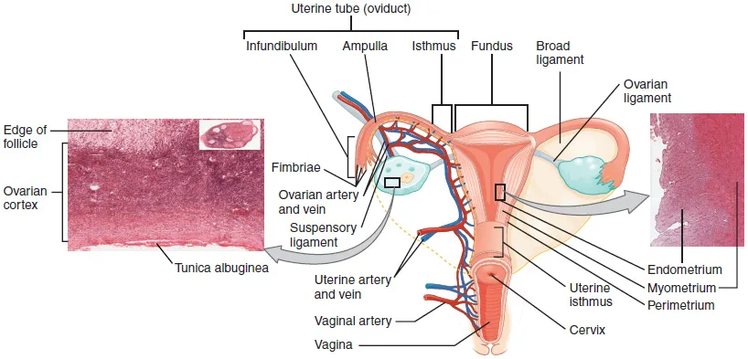
Ovarium:骨盤壁の fossa ovarica(内腸骨動静脈と尿管が後壁を作る)にある扁平な卵形の器官。腹膜に覆われず、表面は生殖上皮——腹腔内に直接露出する唯一の臓器である。
固定は2本の索:lig. ovarii proprium(卵巣 → 子宮角、広間膜の中)と lig. suspensorium ovarii(卵巣 → 骨盤壁、卵巣動静脈を含む)。広間膜との繋がりが mesovarium。
Tuba uterina:約10cm。子宮側から pars uterina → isthmus → ampulla(最も長く広い・受精の場)→ infundibulum(fimbriae を持ち ostium abdominale で腹腔へ開く)。広間膜の上縁のヒダが mesosalpinx。
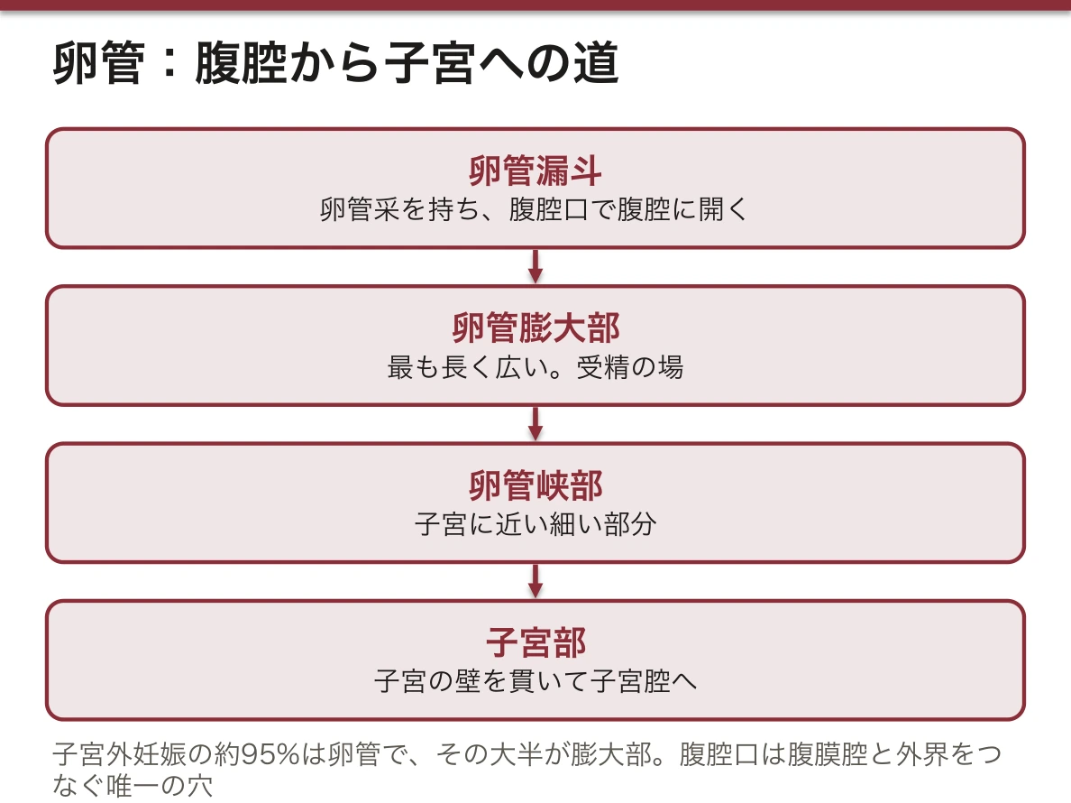
卵管の腹腔口が「腹膜腔と外界を繋ぐ唯一の穴」である。ここから2方向のことが起こる。①排卵した卵子を腹腔から拾う(采が卵巣を掃くように動く)。②腟 → 子宮 → 卵管 → 腹腔と感染が上行する——骨盤内炎症性疾患(PID)、さらに肝周囲へ達すると Fitz-Hugh-Curtis 症候群。
子宮外妊娠の約95%が卵管、その大半が膨大部で起こる。受精が膨大部で起こるのだから、そこで着床が始まってしまえば動きようがない。破裂すると腹腔内出血で Douglas 窩に血液が溜まる——後腟円蓋から穿刺できるのはそのためである。
卵巣の血管は精巣と同じく腹部から来る(卵巣動脈=腹大動脈 L2、静脈は右が下大静脈・左が左腎静脈)。リンパも傍大動脈リンパ節。下降した器官の履歴は男女で共通である。だから卵巣癌の初発症状は骨盤ではなく腹部膨満(腹膜播種と腹水)になる。
卵巣は腹膜に覆われない唯一の臓器で、排卵した卵子はいったん腹腔へ出る。だから腹腔と外界が卵管・子宮・腟を通じて繋がっている——女性だけが上行性の骨盤腹膜炎を起こす解剖学的な理由である。
異所性妊娠の約95%は卵管、なかでも膨大部に着床する。破裂すると腹腔内出血でショックになる。無月経・下腹部痛・性器出血の3つで疑う。
卵巣提索(骨盤漏斗靱帯)の中を卵巣動静脈が走る。卵巣嚢腫茎捻転ではこの索がねじれて突然の下腹部痛と嘔吐を起こす。
卵巣のリンパも傍大動脈リンパ節へ(精巣と同じく、発生の位置が腹部だから)。卵巣癌は腹膜播種で広がり、Douglas 窩に溜まる。腹水と大網の肥厚(omental cake)が典型像である。
English version
Core
Ovarium: a flattened ovoid organ in the fossa ovarica of the pelvic wall (whose posterior wall is formed by the internal iliac vessels and the ureter). It is not covered by peritoneum; its surface is germinal epithelium — the only organ exposed directly in the peritoneal cavity.
It is held by two cords: the lig. ovarii proprium (ovary → cornu of the uterus, within the broad ligament) and the lig. suspensorium ovarii (ovary → pelvic wall, containing the ovarian vessels). Its connection with the broad ligament is the mesovarium.
Tuba uterina: about 10 cm. From the uterus: pars uterina → isthmus → ampulla (the longest and widest; the site of fertilisation) → infundibulum (with the fimbriae, opening into the peritoneal cavity at the ostium abdominale). The fold along the upper margin of the broad ligament is the mesosalpinx.
Going further
The abdominal opening of the tube is the only hole joining the peritoneal cavity to the exterior, and two things follow from it. ① The ovulated oocyte is picked up from the cavity (the fimbriae sweep the ovary). ② Infection ascends vagina → uterus → tube → peritoneal cavity — pelvic inflammatory disease, and reaching the liver, the Fitz-Hugh–Curtis syndrome.
About 95 per cent of ectopic pregnancies are tubal, and most of those in the ampulla. Fertilisation occurs in the ampulla, so once implantation begins there, there is nowhere to go. Rupture bleeds into the peritoneal cavity and the blood collects in the pouch of Douglas — which is why it can be aspirated through the posterior fornix.
The ovarian vessels, like the testicular, come from the abdomen (ovarian artery from the aorta at L2; the vein to the inferior vena cava on the right and the left renal vein on the left), and the lymph goes to the para-aortic nodes. The record of a descended organ is common to both sexes. So the first symptom of ovarian cancer is not pelvic but abdominal distension (peritoneal dissemination and ascites).
In the clinic
The ovary is the only organ not covered by peritoneum, and the ovulated oocyte passes first into the peritoneal cavity. So the cavity communicates with the exterior through tube, uterus and vagina — the anatomical reason why only women develop ascending pelvic peritonitis.
About 95 per cent of ectopic pregnancies are tubal, most implanting in the ampulla. Rupture causes intraperitoneal haemorrhage and shock. Suspect it from the triad of amenorrhoea, lower abdominal pain and vaginal bleeding.
The ovarian vessels run in the suspensory (infundibulopelvic) ligament. In torsion of an ovarian cyst this cord twists and causes sudden lower abdominal pain and vomiting.
The lymph of the ovary also goes to the para-aortic nodes (as with the testis, because it developed in the abdomen). Ovarian cancer spreads by peritoneal dissemination and collects in the pouch of Douglas. Ascites with a thickened omentum (an omental cake) is the typical picture.
卵巣の内部 — 白膜・皮質・髄質Cortex et medulla ovarii
卵巣を切ると、外から次の順に見える。
| 層 | 中身 |
|---|---|
| 表面上皮 | 単層の立方上皮。腹膜の中皮が卵巣門の所で入れ替わったもの(「胚上皮」「生殖上皮」とも呼ばれてきたが、卵子を作るわけではない) |
| tunica albuginea(白膜) | 表面上皮のすぐ下の、密な結合組織の白い膜 |
| cortex ovarii(皮質・実質帯) | 結合組織のあいだに卵胞がいろいろな段階で並ぶ。黄体・白体もここにできる |
| medulla ovarii(髄質・血管帯) | ゆるい結合組織と、卵巣門から入る血管・神経・リンパ管 |
卵胞の数は、出生時に左右で約100〜200万、思春期には数十万に減り、生涯で排卵まで進むのは400〜500個にすぎない。残りは途中で閉鎖する。思春期以降、ほぼ28日ごとに1つの卵胞が成熟して直径約2 cm の胞状卵胞(Graaf 卵胞)になり、表面へ寄って破れる(排卵)。破れた跡は赤体 → 黄体(プロゲステロンとエストロゲンを出す)→ 白体(瘢痕)と変わる。だから子どもの卵巣は表面が滑らかで、排卵が始まると瘢痕でごつごつしてくる。細胞の段階は組織学の「卵巣・子宮」で扱う。
精巣と比べると配置が逆である。精巣は働く部分(精細管)が髄質側に、卵巣は働く部分(卵胞)が皮質側にある。発生の段階で、精巣は奥へ伸びる性索(髄質索)を使い、卵巣は表面近くに新しくできる性索(皮質索)を使うためである。
妊娠すると、胎盤の hCG が黄体を保ち、胎盤がホルモンを作れるようになる妊娠10週ごろまでプロゲステロンを出し続ける。早い時期に黄体を取ると流産しうるのはこのためである。
多嚢胞性卵巣症候群(PCOS)では白膜が厚くなり、成熟しきれない小さな卵胞が皮質の外周に並ぶ(超音波で「ネックレス状」)。排卵が起こらず、月経不順と不妊の原因になる。
卵巣の腫瘍は出身の層で分けて考える:表面の上皮系(最も多い。悪性のものの多くは卵管采から来るという考えが有力)、卵子のもとの胚細胞(若い女性の成熟奇形腫=皮様嚢腫)、間質の細胞。黄体嚢胞が破れると、腹腔内に出血して Douglas 窩に血がたまる。
English version
Core
Cut across, the ovary shows these layers from outside in.
| Layer | Content |
|---|---|
| Surface epithelium | Simple cuboidal epithelium that replaces the peritoneal mesothelium at the hilum (long called “germinal epithelium”, but it does not make eggs) |
| tunica albuginea | White layer of dense connective tissue just under the surface epithelium |
| cortex ovarii (parenchymatous zone) | Follicles at many stages lie in connective tissue; corpus luteum and corpus albicans also form here |
| medulla ovarii (vascular zone) | Loose connective tissue with vessels, nerves and lymphatics entering from the hilum |
Both ovaries hold about 1–2 million follicles at birth, a few hundred thousand at puberty, and only 400–500 ever reach ovulation; the rest undergo atresia. After puberty, roughly every 28 days one follicle matures into a vesicular (Graafian) follicle about 2 cm across, moves to the surface and bursts (ovulation). The site becomes the corpus rubrum, then the corpus luteum (secreting progesterone and oestrogen), then the corpus albicans (a scar). That is why a child’s ovary is smooth and an adult ovary becomes lumpy with scars. Follicle stages are covered in histology, “Ovary and uterus”.
Going further
Compared with the testis, the layout is reversed. The working part of the testis (seminiferous tubules) is on the medullary side; that of the ovary (follicles) is on the cortical side. In development the testis uses sex cords that grow deep (medullary cords), while the ovary uses new cords formed near the surface (cortical cords).
In pregnancy, placental hCG keeps the corpus luteum alive, and it goes on making progesterone until about week 10, when the placenta can take over. Removing the corpus luteum early can therefore cause miscarriage.
In the clinic
In polycystic ovary syndrome (PCOS) the tunica albuginea thickens and small follicles that fail to mature line the edge of the cortex (a “string of pearls” on ultrasound). Ovulation stops, causing irregular periods and infertility.
Ovarian tumours are grouped by the layer they come from: surface epithelial tumours (the most common; many malignant ones are now thought to start in the tubal fimbriae), germ cell tumours (mature teratoma or dermoid cyst in young women) and stromal tumours. A ruptured corpus luteum cyst bleeds into the peritoneal cavity, and blood gathers in the pouch of Douglas.
子宮 — 部位・傾き・支持Uterus
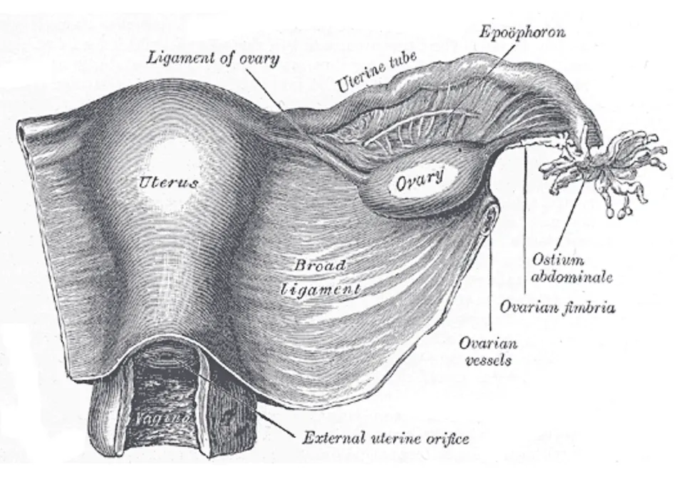
膀胱と直腸のあいだ。約7×5×2.5cm の洋梨形。Fundus uteri(卵管の付着より上)・corpus uteri・isthmus・cervix uteri(portio supravaginalis と portio vaginalis)。2面:facies vesicalis(前)と facies intestinalis(後)。
内腔:三角形の cavitas uteri → canalis cervicis uteri(前後壁に plicae palmatae)→ ostium uteri(外子宮口・前後の labium anterius / posterius)。
壁3層:endometrium(機能層は周期的に剥がれる)・myometrium(最も厚い平滑筋)・perimetrium(漿膜)。
位置:正常は anteversio(腟に対して前へ傾く)+anteflexio(頸に対して体が前へ屈曲)。
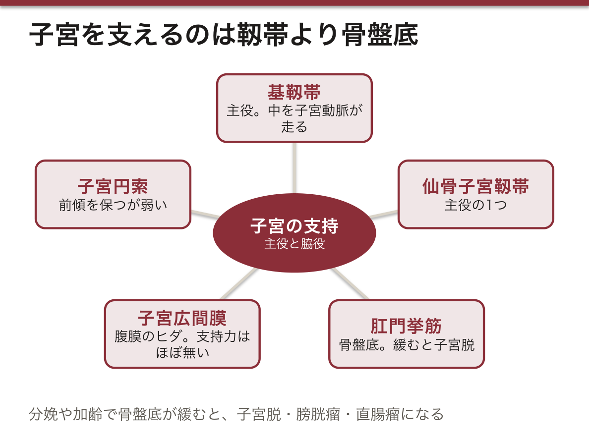
支持は「靱帯より骨盤底」である。Lig. latum uteri(広間膜)は腹膜のヒダにすぎず支持力はほとんど無い。Lig. teres uteri(円索)は前傾を保つが弱い。実際に子宮を支えているのはlig. cardinale(Mackenrodt)・lig. sacrouterinum と、その下のm. levator ani である。だから骨盤底筋が緩むと子宮脱が起こる。
子宮動脈と尿管の関係が、子宮全摘の最大の関門である。子宮動脈は内腸骨動脈から起こり、基靱帯の中を内側へ走って子宮頸の外側約2cm で尿管の前を越える(water under the bridge)。結紮点と尿管が2cm しか離れていない。
Lig. teres uteri は男性の精巣導帯に相当する。子宮角から鼠径管を通って大陰唇に終わる——男性の精巣下降の経路をたどっているが、卵巣は骨盤に留まった。この索に沿って腹膜の突起(Nuck 管)が残ると、女性でも鼠径ヘルニアが起こる。
子宮頸の外子宮口は移行帯(SCJ)で、円柱上皮と重層扁平上皮が入れ替わる。子宮頸癌はほぼここから発生するので、細胞診はこの一点を狙って採る。
子宮頸部は扁平上皮と円柱上皮が出会う場所(SCJ)を持つ。ここが子宮頸癌の発生母地であり、細胞診で狙って擦り取る場所でもある。HPV 感染が原因で、ワクチンと検診で防げる数少ない癌である。
子宮は前傾前屈が正常。後屈だと月経血が出にくく月経困難症の一因になる。
基靱帯(子宮頸横靱帯)の中を子宮動脈が走り、その下を尿管が横切る——water under the bridge。広汎子宮全摘術で尿管損傷が起こるのはここである。
子宮支持の主役は靱帯ではなく骨盤底筋(肛門挙筋)である。分娩や加齢でここが緩むと子宮脱・膀胱瘤・直腸瘤になる。
Douglas 窩は腟後円蓋のすぐ後ろにあるので、腟から穿刺して腹腔内出血を確かめられる。
English version
Core
Between bladder and rectum; pear-shaped, about 7 × 5 × 2.5 cm. Fundus uteri (above the attachment of the tubes), corpus uteri, isthmus, cervix uteri (supravaginal and vaginal portions). Two surfaces: facies vesicalis (front) and facies intestinalis (back).
The cavity: the triangular cavitas uteri → canalis cervicis uteri (with the plicae palmatae on front and back walls) → ostium uteri (with anterior and posterior lips).
Three layers: endometrium (the functional layer shed cyclically), myometrium (the thickest, smooth muscle), perimetrium (serosa).
Position: normally anteversion (tilted forward relative to the vagina) plus anteflexion (the body flexed forward on the cervix).
Going further
Support comes from the pelvic floor, not from the ligaments. The lig. latum uteri is only a fold of peritoneum and gives almost no support; the lig. teres uteri maintains anteversion but is weak. What actually supports the uterus is the lig. cardinale (Mackenrodt’s) and the lig. sacrouterinum, and beneath them the m. levator ani. So when the pelvic floor slackens, prolapse follows.
The relation of uterine artery to ureter is the greatest hazard of hysterectomy. The uterine artery arises from the internal iliac, runs medially within the cardinal ligament and crosses in front of the ureter about 2 cm lateral to the cervix (water under the bridge). Only 2 cm separates the point of ligation from the ureter.
The lig. teres uteri corresponds to the gubernaculum testis of the male. It runs from the cornu of the uterus through the inguinal canal to the labium majus — following the path of testicular descent, though the ovary stayed in the pelvis. If a peritoneal process (the canal of Nuck) persists along it, an inguinal hernia can occur in a woman too.
The external os of the cervix is the transformation zone (SCJ), where columnar and stratified squamous epithelium change over. Almost all cervical carcinoma arises there, so the smear is taken aimed at that one point.
In the clinic
The cervix carries the place where squamous and columnar epithelium meet (the SCJ). It is the seedbed of cervical carcinoma and the place the smear is taken from. Caused by HPV infection, it is one of the few cancers preventable by vaccination and screening.
Anteversion and anteflexion are normal. In retroversion the menstrual blood drains less easily and it contributes to dysmenorrhoea.
The uterine artery runs in the cardinal (transverse cervical) ligament and the ureter passes beneath it — water under the bridge. This is where the ureter is injured in radical hysterectomy.
The principal support of the uterus is not ligament but the pelvic floor muscle (levator ani). Slackening with childbirth and age gives uterine prolapse, cystocele and rectocele.
The pouch of Douglas lies immediately behind the posterior vaginal fornix, so intraperitoneal haemorrhage can be confirmed by puncture through the vagina.
腟と外陰Vagina et organa genitalia externa
Vagina:約8cm の管。前壁は膀胱と尿道、後壁は直腸に接する。粘膜に横走する rugae vaginales。上端は子宮頸を囲んで fornix vaginae(前・後・側部)を作り、pars posterior が最も深い。
外陰(vulva):Labia majora pudendi(大陰唇)・labia minora pudendi(小陰唇)・vestibulum vaginae(小陰唇に囲まれた前庭・ここに ostium urethrae externum と ostium vaginae が開く)。
Clitoris:crura(恥骨下枝に付く)・corpus・glans clitoridis。前庭の深部に bulbus vestibuli(男性の尿道海綿体に相当)、その後端に glandula vestibularis major(Bartholin 腺)。
後腟円蓋の背側が Douglas 窩(直腸子宮窩)である。立位でも仰臥位でも腹腔内で最も低い場所なので、腹腔内の液体(血液・膿・腹水・播種)が最初に溜まる。後腟円蓋からの穿刺(culdocentesis)で腹腔内出血を確認できるのは、腟壁1枚を隔てて腹膜腔に接しているからである。
腟は自浄作用を持つ。重層扁平上皮がエストロゲンの作用でグリコーゲンを蓄え、Döderlein 桿菌がこれを乳酸に変えて pH 4前後を保つ。だからエストロゲンの低い小児と閉経後には腟炎が起こりやすい——上皮が薄くグリコーゲンが乏しく、pH が上がるためである。
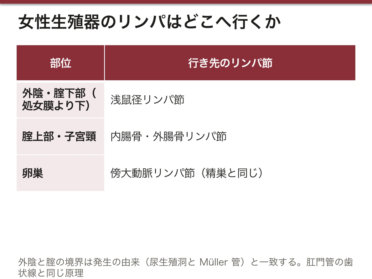
外陰と腟でリンパの行き先が違う。外陰と腟下部(処女膜より下)は浅鼠径リンパ節、腟上部と子宮頸は内腸骨・外腸骨リンパ節。境界は発生の由来(尿生殖洞 vs Müller 管)と一致する——歯状線が肛門管を分けるのと同じ原理である。
Bartholin 腺は男性の尿道球腺に相当する。導管が閉塞すると嚢胞、感染すると膿瘍になり、小陰唇の後方に片側性の腫脹として現れる。位置が決まっているのは、発生上の相同性による。
腟の自浄作用 — エストロゲンが上皮にグリコーゲンを蓄え、Döderlein 桿菌(乳酸桿菌)が乳酸に変えて pH を 4 前後に保つ。だからエストロゲンが低い時期(小児期・閉経後)は感染しやすい。萎縮性腟炎はこの仕組みが働かなくなった状態である。
後腟円蓋は Douglas 窩に接する。ここから穿刺すれば腹腔内の血液や膿を確認できるし、内診で Douglas 窩の腫瘤や圧痛(腹膜播種・子宮内膜症)を触れる。
Bartholin 腺(大前庭腺)は腟口の後外側に開く。導管が詰まると嚢胞・膿瘍になり、坐位困難な痛みを起こす。
会陰裂傷は分娩で最も多い外傷である。III 度以上は外肛門括約筋に及ぶので、確実に修復しないと便失禁が残る。
English version
Core
Vagina: a tube about 8 cm long; the anterior wall against bladder and urethra, the posterior against rectum. The mucosa carries transverse rugae vaginales. Its upper end surrounds the cervix as the fornix vaginae (anterior, posterior and lateral parts), the posterior part being the deepest.
Vulva: labia majora pudendi, labia minora pudendi, vestibulum vaginae (enclosed by the labia minora, into which the external urethral and vaginal orifices open).
Clitoris: crura (attached to the ischiopubic rami), corpus and glans. Deep in the vestibule lie the bulbus vestibuli (corresponding to the male corpus spongiosum) and, at its posterior end, the glandula vestibularis major (Bartholin’s).
Going further
Behind the posterior fornix is the pouch of Douglas (rectouterine pouch). It is the lowest place in the peritoneal cavity, standing or supine, so intraperitoneal fluid (blood, pus, ascites, seedings) collects there first. Puncture through the posterior fornix (culdocentesis) can confirm intraperitoneal bleeding because only one thickness of vaginal wall separates it from the peritoneal cavity.
The vagina cleans itself. Under oestrogen the stratified squamous epithelium stores glycogen, and Döderlein’s bacillus converts it to lactic acid and holds the pH near 4. So vaginitis occurs readily in childhood and after the menopause, when oestrogen is low — the epithelium is thin, the glycogen scanty and the pH rises.
The lymph of vulva and vagina goes to different places. The vulva and the lower vagina (below the hymen) to the superficial inguinal nodes; the upper vagina and the cervix to the internal and external iliac nodes. The boundary coincides with the developmental origin (urogenital sinus versus Müllerian duct) — the same principle as the dentate line dividing the anal canal.
Bartholin’s gland corresponds to the male bulbourethral gland. Obstruction of the duct gives a cyst and infection an abscess, appearing as a unilateral swelling behind the labium minus. Its fixed position follows from the developmental homology.
In the clinic
The self-cleansing of the vagina — oestrogen makes the epithelium store glycogen and Döderlein’s bacillus (lactobacillus) converts it to lactic acid, holding the pH near 4. So infection is easy when oestrogen is low (childhood, after the menopause). Atrophic vaginitis is that mechanism ceasing to work.
The posterior fornix abuts the pouch of Douglas. Puncture there confirms blood or pus in the peritoneal cavity, and on vaginal examination a mass or tenderness in the pouch (peritoneal seeding, endometriosis) can be felt.
Bartholin’s (greater vestibular) gland opens posterolaterally at the introitus. An obstructed duct gives a cyst or abscess and pain that makes sitting impossible.
Perineal tear is the commonest injury of childbirth. Third degree and above involves the external anal sphincter, and unless repaired reliably, faecal incontinence remains.
腟の血管と神経 — 上は子宮と、下は会陰と同じVasa vaginae
動脈はすべて内腸骨動脈の系統から来る。
| 動脈 | 養う所 |
|---|---|
| a. vaginalis(腟動脈:内腸骨動脈または子宮動脈から) | 腟の中部と下部、膀胱の下部 |
| 子宮動脈の腟枝 | 腟の上部と子宮頸。左右の枝が腟の前後の壁で縦につながる |
| 中直腸動脈 | 腟の後壁の中部 |
| 内陰部動脈 | 腟の下端と腟口の周り |
静脈は腟の両側の腟静脈叢に集まり、膀胱静脈叢・直腸静脈叢・子宮静脈叢とつながって内腸骨静脈へ注ぐ。リンパは上部が内・外腸骨リンパ節、下部(処女膜より下)が浅鼠径リンパ節へ。神経は、上部(約3/4)が子宮腟神経叢の自律神経で、伸びる感覚はあるが痛みには鈍い。下部(約1/4)は陰部神経の枝で、痛みを鋭く感じる。
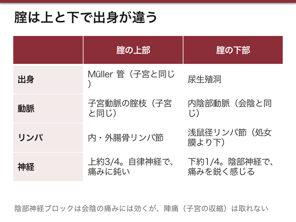
上と下で支配が分かれるのは、出身が違うからである。腟の上部は子宮と同じ Müller 管から、下部は尿生殖洞から作られる。だから上部は子宮と同じ動脈・同じ自律神経を、下部は会陰と同じ内陰部動脈・陰部神経を使う。リンパが上下で別の方向へ行くのも同じ理由である。
分娩のときの痛みは2系統ある。子宮の収縮の痛みは交感神経に伴う求心線維(Th10〜L1)で、腟の下部と会陰の伸びる痛みは陰部神経(S2〜S4)で伝わる。だから陰部神経ブロックは分娩の最後の時期と会陰切開の痛みには効くが、陣痛そのものは取れない。
腟静脈叢は豊かなので、分娩時の腟壁の裂傷や腟壁の血腫は大量出血になりうる。子宮全摘の後に腟の断端から出血するのは、子宮動脈の腟枝・腟動脈の処理が不十分なときである。
English version
Core
All arteries come from the internal iliac system.
| Artery | Area supplied |
|---|---|
| a. vaginalis (from the internal iliac or uterine artery) | Middle and lower vagina, lower bladder |
| Vaginal branches of the uterine artery | Upper vagina and cervix; right and left branches join lengthwise on the front and back walls |
| Middle rectal artery | Middle of the back wall |
| Internal pudendal artery | Lowest vagina and around the vaginal opening |
Veins gather in the vaginal venous plexus on each side, which joins the vesical, rectal and uterine plexuses and drains to the internal iliac vein. Lymph from the upper part goes to the internal and external iliac nodes; from the lower part (below the hymen) to the superficial inguinal nodes. Nerves: the upper part (about three quarters) is supplied by autonomic fibres of the uterovaginal plexus — it senses stretch but is dull to pain. The lower part (about one quarter) gets branches of the pudendal nerve and feels pain sharply.
Going further
The supply divides into upper and lower because the origins differ. The upper vagina comes from the Müllerian duct, like the uterus; the lower part from the urogenital sinus. So the upper part shares the uterus’s arteries and autonomic nerves, and the lower part shares the perineum’s internal pudendal artery and pudendal nerve. Lymph goes in two directions for the same reason.
In the clinic
Labour pain has two routes. Pain of uterine contraction travels with sympathetic afferents (T10–L1); stretch pain of the lower vagina and perineum travels in the pudendal nerve (S2–S4). A pudendal block therefore relieves the end of labour and episiotomy pain but not the contractions themselves.
Because the vaginal plexus is rich, tears of the vaginal wall or a vaginal haematoma at delivery can bleed heavily. Bleeding from the vaginal vault after hysterectomy occurs when the vaginal branches of the uterine artery or the vaginal artery were not secured.
会陰 — 2つの三角と2枚の隔膜Perineum
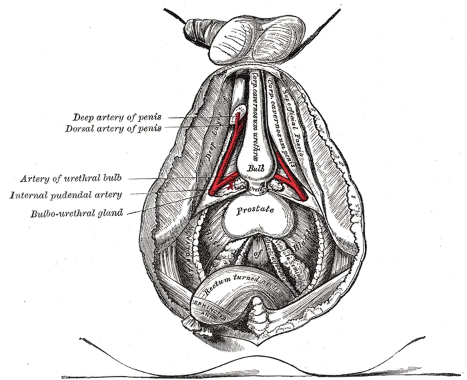
骨盤出口を、両側の坐骨結節を結ぶ線で2つの三角に分ける。
- Trigonum urogenitale(尿生殖三角・前)— 尿生殖隔膜が張る。M. transversus perinei profundus・m. sphincter urethrae externus。浅層に m. ischiocavernosus・m. bulbospongiosus・m. transversus perinei superficialis
- Trigonum anale(肛門三角・後)— M. sphincter ani externus と、その外側の fossa ischioanalis
骨盤隔膜(上)は m. levator ani(恥骨直腸筋・恥骨尾骨筋・腸骨尾骨筋)と m. coccygeus。2枚の隔膜が上下に重なって骨盤の底を作る。
2つの三角の頂点が合う正中の腱性の塊が centrum perinei(会陰腱中心)。
会陰腱中心には、骨盤底と会陰の筋がほとんど全部集まる(外肛門括約筋・球海綿体筋・浅および深会陰横筋・肛門挙筋の一部)。分娩でここが裂けると骨盤底全体の支持が落ち、後年の子宮脱・直腸瘤・便失禁につながる——会陰切開が「あえて制御して切る」処置として行われるのは、この一点を守るためである。正中切開は腱中心を直撃するので、内外斜切開が選ばれる。
Fossa ischioanalis は肛門管の両側の脂肪で満たされた楔形の空間で、排便時に肛門管が広がるための余地である。脂肪組織で血流に乏しく、肛門周囲膿瘍が起こりやすく、左右が肛門の後方で交通するため馬蹄形膿瘍になる。
外側壁の閉鎖筋膜の中を陰部神経管(Alcock 管)が走る。中を通るのは陰部神経(S2〜S4)と内陰部動静脈。会陰の感覚と外尿道・外肛門括約筋はすべてこの1本が担う——だから陰部神経ブロックで会陰の分娩時痛が取れるし、この神経が圧迫されると陰部神経痛になる。
「S2, 3, 4 keep the floor off the floor」——骨盤底の運動と会陰の感覚は S2〜S4 に集約される。馬尾症候群でサドル型感覚障害と膀胱直腸障害が同時に出るのは、この分節がまとめて障害されるからである。
会陰は骨盤底が破綻したときに全部が現れる場所である。
坐骨直腸窩(坐骨肛門窩)は脂肪で満たされた大きな空間で、血流が乏しい。だから肛門周囲膿瘍が広がりやすく、左右が肛門の後ろで交通しているため馬蹄形膿瘍になりうる。切開排膿が原則。
陰部神経(S2〜S4)は坐骨棘の内側を回って Alcock 管(陰部神経管)を通る。陰部神経ブロックは坐骨棘を目標に、腟から刺す——会陰の解剖がそのまま手技になる。
会陰切開は正中側切開が選ばれることが多い。正中切開は外肛門括約筋・直腸へ裂けやすいためである。
Fournier 壊疽 — 会陰の壊死性筋膜炎。糖尿病・免疫不全で起こり、見た目の皮膚所見に不釣り合いな激痛が手がかり。広範なデブリードマンを急ぐ。
English version
Core
The pelvic outlet is divided by the line joining the two ischial tuberosities into two triangles.
- Trigonum urogenitale (anterior) — the urogenital diaphragm is stretched across it: m. transversus perinei profundus and m. sphincter urethrae externus, with m. ischiocavernosus, m. bulbospongiosus and m. transversus perinei superficialis superficially.
- Trigonum anale (posterior) — the m. sphincter ani externus, with the fossa ischioanalis lateral to it.
The pelvic diaphragm above is the m. levator ani (puborectalis, pubococcygeus, iliococcygeus) and the m. coccygeus. Two diaphragms, one above the other, form the floor of the pelvis.
The tendinous mass in the midline where the apices of the two triangles meet is the centrum perinei (perineal body).
Going further
Almost all the muscles of the pelvic floor and perineum converge on the perineal body (external anal sphincter, bulbospongiosus, superficial and deep transverse perineal muscles, part of levator ani). Tearing it in childbirth reduces the support of the whole pelvic floor and leads later to uterine prolapse, rectocele and faecal incontinence — which is why episiotomy is performed as a deliberate, controlled cut, to protect this one point. A midline incision strikes the perineal body directly, so a mediolateral cut is chosen.
The fossa ischioanalis is a wedge-shaped space filled with fat on either side of the anal canal, allowing room for the canal to distend at defaecation. Being fat with a poor blood supply, perianal abscess occurs readily, and because the two sides communicate behind the anus, it becomes a horseshoe abscess.
In the obturator fascia of its lateral wall runs the pudendal canal (of Alcock), carrying the pudendal nerve (S2–S4) and the internal pudendal vessels. Sensation of the perineum and both the external urethral and anal sphincters depend on this one nerve — hence a pudendal block relieves the perineal pain of labour, and compression of it gives pudendal neuralgia.
"S2, 3, 4 keep the floor off the floor" — the movement of the pelvic floor and the sensation of the perineum are concentrated in S2–S4. That saddle anaesthesia and bladder and bowel disturbance occur together in cauda equina syndrome is because those segments are damaged together.
In the clinic
The perineum is where everything appears when the pelvic floor fails.
The ischiorectal (ischioanal) fossa is a large fat-filled space with a poor blood supply. So perianal abscess spreads readily, and because the two sides communicate behind the anus it may become a horseshoe abscess. Incision and drainage is the rule.
The pudendal nerve (S2–S4) hooks round the ischial spine into Alcock’s canal. A pudendal block is given through the vagina, aiming at the ischial spine — the anatomy of the perineum becoming directly the technique.
Episiotomy is usually mediolateral, because a midline cut extends readily into the external anal sphincter and rectum.
Fournier’s gangrene — necrotising fasciitis of the perineum, occurring in diabetes and immunodeficiency; pain out of proportion to the appearance of the skin is the clue. Extensive debridement is urgent.
尿生殖三角の層 — 深会陰隙・会陰膜・浅会陰隙・会陰筋膜Spatium perinei profundum et superficiale
尿生殖三角は、上(骨盤の中)から下(皮膚)へ次の順に重なる:骨盤隔膜 → 深会陰隙 → 会陰膜(membrana perinei)→ 浅会陰隙 → 会陰筋膜(fascia perinei:浅会陰筋を下から包む筋膜)→ 皮下組織(その深い膜様層が Colles 筋膜)→ 皮膚。会陰膜は左右の坐骨恥骨枝のあいだに張る丈夫な線維の膜で、2つの隙の仕切りである。
| 深会陰隙 | 浅会陰隙 | |
|---|---|---|
| 筋 | 深会陰横筋・外尿道括約筋 | 浅会陰横筋・坐骨海綿体筋・球海綿体筋 |
| 男性 | 尿道膜様部・尿道球腺 | 陰茎根(陰茎脚と尿道球)・尿道海綿体部の始まり |
| 女性 | 尿道・腟の一部 | 陰核脚・前庭球・大前庭腺 |
| 血管・神経 | 陰茎(陰核)背動脈・背神経 | 会陰動静脈・会陰神経 |
尿道球や大前庭腺を深い方に数える教材もあるが、標準的な教科書(英語圏・日本)では浅会陰隙に入る。
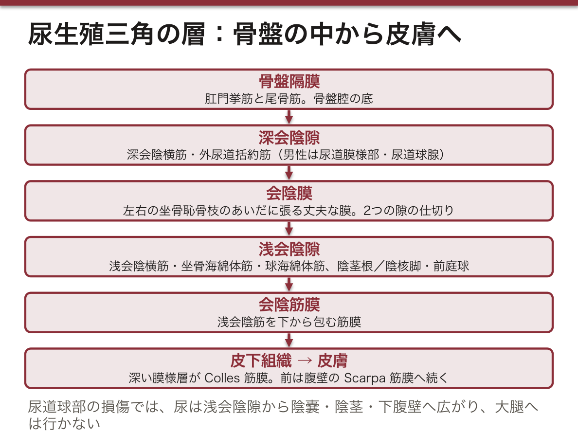
古い見方と新しい見方がある。日本の教科書や古い教科書は、深会陰横筋と外尿道括約筋を上下2枚の筋膜で挟んだ板を尿生殖隔膜と呼び、その下の筋膜が会陰膜にあたる。現在の解剖学用語では尿生殖隔膜の語を外し、深会陰隙は上で閉じておらず、坐骨肛門窩の前の陥凹とつながると考える。どちらの言葉で聞かれても答えられるようにしておく。
Colles 筋膜は、横は坐骨恥骨枝と大腿の筋膜、後ろは会陰膜の後縁に付き、前は陰嚢・陰茎を包んで腹壁の膜様層(Scarpa 筋膜)に続く。この付き方が、漏れた尿や血がどこまで広がるかを決める。
尿道損傷の尿の広がりが典型例である。またがって会陰を打つと(騎乗型損傷)尿道球部が裂け、尿と血は浅会陰隙に出る。Colles 筋膜の付着のため大腿や肛門の方へは行かず、陰嚢・陰茎・下腹壁へ広がる(蝶形の皮下出血)。骨盤骨折で尿道膜様部が裂けると、尿は深会陰隙から骨盤の腹膜外の空間へ漏れ、前立腺が上へ浮く。
同じ通り道を、会陰の壊死性筋膜炎(Fournier 壊疽)も広がる。会陰切開で切るのは浅会陰隙の筋(球海綿体筋・浅会陰横筋)である。
English version
Core
The urogenital triangle is layered from above (inside the pelvis) to below (skin): pelvic diaphragm → deep perineal space → perineal membrane (membrana perinei) → superficial perineal space → perineal fascia (fascia perinei, covering the superficial perineal muscles from below) → subcutaneous tissue (its deep membranous layer is Colles’ fascia) → skin. The perineal membrane is a strong fibrous sheet stretched between the two ischiopubic rami and separates the two spaces.
| Deep perineal space | Superficial perineal space | |
|---|---|---|
| Muscles | Deep transverse perineal, external urethral sphincter | Superficial transverse perineal, ischiocavernosus, bulbospongiosus |
| Male | Membranous urethra, bulbourethral glands | Root of penis (crura and bulb), start of spongy urethra |
| Female | Urethra, part of the vagina | Crura of clitoris, vestibular bulbs, greater vestibular glands |
| Vessels and nerves | Dorsal artery and nerve of penis (clitoris) | Perineal vessels and nerve |
Some course materials place the bulb of the penis or the greater vestibular glands in the deep space, but standard textbooks (English and Japanese) put them in the superficial space.
Going further
There is an old view and a new view. Japanese and older textbooks call the plate of deep transverse perineal muscle and external urethral sphincter, sandwiched between upper and lower fasciae, the urogenital diaphragm; its lower fascia corresponds to the perineal membrane. Current anatomical terminology drops the term: the deep perineal space is not closed above and is continuous with the anterior recess of the ischioanal fossa. Be ready to answer in either language.
Colles’ fascia is attached laterally to the ischiopubic rami and fascia of the thigh, behind to the back edge of the perineal membrane, and in front it wraps the scrotum and penis and continues as the membranous layer of the abdominal wall (Scarpa’s fascia). These attachments decide how far leaked urine or blood can spread.
In the clinic
The spread of urine after urethral injury is the classic example. A straddle injury tears the bulbar urethra, and urine and blood enter the superficial perineal space. Because of the attachments of Colles’ fascia they do not pass to the thighs or the anus but spread to the scrotum, penis and lower abdominal wall (butterfly bruising). When a pelvic fracture tears the membranous urethra, urine leaks from the deep space into the extraperitoneal pelvis, and the prostate rides high.
Necrotising fasciitis of the perineum (Fournier’s gangrene) spreads along the same paths. An episiotomy cuts muscles of the superficial space (bulbospongiosus, superficial transverse perineal).
骨盤隔膜と骨盤底 — 2枚の筋膜と、5つの仕事Diaphragma pelvis
骨盤隔膜は骨盤腔の底を作る漏斗形の筋の板で、肛門挙筋(恥骨尾骨筋・恥骨直腸筋・腸骨尾骨筋)と尾骨筋からなる。ヨーロッパの教材には外肛門括約筋を加えて3筋とするものがあるが、英語圏・日本の多くの教科書は前の2筋とし、外肛門括約筋は肛門三角の会陰筋として扱う。
肛門挙筋は恥骨の後面、肛門挙筋腱弓(内閉鎖筋を覆う閉鎖筋膜が厚くなった線)、坐骨棘から起こり、会陰腱中心・肛門尾骨靱帯・尾骨に付く。前方の左右の恥骨直腸筋のあいだのすき間が尿生殖裂孔で、尿道と(女性では)腟が通る。
上面は上骨盤隔膜筋膜(fascia superior diaphragmatis pelvis:骨盤の壁側筋膜の続き)、下面は下骨盤隔膜筋膜(fascia inferior diaphragmatis pelvis:坐骨肛門窩の内側壁になる)が覆う。上の筋膜の上には、臓器を包む臓側骨盤筋膜と、子宮を支える基靱帯などの結合組織が乗る。神経は仙骨神経叢の直接の枝(S3〜S4)と陰部神経。
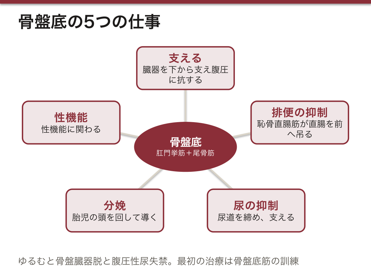
骨盤底の働きは5つ:①骨盤の臓器を下から支え、腹圧に抗する ②排便の抑制(恥骨直腸筋が直腸肛門移行部を前へ吊り、ほぼ直角に曲げる。排便時にはゆるんで角度が開く)③尿の抑制(尿道を締める・支える)④分娩で胎児の頭を回して導く ⑤性機能。
女性で骨盤底の障害が多いのは、尿生殖裂孔が広く3本の管(尿道・腟・直腸)が通ること、そして経腟分娩で肛門挙筋と陰部神経が大きく引き伸ばされることによる。
骨盤底がゆるむと、骨盤臓器脱(膀胱瘤・子宮脱・直腸瘤)と腹圧性尿失禁(せき・くしゃみで漏れる)が起こる。最初の治療は骨盤底筋の訓練で、肛門と腟を締める運動を毎日続けて肛門挙筋を鍛える。排便のときに恥骨直腸筋がかえって縮んでしまうと、出口でふさがる便秘になる。
English version
Core
The pelvic diaphragm is a funnel-shaped sheet of muscle forming the floor of the pelvic cavity, made of levator ani (pubococcygeus, puborectalis, iliococcygeus) and coccygeus. Some European materials add the external anal sphincter to make three muscles, but most English and Japanese textbooks count only the first two and treat the external anal sphincter as a perineal muscle of the anal triangle.
Levator ani arises from the back of the pubis, the tendinous arch of levator ani (a thickened line of the obturator fascia over obturator internus) and the ischial spine, and inserts into the perineal body, anococcygeal ligament and coccyx. The gap in front between the two puborectalis muscles is the urogenital hiatus, through which the urethra and (in women) the vagina pass.
Its upper surface is covered by the fascia superior diaphragmatis pelvis (continuous with the parietal pelvic fascia), its lower surface by the fascia inferior diaphragmatis pelvis (which forms the medial wall of the ischioanal fossa). Above the upper fascia lie the visceral pelvic fascia around the organs and connective tissue such as the cardinal ligament that supports the uterus. Nerves: direct branches of the sacral plexus (S3–S4) and the pudendal nerve.
Going further
The pelvic floor has five jobs: (1) supports pelvic organs from below against abdominal pressure; (2) faecal continence — puborectalis slings the anorectal junction forward into an almost right angle, and relaxes to open the angle during defecation; (3) urinary continence — closes and supports the urethra; (4) turns and guides the fetal head during birth; (5) sexual function.
Pelvic floor problems are more common in women because the urogenital hiatus is wider, three tubes (urethra, vagina, rectum) pass through it, and vaginal birth greatly stretches levator ani and the pudendal nerve.
In the clinic
A weak pelvic floor causes pelvic organ prolapse (cystocele, uterine prolapse, rectocele) and stress urinary incontinence (leaking on coughing or sneezing). First-line treatment is pelvic floor muscle training — daily tightening of the anus and vagina to strengthen levator ani. If puborectalis contracts instead of relaxing during defecation, an outlet-type constipation results.
坐骨肛門窩 — 6方向の壁Fossa ischioanalis
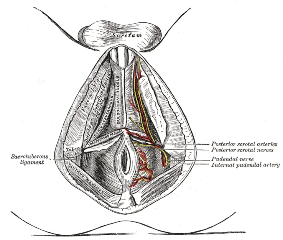
肛門管の両側にある、上が細いくさび形(断面は三角、全体は角柱形)の空間。壁を6方向で言う。
| 壁 | 作るもの |
|---|---|
| 外側 | 坐骨結節、内閉鎖筋とその筋膜(中に陰部神経管=Alcock 管)、仙結節靱帯 |
| 内側 | 肛門挙筋(下骨盤隔膜筋膜)と外肛門括約筋 |
| 前 | 浅・深会陰横筋(その上を前陥凹が恥骨の方へ伸びる) |
| 後 | 尾骨と肛門挙筋の後部(大殿筋の下縁が覆う) |
| 下(底) | 会陰筋膜と皮膚 |
| 上(頂) | 肛門挙筋が閉鎖筋膜から起こる線で、内側と外側の壁が出会う |
中身は坐骨肛門窩脂肪体とゆるい結合組織、そして外側壁から内側へ横切る下直腸動静脈・神経である。
この脂肪は排便のときに押しのけられて、肛門管が広がる余地を作る。前陥凹が深会陰隙の上まで伸びているので、窩の膿は前方の尿生殖三角の奥へも回り込む。左右の窩は肛門の後ろでつながる(馬蹄形の膿瘍の通り道)。
English version
Core
A wedge-shaped space beside the anal canal, narrow at the top (triangular in section, prism-shaped overall). Name its walls in six directions.
| Wall | Formed by |
|---|---|
| Lateral | Ischial tuberosity, obturator internus and its fascia (containing the pudendal canal, Alcock’s canal), sacrotuberous ligament |
| Medial | Levator ani (inferior fascia of the pelvic diaphragm) and external anal sphincter |
| Anterior | Superficial and deep transverse perineal muscles (the anterior recess extends above them toward the pubis) |
| Posterior | Coccyx and the back part of levator ani (overlapped by the lower edge of gluteus maximus) |
| Inferior (floor) | Perineal fascia and skin |
| Superior (apex) | The line where levator ani arises from the obturator fascia, where medial and lateral walls meet |
It contains the ischioanal fat body, loose connective tissue, and the inferior rectal vessels and nerve crossing from the lateral wall to the medial side.
Going further
The fat is pushed aside during defecation, giving the anal canal room to widen. Because the anterior recess extends above the deep perineal space, pus in the fossa can also track forward deep into the urogenital triangle. The two fossae connect behind the anus (the path of a horseshoe abscess).
卵巣Ovarium
骨盤側壁の卵巣窩にある、アーモンド大の腺。腹膜に覆われない唯一の腹腔内臓器——表面は生殖上皮なので、排卵した卵は一度腹腔へ出る。
そのため骨盤内の感染が腹腔へ広がる経路になり、卵管以外への着床(腹腔妊娠)も起こりうる。
English version
Core
An almond-sized gland in the ovarian fossa on the lateral pelvic wall. The only intraperitoneal organ not covered by peritoneum — its surface is germinal epithelium, so the ovulated egg passes first into the peritoneal cavity.
It is therefore a route by which pelvic infection spreads to the abdomen, and implantation outside the tube (abdominal pregnancy) can occur.
卵管端・子宮端Extremitas tubaria, uterina
卵巣の上外側端が卵管端(卵管采と卵巣提索が付く)、下内側端が子宮端(固有卵巣索が付く)。
どちらの端に何が付くかで卵巣の向きが決まるので、標本で見るときはここから始める。
English version
Core
The upper lateral end of the ovary is the tubal extremity (to which the fimbria ovarica and the suspensory ligament attach), the lower medial end the uterine extremity (to which the proper ovarian ligament attaches).
What attaches to which end settles the orientation of the ovary, so work on the specimen begins here.
固有卵巣索Ligamentum ovarii proprium
卵巣の子宮端と子宮の外側縁を結ぶ索。広間膜の中を走る。
子宮円索と合わせて、胎生期の生殖巣導帯の名残——男性の精巣導帯にあたる。
English version
Core
The cord joining the uterine extremity of the ovary to the lateral border of the uterus, running within the broad ligament.
With the round ligament it is the remnant of the fetal gubernaculum — the counterpart of the male gubernaculum testis.
卵巣提索Ligamentum suspensorium ovarii
卵巣の卵管端から骨盤壁へ張る腹膜のヒダ。中を卵巣動静脈が通る。
卵巣手術ではここを結紮する。すぐ下を尿管が走るので損傷に注意——という形で問われる。
English version
Core
The peritoneal fold passing from the tubal extremity of the ovary to the pelvic wall, containing the ovarian vessels. It is ligated in ovarian surgery.
The ureter runs immediately beneath it — which is how the point is asked, as a caution against injury.
卵管Tuba uterina
子宮底の外側角から卵巣へ伸びる約10cm の管。広間膜の上縁に入る。
子宮側から子宮部 → 峡部 → 膨大部 → 漏斗の4部。
English version
Core
The tube about 10 cm long running from the lateral angle of the fundus towards the ovary, enclosed in the upper margin of the broad ligament. Four parts from the uterus: uterine part → isthmus → ampulla → infundibulum.
卵管漏斗Infundibulum tubae uterinae
卵管の外側端の漏斗。縁に卵管采が広がり、その1本(卵巣采)が卵巣に付く。
腹腔に開く卵管腹腔口がここ。女性では腹腔が外界とつながっている——上行感染の道。
English version
Core
The funnel at the lateral end of the tube, its margin fringed with fimbriae, one of which (the fimbria ovarica) attaches to the ovary. The abdominal ostium opening into the peritoneal cavity is here.
In the woman the peritoneal cavity communicates with the exterior — the route of ascending infection.
卵管膨大部Ampulla tubae uterinae
卵管で最も長く太い部分。壁は薄く、粘膜ヒダが複雑に入り組む。
通常の受精はここで起こる。そして子宮外妊娠の最多部位でもある。
English version
Core
The longest and widest part of the tube, thin-walled with intricately folded mucosa. Normal fertilisation occurs here — and it is also the commonest site of ectopic pregnancy.
卵管峡部Isthmus tubae uterinae
子宮に近い、細く壁の厚い部分。卵管結紮はここで行う。
English version
Core
The narrow, thick-walled part near the uterus. Tubal ligation is performed here.
子宮底Fundus uteri
左右の卵管が入る線より上の丸い部分。
妊娠中の子宮底の高さで週数を見るのがここ。
English version
Core
The rounded part above the line at which the tubes enter. The height of the fundus in pregnancy is used to judge the gestation.
子宮体Corpus uteri
子宮底から峡部までの本体。内膜・筋層・外膜の3層で、内膜が周期的に剥がれる。
正常は前傾前屈——体が膀胱の上に前へ倒れ、体と頸の間でさらに前へ折れる。
English version
Core
The main mass from fundus to isthmus, in three layers — endometrium, myometrium and perimetrium — the endometrium being shed cyclically.
Normally anteverted and anteflexed — the body tipped forward over the bladder and flexed further forward on the cervix.
子宮腔Cavitas uteri
子宮体の内腔。前後に扁平な逆三角形のスリットで、上の2角が卵管、下の頂点が内子宮口。
English version
Core
The cavity of the body of the uterus: a flattened, inverted triangular slit, its two upper angles the tubes and its lower apex the internal os.
子宮頸(腟部・腟上部)Cervix uteri
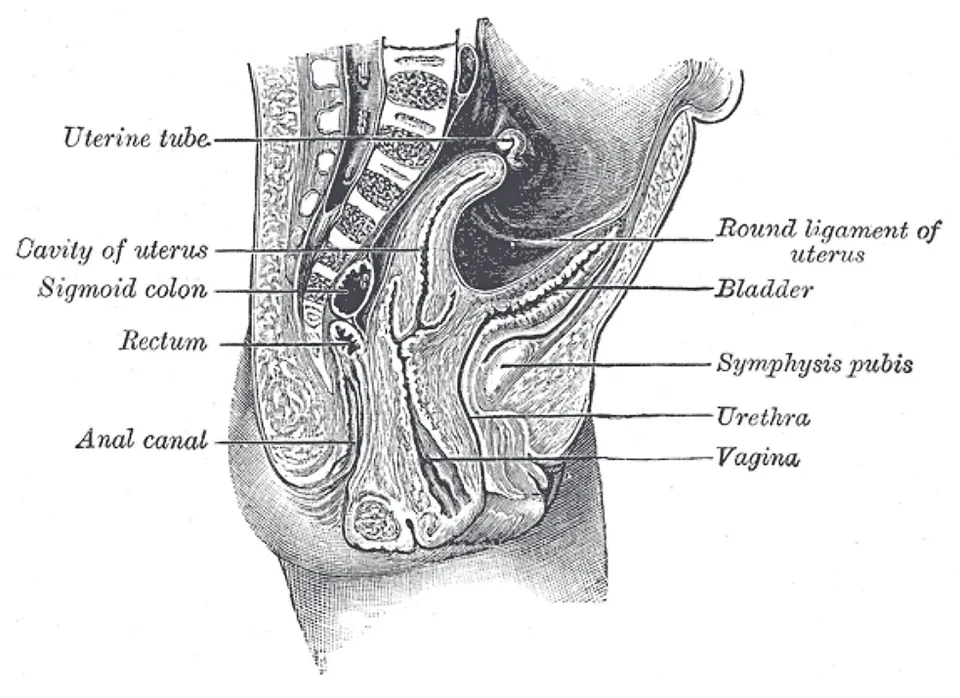
腟が付く線を境に、腟上部と、腟内へ突き出す腟部に分かれる。
腟部の外面は重層扁平上皮、頸管内は円柱上皮で、その境(扁平円柱上皮境界)が子宮頸癌の好発部位——細胞診で狙うのはここ。
English version
Core
Divided at the line of vaginal attachment into the supravaginal portion and the vaginal portion projecting into the vagina.
The outer surface of the vaginal portion is stratified squamous and the canal columnar, and the boundary between them (the squamocolumnar junction) is the site of predilection of cervical carcinoma — the place the smear is aimed at.
外子宮口Ostium uteri
腟部の先端の開口。未産婦では円形、経産婦では横に裂けた形になる。
内診でこの形を見れば分娩歴が分かるという形で問われる。
English version
Core
The opening at the tip of the vaginal portion. Round in the nullipara, a transverse slit in the parous woman.
It is asked in the form that the shape seen on examination tells the obstetric history.
子宮の膀胱面・腸面Facies vesicalis, intestinalis uteri
前面(下)が膀胱面、後面(上)が腸面。前傾しているので、前面は下を向く。
腹膜はこの2面を越えて膀胱子宮窩と直腸子宮窩を作る。
English version
Core
The anterior (lower) surface is the vesical, the posterior (upper) the intestinal. Being anteverted, the anterior surface faces downwards.
The peritoneum passes over these two surfaces to form the vesicouterine and rectouterine pouches.
子宮広間膜Ligamentum latum uteri
子宮から骨盤側壁へ張る二重腹膜のカーテン。上縁に卵管が入る。
中に卵管・固有卵巣索・子宮円索・子宮動静脈・尿管が入る。子宮を支える力はほとんど無い——支えるのは基靭帯と仙骨子宮靭帯、という対比が問われる。
English version
Core
The double curtain of peritoneum from the uterus to the lateral pelvic wall, with the uterine tube in its upper margin. It contains the tube, proper ovarian ligament, round ligament, uterine vessels and the ureter.
It gives almost no support to the uterus — the contrast with the cardinal and uterosacral ligaments, which do, is what is asked.
子宮円索Ligamentum teres uteri
子宮の外側角から広間膜の中を前外側へ走り、鼠径管を通って大陰唇に終わる。
子宮の前傾を保つ。男性の精索に対応する経路をたどるので、女性でも鼠径ヘルニアの通り道になる。
English version
Core
Runs anterolaterally from the lateral angle of the uterus within the broad ligament, passes through the inguinal canal and ends in the labium majus. It maintains anteversion.
Following the path corresponding to the male spermatic cord, it is also the route by which an inguinal hernia can occur in a woman.
腟・腟ヒダVagina, rugae vaginales
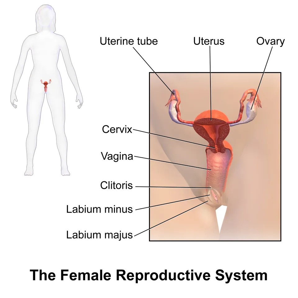
子宮頸から腟前庭までの管。前後に扁平で、内面に横走する腟ヒダがあり伸展を許す。
腺を持たず、Döderlein 桿菌がグリコーゲンを乳酸に変えて酸性(pH 3.8〜4.5)に保つ。
English version
Core
The tube from the cervix to the vestibule, flattened front to back, its inner surface carrying transverse rugae that allow distension.
It has no glands; Döderlein’s bacillus converts glycogen to lactic acid and holds it acid (pH 3.8–4.5).
腟円蓋(後部・前部・側部)Fornix vaginae, pars posterior, anterior, lateralis
腟の上端で、突き出た子宮腟部の周りにできる溝。前・後・左右に分ける。
後円蓋が最も深く、その上が直腸子宮窩(Douglas 窩)。ここから腹腔を穿刺できるのが臨床上の要点。
English version
Core
The gutter formed around the projecting vaginal portion of the cervix at the upper end of the vagina, divided into anterior, posterior and lateral parts.
The posterior fornix is the deepest, and above it lies the rectouterine pouch (of Douglas). That the peritoneal cavity can be punctured from here is the clinical point.
大陰唇Labia majora pudendi
外側の一対の皮膚のヒダ。脂肪に富み、外面に毛が生える。男性の陰嚢に相当する。前で子宮円索が終わる。
English version
Core
The outer pair of skin folds, rich in fat and hair-bearing on the outer aspect. They correspond to the male scrotum. The round ligament of the uterus ends in them anteriorly.
小陰唇Labia minora pudendi
内側の一対の薄いヒダ。毛が無く、脂肪も無い。前で分かれて陰核包皮と小帯を作る。
囲む腔が腟前庭。
English version
Core
The inner pair of thin folds, without hair or fat, dividing in front to form the prepuce and frenulum of the clitoris. The space they enclose is the vestibule.
腟前庭Vestibulum vaginae
小陰唇に囲まれた腔。外尿道口・腟口・大前庭腺(Bartholin 腺)の開口がここに並ぶ。
男性では尿路と生殖路が尿道で合流するのに対し、女性は別々に開く——この違いが問われる。
English version
Core
The space enclosed by the labia minora, into which open, in order, the external urethral orifice, the vaginal orifice and the ducts of the greater vestibular (Bartholin’s) glands.
Where in the man the urinary and genital tracts join in the urethra, in the woman they open separately — and this difference is what is asked.
外尿道口Ostium urethrae externum
腟前庭で陰核の後方、腟口の前方に開く。
女性尿道は約4cm と短く直線的——だから膀胱炎が多く、導尿も容易。
English version
Core
Opens in the vestibule behind the clitoris and in front of the vaginal orifice. The female urethra is short (about 4 cm) and straight — hence the frequency of cystitis and the ease of catheterisation.
腟口Ostium vaginae
外尿道口の後方に開く。縁に処女膜がある。後外側に大前庭腺が開口する。
English version
Core
Opens behind the external urethral orifice, its margin bearing the hymen. The greater vestibular glands open posterolaterally to it.
陰核(脚・体・亀頭)Clitoris
一対の脚が坐骨恥骨枝に付き、合して体となり、先端が亀頭。小陰唇が包皮を作る。
男性の陰茎海綿体に相当するが、尿道海綿体にあたるものを含まず、尿道は通らない——この点が最も問われる。知覚は陰部神経。
English version
Core
A pair of crura attached to the ischiopubic rami unite as the body, ending in the glans; the labia minora form its prepuce.
It corresponds to the male corpora cavernosa but contains nothing corresponding to the corpus spongiosum, and the urethra does not pass through it — the point most often asked. Sensation is by the pudendal nerve.
発生 — Müller 管が合わさって子宮になるDuctus paramesonephricus
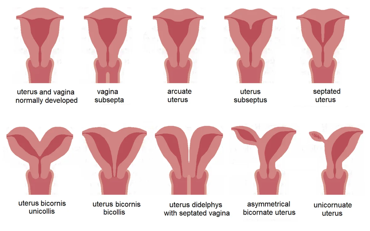
SRY が無いと、生殖腺は第7週以降に卵巣へ進む。最初の性索(髄質索)は消え、表面の上皮から新しく皮質索ができて卵祖細胞を囲み、原始卵胞になる。AMH が無いので Müller 管(中腎傍管)が残り、テストステロンが無いので中腎管は消える(名残:卵巣上体・卵巣傍体、腟の側壁の Gartner 管嚢胞)。
| Müller 管の部分 | できるもの |
|---|---|
| 頭側の開いた端 | 卵管の腹腔口 |
| 頭側の左右別々の部分 | 卵管 |
| 尾側で左右が正中で合わさった部分(子宮腟原基) | 子宮(体と頸)と腟の上部 |
腟の下部は、尿生殖洞から出る洞腟球が作る腟板が中空になってできる。腟と尿生殖洞の境に残る膜が処女膜である。外生殖器は、生殖結節が陰核、尿道ひだが小陰唇、陰唇陰嚢隆起が大陰唇、尿生殖溝が腟前庭になる。左右の Müller 管が正中に寄るとき、腹膜を引き連れてできるひだが子宮広間膜である。
子宮の形の異常は、どの段階でつまずいたかで決まる。①左右が合わさらない → 重複子宮(子宮も頸も2つ)②一部だけ合わさる → 双角子宮 ③合わさったあと中の仕切りが消えない → 中隔子宮(最も多い)④片方の管が育たない → 単角子宮 ⑤両方が育たない → 子宮と腟上部の欠損(MRKH 症候群)。
MRKH 症候群では、卵巣は Müller 管ではなく生殖腺から作られるので正常で、乳房などの二次性徴も普通に出る。月経だけが来ない(原発性無月経)——出身の違いがそのまま症状の形になる。
処女膜閉鎖では、初経が来ても血が出られず腟にたまる。思春期に、月経が無いのに周期的な下腹部痛と、腟口の青紫色のふくらみで見つかる。中隔子宮は流産・早産の原因になり、子宮鏡で中隔を切って治す。Müller 管の異常には、片側の腎の無形成など泌尿器の異常が合併しやすい——どちらも中間中胚葉から隣り合って作られるからである。
English version
Core
Without SRY, the gonad becomes an ovary after week 7. The first sex cords (medullary cords) disappear, and new cortical cords grow from the surface epithelium, surround the oogonia and become primordial follicles. With no AMH, the Müllerian (paramesonephric) duct stays; with no testosterone, the mesonephric duct disappears (remnants: epoophoron, paroophoron, Gartner’s duct cyst in the side wall of the vagina).
| Part of the Müllerian duct | Derivative |
|---|---|
| Open cranial end | Abdominal opening of the uterine tube |
| Separate cranial parts | Uterine tubes |
| Caudal parts fused in the midline (uterovaginal primordium) | Uterus (body and cervix) and upper vagina |
The lower vagina forms when the vaginal plate, made by the sinovaginal bulbs of the urogenital sinus, becomes hollow. The membrane left between vagina and urogenital sinus is the hymen. Externally, the genital tubercle becomes the clitoris, the urethral folds the labia minora, the labioscrotal swellings the labia majora, and the urogenital groove the vestibule. As the two Müllerian ducts move to the midline they drag peritoneum with them, forming the broad ligament.
Going further
The shape of a uterine anomaly depends on the step that failed. (1) The ducts do not fuse → uterus didelphys (two uteri and two cervices); (2) partial fusion → bicornuate uterus; (3) fusion occurs but the inner septum is not removed → septate uterus (the most common); (4) one duct fails to grow → unicornuate uterus; (5) both fail → absence of the uterus and upper vagina (MRKH syndrome).
In MRKH syndrome the ovaries are normal, because they come from the gonad, not the Müllerian duct, so breasts and other secondary sex features develop normally. Only menstruation is missing (primary amenorrhoea) — the difference in origin shapes the symptoms.
In the clinic
With an imperforate hymen, menstrual blood cannot escape and collects in the vagina. It is found at puberty as cyclic lower abdominal pain without periods and a bluish bulge at the vaginal opening. A septate uterus can cause miscarriage and preterm birth and is treated by cutting the septum through a hysteroscope. Müllerian anomalies often come with urinary anomalies such as absence of one kidney, because both are formed side by side from intermediate mesoderm.
まとめ
| 卵巣と卵管 | Ovarium:骨盤壁の fossa ovarica(内腸骨動静脈と尿管が後壁を作る)にある扁平な卵形の器官。 |
|---|---|
| 卵巣の内部 — 白膜・皮質・髄質 | 卵巣を切ると、外から次の順に見える。層中身表面上皮単層の立方上皮。 |
| 子宮 — 部位・傾き・支持 | 膀胱と直腸のあいだ。約7×5×2.5cm の洋梨形。 |
| 腟と外陰 | Vagina:約8cm の管。前壁は膀胱と尿道、後壁は直腸に接する。 |
| 腟の血管と神経 — 上は子宮と、下は会陰と同じ | 動脈はすべて内腸骨動脈の系統から来る。動脈養う所a. vaginalis(腟動脈:内腸骨動脈または子宮動脈から)腟の中部と下部、膀胱の下部子宮動脈の腟枝腟の上部と子宮頸。 |
| 会陰 — 2つの三角と2枚の隔膜 | 骨盤出口を、両側の坐骨結節を結ぶ線で2つの三角に分ける。 |
| 尿生殖三角の層 — 深会陰隙・会陰膜・浅会陰隙・会陰筋膜 | 尿生殖三角は、上(骨盤の中)から下(皮膚)へ次の順に重なる:骨盤隔膜 → 深会陰隙 → 会陰膜(membrana perinei)→ 浅会陰隙 → 会陰筋膜(fascia peri… |
| 骨盤隔膜と骨盤底 — 2枚の筋膜と、5つの仕事 | 骨盤隔膜は骨盤腔の底を作る漏斗形の筋の板で、肛門挙筋(恥骨尾骨筋・恥骨直腸筋・腸骨尾骨筋)と尾骨筋からなる。 |
| 坐骨肛門窩 — 6方向の壁 | 肛門管の両側にある、上が細いくさび形(断面は三角、全体は角柱形)の空間。 |
| 卵巣 | 骨盤側壁の卵巣窩にある、アーモンド大の腺。 |
| 卵管端・子宮端 | 卵巣の上外側端が卵管端(卵管采と卵巣提索が付く)、下内側端が子宮端(固有卵巣索が付く)。 |
| 固有卵巣索 | 卵巣の子宮端と子宮の外側縁を結ぶ索。広間膜の中を走る。 |
| 卵巣提索 | 卵巣の卵管端から骨盤壁へ張る腹膜のヒダ。 |
| 卵管 | 子宮底の外側角から卵巣へ伸びる約10cm の管。 |
| 卵管漏斗 | 卵管の外側端の漏斗。縁に卵管采が広がり、その1本(卵巣采)が卵巣に付く。 |
| 卵管膨大部 | 卵管で最も長く太い部分。壁は薄く、粘膜ヒダが複雑に入り組む。 |
| 卵管峡部 | 子宮に近い、細く壁の厚い部分。卵管結紮はここで行う。 |
| 子宮底 | 左右の卵管が入る線より上の丸い部分。妊娠中の子宮底の高さで週数を見るのがここ。 |
| 子宮体 | 子宮底から峡部までの本体。内膜・筋層・外膜の3層で、内膜が周期的に剥がれる。 |
| 子宮腔 | 子宮体の内腔。前後に扁平な逆三角形のスリットで、上の2角が卵管、下の頂点が内子宮口。 |
| 子宮頸(腟部・腟上部) | 腟が付く線を境に、腟上部と、腟内へ突き出す腟部に分かれる。 |
| 外子宮口 | 腟部の先端の開口。未産婦では円形、経産婦では横に裂けた形になる。 |
| 子宮の膀胱面・腸面 | 前面(下)が膀胱面、後面(上)が腸面。前傾しているので、前面は下を向く。 |
| 子宮広間膜 | 子宮から骨盤側壁へ張る二重腹膜のカーテン。 |
| 子宮円索 | 子宮の外側角から広間膜の中を前外側へ走り、鼠径管を通って大陰唇に終わる。 |
| 腟・腟ヒダ | 子宮頸から腟前庭までの管。前後に扁平で、内面に横走する腟ヒダがあり伸展を許す。 |
| 腟円蓋(後部・前部・側部) | 腟の上端で、突き出た子宮腟部の周りにできる溝。 |
| 大陰唇 | 外側の一対の皮膚のヒダ。脂肪に富み、外面に毛が生える。 |
| 小陰唇 | 内側の一対の薄いヒダ。毛が無く、脂肪も無い。 |
| 腟前庭 | 小陰唇に囲まれた腔。外尿道口・腟口・大前庭腺(Bartholin 腺)の開口がここに並ぶ。 |
| 外尿道口 | 腟前庭で陰核の後方、腟口の前方に開く。女性尿道は約4cm と短く直線的——だから膀胱炎が多く、導尿も容易。 |
| 腟口 | 外尿道口の後方に開く。縁に処女膜がある。 |
| 陰核(脚・体・亀頭) | 一対の脚が坐骨恥骨枝に付き、合して体となり、先端が亀頭。 |
| 発生 — Müller 管が合わさって子宮になる | SRY が無いと、生殖腺は第7週以降に卵巣へ進む。 |
このノートは医学生が授業の内容を自分の言葉でまとめたもので、診療の判断に使うものではありません。誤りに気づいたらお知らせください。