呼吸器は「空気の通る順」に読む——鼻腔 → 咽頭 → 喉頭 → 気管 → 気管支樹 → 肺胞。区間ごとに壁の骨組み(骨・軟骨・平滑筋・無し)が入れ替わり、それが臨床の性質を決める。
この範囲の山場は喉頭である。軟骨3つ+関節2つ+腔の3階建て(前庭・声門・声門下)を、順序立てて示せるようにする。
この範囲の芯は「左右差」である。右主気管支が太く短く垂直だから誤嚥は右へ、右肺が3葉で左が2葉なのは心臓が左にあるから、左肺に心切痕があるのも同じ理由。左右の非対称はすべて心臓の位置から導ける。
- 鼻腔と副鼻腔
- 外鼻 — 上が骨、下が軟骨
- 喉頭 — 軟骨・関節・壁
- 喉頭の壁 — 4層と、2枚の線維弾性膜
- 喉頭腔 — 3階に分かれる
- 気管と気管支樹 — 右へ落ちる理由
- 肺 — 面・縁・葉・門
- 肺区域 — 右10・左8〜10の名前
- 胸膜と縦隔
- 胸膜の血管と神経 — 壁側は胸壁の、臓側は肺の
- 鼻腔
- 上鼻甲介
- 中鼻甲介
- 下鼻甲介
- 中鼻道
- 下鼻道
- 後鼻孔
- 前頭洞・蝶形骨洞・上顎洞
- 甲状軟骨
- 輪状軟骨
- 披裂軟骨
- 喉頭蓋
- 輪状気管靭帯
- 輪状甲状関節
- 輪状披裂関節
- 喉頭口
- 披裂喉頭蓋ひだ
- 喉頭前庭
- 前庭ひだ・前庭裂
- 声門・喉頭室
- 声帯ひだ・声門裂
- 声帯靭帯
- 声帯筋・輪状甲状筋
- 気管分岐部
- 気管軟骨
- 右主気管支・左主気管支
- 肺尖
- 肺底
- 肺の肋骨面(椎骨部)
- 肺の縦隔面
- 肺の横隔面
- 肺の前縁
- 肺の下縁
- 肺門
- 肺根
- 肺の上葉
- 右肺の中葉
- 肺の下葉
- 気管支樹と肺胞 — 壁はどう変わるか
- 肺の体表投影 — 打診と聴診の基準
- 発生 — 前腸の前壁から出る肺芽と、咽頭弓
- 鼻腔と副鼻腔Cavitas nasi et sinus paranasales
- 外鼻 — 上が骨、下が軟骨Nasus externus
- 喉頭 — 軟骨・関節・壁Larynx
- 喉頭の壁 — 4層と、2枚の線維弾性膜Membrana quadrangularis et conus elasticus
- 喉頭腔 — 3階に分かれるCavitas laryngis
- 気管と気管支樹 — 右へ落ちる理由Trachea et arbor bronchialis
- 肺 — 面・縁・葉・門Pulmo
- 肺区域 — 右10・左8〜10の名前Segmenta bronchopulmonalia
- 胸膜と縦隔Pleura et mediastinum
- 胸膜の血管と神経 — 壁側は胸壁の、臓側は肺のVasa et nervi pleurae
- 鼻腔Cavitas nasi
- 上鼻甲介Concha nasalis superior
- 中鼻甲介Concha nasalis media
- 下鼻甲介Concha nasalis inferior
- 中鼻道Meatus nasi medius
- 下鼻道Meatus nasi inferior
- 後鼻孔Choanae
- 前頭洞・蝶形骨洞・上顎洞Sinus frontalis, sphenoidalis, maxillaris
- 甲状軟骨Cartilago thyroidea
- 輪状軟骨Cartilago cricoidea
- 披裂軟骨Cartilago arytenoidea
- 喉頭蓋Epiglottis
- 輪状気管靭帯Ligamentum cricotracheale
- 輪状甲状関節Articulatio cricothyroidea
- 輪状披裂関節Articulatio cricoarytenoidea
- 喉頭口Aditus laryngis
- 披裂喉頭蓋ひだPlica aryepiglottica
- 喉頭前庭Vestibulum laryngis
- 前庭ひだ・前庭裂Plica vestibularis, rima vestibuli
- 声門・喉頭室Glottis, ventriculus laryngis
- 声帯ひだ・声門裂Plica vocalis, rima vocalis
- 声帯靭帯Ligamentum vocale
- 声帯筋・輪状甲状筋Musculus vocalis, musculus cricothyroideus
- 気管分岐部Bifurcatio tracheae
- 気管軟骨Cartilagines tracheales
- 右主気管支・左主気管支Bronchus principalis dexter et sinister
- 肺尖Apex pulmonis
- 肺底Basis pulmonis
- 肺の肋骨面(椎骨部)Facies costalis pulmonis
- 肺の縦隔面Facies mediastinalis pulmonis
- 肺の横隔面Facies diaphragmatica pulmonis
- 肺の前縁Margo anterior pulmonis
- 肺の下縁Margo inferior pulmonis
- 肺門Hilum pulmonis
- 肺根Radix pulmonis
- 肺の上葉Lobus superior pulmonis
- 右肺の中葉Lobus medius pulmonis dextri
- 肺の下葉Lobus inferior pulmonis
- 気管支樹と肺胞 — 壁はどう変わるかArbor alveolaris
- 肺の体表投影 — 打診と聴診の基準Topographia pulmonum
- 発生 — 前腸の前壁から出る肺芽と、咽頭弓Diverticulum respiratorium
- まとめ
鼻腔と副鼻腔Cavitas nasi et sinus paranasales
Nares(外鼻孔)から choanae(後鼻孔)まで。入口の皮膚に覆われた部分が vestibulum nasi、その奥が cavitas nasi propria。中隔で左右に分かれる。
外側壁から3枚の甲介が垂れる:concha nasalis superior・media・inferior。それぞれの下の通路が meatus nasi superior・medius・inferior、後方の共通路が meatus nasopharyngeus。
2つの領域:上部の pars olfactoria(上鼻甲介と対応する中隔・嗅上皮)と、残り大部分の pars respiratoria(多列線毛上皮)。
副鼻腔4対:sinus frontalis・sinus maxillaris・sinus sphenoidalis・cellulae ethmoidales。
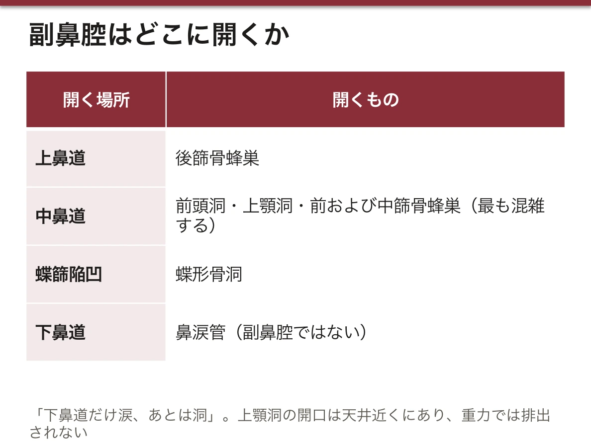
どこに開くかが決まっている。上鼻道=後篩骨蜂巣。中鼻道=前頭洞・上顎洞・前および中篩骨蜂巣(3つも開くので最も混雑する)。蝶篩陥凹=蝶形骨洞。下鼻道=鼻涙管(副鼻腔ではない)。「下鼻道だけ涙、あとは洞」と覚える。
上顎洞の開口部が洞の上部にあることが、副鼻腔炎が最も多い理由である。底ではなく天井近くに穴があるので、重力では排出されず、線毛運動だけが頼り。粘膜が腫れると排出が止まり、膿が溜まる。形の不合理がそのまま病気の頻度になっている数少ない例。
上顎洞の底は上顎臼歯の歯根と接する。だから歯性上顎洞炎が起こるし、抜歯で洞と交通することもある。
甲介があることで乱流ができる。層流のまま通り抜けると加温・加湿・除塵ができないので、あえて流れを乱している。鼻閉が単なる不快でなく「空気の調整の喪失」であるのはこのためである。
副鼻腔の開口部の位置が、炎症の広がりと痛みを決める。
上顎洞の自然口は洞の上方(天井近く)にある。立っている姿勢では重力に逆らって排出しなければならないため、上顎洞炎は最も起こりやすく、最も治りにくい。前かがみで痛みが増すのはこのため。
Kiesselbach 部位(鼻中隔前下部)は4本の動脈が吻合する場所で、鼻出血の約90%がここから起こる。小鼻をつまんで圧迫するのが正しい応急処置である。
篩骨洞は眼窩と紙のように薄い骨(紙様板)で隔てられるだけなので、小児の篩骨洞炎は眼窩蜂窩織炎に進む。眼瞼腫脹・眼球突出・眼球運動障害は緊急である。
嗅裂の障害で嗅覚が落ちる。篩板の骨折では髄液鼻漏——髄膜炎の入口になる。
出典:イラスト解剖学 誌面210頁「副鼻腔の臨床:副鼻腔炎と疼痛」
English version
Core
From the nares to the choanae. The skin-covered part at the entrance is the vestibulum nasi, and beyond it the cavitas nasi propria, divided in two by the septum.
Three conchae hang from the lateral wall: concha nasalis superior, media, inferior, with the meatus nasi superior, medius, inferior beneath them and the common meatus nasopharyngeus behind.
Two regions: the upper pars olfactoria (the superior concha and the septum opposite it; olfactory epithelium) and the remaining, larger pars respiratoria (pseudostratified ciliated epithelium).
Four pairs of paranasal sinuses: sinus frontalis, sinus maxillaris, sinus sphenoidalis, cellulae ethmoidales.
Going further
Where each opens is fixed. Superior meatus = posterior ethmoidal cells. Middle meatus = frontal sinus, maxillary sinus and anterior and middle ethmoidal cells (three of them, so it is the most crowded). Sphenoethmoidal recess = sphenoidal sinus. Inferior meatus = the nasolacrimal duct (not a sinus). Remember: the inferior meatus alone takes tears; the rest take sinuses.
That the opening of the maxillary sinus lies in its upper part is why sinusitis is commonest there. The hole is near the ceiling rather than the floor, so it does not drain by gravity and depends on the cilia alone. When the mucosa swells, drainage stops and pus collects. One of the few cases in which an irrationality of form becomes directly the frequency of a disease.
The floor of the maxillary sinus touches the roots of the upper molars, hence odontogenic maxillary sinusitis, and an extraction may open into the sinus.
The conchae create turbulence. Air passing through in laminar flow could not be warmed, humidified and cleaned, so the flow is deliberately disturbed. This is why nasal obstruction is not mere discomfort but a loss of the conditioning of the air.
In the clinic
The position of a sinus ostium decides the spread of inflammation and the pain.
The natural ostium of the maxillary sinus is high in the sinus, near the ceiling. Standing, it must drain against gravity, so maxillary sinusitis is the commonest and the hardest to resolve. This is why the pain increases on bending forward.
Kiesselbach’s area (the anteroinferior septum) is where four arteries anastomose, the source of about 90 per cent of nosebleeds. Pinching the ala and compressing it is the correct first aid.
The ethmoidal cells are separated from the orbit by only a paper-thin bone (the lamina papyracea), so ethmoiditis in a child progresses to orbital cellulitis. Swelling of the lids, proptosis and limited eye movement are an emergency.
Disease of the olfactory cleft reduces smell. A fracture of the cribriform plate gives CSF rhinorrhoea — a portal for meningitis.
外鼻 — 上が骨、下が軟骨Nasus externus
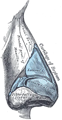
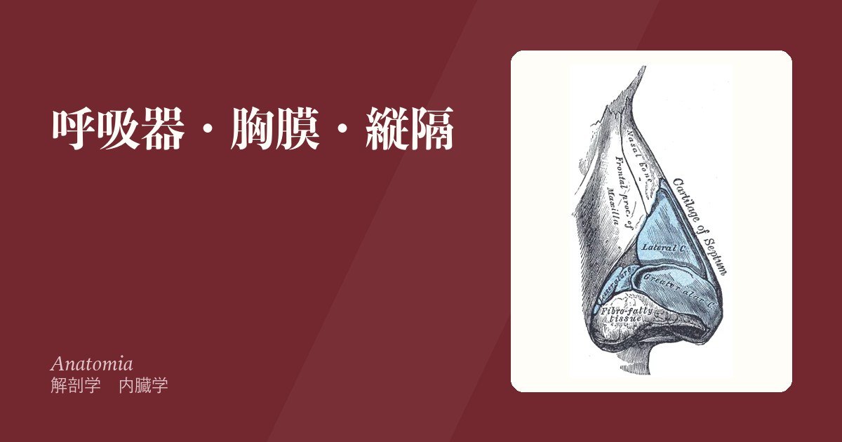
鼻腔の前の壁を作るのが外鼻である。部位は上から radix nasi(鼻根・両眼の間)・dorsum nasi(鼻背)・apex nasi(鼻尖)。両側のふくらみが alae nasi(鼻翼)で、これが外鼻孔 nares の縁を作る。
骨組みは上1/3が骨、下2/3が軟骨である。
| 部分 | 骨組み |
|---|---|
| 鼻根・鼻背の上部 | ossa nasalia(鼻骨)・maxilla の processus frontalis |
| 鼻背の下部・側面 | cartilago nasi lateralis(外側鼻軟骨) |
| 鼻尖・鼻翼 | cartilago alaris major(大鼻翼軟骨:内側脚と外側脚)・cartilagines alares minores |
| 中央の支え | cartilago septi nasi(鼻中隔軟骨) |
表面は m. nasalis(鼻筋)と皮膚で覆われる。鼻尖・鼻翼の皮膚は厚く、皮脂腺が多い。
上が骨で下が軟骨なのは、ぶつかったときに曲がって逃げるためである。軟骨の部分はしなるので折れにくく、骨折は動かない上部の鼻骨に起こる。
鼻翼の軟骨は外鼻孔がつぶれないよう形を保ち、m. nasalis の鼻翼部は鼻孔を広げる。鼻中隔と外側鼻軟骨の下縁のあいだのすき間(内鼻弁)は鼻の気道で最も狭く、ここが少し狭くなるだけで鼻づまりが強くなる。
鼻骨骨折は顔の骨折で最も多い。鼻中隔に血がたまった鼻中隔血腫は、すぐに切って出す。鼻中隔軟骨には血管が無く、表面の軟骨膜から栄養を受けているので、血腫で軟骨膜がはがれると軟骨が死んで鼻背がへこむ(鞍鼻)からである。
乳児や呼吸困難の患者で鼻翼が呼吸ごとに広がる(鼻翼呼吸)のは、鼻筋で気道を広げようとする努力のサインである。鼻から上唇にかけての皮膚の感染は、顔面静脈・眼静脈を通じて海綿静脈洞に広がりうるので、むやみにつぶさない。
English version
Core
The external nose forms the front wall of the nasal cavity. From above: radix nasi (root, between the eyes), dorsum nasi (bridge) and apex nasi (tip). The rounded sides are the alae nasi, which form the edges of the nostrils (nares).
Its framework is bone in the upper third and cartilage in the lower two thirds.
| Part | Framework |
|---|---|
| Root and upper bridge | ossa nasalia and the processus frontalis of the maxilla |
| Lower bridge and sides | cartilago nasi lateralis |
| Tip and alae | cartilago alaris major (medial and lateral crura) and cartilagines alares minores |
| Central support | cartilago septi nasi |
It is covered by m. nasalis and skin. The skin over the tip and alae is thick and rich in sebaceous glands.
Going further
Bone above and cartilage below lets the nose bend away when it is hit. The cartilage flexes and seldom breaks, so fractures occur in the fixed nasal bones above.
The alar cartilages keep the nostrils from collapsing, and the alar part of m. nasalis widens them. The gap between the septum and the lower edge of the lateral cartilage (internal nasal valve) is the narrowest point of the nasal airway, so even a small narrowing here causes a blocked nose.
In the clinic
Nasal fracture is the most common facial fracture. A septal haematoma must be drained at once: the septal cartilage has no vessels and is fed by its perichondrium, so if blood lifts the perichondrium the cartilage dies and the bridge collapses (saddle nose).
Flaring of the alae with each breath in infants or breathless patients shows effort to widen the airway with m. nasalis. Skin infection between the nose and upper lip can spread through the facial and ophthalmic veins to the cavernous sinus, so such spots should not be squeezed.
喉頭 — 軟骨・関節・壁Larynx
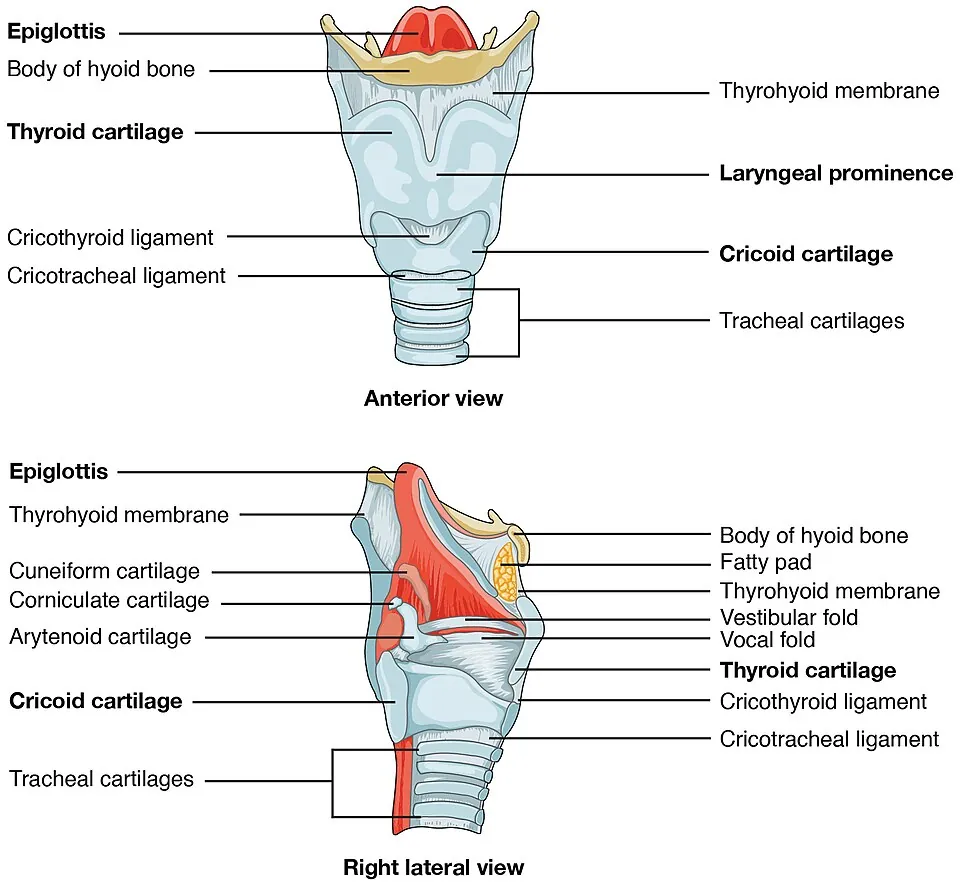
C3〜C6の高さ、舌骨の下、気管の上。3つの単一軟骨+3対の対性軟骨。
- Cartilago thyroidea(甲状軟骨)— 2枚の板が前で合し、上縁に prominentia laryngea(喉頭隆起)
- Cartilago cricoidea(輪状軟骨)— 気道で唯一の完全な輪。前が低く後が高い印章形
- Epiglottis(喉頭蓋)— 弾性軟骨の葉状
- Cartilagines arytenoideae(披裂軟骨・対)— 輪状軟骨板の上に乗り、声帯突起と筋突起を持つ
関節2つ:art. cricothyroidea(甲状軟骨が前傾して声帯を伸ばす=高い声)と art. cricoarytenoidea(披裂軟骨が回転・滑走して声門を開閉する)。
膜:membrana thyrohyoidea(舌骨と甲状軟骨)・lig. cricothyroideum(輪状甲状靱帯)。
内喉頭筋は「声門を閉じる筋が多数、開く筋は1つ」である。開大するのは m. cricoarytenoideus posterior のみ——両側が麻痺すると声門が開かず窒息するため、「唯一の外転筋」として必ず問われる。
神経支配も1つだけ例外。内喉頭筋はすべて反回神経(n. laryngeus recurrens)だが、m. cricothyroideus だけ上喉頭神経外枝。感覚は声帯より上が上喉頭神経内枝、下が反回神経。「輪状甲状筋だけ違う」。
反回神経の走行に左右差がある。右は右鎖骨下動脈を、左は大動脈弓(動脈管索)を回って上行する。左のほうが長く胸腔を通るので、肺癌・大動脈瘤・僧帽弁狭窄による左房拡大で左反回神経麻痺(嗄声)が起こる。嗄声が胸部疾患の徴候になるのは、この走行のためである。
輪状甲状靱帯は緊急気道確保の窓である。皮膚のすぐ下にあり、上下を軟骨に挟まれて位置が確実で、その高さには甲状腺も大血管も無い。輪状甲状靱帯切開が「輪状軟骨と甲状軟骨のあいだ」で行われる理由は、この3つが揃うのがそこだけだからである。
喉頭の筋はすべて反回神経が支配する——輪状甲状筋だけが上喉頭神経外枝。この1つの例外が試験でも臨床でも要になる。
反回神経麻痺の主症状は嗄声である。声門を閉じる筋(甲状披裂筋・外側輪状披裂筋・横披裂筋)のどれが麻痺しても声門が閉じきらないためである。
声門を開く筋は後輪状披裂筋ただ1つ。だから両側反回神経麻痺では声門が開かず、呼吸困難・吸気性喘鳴になり、気管切開を要する。「開く筋は1つしかない」ことが緊急性の理由である。
左反回神経は大動脈弓を、右は鎖骨下動脈を回る。左は経路が長く胸腔内を通るため、肺癌(左上葉)・大動脈瘤・左房拡大・縦隔リンパ節転移で侵されやすい。原因不明の嗄声は胸部を調べる。
出典:イラスト解剖学 誌面477頁「声帯の臨床関連事項」
English version
Core
At the level of C3–C6, below the hyoid and above the trachea. Three unpaired plus three paired cartilages.
- Cartilago thyroidea — two laminae meeting in front, with the prominentia laryngea at the upper border.
- Cartilago cricoidea — the only complete ring in the airway, signet-shaped, low in front and high behind.
- Epiglottis — a leaf of elastic cartilage.
- Cartilagines arytenoideae (paired) — riding on the lamina of the cricoid, with vocal and muscular processes.
Two joints: the art. cricothyroidea (the thyroid cartilage tilts forward and stretches the vocal folds = a higher voice) and the art. cricoarytenoidea (the arytenoid rotates and slides to open and close the glottis).
Membranes: membrana thyrohyoidea and lig. cricothyroideum.
Going further
Among the intrinsic muscles many close the glottis and only one opens it. The only abductor is the m. cricoarytenoideus posterior — and because bilateral paralysis leaves the glottis shut and asphyxiates, "the only abductor" is invariably asked.
The innervation too has a single exception. All the intrinsic muscles are supplied by the recurrent laryngeal nerve except the m. cricothyroideus, by the external branch of the superior laryngeal. Sensation above the vocal folds is by the internal branch of the superior laryngeal, below by the recurrent. "Only the cricothyroid is different."
The course of the recurrent laryngeal nerve is asymmetrical: the right hooks round the right subclavian artery, the left round the aortic arch (the ligamentum arteriosum). The left is longer and passes through the thorax, so left recurrent nerve palsy (hoarseness) follows lung cancer, aortic aneurysm and left atrial enlargement in mitral stenosis. That hoarseness can be a sign of thoracic disease is due to this course.
The cricothyroid ligament is the window for emergency airway access. It lies immediately beneath the skin, its position is certain between two cartilages, and at that level there is neither thyroid gland nor great vessel. Cricothyroidotomy is done between cricoid and thyroid cartilage because that is the only place where all three hold.
In the clinic
All the muscles of the larynx are supplied by the recurrent laryngeal nerve — except the cricothyroid, by the external branch of the superior laryngeal. This one exception is the crux both in the examination and in the clinic.
The chief symptom of recurrent nerve palsy is hoarseness, because paralysis of any of the adductors (thyroarytenoid, lateral cricoarytenoid, transverse arytenoid) leaves the glottis incompletely closed.
There is only one abductor, the posterior cricoarytenoid. So in bilateral recurrent nerve palsy the glottis will not open, giving respiratory distress and inspiratory stridor and requiring tracheostomy. The reason for the urgency is that there is only one muscle that opens it.
The left recurrent nerve hooks round the aortic arch, the right round the subclavian artery. The left, having a longer course through the thorax, is readily involved by carcinoma of the lung (left upper lobe), aortic aneurysm, left atrial enlargement and mediastinal nodal metastases. Unexplained hoarseness calls for investigation of the chest.
喉頭の壁 — 4層と、2枚の線維弾性膜Membrana quadrangularis et conus elasticus
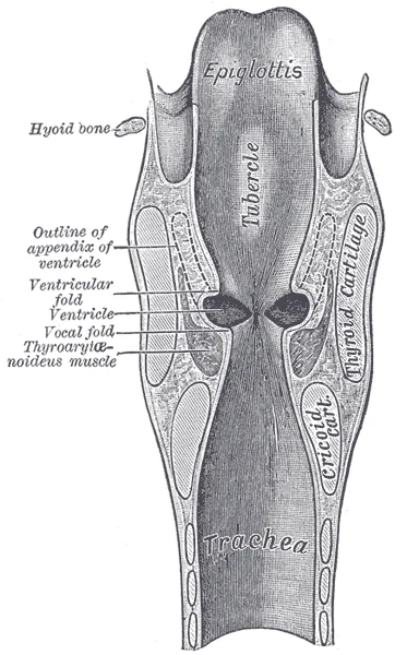
喉頭の壁は内から4層である。
| 層 | 中身 |
|---|---|
| ① 粘膜 | 大部分は多列線毛上皮。声帯ひだと喉頭蓋の上部だけは重層扁平上皮。喉頭腺とリンパ小節を含む |
| ② 粘膜下組織(線維弾性膜) | 軟骨の内側に張る membrana fibroelastica。上部が membrana quadrangularis(四角膜)、下部が conus elasticus(弾性円錐) |
| ③ 軟骨と筋 | 喉頭軟骨と内喉頭筋 |
| ④ 外膜 | 周りの頸部の組織とつなぐ結合組織 |
四角膜は喉頭蓋の外側縁と甲状軟骨から披裂軟骨へ張る。上縁は披裂喉頭蓋ひだの芯、下縁が厚くなって lig. vestibulare(前庭ひだの芯)になる。弾性円錐は輪状軟骨の上縁から上へすぼまり、上縁が厚くなって lig. vocale(甲状軟骨の内面 → 披裂軟骨の声帯突起)になる。前の正中で厚い部分が lig. cricothyroideum medianum である。
内喉頭筋は働きで3群に分ける:喉頭口を開閉する筋(披裂喉頭蓋筋・甲状喉頭蓋筋)、声門裂を開閉する筋(開くのは後輪状披裂筋だけ。閉じるのは外側輪状披裂筋・横/斜披裂筋・甲状披裂筋)、声帯の張りを変える筋(張る輪状甲状筋、ゆるめる声帯筋)。
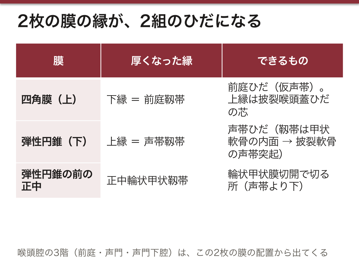
2枚の膜の自由縁が、そのまま2組のひだになる。上の膜の下縁が前庭ひだ(仮声帯)、下の膜の上縁が声帯ひだ。だから喉頭腔の3階(前庭・声門・声門下腔)は、膜の配置を描けば自然に出てくる。
声帯ひだの上皮が重層扁平なのは、1秒に100回以上ぶつかり合う摩擦に耐えるためである。その下の粘膜固有層の浅い所はとても柔らかく(Reinke 腔)、ここが波打つことで声が出る。
緊急の気道確保(輪状甲状膜切開)で切るのは lig. cricothyroideum medianum、つまり弾性円錐の前の部分である。声帯より下なので、声門が腫れてふさがっていても空気の通り道を作れる。
喫煙や声の使いすぎで Reinke 腔に水がたまると声帯がぶよぶよに腫れ(Reinke 浮腫)、声が低くかすれる。声帯の扁平上皮から出る癌は扁平上皮癌である。
English version
Core
The laryngeal wall has four layers from inside out.
| Layer | Content |
|---|---|
| (1) Mucosa | Mostly ciliated pseudostratified epithelium; stratified squamous only on the vocal folds and the upper epiglottis. Contains laryngeal glands and lymphoid nodules |
| (2) Submucosa (fibroelastic membrane) | The membrana fibroelastica inside the cartilages. Upper part: membrana quadrangularis; lower part: conus elasticus |
| (3) Cartilages and muscles | Laryngeal cartilages and intrinsic muscles |
| (4) Adventitia | Connective tissue joining the larynx to the neck |
The quadrangular membrane runs from the epiglottis edge and thyroid cartilage to the arytenoid. Its upper edge is the core of the aryepiglottic fold, and its thickened lower edge is the lig. vestibulare (core of the vestibular fold). The conus elasticus rises from the upper edge of the cricoid and narrows upward; its thickened upper edge is the lig. vocale (thyroid cartilage → vocal process of arytenoid). Its thick front midline part is the lig. cricothyroideum medianum.
Intrinsic muscles fall into three groups by action: those acting on the laryngeal inlet (aryepiglottic, thyroepiglottic); those opening or closing the rima glottidis (only the posterior cricoarytenoid opens; lateral cricoarytenoid, transverse/oblique arytenoid and thyroarytenoid close); and those changing vocal fold tension (cricothyroid tightens, vocalis relaxes).
Going further
The free edges of the two membranes become the two pairs of folds. The lower edge of the upper membrane is the vestibular (false) fold; the upper edge of the lower membrane is the vocal fold. Draw the membranes and the three levels of the laryngeal cavity (vestibule, glottis, infraglottic cavity) appear by themselves.
The vocal fold has squamous epithelium because it must survive friction from more than 100 collisions a second. Below it, the superficial lamina propria is very soft (Reinke’s space), and its wave-like movement produces the voice.
In the clinic
In an emergency airway (cricothyroidotomy) the cut goes through the lig. cricothyroideum medianum — the front part of the conus elasticus. It lies below the vocal folds, so an airway can be made even when the glottis is swollen shut.
Smoking or voice overuse can fill Reinke’s space with fluid (Reinke’s oedema), making the voice low and hoarse. Cancer arising from the squamous epithelium of the vocal fold is squamous cell carcinoma.
喉頭腔 — 3階に分かれるCavitas laryngis
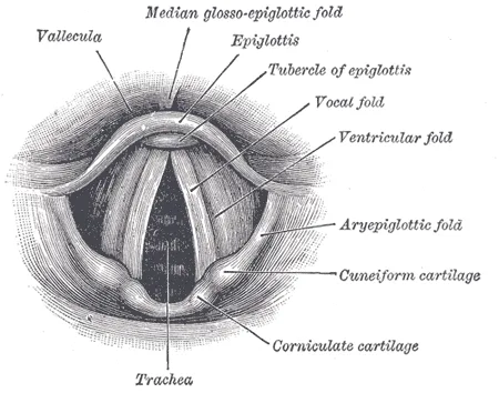
上の入口が aditus laryngis(前は喉頭蓋、側方は plica aryepiglottica、後ろは披裂間切痕)。ここから下へ3階。
| 階 | 範囲 | 境界 |
|---|---|---|
| Vestibulum laryngis | 喉頭口 〜 前庭ヒダ | 下端が plica vestibularis(仮声帯)と rima vestibuli |
| Ventriculus laryngis | 前庭ヒダと声帯ヒダのあいだの外側の窪み | — |
| Glottis | Plica vocalis(声帯ヒダ)と rima glottidis(声門裂) | 気道で最も狭い |
| Cavitas infraglottica | 声門 〜 輪状軟骨下縁 | 気管へ続く |
Plica vocalis の中身が lig. vocale(声帯靱帯)と m. vocalis。前は甲状軟骨の内面、後ろは披裂軟骨の声帯突起に付く。
声帯ヒダが白く見えるのは、粘膜が薄く血管が乏しいうえ、下の靱帯が透けるから。前庭ヒダは赤い(血管に富む)。喉頭鏡で「白い方が本物」と見分ける。
声門が最も狭いことは麻酔と救急で重要である。ただし小児では輪状軟骨部が最狭——輪状軟骨が完全な輪で拡張しないため。だからカフ無しチューブが小児で使われてきた。
Ventriculus laryngis(Morgagni 洞)は声帯の潤滑を担う粘液腺の袋。ここが外側へ膨れると喉頭瘤(laryngocele)になる。
声帯より上と下でリンパの流れが違う。声帯上部は上喉頭動脈に伴って上深頸リンパ節へ、下部は気管前・下深頸リンパ節へ。声帯そのものはリンパ管がほとんど無い——だから声門癌は転移が遅く、嗄声で早く見つかることもあって予後が最も良い喉頭癌になる。解剖の乏しさが予後の良さになっている。
声門下腔は輪状軟骨という完全な輪に囲まれている。気道の中で唯一、全周が軟骨で囲まれた場所なので、腫れても外へ逃げられず、内腔が狭くなるしかない。
だから小児のクループ(急性声門下喉頭炎)では犬吠様咳嗽・吸気性喘鳴が出て、X 線正面像で気管が先細りに見える(steeple sign)。小児の気道最狭窄部が声門下であることが、小児でカフなし気管チューブを使ってきた理由でもある。
喉頭浮腫(アナフィラキシー・血管性浮腫)も同じ理由で急速に致命的になる。
声門上と声門下でリンパ流が違う。声帯そのものはリンパ管が乏しいため、声門癌は転移しにくく予後がよい。声門上癌は早期から頸部リンパ節へ転移する。
English version
Core
The upper opening is the aditus laryngis (epiglottis in front, plicae aryepiglotticae at the sides, interarytenoid notch behind). Below it, three storeys.
| Storey | Extent | Boundary |
|---|---|---|
| Vestibulum laryngis | Inlet to the vestibular folds | Below, the plica vestibularis (false cord) and the rima vestibuli |
| Ventriculus laryngis | The lateral recess between vestibular and vocal folds | — |
| Glottis | The plica vocalis and the rima glottidis | The narrowest part of the airway |
| Cavitas infraglottica | Glottis to the lower border of the cricoid | Continues into the trachea |
The substance of the vocal fold is the lig. vocale and the m. vocalis, attached in front to the inner surface of the thyroid cartilage and behind to the vocal process of the arytenoid.
Going further
The vocal folds look white because the mucosa is thin and poorly vascularised and the underlying ligament shows through. The vestibular folds are red (well vascularised). At laryngoscopy, the white one is the true cord.
That the glottis is the narrowest part matters in anaesthesia and in emergency care. In children, however, the narrowest part is at the cricoid — because the cricoid is a complete ring and cannot expand. This is why uncuffed tubes were used in children.
The ventricle (of Morgagni) is a pouch of mucous glands lubricating the cords. Its lateral herniation is a laryngocele.
The lymphatic drainage differs above and below the cords. Above, with the superior laryngeal artery to the upper deep cervical nodes; below, to the pretracheal and lower deep cervical nodes. The cords themselves have almost no lymphatics — so glottic carcinoma metastasises late and, being found early through hoarseness, has the best prognosis of the laryngeal cancers. A poverty of anatomy becoming a good prognosis.
In the clinic
The infraglottic space is surrounded by the cricoid, a complete ring. It is the only place in the airway enclosed by cartilage all the way round, so swelling has nowhere to escape outwards and can only narrow the lumen.
Hence in croup (acute subglottic laryngitis) in children there is a barking cough and inspiratory stridor, and on the AP radiograph the airway tapers (the steeple sign). That the narrowest part of the child’s airway is subglottic is also why uncuffed tracheal tubes have been used in children.
Laryngeal oedema (anaphylaxis, angio-oedema) becomes fatal rapidly for the same reason.
Lymphatic drainage differs above and below the glottis. The cords themselves have few lymphatics, so glottic carcinoma metastasises little and has a good prognosis; supraglottic carcinoma spreads early to the cervical nodes.
気管と気管支樹 — 右へ落ちる理由Trachea et arbor bronchialis
Trachea:C6(輪状軟骨下縁)から Th4〜5(胸骨角の高さ)まで約10〜12cm。16〜20個の C字型 cartilagines tracheales が ligg. anularia で繋がる。後壁は軟骨を欠き paries membranaceus(膜性壁)で、m. trachealis と食道が接する。
下端の bifurcatio tracheae で左右の bronchus principalis に分かれる。分岐部の内面の稜が carina tracheae。
| 右主気管支 | 左主気管支 | |
|---|---|---|
| 太さ | 太い | 細い |
| 長さ | 短い(約2.5cm) | 長い(約5cm) |
| 傾き | 垂直に近い(約25°) | 水平に近い(約45°) |
以下 bronchi lobares(右3・左2)→ bronchi segmentales(右10・左8〜10)→ … → bronchioli(軟骨が消える)→ bronchioli terminales → bronchioli respiratorii → ductuli alveolares → alveoli。
誤嚥物は右へ落ちる。右主気管支が太く短く垂直だからで、仰臥位では右下葉の上区、坐位では右下葉の底区に落ちる。誤嚥性肺炎の好発部位が体位で変わるのは、この幾何の帰結である。
後壁に軟骨が無いのは食道のため。嚥下のたびに食塊が通れるよう、後ろだけ柔らかくしてある。気管切開が前面から行われるのも、後壁を突き破れば食道に届いてしまうためである(気管食道瘻)。
軟骨が消える所が「気管支」と「細気管支」の境である。以降は壁の平滑筋だけが内腔を決めるので、気管支喘息で狭窄するのは細気管支。逆に軟骨のある太い気管支は攣縮しない。「軟骨があるかどうか」で、狭くなる場所が決まる。
肺区域は気管支と動脈が一組で支配する独立の単位で、区域間に静脈が走る。だから区域切除(segmentectomy)が成立する——解剖の単位がそのまま術式の単位になっている。
右主気管支は太く・短く・垂直に近い。だから誤嚥物と気管チューブは右へ落ちる。
誤嚥性肺炎は右下葉に多い(臥位では右上葉後区・下葉上区)。気管チューブを深く入れすぎると右主気管支に入り、左肺が換気されない——挿管後に左右の呼吸音を必ず比べるのはこのためである。
気管分岐部(気管竜骨)は胸骨角(Th4/5)の高さ。気管支鏡でも胸部 X 線でも基準点になる。左房が拡大すると分岐角が開く(僧帽弁狭窄症の間接所見)。
気管の後壁は軟骨を欠き、平滑筋と食道に接する。だから食道癌が気管へ穿破して気管食道瘻をつくりうるし、気管切開のときに後壁を突き抜けると食道を傷つける。
English version
Core
Trachea: from C6 (the lower border of the cricoid) to Th4–5 (the level of the sternal angle), 10–12 cm. Sixteen to twenty C-shaped cartilagines tracheales joined by anular ligaments. The posterior wall lacks cartilage and is the paries membranaceus, with the trachealis muscle and the oesophagus against it.
At the bifurcatio tracheae it divides into the two bronchi principales, with the ridge of the carina tracheae on the inner surface.
| Right main bronchus | Left main bronchus | |
|---|---|---|
| Calibre | Wide | Narrow |
| Length | Short (about 2.5 cm) | Long (about 5 cm) |
| Angle | Nearly vertical (about 25°) | Nearer horizontal (about 45°) |
Below: bronchi lobares (3 right, 2 left) → bronchi segmentales (10 right, 8–10 left) → … → bronchioli (cartilage disappears) → terminal bronchioles → respiratory bronchioles → alveolar ducts → alveoli.
Going further
Aspirated material falls to the right, the right main bronchus being wider, shorter and more vertical. Supine it goes to the superior segment of the right lower lobe, sitting to the basal segments — the site of aspiration pneumonia changes with posture as a consequence of that geometry.
There is no cartilage in the posterior wall because of the oesophagus: it is left soft behind so a bolus can pass at every swallow. Tracheostomy is performed from the front for the same reason — pierce the posterior wall and you are in the oesophagus (a tracheo-oesophageal fistula).
Where the cartilage disappears is the boundary between "bronchus" and "bronchiole". Beyond it the lumen is set by the smooth muscle of the wall alone, so what narrows in asthma is the bronchioles; the larger, cartilaginous bronchi do not go into spasm. Whether there is cartilage decides where the narrowing occurs.
A bronchopulmonary segment is an independent unit supplied by a bronchus with its artery, and the veins run between segments. This is why segmentectomy is possible — a unit of anatomy becoming a unit of operation.
In the clinic
The right main bronchus is wide, short and near-vertical. So aspirated material and tracheal tubes go to the right. Aspiration pneumonia is commonest in the right lower lobe (supine, in the posterior segment of the upper lobe and the superior segment of the lower). A tube passed too far enters the right main bronchus and the left lung is not ventilated — which is why the breath sounds are always compared after intubation.
The carina lies at the level of the sternal angle (Th4/5), a reference point at bronchoscopy and on the chest film. Enlargement of the left atrium widens the angle of bifurcation (an indirect sign of mitral stenosis).
The posterior wall of the trachea lacks cartilage and lies against smooth muscle and the oesophagus. So oesophageal carcinoma may erode into the trachea and form a fistula, and piercing the posterior wall at tracheostomy injures the oesophagus.
肺 — 面・縁・葉・門Pulmo
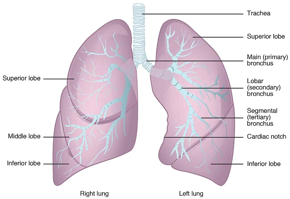
Apex pulmonis は鎖骨より2〜3cm 上へ出る。Basis pulmonis は横隔膜に乗る。
3面:facies costalis(肋骨面)・facies mediastinalis(縦隔面・hilum pulmonis がある)・facies diaphragmatica。2縁:鋭い margo anterior と margo inferior。
| 右肺 | 左肺 | |
|---|---|---|
| 葉 | 3(上・中・下) | 2(上・下) |
| 裂 | Fissura obliqua + horizontalis | Fissura obliqua のみ |
| 特徴 | 大きく短く幅広い | Incisura cardiaca と lingula |
Radix pulmonis(肺根)に含まれるのは主気管支・肺動脈・2本の肺静脈・気管支動静脈・神経・リンパ管。
肺門の中の配置に左右差がある。右は上から:気管支 → 動脈 → 静脈(eparterial bronchus が動脈の上を越える)、左は上から:動脈 → 気管支 → 静脈。「右は気管支が上、左は動脈が上」——胸部CTで左右を判定する所見として実際に使う。
左肺の心切痕と舌区は、心臓が左に寄っているためにできた「よけ」である。左肺が2葉なのも、上葉の一部が舌区として下へ伸びるのも、同じ原因の別の表れ。左右差を1つの理由でまとめられる。
肺には2系統の血管がある。機能血管=肺動脈・肺静脈(ガス交換のため、静脈血を運ぶ)と、栄養血管=気管支動脈(胸大動脈から、動脈血で肺実質を養う)。肺塞栓で必ずしも梗塞にならないのは、気管支動脈からの二重支配があるからである。
水平裂があるのは右だけで、正面の胸部X線で右の第4肋骨の高さに一本の線として見える。この線の位置がずれていれば無気肺や胸水を疑う。
右3葉・左2葉。左が2葉なのは心臓が左に寄っているためで、同じ理由で左肺には心切痕と小舌(lingula)がある。
肺区域(S1〜S10)は気管支と肺動脈を単位とするので、区域切除ができる。肺癌の手術も結核の病巣も区域で語る。
シルエットサイン — X 線で心臓や横隔膜の輪郭が消えていれば、その構造と接する肺に病変がある。右心縁が消えれば右中葉、左心縁なら舌区、横隔膜なら下葉。正面像1枚で病変の前後が読めるのは、この解剖の応用である。
肺尖は鎖骨より2〜3cm 上へ出る。だから鎖骨上窩の穿刺や中心静脈カテーテル挿入で気胸を起こしうる。肺尖部の腫瘍(Pancoast 腫瘍)は腕神経叢(C8・Th1)と交感神経幹を侵し、上肢の疼痛・筋萎縮と Horner 症候群を起こす。
出典:イラスト解剖学 誌面490頁「肺のX線像:シルエットサイン」
English version
Core
The apex pulmonis rises 2–3 cm above the clavicle; the basis pulmonis rests on the diaphragm.
Three surfaces: facies costalis, facies mediastinalis (bearing the hilum pulmonis) and facies diaphragmatica. Two borders: the sharp margo anterior and margo inferior.
| Right lung | Left lung | |
|---|---|---|
| Lobes | 3 (upper, middle, lower) | 2 (upper, lower) |
| Fissures | Oblique + horizontal | Oblique only |
| Features | Larger, shorter, broader | Cardiac notch and lingula |
The radix pulmonis contains the main bronchus, pulmonary artery, two pulmonary veins, bronchial vessels, nerves and lymphatics.
Going further
The arrangement within the hilum differs between the sides. On the right, from above: bronchus → artery → vein (the eparterial bronchus passes above the artery); on the left, from above: artery → bronchus → vein. "Bronchus uppermost on the right, artery uppermost on the left" — used in practice on CT to decide which side is which.
The cardiac notch and lingula of the left lung are the "give" made because the heart lies to the left. That the left lung has two lobes, and that part of its upper lobe extends down as the lingula, are two expressions of the same cause. The left–right differences can be gathered under one reason.
The lung has two vascular systems. Functional = the pulmonary arteries and veins (for gas exchange, carrying venous blood); nutritive = the bronchial arteries (from the thoracic aorta, arterial blood nourishing the parenchyma). That pulmonary embolism does not necessarily infarct is because of this dual supply.
Only the right has a horizontal fissure, seen on a frontal chest film as a line at the level of the fourth rib. If that line is displaced, suspect collapse or effusion.
In the clinic
Three lobes on the right, two on the left. The left has two because the heart lies to the left, and for the same reason the left lung has a cardiac notch and a lingula.
The bronchopulmonary segments (S1–S10) take the bronchus and pulmonary artery as their unit, so segmentectomy is possible. Both lung cancer surgery and tuberculous lesions are described by segment.
The silhouette sign — if the outline of the heart or diaphragm is lost on the film, the lesion lies in the lung touching that structure. Loss of the right heart border = the right middle lobe; the left border = the lingula; the diaphragm = a lower lobe. That a single frontal film can read the front-to-back position of a lesion is an application of this anatomy.
The apex rises 2–3 cm above the clavicle, so puncture of the supraclavicular fossa or central venous cannulation can cause a pneumothorax. A tumour of the apex (Pancoast) involves the brachial plexus (C8, Th1) and the sympathetic trunk, giving pain and wasting in the arm with Horner’s syndrome.
肺区域 — 右10・左8〜10の名前Segmenta bronchopulmonalia
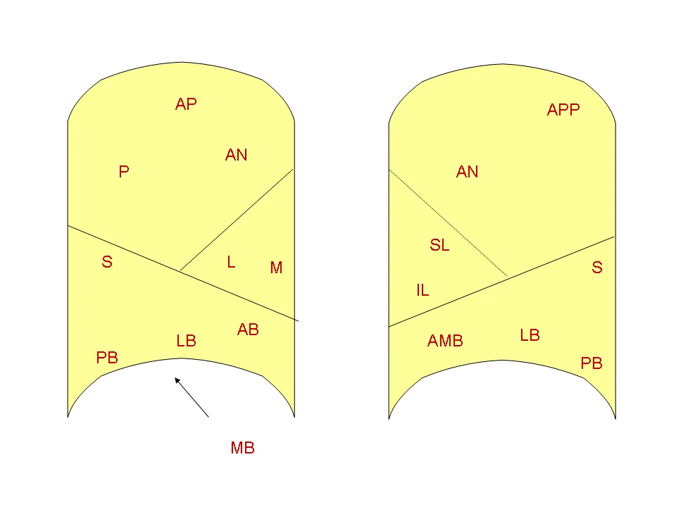
肺区域は1本の区域気管支と、それに伴う肺動脈の枝が支配する、ピラミッド形の単位である。頂点が肺門、底が肺の表面を向く。気管支と動脈は区域の中心を、静脈は区域と区域の境を走る。番号(S1〜S10)は日本の臨床でそのまま使う。
| 葉 | 右肺 | 左肺 |
|---|---|---|
| 上葉 | S1 apicale(肺尖区) S2 posterius(後上葉区) S3 anterius(前上葉区) | S1+2 apicoposterius(肺尖後区) S3 anterius(前上葉区) |
| 中葉 / 舌区 | S4 laterale(外側中葉区) S5 mediale(内側中葉区) | S4 lingulare superius(上舌区) S5 lingulare inferius(下舌区) |
| 下葉 | S6 superius(上-下葉区) S7 basale mediale(内側肺底区) S8 basale anterius(前肺底区) S9 basale laterale(外側肺底区) S10 basale posterius(後肺底区) | S6 superius (S7 は小さいか、S8 と一緒になる) S8 basale anterius S9 basale laterale S10 basale posterius |
左肺の区域の数は数え方で変わる。S1 と S2 を別に数え S7 も置けば10(解剖学用語の数え方)、S1+2 を1つにして S7 を残せば9(ヨーロッパの教科書に多い)、S1+2 を1つにして S7 を数えなければ8(日本の臨床で多い)。右は10で動かない。
覚え方は「上葉は頂・後・前の3つ、中葉は外・内の2つ(左では舌区の上・下に置き換わる)、下葉は上に1つ(S6)と底に4つ(内・前・外・後)」。左では心臓が場所を取るので内側肺底区(S7)が育ちにくく、上葉の S1 と S2 も1本の気管支から出る——左右差はこの2か所だけである。
区域の境を静脈が走ることが、区域を外科の単位にしている。区域気管支と動脈を根元で処理し、区域間静脈に沿って切り離せば、隣の区域を傷つけずにその区域だけを取れる(区域切除)。
病変の場所は区域で言う。結核の再燃は S1・S2・S6 に多い——上葉の後ろ寄りと下葉の上部は換気に比べて血流が少なく酸素分圧が高いので、結核菌が育ちやすい。仰臥位での誤嚥は背中側の S2・S6 に、坐位・立位では下葉の肺底区(とくに右)に落ちる。小さな肺癌では、肺葉を丸ごと取らずに区域切除を選ぶことがある。
English version
Core
A bronchopulmonary segment is a pyramid-shaped unit supplied by one segmental bronchus and its branch of the pulmonary artery. Its apex points to the hilum and its base to the lung surface. Bronchus and artery run in the centre of the segment; veins run in the borders between segments. The numbers S1–S10 are used directly in Japanese clinical practice.
| Lobe | Right lung | Left lung |
|---|---|---|
| Upper | S1 apicale S2 posterius S3 anterius | S1+2 apicoposterius S3 anterius |
| Middle / lingula | S4 laterale S5 mediale | S4 lingulare superius S5 lingulare inferius |
| Lower | S6 superius S7 basale mediale S8 basale anterius S9 basale laterale S10 basale posterius | S6 superius (S7 small or joined with S8) S8 basale anterius S9 basale laterale S10 basale posterius |
The number of left segments depends on how you count: 10 if S1 and S2 are separate and S7 is kept (Terminologia Anatomica), 9 if S1+2 is one and S7 is kept (common in European textbooks), 8 if S1+2 is one and S7 is not counted (common in Japanese clinical use). The right lung always has 10.
Going further
A way to remember: upper lobe = apical, posterior, anterior; middle lobe = lateral, medial (on the left replaced by superior and inferior lingular); lower lobe = one on top (S6) and four at the base (medial, anterior, lateral, posterior). On the left the heart takes space, so the medial basal segment (S7) hardly develops, and S1 and S2 come off one bronchus. These are the only two left–right differences.
Because veins run in the borders, the segment is a surgical unit. Divide the segmental bronchus and artery at their roots and separate along the intersegmental veins, and one segment can be removed without harming its neighbours (segmentectomy).
In the clinic
Lesions are located by segment. Reactivated tuberculosis favours S1, S2 and S6: the back of the upper lobe and the top of the lower lobe have little blood flow compared with ventilation, so oxygen pressure is high and the bacteria grow well. Aspiration lying on the back goes to the dorsal S2 and S6; sitting or standing, to the basal segments of the lower lobe (especially on the right). For a small lung cancer, a segmentectomy may be chosen instead of removing the whole lobe.
胸膜と縦隔Pleura et mediastinum
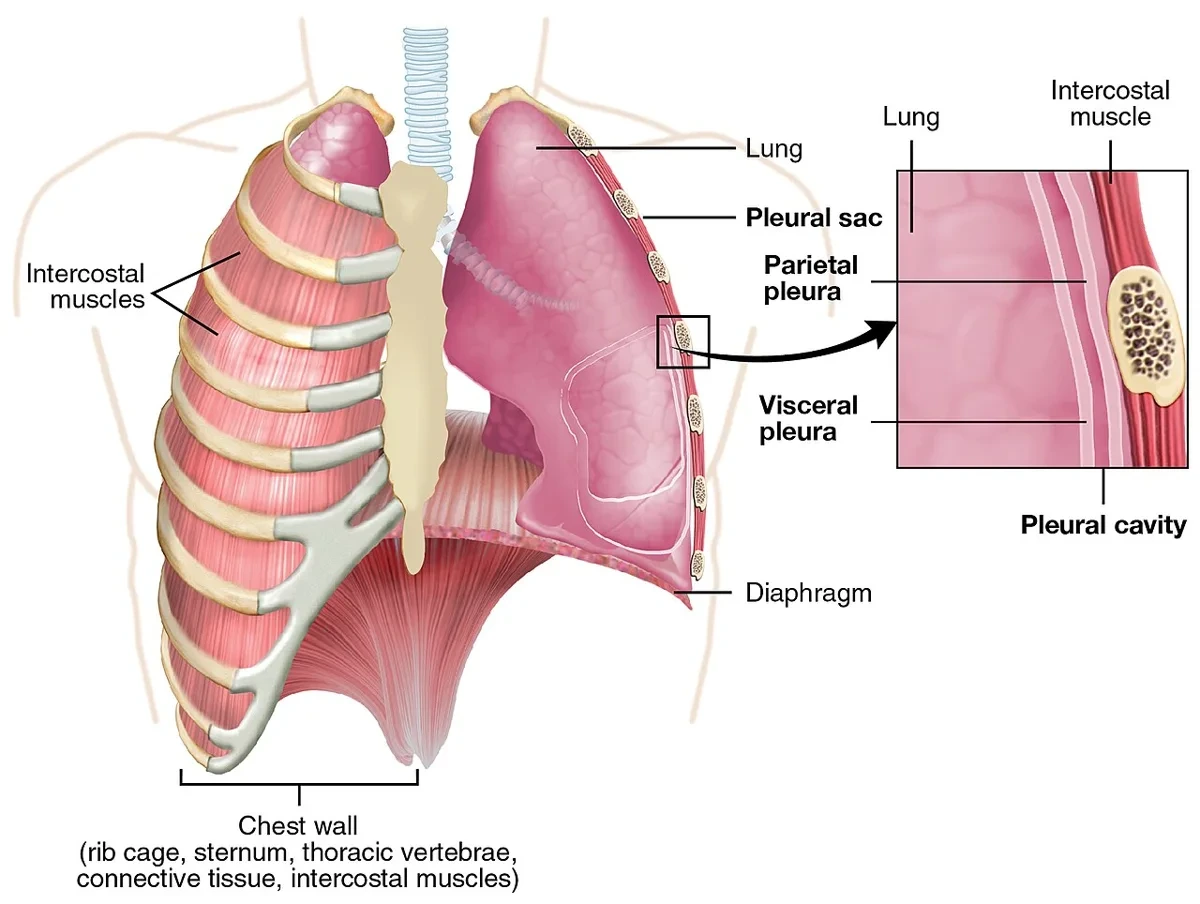
Pleura visceralis (pulmonalis) は肺の表面に密着し、裂の中まで入り込む。肺門で折り返して pleura parietalis となり胸壁を裏打ちする。壁側胸膜は場所で pars costalis・diaphragmatica・mediastinalis・cupula pleurae に分けられる。
あいだが cavitas pleuralis(少量の漿液・陰圧)。肺が入り込まない袋の隅が recessus で、最大が recessus costodiaphragmaticus(肋骨横隔洞)。
境界の高さ(鎖骨中線 / 中腋窩線 / 肩甲線):肺の下縁=6 / 8 / 10 肋骨、胸膜の下縁=8 / 10 / 12 肋骨。常に胸膜が2肋骨ぶん下——この差が肋骨横隔洞である。
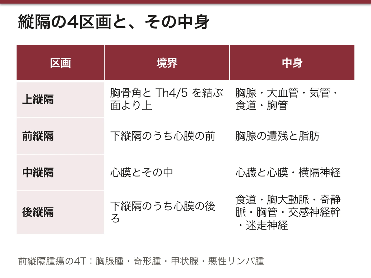
Mediastinum:胸骨角と Th4/5 を結ぶ面で上縦隔と下縦隔に分け、下縦隔を心膜で前・中・後に分ける。
肺と胸膜の下縁が2肋骨ずれることが、胸腔穿刺の高さを決める。胸水は肋骨横隔洞に溜まるので、肩甲線で第7〜9肋間を穿刺する。肋骨の上縁に沿って針を入れる——肋間動静脈と神経が肋骨の下縁の溝を走るためである。
胸膜腔が陰圧であることが気胸を説明する。肺は自らの弾性で縮もうとし、胸壁は広がろうとする。この綱引きが陰圧を生む。壁に穴が開けば陰圧が消え、肺は縮む。緊張性気胸は穴が弁になって吸気ごとに空気が溜まり、縦隔が健側へ押される——縦隔が可動な仕切りにすぎないからである。
縦隔の中身は区画で言えるようにする。上縦隔=胸腺・大血管・気管・食道・胸管。前縦隔=胸腺の遺残と脂肪。中縦隔=心臓と心膜・横隔神経。後縦隔=食道・胸大動脈・奇静脈・胸管・交感神経幹・迷走神経。前縦隔腫瘍の「4T」(Thymoma・Teratoma・Thyroid・Terrible lymphoma)は、そこに何があるかから直接出る。
壁側胸膜と臓側胸膜で痛みが違う。壁側は肋間神経と横隔神経が支配し鋭く痛む(胸膜痛・横隔膜の中心部は肩へ放散)。臓側は自律神経で痛みを感じない。「胸膜炎が痛いのは壁側だけ」。
肋骨横隔洞は立位で胸腔の最下点である。胸水はここに溜まるので、胸腔穿刺は中腋窩線上の第7〜9肋間で行う。肋骨の上縁に沿って刺す——肋間動静脈と神経は肋骨の下縁の溝を走るからである。
臓側胸膜には痛覚が無い。痛みを感じるのは壁側胸膜で、肋間神経(体性)が支配する部分はその場所の鋭い痛み、横隔胸膜の中央部(横隔神経 C3〜C5)は肩へ放散する。肺の中の病変が痛くないのに、胸膜に達すると痛むのはこのため。
緊張性気胸 — 縦隔が健側へ偏位し、静脈還流が絶たれる。画像を待たずに脱気する。
縦隔の区分は臨床では上・前・中・後で語る。前縦隔腫瘍の4T(胸腺腫 Thymoma・奇形腫 Teratoma・甲状腺 Thyroid・悪性リンパ腫 Terrible lymphoma)。
出典:イラスト解剖学 誌面311頁「臨床における縦隔の区分」
English version
Core
The pleura visceralis is applied to the surface of the lung and runs into the fissures; at the hilum it reflects to become the pleura parietalis lining the chest wall. The parietal pleura is described by region as pars costalis, diaphragmatica, mediastinalis and cupula pleurae. Between them is the cavitas pleuralis (a little serous fluid, at negative pressure).
The corners of the sac that the lung does not enter are the recessus, the largest being the recessus costodiaphragmaticus.
Levels of the borders (midclavicular / midaxillary / scapular lines): the lower border of the lung = ribs 6 / 8 / 10; of the pleura = ribs 8 / 10 / 12. The pleura is always two ribs lower — and that difference is the costodiaphragmatic recess.
Mediastinum: divided by the plane from the sternal angle to Th4/5 into superior and inferior, and the inferior divided by the pericardium into anterior, middle and posterior.
Going further
That the borders of lung and pleura differ by two ribs decides the level of a pleural tap. Effusion collects in the costodiaphragmatic recess, so the needle is passed in the seventh to ninth space at the scapular line, along the upper border of a rib — because the intercostal vessels and nerve run in the groove on the lower border.
That the pleural cavity is at negative pressure explains pneumothorax. The lung tends to recoil by its own elasticity and the chest wall to spring outwards; the tug-of-war creates the negative pressure. Make a hole in the wall and the negative pressure is gone and the lung collapses. In a tension pneumothorax the hole acts as a valve, air accumulates with each breath and the mediastinum is pushed to the sound side — because the mediastinum is only a mobile partition.
Be able to give the contents by compartment. Superior = thymus, great vessels, trachea, oesophagus, thoracic duct. Anterior = the remnant of the thymus and fat. Middle = heart and pericardium, phrenic nerves. Posterior = oesophagus, thoracic aorta, azygos vein, thoracic duct, sympathetic trunk, vagus. The four Ts of anterior mediastinal masses (thymoma, teratoma, thyroid, terrible lymphoma) come straight out of what is there.
Pain differs between parietal and visceral pleura. The parietal is supplied by the intercostal and phrenic nerves and hurts sharply (pleuritic pain; the central diaphragm refers to the shoulder). The visceral is autonomic and feels no pain. "Pleurisy hurts only from the parietal side."
In the clinic
The costodiaphragmatic recess is the lowest point of the pleural cavity when standing. Effusion collects there, so thoracocentesis is performed in the seventh to ninth space at the midaxillary line, along the upper border of a rib — the intercostal vessels and nerve running in the groove on the lower border.
The visceral pleura has no pain sensation. What hurts is the parietal pleura: the part supplied by the intercostal nerves (somatic) gives sharp pain at that site; the central part of the diaphragmatic pleura (phrenic, C3–C5) refers to the shoulder. This is why a lesion within the lung is painless until it reaches the pleura.
Tension pneumothorax — the mediastinum shifts to the sound side and venous return is cut off. Decompress without waiting for imaging.
Clinically the mediastinum is described as superior, anterior, middle and posterior. The four Ts of anterior mediastinal masses (thymoma, teratoma, thyroid, terrible lymphoma).
胸膜の血管と神経 — 壁側は胸壁の、臓側は肺のVasa et nervi pleurae
胸膜の血管と神経も、腹膜と同じく「壁側は体壁のもの、臓側は臓器のもの」で決まる。
| 壁側胸膜 | 臓側胸膜 | |
|---|---|---|
| 動脈 | 胸壁の動脈:肋間動脈(胸大動脈の枝)と内胸動脈の枝(前肋間枝・心膜横隔動脈・筋横隔動脈)、上横隔動脈 | 気管支動脈(と、肺循環の毛細血管) |
| 静脈 | 奇静脈系・内胸静脈(体循環)へ | 肺静脈・気管支静脈へ |
| リンパ | 胸骨傍・肋間・横隔・後縦隔リンパ節へ | 胸膜下のリンパ網から肺門のリンパ節へ |
| 神経 | 体性:肋骨部と横隔膜の周辺部=肋間神経、縦隔部と横隔膜の中央部=横隔神経 | 自律神経(肺神経叢)。痛覚は無い |
規則の理由は発生である。壁側胸膜は体壁の一部として、臓側胸膜は肺の表面として作られるので、血管も神経もそれぞれの持ち主から受ける。
胸水は主に壁側胸膜から出て、壁側胸膜から吸収される。壁側の毛細血管は体循環なので圧が高く液を押し出し、壁側胸膜(とくに横隔膜と縦隔の面)にあるリンパ管への小孔が液を回収する。どちらかのバランスが崩れると胸水がたまる。
胸腔穿刺やドレーン挿入の局所麻酔は、壁側胸膜までしっかり効かせる。痛むのは皮膚と壁側胸膜で、肺の表面(臓側胸膜)は刺されても痛まない。
縦隔のリンパ節に癌が広がると、壁側胸膜からのリンパの出口がふさがれて胸水が引かなくなる。心不全の胸水は毛細血管の圧が上がって出る液が増えたもの、癌性胸水は出口がふさがったもの——同じ「胸水」でも、解剖のどちら側の問題かで性質が変わる。
English version
Core
Like the peritoneum, the pleura follows the rule “parietal layer takes the body wall’s supply, visceral layer takes the organ’s supply”.
| Parietal pleura | Visceral pleura | |
|---|---|---|
| Arteries | Chest-wall arteries: intercostal arteries (from the thoracic aorta) and branches of the internal thoracic artery (anterior intercostal, pericardiacophrenic, musculophrenic), superior phrenic arteries | Bronchial arteries (and capillaries of the pulmonary circulation) |
| Veins | To the azygos system and internal thoracic veins (systemic) | To pulmonary and bronchial veins |
| Lymph | To parasternal, intercostal, diaphragmatic and posterior mediastinal nodes | Through the subpleural network to the hilar nodes |
| Nerves | Somatic: costal part and edge of the diaphragm = intercostal nerves; mediastinal part and centre of the diaphragm = phrenic nerve | Autonomic (pulmonary plexus). No pain sensation |
Going further
The reason is development. The parietal pleura forms as part of the body wall, the visceral pleura as the surface of the lung, so each gets its vessels and nerves from its owner.
Pleural fluid is mainly produced by and absorbed through the parietal pleura. Parietal capillaries are systemic and have high pressure, so they push fluid out; small openings into lymphatics in the parietal pleura (especially over the diaphragm and mediastinum) take it back. If either side of this balance fails, fluid collects.
In the clinic
For thoracentesis or a chest drain, local anaesthetic must reach down to the parietal pleura. The skin and parietal pleura hurt; the lung surface (visceral pleura) does not.
When cancer spreads to mediastinal lymph nodes, the lymph exit from the parietal pleura is blocked and the effusion does not clear. Effusion in heart failure is more fluid pushed out by raised capillary pressure; malignant effusion is a blocked exit. Both are “pleural effusion”, but the problem sits on a different side of the anatomy.
鼻腔Cavitas nasi
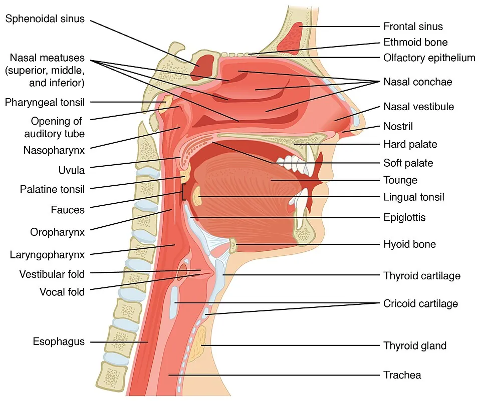
鼻中隔で左右に分かれ、前は鼻孔、後ろは後鼻孔で咽頭鼻部に開く。
嗅部は上鼻甲介と対する天井のわずかな範囲だけで、残りは呼吸部。吸気を温め・湿らせ・濾す。
English version
Core
Divided in two by the nasal septum, opening in front at the nares and behind through the choanae into the nasopharynx.
The olfactory region is only a small area of the roof opposite the superior concha; the rest is respiratory. It warms, moistens and filters the inspired air.
上鼻甲介Concha nasalis superior
篩骨の一部。上鼻道を作り、そこに後篩骨洞が開く。すぐ上の蝶篩陥凹に蝶形骨洞が開く。
English version
Core
Part of the ethmoid bone. It forms the superior meatus, into which the posterior ethmoidal cells open. The sphenoidal sinus opens into the sphenoethmoidal recess just above it.
中鼻甲介Concha nasalis media
これも篩骨の一部。中鼻道を作る。
English version
Core
Also part of the ethmoid bone, forming the middle meatus.
下鼻甲介Concha nasalis inferior
3つのうちここだけが独立した骨(他の2つは篩骨の突起)。下鼻道を作る。
「上と中は篩骨、下だけ独立骨」——標本で必ず確かめておく。
English version
Core
Of the three, this alone is an independent bone (the other two are processes of the ethmoid). It forms the inferior meatus.
"Superior and middle from the ethmoid, inferior alone a separate bone" — always confirm it on the specimen.
中鼻道Meatus nasi medius
中鼻甲介の下の通路。半月裂孔という溝があり、そこへ前頭洞・上顎洞・前および中篩骨洞が開く。
副鼻腔の大半がここへ開くので、中鼻道は副鼻腔炎の要になる。
English version
Core
The passage beneath the middle concha. It contains the semilunar hiatus, into which open the frontal sinus, the maxillary sinus and the anterior and middle ethmoidal cells.
Most of the sinuses open here, so the middle meatus is the crux of sinusitis.
下鼻道Meatus nasi inferior
下鼻甲介の下の通路。ここに開くのは鼻涙管だけ。
「泣くと鼻が出る」のはこの管のため。開口部の覚え方は「上=後篩骨、中=ほぼ全部、下=鼻涙管」。
English version
Core
The passage beneath the inferior concha. The only thing opening here is the nasolacrimal duct — which is why crying makes the nose run.
Learn the openings as: superior = posterior ethmoidal, middle = almost everything, inferior = the nasolacrimal duct.
後鼻孔Choanae
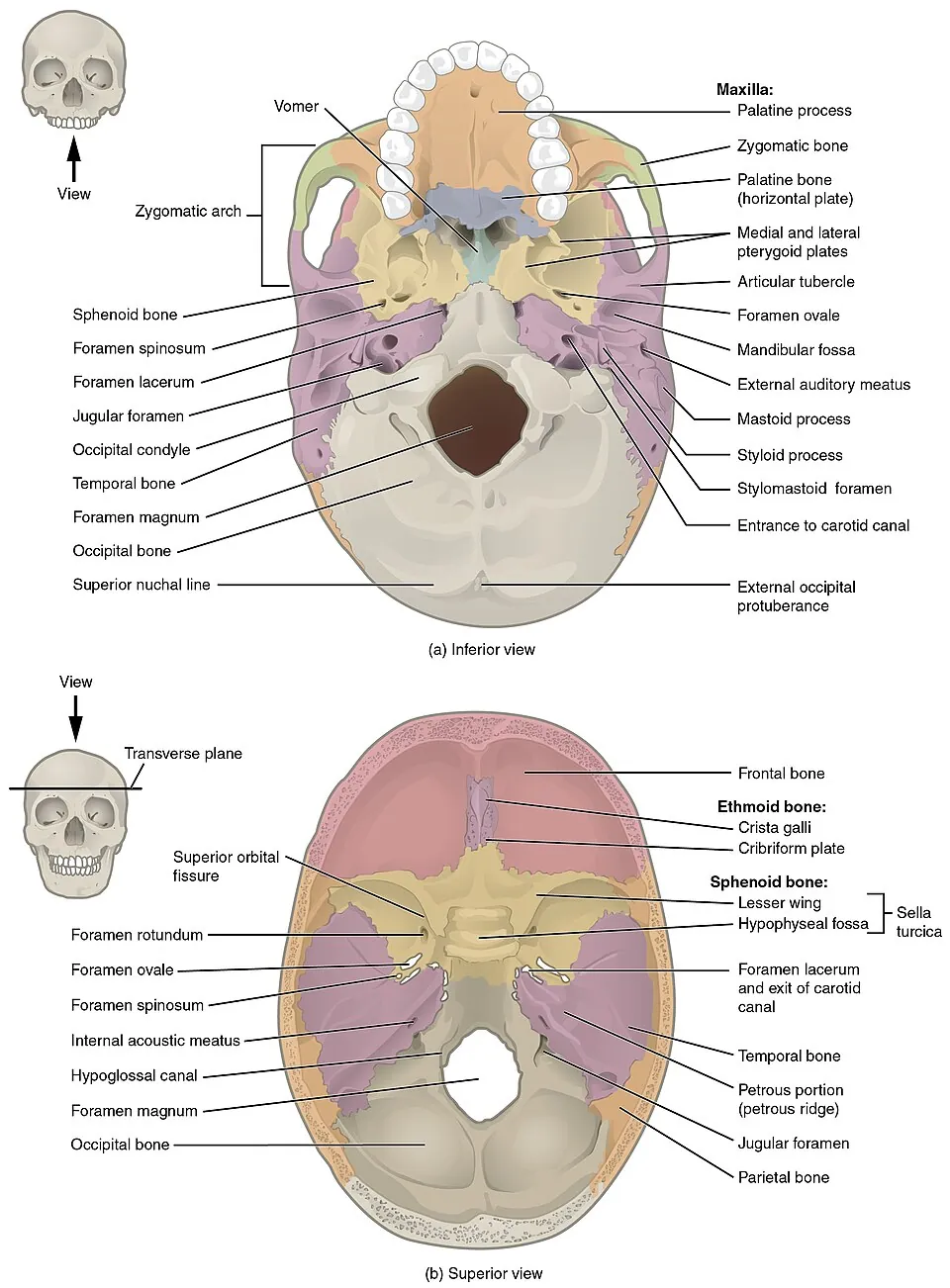
鼻腔の後方の出口で、咽頭鼻部へ開く一対の孔。鋤骨と蝶形骨、口蓋骨が縁を作る。
English version
Core
The pair of posterior openings of the nasal cavity into the nasopharynx, bounded by the vomer, the sphenoid and the palatine bones.
前頭洞・蝶形骨洞・上顎洞Sinus frontalis, sphenoidalis, maxillaris
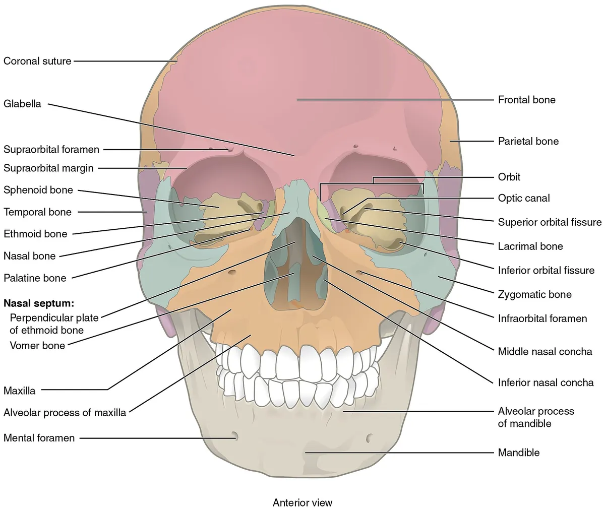
副鼻腔は4対(前頭・篩骨・蝶形骨・上顎)。いずれも鼻腔の粘膜が骨の中へ入り込んだもの。
上顎洞が最大で、開口部が洞の天井近くにある——底より高い位置なので立位では排出されにくく、炎症が長引く。上壁は眼窩底、下壁は上顎歯根に接するので、歯性上顎洞炎が起こる。
English version
Core
There are four pairs of paranasal sinus (frontal, ethmoidal, sphenoidal, maxillary), each the nasal mucosa grown into a bone.
The maxillary is the largest, and its opening lies near its roof — higher than its floor, so it drains poorly in the standing position and inflammation is protracted. Its roof is the floor of the orbit and its floor lies against the roots of the upper teeth, hence odontogenic maxillary sinusitis.
甲状軟骨Cartilago thyroidea
喉頭で最大の軟骨。2枚の板が正中で合わさり、上端の突出が喉頭隆起(のどぼとけ)。硝子軟骨。
男性で角が鋭く前へ出る。上角・下角を持ち、下角が輪状軟骨と関節する。
English version
Core
The largest cartilage of the larynx, two laminae meeting in the midline with the laryngeal prominence at the upper end (the Adam’s apple). Hyaline. In the male the angle is sharper and projects further forward. It has superior and inferior horns, the inferior articulating with the cricoid.
輪状軟骨Cartilago cricoidea
気道で唯一、全周が完全な輪になっている軟骨。前が細い弓、後ろが広い板の印章指輪型。C6 の高さ。
完全な輪だからこそ外から押せば食道を閉じられる(輪状軟骨圧迫)。小児では気道の最狭部でもある。
English version
Core
The only cartilage in the airway that is a complete ring all the way round, shaped like a signet ring with a narrow arch in front and a broad lamina behind, at the level of C6.
Being a complete ring it can be pressed from outside to occlude the oesophagus (cricoid pressure). It is also the narrowest part of the child’s airway.
披裂軟骨Cartilago arytenoidea
輪状軟骨板の上に乗る左右一対の三角錐。前の声帯突起に声帯靭帯が、外側の筋突起に筋が付く。
声帯を動かすのはこの軟骨の回転と滑走。喉頭の運動を説明するときの中心。
English version
Core
The pair of triangular pyramids riding on the lamina of the cricoid. The vocal ligament attaches to the vocal process in front, the muscles to the muscular process laterally.
What moves the vocal folds is the rotation and gliding of this cartilage — the centre of any account of laryngeal movement.
喉頭蓋Epiglottis
甲状軟骨の裏から立ち上がる木の葉形の弾性軟骨。
嚥下のとき喉頭が引き上げられ、その動きで受動的に倒れて喉頭口を塞ぐ——自分で倒れるのではない点が問われる。
English version
Core
The leaf-shaped elastic cartilage rising from behind the thyroid cartilage.
On swallowing, the larynx is drawn up and that movement tips it passively over the laryngeal inlet — that it does not fall of its own accord is what is asked.
輪状気管靭帯Ligamentum cricotracheale
輪状軟骨の下縁と第1気管軟骨を結ぶ膜。喉頭と気管の継ぎ目。
English version
Core
The membrane joining the lower border of the cricoid to the first tracheal cartilage — the seam between larynx and trachea.
輪状甲状関節Articulatio cricothyroidea
甲状軟骨の下角と輪状軟骨の関節。横軸まわりの回転で甲状軟骨が前へ傾く。
この傾きが声帯を引き伸ばして高い声を出す。動かすのは輪状甲状筋。
English version
Core
The joint between the inferior horn of the thyroid cartilage and the cricoid. Rotation about a transverse axis tilts the thyroid cartilage forwards. That tilt stretches the vocal folds and raises the pitch. It is moved by the cricothyroid muscle.
輪状披裂関節Articulatio cricoarytenoidea
輪状軟骨板の上面と披裂軟骨の底の関節。回転と滑走で声帯突起が内外へ動く。
声門を開け閉めするのはこの関節。輪状甲状関節(張力)と役割を分けて覚える。
English version
Core
The joint between the upper surface of the lamina of the cricoid and the base of the arytenoid. Rotation and gliding move the vocal process in and out. This is the joint that opens and closes the glottis.
Learn its role separately from the cricothyroid joint (tension).
喉頭口Aditus laryngis
咽頭喉頭部から喉頭へ入る口。前を喉頭蓋、両側を披裂喉頭蓋ひだ、後ろを披裂間切痕が囲む。
English version
Core
The opening from the laryngopharynx into the larynx, bounded in front by the epiglottis, at the sides by the aryepiglottic folds and behind by the interarytenoid notch.
披裂喉頭蓋ひだPlica aryepiglottica
喉頭蓋の外側縁と披裂軟骨を結ぶひだ。喉頭口の側壁を作り、外側が梨状陥凹になる。
English version
Core
The fold joining the lateral margin of the epiglottis to the arytenoid cartilage. It forms the lateral wall of the laryngeal inlet, with the piriform recess outside it.
喉頭前庭Vestibulum laryngis
喉頭口から前庭ひだまでの、喉頭腔の上の部分。
喉頭腔は前庭 → 喉頭室 → 声門下腔の3段。2組のひだが3つに区切ると押さえる。
English version
Core
The upper part of the laryngeal cavity, from the inlet to the vestibular folds.
The laryngeal cavity is three storeys — vestibule → ventricle → infraglottic space. Fix that two pairs of folds divide it into three.
前庭ひだ・前庭裂Plica vestibularis, rima vestibuli
上の一対のひだ(仮声帯)と、その間の裂。発声には関与しないが、息こらえのとき閉じる。
English version
Core
The upper pair of folds (the false cords) and the fissure between them. They take no part in phonation but close in the Valsalva manoeuvre.
声門・喉頭室Glottis, ventriculus laryngis
声門=声帯ひだ+声帯突起+声門裂の総称、つまり発声装置そのもの。
喉頭室は前庭ひだと声帯ひだの間の外側へ入り込む窪み。声帯の潤滑に働く。
English version
Core
The glottis = the vocal folds, the vocal processes and the rima glottidis together — that is, the vocal apparatus itself. The ventricle is the recess passing laterally between vestibular and vocal folds, which lubricates the cords.
声帯ひだ・声門裂Plica vocalis, rima vocalis
下の一対のひだと、その間の裂。気道でいちばん狭い場所。
白く見えるのは血管に乏しい重層扁平上皮のため。前庭ひだ(上・赤い)との対比が標本での見分けどころ。
English version
Core
The lower pair of folds and the fissure between them. The narrowest part of the airway. They look white because the stratified squamous epithelium is poorly vascularised.
The contrast with the vestibular folds (above, red) is how they are told apart on the specimen.
声帯靭帯Ligamentum vocale
甲状軟骨の内面と披裂軟骨の声帯突起を結ぶ弾性線維の索。声帯ひだの芯になる。
English version
Core
The band of elastic fibres joining the inner surface of the thyroid cartilage to the vocal process of the arytenoid. It is the core of the vocal fold.
声帯筋・輪状甲状筋Musculus vocalis, musculus cricothyroideus
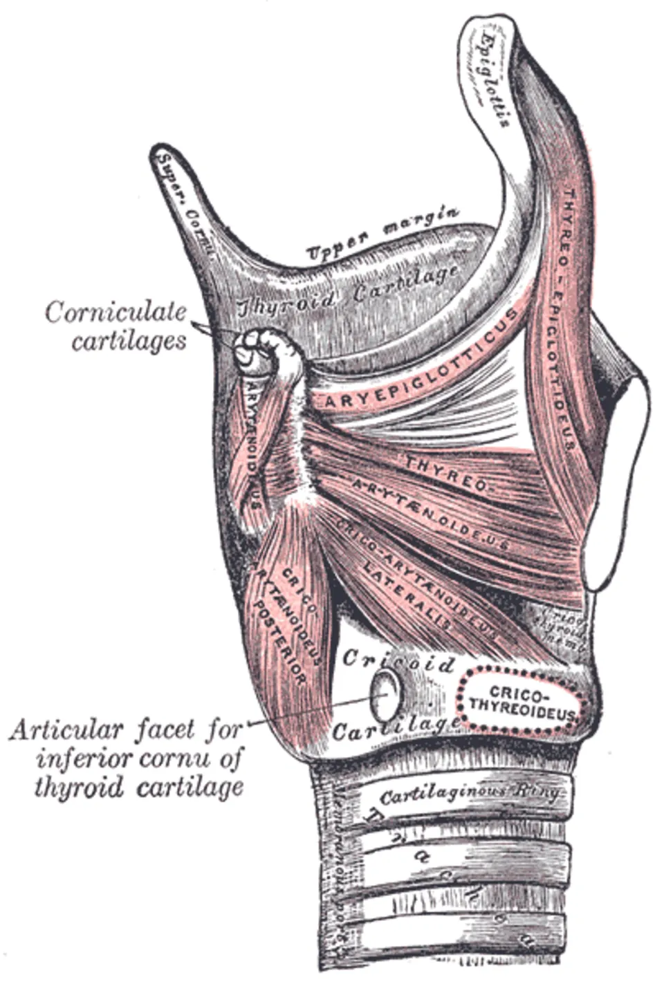
声帯筋は声帯靭帯に沿う内喉頭筋で、細かく張りを調節する。
輪状甲状筋だけが上喉頭神経外枝の支配で、他の内喉頭筋はすべて反回神経。この1つだけの例外が最頻出。甲状軟骨を前傾させて声帯を伸ばし、声を高くする。
English version
Core
The vocalis is the intrinsic muscle alongside the vocal ligament, making fine adjustments of tension.
The cricothyroid alone is supplied by the external branch of the superior laryngeal nerve; every other intrinsic muscle is supplied by the recurrent laryngeal. This single exception is the commonest of all questions. It tilts the thyroid cartilage forward, stretches the cords and raises the pitch.
気管分岐部Bifurcatio tracheae
気管が左右の主気管支に分かれる場所。Th4/5(胸骨角の高さ)。
内腔から見ると正中に鋭い稜(気管竜骨 carina)があり、気管支鏡の基準点になる。
English version
Core
Where the trachea divides into the two main bronchi, at Th4/5 (the level of the sternal angle).
Seen from within there is a sharp median ridge, the carina, the reference point at bronchoscopy.
気管軟骨Cartilagines tracheales
16〜20個のC字(馬蹄形)の硝子軟骨。後ろは開いていて膜性壁になり、そこに平滑筋と食道が接する。
後ろが開いているから食塊が通れる——輪状軟骨が完全輪であるのと対にして覚える。
English version
Core
Sixteen to twenty C-shaped (horseshoe) hyaline cartilages. They are open behind, where the membranous wall lies with the smooth muscle and the oesophagus against it.
Being open behind is what lets a bolus pass — learn it as a pair with the cricoid, which is a complete ring.
右主気管支・左主気管支Bronchus principalis dexter et sinister
右は太く・短く・垂直に近い(約25度)。左は細く・長く・水平に近い(約45度)。
誤嚥した異物が右へ落ちるのはこのため。標本でも説明でも必ず押さえる非対称。
English version
Core
The right is wide, short and nearly vertical (about 25°); the left is narrow, long and nearer horizontal (about 45°). This is why an aspirated foreign body goes to the right.
An asymmetry always to be known, on the specimen and in explanation.
肺尖Apex pulmonis
肺の上端で、鎖骨の内側1/3の上方2〜3cmまで、胸郭の外へ出る。
ここに接する構造の関係で、肺尖部の腫瘍(Pancoast 腫瘍)は腕神経叢と交感神経幹を巻き込む。鎖骨上窩の穿刺で気胸を起こしやすいのもこのため。
English version
Core
The upper end of the lung, projecting 2–3 cm above the medial third of the clavicle and out of the thoracic cage.
Because of what it touches there, an apical tumour (Pancoast) involves the brachial plexus and the sympathetic trunk. It is also why puncture of the supraclavicular fossa readily causes a pneumothorax.
肺底Basis pulmonis
横隔膜に乗る下面。凹面で、横隔膜のドームに合う。
English version
Core
The lower surface resting on the diaphragm, concave to fit its dome.
肺の肋骨面(椎骨部)Facies costalis pulmonis
肋骨と肋間筋に接する広い凸面。後方で椎体に接する部分を椎骨部という。
English version
Core
The broad convex surface against the ribs and intercostal muscles. The part touching the vertebral bodies behind is the vertebral part.
肺の縦隔面Facies mediastinalis pulmonis
縦隔に向かう内側面。肺門があり、心臓による心圧痕が刻まれる。
心圧痕は左で深い(心が左に寄るため)ので、左右の肺を見分ける手掛かりになる。
English version
Core
The medial surface facing the mediastinum, bearing the hilum and the cardiac impression.
The cardiac impression is deeper on the left (the heart lying to the left), and so is a clue for telling the lungs apart.
肺の横隔面Facies diaphragmatica pulmonis
肺底そのもの。横隔膜を介して、右は肝臓、左は胃と脾に接する。
English version
Core
The base of the lung itself, related through the diaphragm to the liver on the right and to the stomach and spleen on the left.
肺の前縁Margo anterior pulmonis
肋骨面と縦隔面の境の鋭い縁。左肺ではここに心切痕があり、その下に小舌が垂れる。
心切痕があるのが左——左右を見分ける最も確実な目印。
English version
Core
The sharp border between the costal and mediastinal surfaces. In the left lung it carries the cardiac notch, with the lingula hanging below it.
The presence of the cardiac notch means the left — the surest way of telling the sides apart.
肺の下縁Margo inferior pulmonis
肋骨面と横隔面の境の鋭い縁。肋骨横隔洞へ入り込み、深吸気で下がる。
English version
Core
The sharp border between the costal and diaphragmatic surfaces. It enters the costodiaphragmatic recess and descends on deep inspiration.
肺門Hilum pulmonis
縦隔面のくぼみで、気管支・血管・神経・リンパ管が出入りする入口そのもの。
English version
Core
The depression on the mediastinal surface — the entrance itself, through which bronchus, vessels, nerves and lymphatics pass.
肺根Radix pulmonis
肺門を出入りする構造の束(肺門は場所、肺根は中身)。胸膜に包まれる。
並びは右が「上から 気管支・肺動脈・肺静脈」、左が「上から 肺動脈・気管支・肺静脈」。この左右差が定番の設問。
English version
Core
The bundle of structures entering and leaving the hilum (the hilum is the place, the root is the contents), enclosed in pleura.
The arrangement is, on the right from above, bronchus, pulmonary artery, pulmonary vein; on the left from above, pulmonary artery, bronchus, pulmonary vein. This asymmetry is a standard question.
肺の上葉Lobus superior pulmonis
左右ともにある。左の上葉には小舌が付き、これが右の中葉に相当する。
English version
Core
Present on both sides. The left upper lobe carries the lingula, which corresponds to the right middle lobe.
右肺の中葉Lobus medius pulmonis dextri
右にしかない葉。斜裂と水平裂に挟まれる。
右は2本の裂で3葉、左は1本の裂で2葉。左が2葉なのは心臓が場所を取るため。
English version
Core
A lobe found only on the right, between the oblique and horizontal fissures.
Two fissures and three lobes on the right; one fissure and two lobes on the left. The left has two because the heart takes the room.
肺の下葉Lobus inferior pulmonis
斜裂より後下方の葉。背側の大部分を占めるので、背中から聴くとほぼ下葉の音になる。
English version
Core
The lobe behind and below the oblique fissure. It occupies most of the dorsal aspect, so listening from the back gives almost entirely lower lobe sounds.
気管支樹と肺胞 — 壁はどう変わるかArbor alveolaris
気道を下るにつれて壁が段階的に変わる——これが答えの骨格。
①軟骨:気管はC字 → 主気管支もC字 → 肺内気管支では不規則な板状になり → 細気管支で消える。
②平滑筋:軟骨が減るほど相対的に増える。細気管支では全周を取り巻く——だから気管支喘息は細気管支で閉塞する。
③上皮:多列線毛円柱 → 単層線毛円柱 → 終末細気管支で単層立方(線毛が消え、Clara 細胞が出る) → 肺胞で単層扁平。
④杯細胞は細気管支で消える。
呼吸細気管支から先が「呼吸部」(壁に肺胞が付き、ガス交換が始まる)。肺胞管 → 肺胞嚢 → 肺胞。
血液空気関門は①I型肺胞上皮 ②融合した基底膜 ③毛細血管内皮の3層、約0.5μm。
II型肺胞上皮がサーファクタントを出して表面張力を下げる。早産児でこれが足りないのが新生児呼吸窮迫症候群。肺胞孔(Kohn 孔)が肺胞間の側副換気を担う。
English version
Core
The wall changes in stages as the airway descends — this is the skeleton of the answer.
- Cartilage: C-shaped in the trachea → C-shaped in the main bronchi → irregular plates in the intrapulmonary bronchi → gone at the bronchioles.
- Smooth muscle: relatively more as the cartilage decreases; in the bronchioles it encircles the whole wall — which is why asthma obstructs at the bronchiole.
- Epithelium: pseudostratified ciliated columnar → simple ciliated columnar → simple cuboidal at the terminal bronchiole (cilia lost, club cells appear) → simple squamous in the alveolus.
- Goblet cells disappear at the bronchioles.
From the respiratory bronchiole onwards is the respiratory portion (alveoli on the wall, gas exchange beginning): alveolar duct → alveolar sac → alveolus.
The blood–air barrier is three layers — type I alveolar epithelium, the fused basal laminae and the capillary endothelium — about 0.5 µm.
Going further
Type II alveolar cells secrete surfactant and lower the surface tension. Insufficiency of it in the preterm infant is neonatal respiratory distress syndrome.
The alveolar pores (of Kohn) provide collateral ventilation between alveoli.
肺の体表投影 — 打診と聴診の基準Topographia pulmonum
肺尖——鎖骨の内側1/3の上方2〜3cm。
下界(安静呼気時)——鎖骨中線で第6肋骨、中腋窩線で第8肋骨、肩甲線で第10肋骨、脊柱線で Th11 棘突起。この4点が定番。深吸気で2〜3cm 下がる。
胸膜の下界は肺より2肋骨低い(鎖骨中線で第8、中腋窩線で第10、肩甲線で第12)。その差が肋骨横隔洞で、胸水はここに溜まる。
葉の境——斜裂は Th3 棘突起から前下方へ走り、鎖骨中線で第6肋骨に達する。水平裂(右のみ)は第4肋骨の高さで前方を水平に走る。
だから中葉は前面からしか聴こえず、背中から聴くのはほぼ下葉——聴診の基本がここから出る。
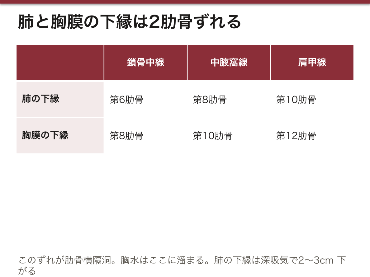
胸腔穿刺は肋骨の上縁に沿って刺す(肋間神経・血管は肋骨の下縁を走るため)。通常は中腋窩線の第7〜9肋間。
English version
Core
Apex — 2–3 cm above the medial third of the clavicle.
Lower border (in quiet expiration) — rib 6 at the midclavicular line, rib 8 at the midaxillary, rib 10 at the scapular, the spine of Th11 at the vertebral line. These four points are standard. It descends 2–3 cm on deep inspiration.
The lower border of the pleura is two ribs lower than the lung (rib 8 at the midclavicular, 10 at the midaxillary, 12 at the scapular). That difference is the costodiaphragmatic recess, where effusion collects.
The fissures — the oblique runs from the spine of Th3 down and forwards to reach rib 6 at the midclavicular line; the horizontal (right only) runs forward horizontally at the level of rib 4. So the middle lobe can be heard only from the front, and what is heard from the back is almost entirely lower lobe — the basis of auscultation comes from this.
Going further
A pleural tap is made along the upper border of a rib (the intercostal nerve and vessels running on the lower border), usually in the seventh to ninth space at the midaxillary line.
発生 — 前腸の前壁から出る肺芽と、咽頭弓Diverticulum respiratorium
第4週、前腸の前壁に溝(喉頭気管溝)ができ、そこから呼吸憩室(肺芽)が前下方へ伸びる。両側から気管食道ひだが寄って気管食道中隔となり、前の喉頭気管管と後ろの食道を分ける。だから気道の上皮と腺は内胚葉、軟骨・平滑筋・結合組織は周りの臓側中胚葉からできる。
肺芽は第5週に左右の気管支芽に分かれ、右は3本・左は2本の葉気管支を出し、続いて区域気管支(右10・左8〜9)に分かれる。肺の成熟は4期で進む。
| 時期 | おおよその週 | 起きること |
|---|---|---|
| 偽腺期 | 5〜16週 | 気管支が終末細気管支まで枝分かれする。まだガス交換はできない |
| 細管期 | 16〜26週 | 呼吸細気管支ができ、毛細血管が近づく |
| 終末嚢期 | 26週〜出生 | 薄い終末嚢(原始肺胞)が増え、II 型細胞がサーファクタントを作り始める |
| 肺胞期 | 胎生末期〜小児期 | 本当の肺胞が増え続ける(生後も数年続く) |
喉頭の軟骨と内喉頭筋は、第4・第6咽頭弓の間葉からできる。
咽頭弓の筋と神経の対応は、喉頭の神経の謎を解く。
| 咽頭弓 | 主な筋 | 神経 |
|---|---|---|
| 第1(下顎弓) | 咀嚼筋・顎二腹筋前腹・顎舌骨筋・鼓膜張筋・口蓋帆張筋 | 三叉神経(V3) |
| 第2(舌骨弓) | 表情筋・アブミ骨筋・茎突舌骨筋・顎二腹筋後腹 | 顔面神経 |
| 第3 | 茎突咽頭筋 | 舌咽神経 |
| 第4 | 輪状甲状筋・口蓋帆挙筋・咽頭収縮筋 | 迷走神経(上喉頭神経) |
| 第6 | 輪状甲状筋以外の内喉頭筋 | 迷走神経(反回神経) |
内喉頭筋で輪状甲状筋だけ支配が違うのは、それだけが第4弓の出身だからである。反回神経の左右差も弓の動脈で決まる。左は第6弓動脈の名残(動脈管索)の下をくぐるので大動脈弓を回り、右は第6弓動脈の遠位部が消えるので、1つ上の第4弓動脈(右鎖骨下動脈)を回る。
食道閉鎖と気管食道瘻は気管食道中隔がうまく分かれないことで起こる(およそ3,000〜4,500出生に1人)。最も多いのは、食道の上部が盲端で終わり、下部が気管とつながる型である。胎児は羊水を飲み込めないので羊水過多になり、生まれると泡状の唾液があふれ、初めての授乳でむせる。胃管が食道の途中で反転して止まることで疑う。
早産児では終末嚢期が十分に進んでおらず、サーファクタントが足りずに肺胞がつぶれる(新生児呼吸窮迫症候群)。早産が予測されるとき母体にステロイドを投与するのは、II 型細胞の成熟を早めるためである。
English version
Core
In week 4 a groove (laryngotracheal groove) forms in the front wall of the foregut, and the respiratory diverticulum (lung bud) grows forward and downward from it. Tracheoesophageal folds meet from both sides as the tracheoesophageal septum, separating the laryngotracheal tube in front from the oesophagus behind. So the epithelium and glands of the airway come from endoderm, while cartilage, smooth muscle and connective tissue come from the surrounding splanchnic mesoderm.
In week 5 the lung bud splits into right and left bronchial buds; the right gives three lobar bronchi and the left two, then segmental bronchi (10 on the right, 8–9 on the left). The lung matures in four periods.
| Period | Approx. weeks | What happens |
|---|---|---|
| Pseudoglandular | 5–16 | Bronchi branch down to terminal bronchioles; no gas exchange yet |
| Canalicular | 16–26 | Respiratory bronchioles form; capillaries come close |
| Terminal sac | 26 to birth | Thin terminal sacs (primitive alveoli) increase; type II cells begin to make surfactant |
| Alveolar | Late fetal life to childhood | True alveoli keep increasing for several years after birth |
The laryngeal cartilages and intrinsic muscles come from the mesenchyme of the 4th and 6th pharyngeal arches.
Going further
The muscles and nerves of the pharyngeal arches solve the puzzle of the laryngeal nerves.
| Arch | Main muscles | Nerve |
|---|---|---|
| 1st (mandibular) | Muscles of mastication, anterior belly of digastric, mylohyoid, tensor tympani, tensor veli palatini | Trigeminal (V3) |
| 2nd (hyoid) | Muscles of facial expression, stapedius, stylohyoid, posterior belly of digastric | Facial |
| 3rd | Stylopharyngeus | Glossopharyngeal |
| 4th | Cricothyroid, levator veli palatini, pharyngeal constrictors | Vagus (superior laryngeal) |
| 6th | All intrinsic laryngeal muscles except cricothyroid | Vagus (recurrent laryngeal) |
Only the cricothyroid has a different nerve among the laryngeal muscles because it alone comes from the 4th arch. The left–right difference of the recurrent nerve is set by the arch arteries: on the left it hooks under the remnant of the 6th arch artery (ligamentum arteriosum) and so around the aortic arch; on the right the distal 6th arch artery disappears, so the nerve hooks around the next one up, the 4th arch artery (right subclavian).
In the clinic
Oesophageal atresia with tracheoesophageal fistula results from faulty division by the tracheoesophageal septum (about 1 in 3,000–4,500 births). The most common type has a blind upper oesophagus and a lower oesophagus joined to the trachea. The fetus cannot swallow amniotic fluid, causing polyhydramnios; after birth, frothy saliva overflows and the baby chokes at the first feed. A nasogastric tube that turns back and stops in the oesophagus suggests it.
In preterm babies the terminal sac period is not complete, surfactant is lacking and alveoli collapse (neonatal respiratory distress syndrome). Steroids are given to the mother when preterm birth is expected to speed the maturation of type II cells.
まとめ
| 鼻腔と副鼻腔 | Nares(外鼻孔)から choanae(後鼻孔)まで。 |
|---|---|
| 外鼻 — 上が骨、下が軟骨 | 鼻腔の前の壁を作るのが外鼻である。部位は上から radix nasi(鼻根・両眼の間)・dorsum nasi(鼻背)・apex nasi(鼻尖)。 |
| 喉頭 — 軟骨・関節・壁 | C3〜C6の高さ、舌骨の下、気管の上。3つの単一軟骨+3対の対性軟骨。 |
| 喉頭の壁 — 4層と、2枚の線維弾性膜 | 喉頭の壁は内から4層である。層中身① 粘膜大部分は多列線毛上皮。 |
| 喉頭腔 — 3階に分かれる | 上の入口が aditus laryngis(前は喉頭蓋、側方は plica aryepiglottica、後ろは披裂間切痕)。 |
| 気管と気管支樹 — 右へ落ちる理由 | Trachea:C6(輪状軟骨下縁)から Th4〜5(胸骨角の高さ)まで約10〜12cm。 |
| 肺 — 面・縁・葉・門 | Apex pulmonis は鎖骨より2〜3cm 上へ出る。 |
| 肺区域 — 右10・左8〜10の名前 | 肺区域は1本の区域気管支と、それに伴う肺動脈の枝が支配する、ピラミッド形の単位である。 |
| 胸膜と縦隔 | Pleura visceralis (pulmonalis) は肺の表面に密着し、裂の中まで入り込む。 |
| 胸膜の血管と神経 — 壁側は胸壁の、臓側は肺の | 胸膜の血管と神経も、腹膜と同じく「壁側は体壁のもの、臓側は臓器のもの」で決まる。 |
| 鼻腔 | 鼻中隔で左右に分かれ、前は鼻孔、後ろは後鼻孔で咽頭鼻部に開く。 |
| 上鼻甲介 | 篩骨の一部。上鼻道を作り、そこに後篩骨洞が開く。 |
| 中鼻甲介 | これも篩骨の一部。中鼻道を作る。 |
| 下鼻甲介 | 3つのうちここだけが独立した骨(他の2つは篩骨の突起)。 |
| 中鼻道 | 中鼻甲介の下の通路。半月裂孔という溝があり、そこへ前頭洞・上顎洞・前および中篩骨洞が開く。 |
| 下鼻道 | 下鼻甲介の下の通路。ここに開くのは鼻涙管だけ。 |
| 後鼻孔 | 鼻腔の後方の出口で、咽頭鼻部へ開く一対の孔。 |
| 前頭洞・蝶形骨洞・上顎洞 | 副鼻腔は4対(前頭・篩骨・蝶形骨・上顎)。 |
| 甲状軟骨 | 喉頭で最大の軟骨。2枚の板が正中で合わさり、上端の突出が喉頭隆起(のどぼとけ)。 |
| 輪状軟骨 | 気道で唯一、全周が完全な輪になっている軟骨。 |
| 披裂軟骨 | 輪状軟骨板の上に乗る左右一対の三角錐。前の声帯突起に声帯靭帯が、外側の筋突起に筋が付く。 |
| 喉頭蓋 | 甲状軟骨の裏から立ち上がる木の葉形の弾性軟骨。 |
| 輪状気管靭帯 | 輪状軟骨の下縁と第1気管軟骨を結ぶ膜。喉頭と気管の継ぎ目。 |
| 輪状甲状関節 | 甲状軟骨の下角と輪状軟骨の関節。横軸まわりの回転で甲状軟骨が前へ傾く。 |
| 輪状披裂関節 | 輪状軟骨板の上面と披裂軟骨の底の関節。回転と滑走で声帯突起が内外へ動く。 |
| 喉頭口 | 咽頭喉頭部から喉頭へ入る口。前を喉頭蓋、両側を披裂喉頭蓋ひだ、後ろを披裂間切痕が囲む。 |
| 披裂喉頭蓋ひだ | 喉頭蓋の外側縁と披裂軟骨を結ぶひだ。喉頭口の側壁を作り、外側が梨状陥凹になる。 |
| 喉頭前庭 | 喉頭口から前庭ひだまでの、喉頭腔の上の部分。 |
| 前庭ひだ・前庭裂 | 上の一対のひだ(仮声帯)と、その間の裂。 |
| 声門・喉頭室 | 声門=声帯ひだ+声帯突起+声門裂の総称、つまり発声装置そのもの。 |
| 声帯ひだ・声門裂 | 下の一対のひだと、その間の裂。気道でいちばん狭い場所。 |
| 声帯靭帯 | 甲状軟骨の内面と披裂軟骨の声帯突起を結ぶ弾性線維の索。 |
| 声帯筋・輪状甲状筋 | 声帯筋は声帯靭帯に沿う内喉頭筋で、細かく張りを調節する。 |
| 気管分岐部 | 気管が左右の主気管支に分かれる場所。Th4/5(胸骨角の高さ)。 |
| 気管軟骨 | 16〜20個のC字(馬蹄形)の硝子軟骨。 |
| 右主気管支・左主気管支 | 右は太く・短く・垂直に近い(約25度)。 |
| 肺尖 | 肺の上端で、鎖骨の内側1/3の上方2〜3cmまで、胸郭の外へ出る。 |
| 肺底 | 横隔膜に乗る下面。凹面で、横隔膜のドームに合う。 |
| 肺の肋骨面(椎骨部) | 肋骨と肋間筋に接する広い凸面。後方で椎体に接する部分を椎骨部という。 |
| 肺の縦隔面 | 縦隔に向かう内側面。肺門があり、心臓による心圧痕が刻まれる。 |
| 肺の横隔面 | 肺底そのもの。横隔膜を介して、右は肝臓、左は胃と脾に接する。 |
| 肺の前縁 | 肋骨面と縦隔面の境の鋭い縁。左肺ではここに心切痕があり、その下に小舌が垂れる。 |
| 肺の下縁 | 肋骨面と横隔面の境の鋭い縁。肋骨横隔洞へ入り込み、深吸気で下がる。 |
| 肺門 | 縦隔面のくぼみで、気管支・血管・神経・リンパ管が出入りする入口そのもの。 |
| 肺根 | 肺門を出入りする構造の束(肺門は場所、肺根は中身)。 |
| 肺の上葉 | 左右ともにある。左の上葉には小舌が付き、これが右の中葉に相当する。 |
| 右肺の中葉 | 右にしかない葉。斜裂と水平裂に挟まれる。 |
| 肺の下葉 | 斜裂より後下方の葉。背側の大部分を占めるので、背中から聴くとほぼ下葉の音になる。 |
| 気管支樹と肺胞 — 壁はどう変わるか | 気道を下るにつれて壁が段階的に変わる——これが答えの骨格。 |
| 肺の体表投影 — 打診と聴診の基準 | 肺尖——鎖骨の内側1/3の上方2〜3cm。 |
| 発生 — 前腸の前壁から出る肺芽と、咽頭弓 | 第4週、前腸の前壁に溝(喉頭気管溝)ができ、そこから呼吸憩室(肺芽)が前下方へ伸びる。 |
このノートは医学生が授業の内容を自分の言葉でまとめたもので、診療の判断に使うものではありません。誤りに気づいたらお知らせください。