心臓は「4つの部屋を、血の流れる順に1本の道として読む」のが最も間違えにくい。上大静脈 → 右房 → 三尖弁 → 右室 → 肺動脈弁 → 肺 → 左房 → 僧帽弁 → 左室 → 大動脈弁 → 全身、と指でなぞりながら構造を挙げていく。
標本では心房と心室の内面を見分けられるようにしておく。櫛状筋があれば心房、肉柱と乳頭筋があれば心室。右と左は壁の厚さ(左は右の約3倍)で決まる。
この範囲の芯は「弁の解剖学的位置と、聴診する場所が一致しない」ことである。音は血流に乗って伝わるので、聴くのは弁の真上ではなく血液が飛んでいく先。この一点を理解していないと、聴診部位はただの丸暗記になる。
- 心臓 — 外形と溝
- 右心房と右心室 — 内面の構造
- 左心房と左心室
- 心臓の弁 — 位置と聴診部位が違う
- 刺激伝導系 — 特殊心筋の道
- 心膜 — 2枚と2つの洞
- 心尖
- 胸肋面(前面)
- 横隔面(下面)
- 肺面(左側面)
- 右心耳・左心耳
- 冠状溝
- 前室間溝・後室間溝
- 上大静脈口
- 下大静脈口
- 櫛状筋
- 右房室口・左房室口
- 三尖弁
- 僧帽弁
- 腱索
- 乳頭筋
- 肉柱
- 肺動脈幹口
- 肺動脈幹
- 肺動脈弁
- 大動脈口
- 大動脈弁
- 肺静脈
- 体循環と肺循環
- 心臓の壁 — 3層
- 心臓の位置と体表投影
- 局所解剖線 — 体の表面に引く11本の縦線
- 心臓 — 外形と溝Cor
- 右心房と右心室 — 内面の構造Atrium dextrum et ventriculus dexter
- 左心房と左心室Atrium sinistrum et ventriculus sinister
- 心臓の弁 — 位置と聴診部位が違うValvae cordis
- 刺激伝導系 — 特殊心筋の道Systema conducens cordis
- 心膜 — 2枚と2つの洞Pericardium
- 心尖Apex cordis
- 胸肋面(前面)Facies sternocostalis
- 横隔面(下面)Facies diaphragmatica
- 肺面(左側面)Facies pulmonalis
- 右心耳・左心耳Auricula dextra, sinistra
- 冠状溝Sulcus coronarius
- 前室間溝・後室間溝Sulcus interventricularis anterior, posterior
- 上大静脈口Ostium venae cavae superioris
- 下大静脈口Ostium venae cavae inferioris
- 櫛状筋Musculi pectinati
- 右房室口・左房室口Ostium atrioventriculare dextrum, sinistrum
- 三尖弁Valva tricuspidalis
- 僧帽弁Valva atrioventricularis sinistra sive mitralis
- 腱索Chordae tendineae
- 乳頭筋Musculi papillares
- 肉柱Trabeculae carneae
- 肺動脈幹口Ostium trunci pulmonalis
- 肺動脈幹Truncus pulmonalis
- 肺動脈弁Valva trunci pulmonalis
- 大動脈口Ostium aortae
- 大動脈弁Valva aortae
- 肺静脈Venae pulmonales
- 体循環と肺循環Circulatio sanguinis
- 心臓の壁 — 3層Paries cordis
- 心臓の位置と体表投影Topographia cordis
- 局所解剖線 — 体の表面に引く11本の縦線Lineae topographicae
- まとめ
心臓 — 外形と溝Cor
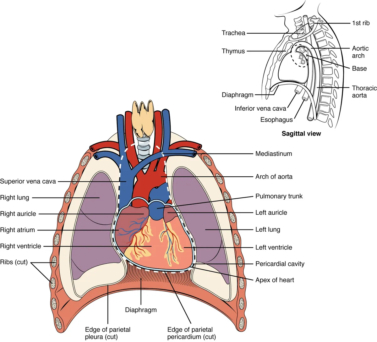
縦隔中部、心膜の中。底 basis cordis は右上後方(大血管が出入りする)、apex cordis は左下前方(第5肋間・左鎖骨中線で拍動を触れる)。
3面:facies sternocostalis(前・胸骨と肋軟骨に接する。主に右室)・facies diaphragmatica(下・主に左室)・facies pulmonalis(左・左室)。Margo dexter は鋭い縁で右房が作る。
溝が部屋の境界を外から示す:sulcus coronarius(冠状溝・房室境界を輪状に回る)・sulcus interventricularis anterior / posterior(心室中隔の位置)。溝の中を冠状動脈と心臓静脈が走る。
左右の心房から前上方へ突き出す耳状の部分が auricula dextra / sinistra。
「胸骨の後ろにあるのは主に右室」という事実が2つのことを説明する。①胸骨圧迫で圧迫されるのは右室。②心タンポナーデや外傷で最初に傷むのも右室。心臓は左にあるという直感と、実際に前面を占める部屋が違う。
左心耳は血栓ができる場所である。櫛状筋が発達した袋小路で、心房細動になると血流がよどむ。心原性脳塞栓の血栓の約90%がここで生じる——だから左心耳閉鎖術という治療が成立する。
冠状動脈は溝を走る。A. coronaria sinistra は前室間枝(前室間溝)と回旋枝(左の冠状溝)に、a. coronaria dextra は右の冠状溝を回って後室間枝になる。後室間枝をどちらが出すかで優位性が決まる(右優位が約85%)。心筋梗塞の責任血管を心電図の誘導から言えるのは、この走行が決まっているからである。
心尖拍動は第5肋間・左鎖骨中線で触れる。外下方へずれていれば左室拡大——触診だけで心拡大が分かる。
心嚢穿刺は剣状突起の左下から左肩へ向けて刺す(剣状突起下アプローチ)。心タンポナーデは Beck 3徴(血圧低下・頸静脈怒張・心音減弱)と奇脈で診断し、心嚢液を抜けば劇的に改善する。
冠状溝と前室間溝には冠動脈が走る。前室間溝=左前下行枝(LAD)で、ここが詰まると前壁中隔梗塞——V1〜V4 の ST 上昇になる。溝の名前と心電図の誘導が一対一で対応する。
胸部 X 線の心胸郭比(CTR)は正常50%以下(立位 PA 像)。AP 像では心臓が拡大して写るので、この基準を当てはめてはいけない。
出典:イラスト解剖学 誌面316頁「心タンポナーデ・心嚢穿刺」
English version
Core
In the middle mediastinum, within the pericardium. The basis cordis faces up, back and to the right (where the great vessels enter and leave); the apex cordis down, forward and to the left (its beat palpable in the fifth intercostal space at the midclavicular line).
Three surfaces: facies sternocostalis (anterior, against sternum and costal cartilages; chiefly right ventricle), facies diaphragmatica (inferior; chiefly left ventricle), facies pulmonalis (left; left ventricle). The sharp margo dexter is formed by the right atrium.
The sulci show the boundaries of the chambers from outside: sulcus coronarius (encircling the atrioventricular junction) and the anterior and posterior sulci interventriculares (the position of the interventricular septum). The coronary arteries and cardiac veins run in the sulci.
The ear-shaped projections from the atria are the auricula dextra / sinistra.
Going further
The fact that what lies behind the sternum is chiefly the right ventricle explains two things. ① What is compressed in chest compressions is the right ventricle. ② What is injured first in tamponade and in trauma is also the right ventricle. The intuition that the heart is on the left and the chamber that actually occupies the front are two different things.
The left auricle is where thrombus forms. It is a cul-de-sac with well-developed pectinate muscles, and in atrial fibrillation the flow stagnates there. About 90 per cent of the thrombi of cardiogenic cerebral embolism arise there — which is why left atrial appendage occlusion exists as a treatment.
The coronary arteries run in the sulci. The a. coronaria sinistra divides into the anterior interventricular branch (in the anterior interventricular sulcus) and the circumflex (in the left coronary sulcus); the a. coronaria dextra runs round the right coronary sulcus and becomes the posterior interventricular branch. Which artery gives the posterior interventricular branch defines dominance (right-dominant in about 85 per cent). That the culprit vessel of an infarct can be named from the ECG leads is because this course is fixed.
In the clinic
The apex beat is felt in the fifth intercostal space at the left midclavicular line. Displaced down and out, it means left ventricular enlargement — cardiac enlargement detected by palpation alone.
Pericardiocentesis is performed from below and left of the xiphoid, aiming at the left shoulder (the subxiphoid approach). Cardiac tamponade is diagnosed by Beck’s triad (hypotension, distended neck veins, muffled heart sounds) with pulsus paradoxus, and improves dramatically when the fluid is drawn off.
The coronary arteries run in the coronary and anterior interventricular sulci. The anterior interventricular sulcus = the left anterior descending artery, occlusion of which gives an anteroseptal infarct — ST elevation in V1–V4. The name of the sulcus and the ECG leads correspond one to one.
The cardiothoracic ratio on the chest film is normally 50 per cent or less (erect PA). On an AP film the heart is magnified, and this criterion must not be applied.
右心房と右心室 — 内面の構造Atrium dextrum et ventriculus dexter
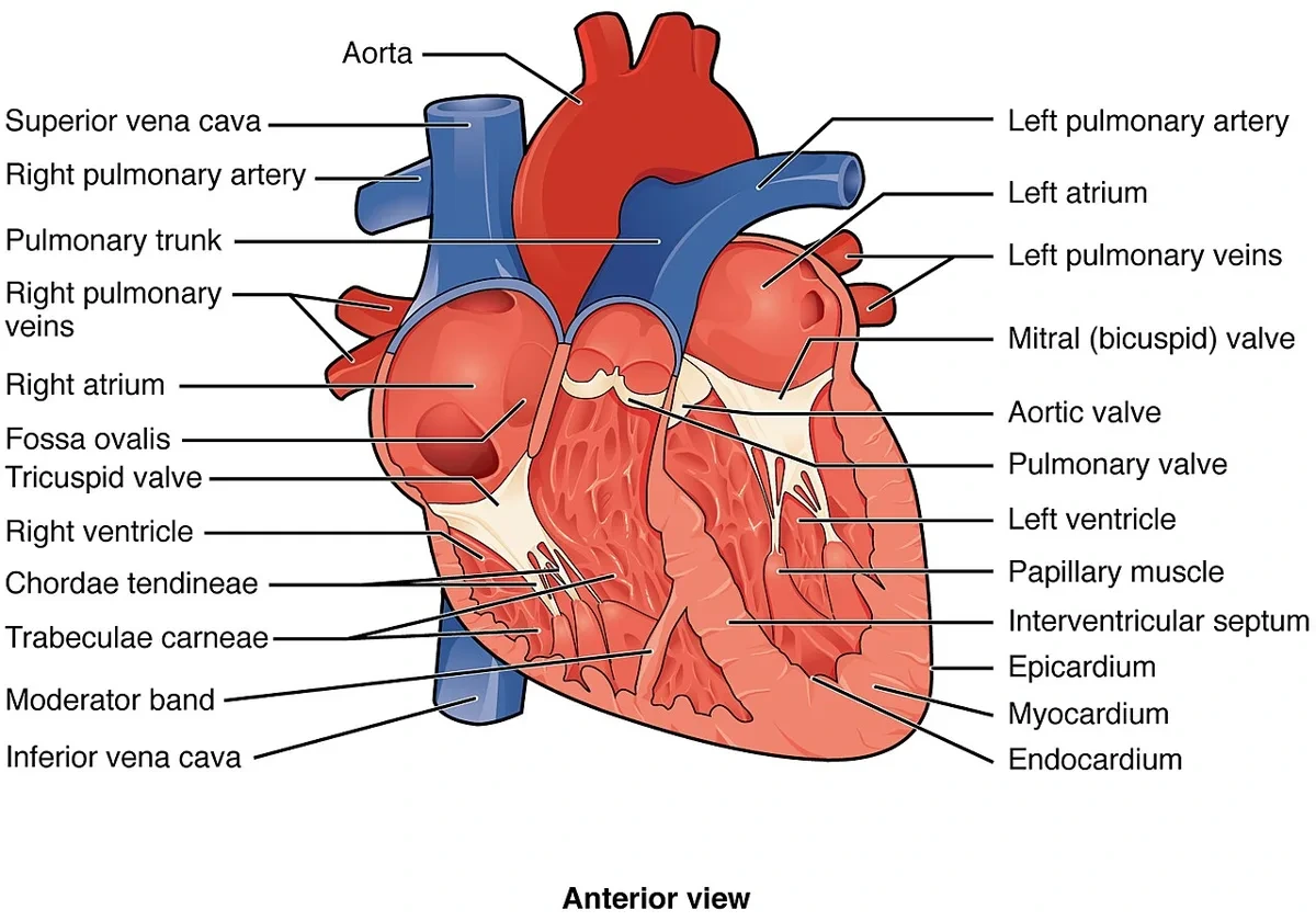
右心房に入るもの3つ:ostium venae cavae superioris・ostium venae cavae inferioris(Eustachi 弁を伴う)・ostium sinus coronarii(Thebesius 弁)。
内面は2領域に分かれる。後方の平滑な sinus venarum cavarum(静脈洞由来)と、前方の mm. pectinati(櫛状筋)のある粗い部分(原始心房由来)。境界の稜が crista terminalis(外面では sulcus terminalis)。
心房中隔に楕円の窪み fossa ovalis、その上前縁の隆起が limbus fossae ovalis。
右心室:入口は valva tricuspidalis(三尖弁)、出口は valva trunci pulmonalis。内面に trabeculae carneae(肉柱)と3群の mm. papillares があり、chordae tendineae で弁尖を引く。流出路の平滑な部分が conus arteriosus (infundibulum)。
Crista terminalis の上端に洞房結節がある。上大静脈と右心房の接合部で、外面の sulcus terminalis の上端にあたる。「解剖学的な稜の上端=ペースメーカー」という対応は、電気生理の検査やアブレーションでそのまま使われる。
Fossa ovalis は卵円孔の跡。胎児期には右房から左房へ血液が抜けており、生後に左房圧が上がって一次中隔が押しつけられ閉じる。約25%で癒合が不完全(卵円孔開存)で、通常は無症状だが奇異性塞栓の経路になりうる。
右室の壁は薄い(約3〜5mm)。肺循環の抵抗が体循環の約1/6 しかないためで、圧に対しては弱く、容量負荷には強い。だから肺塞栓のような急な圧負荷では容易に拡張・破綻する(急性肺性心)が、心房中隔欠損の慢性容量負荷には長年耐える。
Trabecula septomarginalis(moderator band)は中隔から前乳頭筋へ渡る肉柱で、右脚の一部を運ぶ。右室を同定する所見としても使われる。
卵円窩は心房中隔の弱点である。卵円孔開存(PFO)は成人の約4分の1に残り、ふだんは無症状だが、右房圧が上がったときに右→左シャントができる。下肢の静脈血栓が肺を素通りして動脈系へ回る奇異性塞栓の経路で、若年者の原因不明の脳梗塞で必ず探される。
心房中隔欠損症(ASD) — 左→右シャント。肺血流が増えて II 音の固定性分裂。長く続くと肺高血圧からシャントが逆転する(Eisenmenger 症候群)。
三尖弁は中心静脈カテーテルとペースメーカーリードの通り道である。右心系の感染性心内膜炎は静注薬物使用者・カテーテル留置例に多く、敗血症性肺塞栓を起こす。
分界稜の上端に洞房結節がある。右房の手術やカテーテル操作で傷つけると洞不全症候群になる。
English version
Core
Three things enter the right atrium: the ostium venae cavae superioris, the ostium venae cavae inferioris (with the Eustachian valve) and the ostium sinus coronarii (with the Thebesian valve).
The interior falls into two regions: the smooth posterior sinus venarum cavarum (from the sinus venosus) and the rough anterior part with the mm. pectinati (from the primitive atrium). The ridge between them is the crista terminalis (externally the sulcus terminalis).
In the interatrial septum is the oval depression, the fossa ovalis, with the limbus fossae ovalis at its upper anterior margin.
Right ventricle: the inlet is the valva tricuspidalis, the outlet the valva trunci pulmonalis. The interior carries trabeculae carneae and three groups of mm. papillares, which pull on the cusps through the chordae tendineae. The smooth outflow tract is the conus arteriosus (infundibulum).
Going further
The sinuatrial node lies at the upper end of the crista terminalis, at the junction of superior vena cava and right atrium, corresponding externally to the upper end of the sulcus terminalis. The correspondence "the upper end of an anatomical ridge = the pacemaker" is used as it stands in electrophysiological study and ablation.
The fossa ovalis is the trace of the foramen ovale. In fetal life blood passed from right atrium to left, and after birth the pressure in the left atrium rises and presses the septum primum against it to close it. In about 25 per cent the fusion is incomplete (patent foramen ovale), usually silent but a possible route for paradoxical embolism.
The wall of the right ventricle is thin (3–5 mm), the resistance of the pulmonary circulation being about a sixth of the systemic. So it is weak against pressure but tolerant of volume: it dilates and fails readily under an acute pressure load such as pulmonary embolism (acute cor pulmonale), yet tolerates the chronic volume load of an atrial septal defect for years.
The trabecula septomarginalis (moderator band) runs from the septum to the anterior papillary muscle and carries part of the right bundle branch. It is also used as a finding to identify the right ventricle.
In the clinic
The fossa ovalis is the weak point of the interatrial septum. A patent foramen ovale persists in about a quarter of adults; usually silent, it opens a right-to-left shunt when the right atrial pressure rises. It is the route of paradoxical embolism, by which a leg vein thrombus bypasses the lung into the arterial system, and is always sought in unexplained stroke in the young.
Atrial septal defect — a left-to-right shunt. Pulmonary flow increases and the second sound is fixedly split. If it continues long, pulmonary hypertension reverses the shunt (Eisenmenger’s syndrome).
The tricuspid valve is the route of central venous catheters and pacing leads. Right-sided infective endocarditis is commonest in intravenous drug users and in patients with indwelling catheters, and causes septic pulmonary emboli.
The sinuatrial node lies at the upper end of the crista terminalis. Injury during right atrial surgery or catheter manipulation gives sick sinus syndrome.
左心房と左心室Atrium sinistrum et ventriculus sinister
左心房:後壁に ostia venarum pulmonalium(4つ・左右上下)。内面はほぼ平滑で、櫛状筋は左心耳の中だけにある。中隔面に卵円窩の弁の跡(valvula foraminis ovalis)。
左心室:入口は valva mitralis (bicuspidalis)、出口は valva aortae。壁は右室の約3倍(8〜12mm)。Trabeculae carneae は右室より細かく密。2群の mm. papillares(前外側・後内側)が chordae tendineae で2尖を引く。
Septum interventriculare は大部分が厚い pars muscularis、上後部のごく一部が薄い pars membranacea。
乳頭筋の数が弁尖の数と対応する(右室3群→三尖、左室2群→二尖)。腱索は隣り合う2つの乳頭筋から1つの弁尖へ渡るので、1本の乳頭筋が断裂しても弁全体が翻らない設計になっている。
後内側乳頭筋は右冠状動脈のみから栄養される(前外側は左前下行枝と回旋枝の二重支配)。だから下壁梗塞では後内側乳頭筋が断裂しやすく、急性僧帽弁閉鎖不全を起こす。単一支配か二重支配かという解剖の差が、合併症の左右差を作る。
Pars membranacea は心室中隔欠損の好発部位である。発生の最後に、心内膜床・球稜・筋性中隔の3方向から膜が寄って閉じるため、最も閉じ損ねやすい。膜性部の上を房室束(His 束)が通るので、この部位の手術は伝導障害と隣り合わせになる。
左心耳は心房細動における血栓の巣である。心原性脳塞栓の血栓の大半はここでできるので、経食道心エコーで必ず観察し、抗凝固ができない例では左心耳閉鎖術が行われる。左房のすぐ後ろが食道だから経食道で見えるのであり、同じ理由で左房が拡大すると嚥下障害が起こる。
僧帽弁狭窄症で左房が拡大すると、左反回神経が引き伸ばされて嗄声(Ortner 症候群)。
左室壁は右室の約3倍厚い。体循環に対して拍出するためで、心筋梗塞のほとんどが左室に起こるのもここに需要が集中するからである。
心室中隔欠損症(VSD)は最も多い先天性心疾患。膜性部に好発し、小さいほど雑音は大きい。
出典:イラスト解剖学 誌面367頁「心室中隔欠損症」
English version
Core
Left atrium: the ostia venarum pulmonalium (four, upper and lower on each side) open in its posterior wall. The interior is almost smooth, the pectinate muscles being confined to the auricle. On the septal surface is the trace of the valve of the foramen ovale.
Left ventricle: the inlet is the valva mitralis (bicuspidalis), the outlet the valva aortae. The wall is about three times that of the right ventricle (8–12 mm). The trabeculae carneae are finer and denser than on the right. Two groups of mm. papillares (anterolateral and posteromedial) pull the two cusps through the chordae.
The septum interventriculare is mostly a thick pars muscularis, with a small thin pars membranacea above and behind.
Going further
The number of papillary muscles matches the number of cusps (three groups in the right ventricle → three cusps; two in the left → two). The chordae from two adjacent papillary muscles pass to one cusp, so the design is such that rupture of a single papillary muscle does not evert the whole valve.
The posteromedial papillary muscle is supplied by the right coronary artery alone (the anterolateral has a dual supply from the left anterior descending and the circumflex). So in inferior infarction the posteromedial muscle ruptures readily and causes acute mitral regurgitation. The anatomical difference between a single and a dual supply creates the asymmetry of the complication.
The pars membranacea is the site of predilection of ventricular septal defect. It closes last, as membrane converges from three directions — endocardial cushions, bulbar ridges and muscular septum — and so it fails to close most readily. The bundle of His runs over the membranous part, so surgery there sits next door to conduction block.
In the clinic
The left auricle is the nest of thrombus in atrial fibrillation. Most of the thrombi of cardiogenic stroke form there, so it is always examined on transoesophageal echocardiography, and left atrial appendage occlusion is performed where anticoagulation is impossible. It can be seen transoesophageally because the oesophagus lies directly behind the left atrium — and for the same reason an enlarged left atrium causes dysphagia.
When the left atrium enlarges in mitral stenosis, the left recurrent laryngeal nerve is stretched and hoarseness follows (Ortner’s syndrome).
The left ventricular wall is about three times the right, having to eject against the systemic circulation. That almost all myocardial infarction occurs in the left ventricle is because demand is concentrated there.
Ventricular septal defect is the commonest congenital heart disease, with a predilection for the membranous part. The smaller it is, the louder the murmur.
心臓の弁 — 位置と聴診部位が違うValvae cordis
4つの弁は同じ線維性の面(心臓の骨格)に並ぶ。
| 弁 | 尖の数 | 解剖学的な位置 | 聴診する場所 |
|---|---|---|---|
| Valva tricuspidalis | 3 | 胸骨の後ろ・第4肋間の高さ | 胸骨左縁 第4肋間 |
| Valva mitralis | 2 | 第4肋間の胸骨左縁の後ろ | 心尖部(第5肋間・左鎖骨中線) |
| Valva trunci pulmonalis | 3(半月弁) | 第3肋軟骨の高さ・胸骨左縁 | 胸骨左縁 第2肋間 |
| Valva aortae | 3(半月弁) | 第3肋間の胸骨の後ろ | 胸骨右縁 第2肋間 |
半月弁の弁尖の後ろの膨らみが sinus aortae(Valsalva 洞)で、左右の洞から冠状動脈が起こる。
なぜ位置と聴診部位がずれるのか。音は弁そのものからではなく、血液が流れていく方向へ伝わる。大動脈弁を通った血液は上行大動脈を右上へ向かうので右第2肋間、肺動脈弁の血液は左上へ向かうので左第2肋間、僧帽弁の逆流は左房を経て心尖へ抜けるので心尖部。丸暗記ではなく血流の向きで導ける。
房室弁が翻らないのは3つの仕掛けによる。①腱索が弁尖の縁を引く、②乳頭筋が収縮期に短縮して腱索の張力を保つ、③弁尖が互いに重なり合って接する。収縮期に乳頭筋が縮むのは、弁を閉じるためではなく「開かせないため」という点が理解の分かれ目である。
Valsalva 洞から冠状動脈が出ることには意味がある。収縮期には弁尖が開いて洞の入口をふさぐ形になり、拡張期に弁が閉じると洞に血液が溜まって冠状動脈へ流れる。心筋が拡張期にしか灌流されないのはこの構造による——頻脈が心筋虚血を悪化させる理由でもある。
聴診部位は弁の解剖学的な位置とずれる。血流が下流へ運ばれる方向に音が伝わるからで、「音は血の流れる先で聴く」と理解すると丸暗記が要らなくなる。
大動脈弁=第2肋間胸骨右縁、肺動脈弁=第2肋間胸骨左縁、三尖弁=第4肋間胸骨左縁、僧帽弁=第5肋間左鎖骨中線(心尖部)。
大動脈弁狭窄症 — 収縮期駆出性雑音が頸部へ放散。狭心痛・失神・心不全が出たら予後が急に悪くなる。
僧帽弁閉鎖不全症 — 全収縮期雑音が左腋窩へ放散。
弁は無血管で、線維輪に付く心内膜のひだである。血流に直接さらされ、血流が届きにくいため、感染性心内膜炎の疣贅は弁にできる。
出典:イラスト解剖学 誌面322頁「心音とその聴診」
English version
Core
The four valves lie on the same fibrous plane (the skeleton of the heart).
| Valve | Cusps | Anatomical position | Where it is auscultated |
|---|---|---|---|
| Valva tricuspidalis | 3 | Behind the sternum, at the level of the 4th space | Left sternal edge, 4th space |
| Valva mitralis | 2 | Behind the left sternal edge in the 4th space | The apex (5th space, midclavicular line) |
| Valva trunci pulmonalis | 3 (semilunar) | Level of the 3rd costal cartilage, left sternal edge | Left sternal edge, 2nd space |
| Valva aortae | 3 (semilunar) | Behind the sternum in the 3rd space | Right sternal edge, 2nd space |
The dilatation behind a semilunar cusp is the sinus aortae (of Valsalva), and the coronary arteries arise from the right and left sinuses.
Going further
Why the position and the site of auscultation differ. The sound is conducted not from the valve itself but in the direction the blood is travelling. Blood through the aortic valve goes up and to the right in the ascending aorta, hence the right second space; blood through the pulmonary valve goes up and to the left, hence the left second space; mitral regurgitation passes through the left atrium towards the apex, hence the apex. It can be derived from the direction of flow instead of being memorised.
The atrioventricular valves do not evert, for three reasons: ① the chordae pull on the margins of the cusps ② the papillary muscles shorten in systole and keep the chordae taut ③ the cusps overlap and coapt. That the papillary muscles contract in systole not to close the valve but to prevent it opening backwards is the point at which understanding divides.
That the coronary arteries arise from the sinuses of Valsalva has a meaning. In systole the open cusps come to cover the mouths of the sinuses; in diastole the valve closes, blood collects in the sinuses and flows into the coronary arteries. This structure is why the myocardium is perfused only in diastole — and also why tachycardia worsens myocardial ischaemia.
In the clinic
The sites of auscultation are displaced from the anatomical positions of the valves, because the sound is carried in the direction the blood flows downstream. Understand it as "listen where the blood is going" and no memorisation is required.
Aortic valve = right sternal edge 2nd space; pulmonary = left sternal edge 2nd space; tricuspid = left sternal edge 4th space; mitral = 5th space midclavicular line (the apex).
Aortic stenosis — an ejection systolic murmur radiating to the neck. Once angina, syncope or heart failure appears, the prognosis worsens abruptly.
Mitral regurgitation — a pansystolic murmur radiating to the left axilla.
The valves are avascular, folds of endocardium attached to the fibrous rings. Exposed directly to the blood stream and poorly reached by it, they are where the vegetations of infective endocarditis form.
刺激伝導系 — 特殊心筋の道Systema conducens cordis
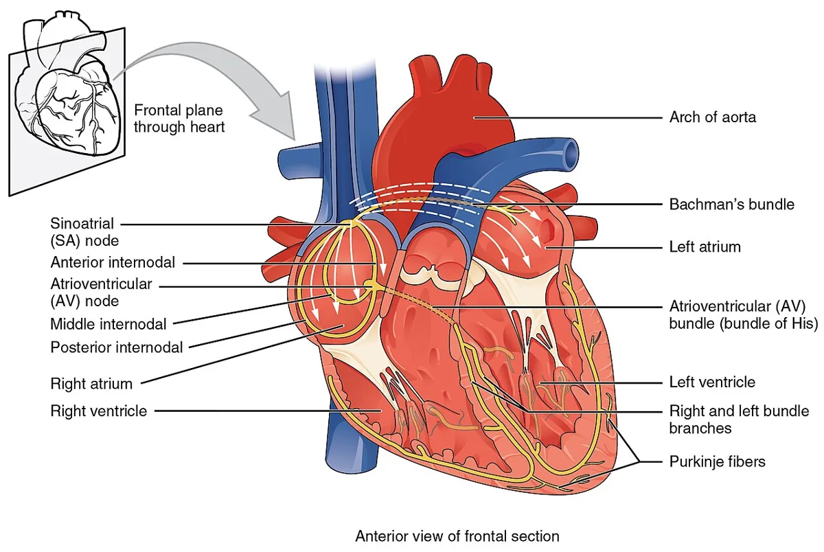
収縮するためではなく興奮を作って伝えるために分化した心筋。順に:
- Nodus sinuatrialis(洞房結節・Keith-Flack)— 上大静脈と右房の境、分界稜の上端。固有頻度 60〜100/分
- Nodus atrioventricularis(房室結節・Aschoff-Tawara)— Koch 三角の中(冠状静脈洞口・三尖弁中隔尖・Todaro 腱で囲まれる)。40〜60/分
- Fasciculus atrioventricularis(His 束)— 心室中隔膜性部を貫く
- Crus dextrum / sinistrum(右脚・左脚)
- Rami subendocardiales(Purkinje 線維)— 20〜40/分
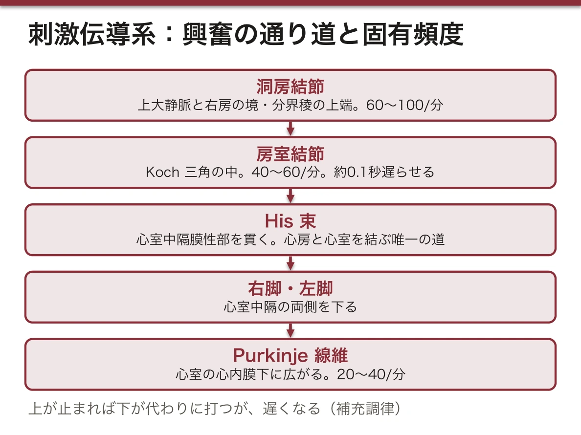
心房と心室のあいだは線維輪で電気的に完全に絶縁されている。唯一の通り道が His 束である。だから房室結節でしか興奮は心室へ渡れない——心房細動でも心室が同じ速さで拍動しないのは、房室結節が「関所」として伝導を遅らせるからである。
房室結節の遅れ(約0.1秒)には意味がある。心房が収縮し終えてから心室が収縮するための時間で、心電図の PQ 間隔そのもの。この遅れが無ければ心房収縮による心室充満(atrial kick)が失われる。
血管支配が不整脈の型を決める。洞房結節は約60%で右冠状動脈、房室結節は約90%で右冠状動脈から栄養される。だから下壁梗塞(右冠状動脈)では徐脈と房室ブロックが出やすい。一方、脚は左前下行枝の中隔枝が養うので、前壁梗塞では脚ブロックと完全房室ブロックが出る——後者のほうが予後が悪い。
Koch 三角はアブレーションの標識である。房室結節回帰性頻拍で遅伝導路を焼くのはこの三角の下方で、三角の外に出ると房室ブロックを作ってしまう。
階層構造がそのまま補充調律の速さになる。洞房結節 60〜100/分 → 房室結節 40〜60/分 → His–Purkinje 20〜40/分。上が止まれば下が代わりに打つが、遅くなる。
房室結節は心房と心室を電気的につなぐ唯一の道である。だから心房細動でも心室が同じ速さで動かずに済む(生理的なフィルター)。副伝導路(Kent 束)があるとこの関門を迂回してしまい、WPW 症候群——デルタ波・PQ 短縮になる。
房室結節は右冠動脈から栄養される(多くの人で)。だから下壁梗塞では房室ブロックと徐脈を合併しやすい。
洞房結節は上大静脈と右房の境にある。洞不全症候群ではめまい・失神(Adams–Stokes 発作)を起こし、ペースメーカーの適応になる。
English version
Core
Myocardium differentiated not to contract but to generate and conduct excitation. In order:
- Nodus sinuatrialis (of Keith and Flack) — at the junction of superior vena cava and right atrium, at the upper end of the crista terminalis. Intrinsic rate 60–100/min.
- Nodus atrioventricularis (of Aschoff and Tawara) — within the triangle of Koch (bounded by the coronary sinus orifice, the septal cusp of the tricuspid valve and the tendon of Todaro). 40–60/min.
- Fasciculus atrioventricularis (bundle of His) — pierces the membranous part of the septum.
- Crus dextrum / sinistrum — the bundle branches.
- Rami subendocardiales (Purkinje fibres) — 20–40/min.
Going further
Atria and ventricles are completely insulated electrically by the fibrous rings, and the only way through is the bundle of His. So excitation can reach the ventricle only at the atrioventricular node — which is why the ventricles do not beat at the same rate in atrial fibrillation: the node acts as a check-point and delays conduction.
The delay at the node (about 0.1 s) has a purpose. It is the time for the atria to finish contracting before the ventricles do, and is itself the PQ interval of the ECG. Without it, ventricular filling from atrial contraction (the atrial kick) is lost.
The blood supply decides the type of arrhythmia. The sinuatrial node is supplied by the right coronary artery in about 60 per cent and the atrioventricular node in about 90 per cent. So inferior infarction (right coronary) readily gives bradycardia and atrioventricular block. The bundle branches, by contrast, are supplied by the septal branches of the left anterior descending, so anterior infarction gives bundle branch block and complete heart block — and the latter carries the worse prognosis.
The triangle of Koch is the landmark for ablation. The slow pathway is ablated at its lower part in AV nodal re-entrant tachycardia, and going outside the triangle creates heart block.
In the clinic
The hierarchy is itself the rate of the escape rhythm. Sinuatrial node 60–100/min → atrioventricular node 40–60/min → His–Purkinje 20–40/min. If the one above stops, the one below takes over — more slowly.
The atrioventricular node is the only electrical connection between atria and ventricles, which is why the ventricles need not follow the rate of atrial fibrillation (a physiological filter). An accessory pathway (the bundle of Kent) bypasses that gate — WPW syndrome, with a delta wave and a short PQ interval.
The atrioventricular node is supplied by the right coronary artery in most people, so inferior infarction is readily complicated by heart block and bradycardia.
The sinuatrial node lies at the junction of superior vena cava and right atrium. In sick sinus syndrome it causes dizziness and syncope (Adams–Stokes attacks) and is an indication for a pacemaker.
心膜 — 2枚と2つの洞Pericardium
Pericardium fibrosum(線維性心膜)— 外側の丈夫な袋。下は横隔膜の腱中心に癒合、上は大血管の外膜へ移行。伸びない。
Pericardium serosum(漿膜性心膜)— 内側。lamina parietalis(線維性心膜の内面を裏打ち)と lamina visceralis (epicardium)(心表面)が大血管の根元で折り返して連続する。あいだの cavitas pericardiaca に 15〜50mL の漿液。
折り返しのためにできる袋小路が2つ:Sinus transversus pericardii(前の動脈幹〈大動脈・肺動脈幹〉と後ろの静脈〈上大静脈・肺静脈〉のあいだ)と sinus obliquus pericardii(左房の後ろ・肺静脈に囲まれた行き止まり)。
線維性心膜が伸びないことが、心タンポナーデを作る。心嚢に急に血液が溜まると、袋が広がれないので圧がそのまま心臓を押しつぶす。最初に潰れるのは壁の薄い右房と右室——静脈還流が落ちて Beck の三徴(低血圧・頸静脈怒張・心音減弱)と奇脈が出る。逆に慢性の心嚢液貯留は1L 以上溜まっても症状が出ないことがある(袋がゆっくり伸びるため)。「溜まった量ではなく、溜まる速さが症状を決める」。
Sinus transversus は心臓外科で日常的に使われる。ここに指を通して大動脈と肺動脈幹の後ろへ回り、テープをかけて遮断する。解剖の袋小路が、そのまま手術の通り道になっている。
心膜の痛みは横隔神経(C3〜C5)が運ぶ。だから心膜炎の痛みは左肩や僧帽筋の縁へ放散し、前かがみで軽くなる。心筋虚血の痛み(自律神経・Th1〜Th4)とは経路が違うので、放散する場所が異なる。
心膜腔にはふだん 15〜50mL の液しかない。線維性心膜は伸びないので、急に溜まれば 100〜200mL でもタンポナーデになり、ゆっくりなら 1L でも耐える。「量ではなく速さ」が決めるという要点は、この膜の性質から出る。
心タンポナーデ — Beck 3徴(血圧低下・頸静脈怒張・心音減弱)、奇脈、心電図の低電位・電気的交互脈。心嚢穿刺で救命する。
心膜横洞は上行大動脈・肺動脈幹の後ろを通る通路で、心臓手術で大血管に鉗子をかけるときの指の通り道になる。
収縮性心膜炎では心膜が硬化して心臓を締めつけ、右心不全(浮腫・肝腫大・腹水)が前面に出る。結核が主要な原因である。
出典:イラスト解剖学 誌面316頁「心タンポナーデ・心嚢穿刺」
English version
Core
- Pericardium fibrosum — the tough outer sac, fused below with the central tendon of the diaphragm and continuous above with the adventitia of the great vessels. It does not stretch.
- Pericardium serosum — within: the lamina parietalis lining the fibrous pericardium and the lamina visceralis (epicardium) on the surface of the heart, continuous with each other where they reflect at the roots of the great vessels. Between them the cavitas pericardiaca holds 15–50 mL of serous fluid.
The reflections create two culs-de-sac: the sinus transversus pericardii (between the arterial trunks in front — aorta and pulmonary trunk — and the veins behind — superior vena cava and pulmonary veins) and the sinus obliquus pericardii (behind the left atrium, a blind end surrounded by the pulmonary veins).
Going further
That the fibrous pericardium does not stretch creates cardiac tamponade. When blood collects suddenly in the sac, the bag cannot expand and the pressure crushes the heart. What collapses first is the thin-walled right atrium and right ventricle — venous return falls and Beck’s triad (hypotension, distended neck veins, muffled sounds) with pulsus paradoxus follows.
Conversely a chronic effusion may exceed a litre without symptoms (the sac stretches slowly). "It is not the volume but the speed of accumulation that decides the symptoms."
The transverse sinus is used daily in cardiac surgery. A finger is passed through it behind the aorta and pulmonary trunk to sling and clamp them. A cul-de-sac of anatomy that has become a passage of surgery.
The pain of the pericardium is carried by the phrenic nerve (C3–C5). So pericarditic pain radiates to the left shoulder and the border of the trapezius and eases on leaning forward. The pain of myocardial ischaemia travels by a different route (autonomic, Th1–Th4) and radiates elsewhere.
In the clinic
The pericardial cavity normally holds only 15–50 mL. The fibrous pericardium does not stretch, so 100–200 mL accumulating quickly causes tamponade while a litre accumulating slowly is tolerated. The point that it is decided "by speed, not by volume" comes from the nature of this membrane.
Cardiac tamponade — Beck’s triad (hypotension, distended neck veins, muffled sounds), pulsus paradoxus, low voltage and electrical alternans on the ECG. Pericardiocentesis is life-saving.
The transverse sinus is the passage behind the ascending aorta and pulmonary trunk, and the way a finger goes when clamping the great vessels in cardiac surgery.
In constrictive pericarditis the pericardium hardens and constricts the heart, and right heart failure (oedema, hepatomegaly, ascites) dominates. Tuberculosis is the principal cause.
心尖Apex cordis
左心室が作る、左前下方を向いた尖端。第5肋間・左鎖骨中線のやや内側に位置する。
心尖拍動を触れる場所で、心の大きさの目安になる。ここが外下方へずれていれば左室拡大。
English version
Core
The apex formed by the left ventricle, pointing down, forwards and to the left, in the fifth intercostal space a little medial to the left midclavicular line.
It is where the apex beat is felt and is the measure of the size of the heart: displaced down and out, it means left ventricular enlargement.
胸肋面(前面)Facies sternocostalis
胸骨と肋軟骨に向かう前面。主に右心房と右心室が作る。
前面がほとんど右心系であることが、胸部外傷でまず右室が傷つく理由。
English version
Core
The anterior surface, facing the sternum and costal cartilages, formed chiefly by the right atrium and right ventricle.
That the front is almost entirely right heart is why the right ventricle is injured first in chest trauma.
横隔面(下面)Facies diaphragmatica
横隔膜に乗る下面。主に左心室と一部右心室が作る。後室間溝が走る。
English version
Core
The lower surface, resting on the diaphragm, formed chiefly by the left ventricle with part of the right. The posterior interventricular sulcus runs on it.
肺面(左側面)Facies pulmonalis
左肺の心圧痕に向かう左面。左心室が作る。
3面は「前が右室、下が左室、左が左室」——どの面をどの部屋が作るかが定番の設問。
English version
Core
The left surface, facing the cardiac impression of the left lung, formed by the left ventricle.
The three surfaces are "front right ventricle, below left ventricle, left left ventricle" — which chamber forms which surface is a standard question.
右心耳・左心耳Auricula dextra, sinistra
心房の前上部から前へ突き出す耳たぶ状の袋。原始心房の遺残で、内面に櫛状筋がある。
心房細動で血栓ができるのは、ほとんどが左心耳——流れが澱むため。ここから飛んだ血栓が心原性脳塞栓を起こす。
English version
Core
The ear-shaped pouches projecting forwards from the upper front of the atria, remnants of the primitive atrium, with pectinate muscles inside.
Thrombus in atrial fibrillation forms almost always in the left auricle — because the flow stagnates there. A thrombus flying from it causes cardiogenic cerebral embolism.
冠状溝Sulcus coronarius
心をぐるりと取り巻く溝で、心房と心室の境。中を右冠状動脈と冠状静脈洞が走る。
標本ではまずこの溝で心房と心室を分けるのが順序。
English version
Core
The sulcus encircling the heart, the boundary between atria and ventricles. The right coronary artery and the coronary sinus run in it.
On the specimen, dividing atrium from ventricle at this sulcus is the first step.
前室間溝・後室間溝Sulcus interventricularis anterior, posterior
前面と下面を心尖へ向かって走る溝で、左右の心室の境。
前を左前下行枝、後ろを後下行枝が走る。2本が心尖で出会う。溝の位置がそのまま冠状動脈の走行なので、梗塞部位の推定に直結する。
English version
Core
The sulci running towards the apex on the anterior and inferior surfaces, the boundary between the two ventricles. The left anterior descending artery runs in the anterior, the posterior descending in the posterior; the two meet at the apex.
The position of the sulcus is precisely the course of the coronary artery, and so bears directly on inferring the site of an infarct.
上大静脈口Ostium venae cavae superioris
右心房の上後部に開く。弁を持たない。
この口の近くの心房壁に洞房結節がある。
English version
Core
Opens into the upper posterior part of the right atrium. It has no valve. The sinuatrial node lies in the atrial wall near this opening.
下大静脈口Ostium venae cavae inferioris
右心房の下後部に開く。下大静脈弁(Eustachio 弁)という遺残の弁を伴う。
胎生期にはこの弁が血流を卵円孔へ導いていた。
English version
Core
Opens into the lower posterior part of the right atrium, with the vestigial Eustachian valve.
In fetal life this valve directed the flow towards the foramen ovale.
櫛状筋Musculi pectinati
心房の内面に並ぶ櫛の歯状の筋の隆起。心耳と、右心房では分界稜より前の部分にある。
後方の滑らかな壁(静脈洞由来)と、前方の櫛状筋のある壁(原始心房由来)の境が分界稜——発生の違いが内面にそのまま残っている。
English version
Core
The comb-like muscular ridges on the inner surface of the atria, found in the auricles and, in the right atrium, in the part in front of the crista terminalis.
The boundary between the smooth posterior wall (from the sinus venosus) and the ridged anterior wall (from the primitive atrium) is the crista terminalis — a difference of development left as it is on the inner surface.
右房室口・左房室口Ostium atrioventriculare dextrum, sinistrum
心房から心室へ血が入る口。右は三尖弁、左は僧帽弁で守られる。
English version
Core
The orifices through which blood passes from atrium to ventricle, guarded on the right by the tricuspid valve and on the left by the mitral.
三尖弁Valva tricuspidalis
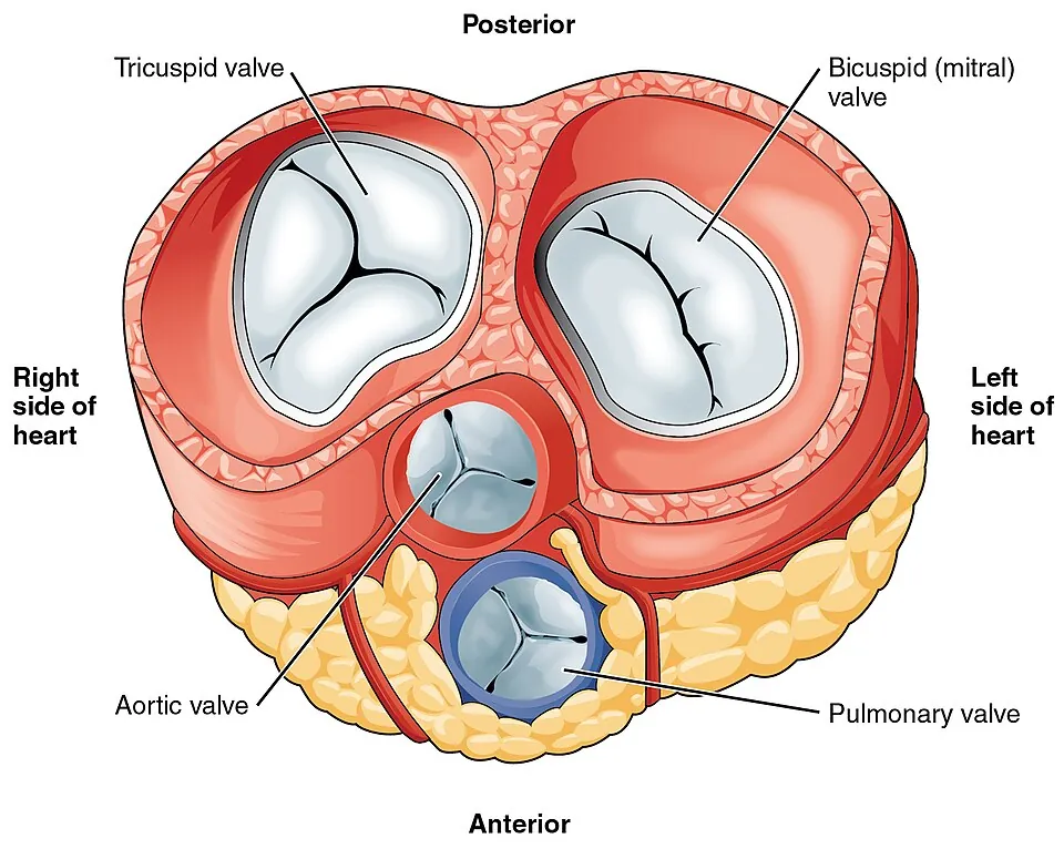
右房室口の弁。前尖・後尖・中隔尖の3枚。腱索と乳頭筋で支えられる。
聴診は胸骨左縁第4肋間。
English version
Core
The valve of the right atrioventricular orifice, with anterior, posterior and septal cusps, supported by chordae tendineae and papillary muscles. Auscultated at the left sternal edge in the fourth space.
僧帽弁Valva atrioventricularis sinistra sive mitralis
左房室口の弁。前尖と後尖の2枚だけで、司教冠(mitra)に似ることが名の由来。
聴診は心尖部。「右は3枚、左は2枚」が最も基本の対比。リウマチ熱で最も侵されるのがこの弁。
English version
Core
The valve of the left atrioventricular orifice, with only two cusps, anterior and posterior; named from its resemblance to a bishop’s mitre. Auscultated at the apex.
"Three on the right, two on the left" is the most basic of the contrasts. It is the valve most affected in rheumatic fever.
腱索Chordae tendineae
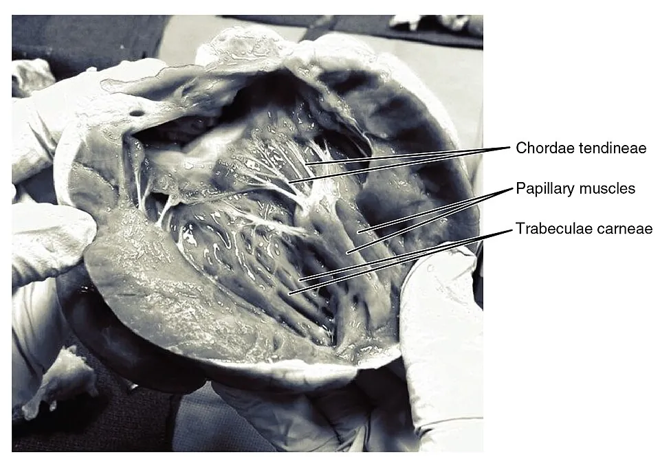
乳頭筋と房室弁の尖を結ぶ細い腱の糸。
弁を開く力ではなく、収縮期に弁が心房側へ翻るのを防ぐ「引き綱」。断裂すると急性の弁逆流になる。
English version
Core
The fine tendinous threads joining the papillary muscles to the cusps of the atrioventricular valves. They are not what opens the valve but the guy-ropes that prevent it everting into the atrium in systole.
Rupture gives acute valvular regurgitation.
乳頭筋Musculi papillares
心室壁から突き出す円錐状の筋。右室に3個、左室に2個(前乳頭筋・後乳頭筋)。
心室の収縮と同時に縮んで腱索を引く。心筋梗塞で断裂すると急性僧帽弁閉鎖不全になり、後乳頭筋は血流が1本しかないため壊死しやすい。
English version
Core
The conical muscles projecting from the ventricular wall — three in the right ventricle, two in the left (anterior and posterior). They shorten as the ventricle contracts and pull on the chordae.
Rupture after myocardial infarction gives acute mitral regurgitation, and the posterior papillary muscle, having only one blood supply, is the more liable to necrose.
肉柱Trabeculae carneae
心室内面の不規則な筋の隆起。心房の櫛状筋よりずっと太く粗い。
右室では中隔縁柱(節制帯)が特に太く、右脚を乳頭筋へ運ぶ。「心房=櫛状筋、心室=肉柱」で対にする。
English version
Core
The irregular muscular ridges on the inner surface of the ventricles, much coarser and thicker than the pectinate muscles of the atria. In the right ventricle the septomarginal trabecula (moderator band) is particularly thick and carries the right bundle branch to the papillary muscle.
Pair them as "atrium = pectinate muscles, ventricle = trabeculae carneae".
肺動脈幹口Ostium trunci pulmonalis
右心室の流出路(動脈円錐)の出口。
English version
Core
The outlet of the outflow tract (the conus arteriosus) of the right ventricle.
肺動脈幹Truncus pulmonalis
右心室から出て左後上方へ向かい、大動脈弓の下で左右の肺動脈に分かれる。
分岐部と大動脈弓の間に動脈管索(胎生期の動脈管の遺残)が張る。静脈血を運ぶ動脈である点が問われる。
English version
Core
Leaves the right ventricle, passes up, back and to the left, and divides beneath the aortic arch into the two pulmonary arteries. Between the bifurcation and the arch stretches the ligamentum arteriosum (the remnant of the ductus arteriosus).
That it is an artery carrying venous blood is what is asked.
肺動脈弁Valva trunci pulmonalis
肺動脈幹の起始にある3枚の半月弁。腱索を持たない。
聴診は胸骨左縁第2肋間。
English version
Core
The three semilunar cusps at the origin of the pulmonary trunk. They have no chordae. Auscultated at the left sternal edge in the second space.
大動脈口Ostium aortae
左心室の流出路の出口。僧帽弁の前尖が、流入路と流出路の仕切りになっている。
English version
Core
The outlet of the outflow tract of the left ventricle. The anterior cusp of the mitral valve is the partition between the inflow and the outflow tract.
大動脈弁Valva aortae
大動脈起始の3枚の半月弁(右冠尖・左冠尖・無冠尖)。弁の上のふくらみが Valsalva 洞。
右冠状動脈と左冠状動脈は、この洞から出る——だから冠状動脈は拡張期に灌流される。聴診は胸骨右縁第2肋間。
English version
Core
The three semilunar cusps at the origin of the aorta (right coronary, left coronary and non-coronary). The dilatations above them are the sinuses of Valsalva.
The right and left coronary arteries arise from these sinuses — which is why the coronary arteries are perfused in diastole. Auscultated at the right sternal edge in the second space.
肺静脈Venae pulmonales
左右2本ずつ、計4本が左心房の後壁に開く。弁を持たない。
動脈血を運ぶ静脈。心房細動の異所性興奮はこの流入部から起こることが多く、カテーテルアブレーションの標的になる。
English version
Core
Two on each side, four in all, opening into the posterior wall of the left atrium. They have no valves. They are veins carrying arterial blood.
The ectopic activity of atrial fibrillation commonly arises at these openings, and they are the target of catheter ablation.
体循環と肺循環Circulatio sanguinis
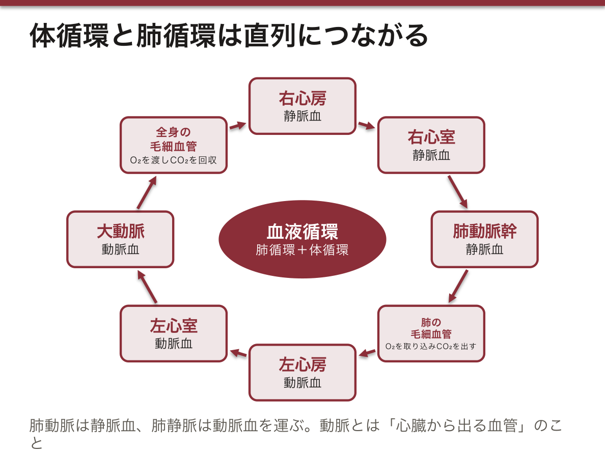
体循環(大循環)——左心室 → 大動脈 → 全身の毛細血管 → 上下大静脈 → 右心房。動脈血で出て、静脈血で戻る。
肺循環(小循環)——右心室 → 肺動脈幹 → 肺の毛細血管 → 肺静脈 → 左心房。静脈血で出て、動脈血で戻る。
「動脈=動脈血」ではない——肺動脈は静脈血、肺静脈は動脈血を運ぶ。動脈とは「心臓から出る血管」の意味である。ここが最頻出の引っかけ。
2つは直列なので、1分間に両心室を通る血液量は等しい。ただし圧が全く違う——体循環は約120/80 mmHg、肺循環は約25/8 mmHg。だから左室壁は右室壁の約3倍厚い。
第3の系として門脈系がある——毛細血管を2回通る(腸の毛細血管 → 門脈 → 肝の類洞)。経口薬の初回通過効果の解剖学的な理由。
English version
Core
Systemic (greater) circulation — left ventricle → aorta → the capillaries of the body → superior and inferior venae cavae → right atrium. Leaves as arterial blood, returns as venous.
Pulmonary (lesser) circulation — right ventricle → pulmonary trunk → the capillaries of the lung → pulmonary veins → left atrium. Leaves as venous blood, returns as arterial.
"Artery" does not mean arterial blood — the pulmonary artery carries venous blood and the pulmonary veins arterial blood. An artery means a vessel leaving the heart. This is the commonest trick of all.
The two are in series, so the volume passing through the two ventricles in a minute is equal. But the pressures are wholly different — about 120/80 mmHg systemically, about 25/8 mmHg in the lung. Hence the left ventricular wall is about three times the right.
Going further
There is a third system, the portal system — passing through capillaries twice (the capillaries of the gut → portal vein → the hepatic sinusoids). The anatomical reason for the first-pass effect of oral drugs.
心臓の壁 — 3層Paries cordis
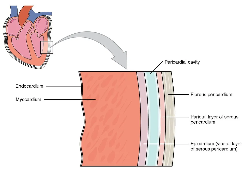
内から心内膜・心筋層・心外膜の3層。
心内膜 endocardium——内皮+結合組織。弁は心内膜のひだであり、血管の内膜と連続する。感染性心内膜炎はここに起こる。
心筋層 myocardium——最も厚い層。厚さは仕事量で決まる——左室 > 右室 > 心房。心房と心室の筋は線維骨格で完全に隔てられており、興奮は房室束を通してしか伝わらない。
心外膜 epicardium——漿膜性心膜の臓側板そのもの。下に脂肪と冠状動脈が走る。
線維骨格(4つの弁を囲む線維輪)は、①弁の支え ②心房筋と心室筋の絶縁 ③筋の付着部、の3役を果たす。
心筋の走行は3層(浅層は斜め、中層は輪状、深層は縦)で、収縮は「絞る」動きになる。心尖から捻れるように駆出する。
English version
Core
Three layers from within: endocardium, myocardium, epicardium.
- Endocardium — endothelium with connective tissue. The valves are folds of endocardium, continuous with the intima of the vessels. Infective endocarditis occurs here.
- Myocardium — the thickest layer. Its thickness is set by the work done: left ventricle > right ventricle > atria. Atrial and ventricular muscle are completely separated by the fibrous skeleton, and excitation can pass only through the atrioventricular bundle.
- Epicardium — the visceral layer of the serous pericardium itself, with fat and the coronary arteries beneath it.
The fibrous skeleton (the rings around the four valves) does three jobs: ① supports the valves ② insulates atrial from ventricular muscle ③ provides attachment for the muscle.
Going further
The myocardium runs in three layers (superficial oblique, middle circular, deep longitudinal), so the contraction is a wringing movement, ejecting with a twist from the apex.
心臓の位置と体表投影Topographia cordis
中縦隔に、心膜に包まれて位置する。約2/3が正中より左。前は胸骨と第2〜6肋軟骨、後ろは Th5〜Th8、下は横隔膜。
体表投影の4点——①右上:右第3肋軟骨上縁、胸骨右縁から1cm ②右下:右第6肋軟骨、胸骨右縁から1cm ③左上:左第2肋間、胸骨左縁から2cm ④左下=心尖:左第5肋間、鎖骨中線の内側1〜2cm。
弁の位置と聴診部位は違う——弁は胸骨の裏に集まっているが、聴診は血流の下流で聴く。大動脈弁=右第2肋間、肺動脈弁=左第2肋間、三尖弁=胸骨左縁第4肋間、僧帽弁=心尖部。この食い違いの理由まで言えると強い。
心膜穿刺は剣状突起の左下から左肩へ向けて刺す(Larrey 点)。心膜腔に液が溜まると心タンポナーデになる。
English version
Core
In the middle mediastinum, enclosed in the pericardium, about two thirds of it to the left of the midline. In front, the sternum and the second to sixth costal cartilages; behind, Th5–Th8; below, the diaphragm.
Four points of the surface projection — ① upper right: the upper border of the right third costal cartilage, 1 cm from the right sternal edge ② lower right: the right sixth costal cartilage, 1 cm from the right sternal edge ③ upper left: the left second intercostal space, 2 cm from the left sternal edge ④ lower left = the apex: the left fifth space, 1–2 cm medial to the midclavicular line.
The positions of the valves and the sites of auscultation differ — the valves are gathered behind the sternum, but you listen downstream of the flow. Aortic = right second space; pulmonary = left second space; tricuspid = left sternal edge fourth space; mitral = the apex. Being able to give the reason for the discrepancy is what counts.
Going further
Pericardiocentesis is performed from below and left of the xiphoid, aiming at the left shoulder (Larrey’s point). Fluid collecting in the pericardial cavity gives cardiac tamponade.
局所解剖線 — 体の表面に引く11本の縦線Lineae topographicae
見えない臓器の位置を体の表面で言うために、縦の線を引き、それと肋骨・肋間・椎骨の高さを組み合わせて座標にする。前・後ろ・横で11本ある(右と左がある線は dx, sin を付けて呼ぶ)。
| 線 | 日本語 | どこを通るか | |
|---|---|---|---|
| 前 | linea mediana anterior | 前正中線 | 胸骨の中央 |
| 前 | linea sternalis | 胸骨線 | 胸骨の外側縁 |
| 前 | linea parasternalis | 胸骨傍線 | 胸骨線と鎖骨中線の中間 |
| 前 | linea medioclavicularis | 鎖骨中線 | 鎖骨の中点(男性ではほぼ乳頭を通る) |
| 横 | linea axillaris anterior | 前腋窩線 | 腋窩の前縁(大胸筋が作る前腋窩ひだ) |
| 横 | linea axillaris media | 中腋窩線 | 前腋窩線と後腋窩線の中間 |
| 横 | linea axillaris posterior | 後腋窩線 | 腋窩の後縁(広背筋・大円筋が作る後腋窩ひだ) |
| 後 | linea scapularis | 肩甲線 | 肩甲骨の下角(腕を下ろした姿勢で) |
| 後 | linea paravertebralis | 脊柱傍線 | 椎骨線と肩甲線の中間 |
| 後 | linea vertebralis | 椎骨線(脊柱線) | 脊柱の外側縁 |
| 後 | linea mediana posterior | 後正中線 | 棘突起の列 |
脊柱の近くの2本は、本によって名前と位置の対応が少し違う(横突起の先端を通る線そのものを脊柱傍線と呼ぶ本もある)。使う本の定義を一度確かめておく。
線は「縦の線 × 肋骨または肋間」で1点を決めるための座標軸である。肋骨を数える起点は、前では胸骨角(第2肋骨が付く)、後ろでは隆椎(C7 棘突起)と、腕を下ろしたときの肩甲骨下角(第7肋骨・Th7 の高さ)。
この座標で体表投影がそろって書ける。心尖=左第5肋間・鎖骨中線のやや内側、肺の下縁=鎖骨中線・中腋窩線・肩甲線で第6・8・10肋骨、胸膜の下縁はその2肋骨下、肝の上界=右鎖骨中線で第4肋間、胆嚢底=右鎖骨中線と第9肋軟骨の交点。線を先に覚えると、臓器ごとの数字が同じ形式で並ぶ。
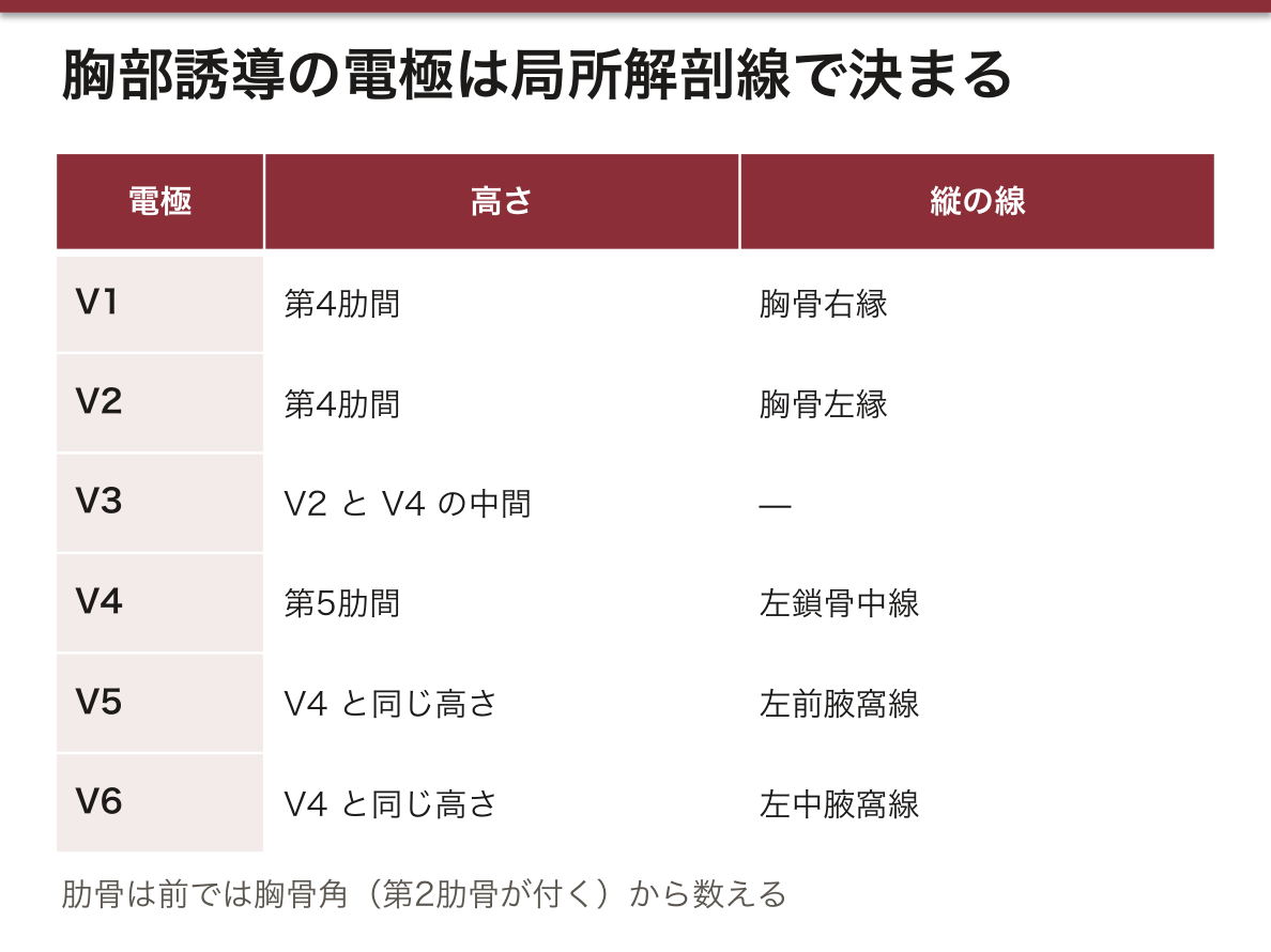
胸部誘導心電図の電極の位置は、この線で定義されている。V1=第4肋間・胸骨右縁、V2=第4肋間・胸骨左縁、V4=第5肋間・左鎖骨中線、V3=V2 と V4 の中間、V5=V4 の高さの前腋窩線、V6=同じ高さの中腋窩線。
胸腔ドレーンは、前腋窩線と中腋窩線のあいだの第4〜5肋間(大胸筋の外側縁・広背筋の前縁・乳頭の高さで囲まれる「安全三角」)から入れる。胸腔穿刺は肩甲線や中腋窩線で、肋骨の上縁に沿って行う。どれも線を正しく引けることが前提である。
English version
Core
To describe the position of hidden organs on the body surface, we draw vertical lines and combine them with the level of ribs, intercostal spaces and vertebrae as coordinates. There are eleven lines at the front, side and back (paired lines are named dx and sin).
| Line | Japanese | Where it runs | |
|---|---|---|---|
| Front | linea mediana anterior | 前正中線 | Middle of the sternum |
| Front | linea sternalis | 胸骨線 | Lateral edge of the sternum |
| Front | linea parasternalis | 胸骨傍線 | Midway between sternal and midclavicular lines |
| Front | linea medioclavicularis | 鎖骨中線 | Midpoint of the clavicle (in men it passes close to the nipple) |
| Side | linea axillaris anterior | 前腋窩線 | Front edge of the axilla (anterior axillary fold, pectoralis major) |
| Side | linea axillaris media | 中腋窩線 | Midway between anterior and posterior axillary lines |
| Side | linea axillaris posterior | 後腋窩線 | Back edge of the axilla (posterior axillary fold, latissimus dorsi and teres major) |
| Back | linea scapularis | 肩甲線 | Inferior angle of the scapula (arms at the sides) |
| Back | linea paravertebralis | 脊柱傍線 | Midway between vertebral and scapular lines |
| Back | linea vertebralis | 椎骨線 | Lateral edge of the vertebral column |
| Back | linea mediana posterior | 後正中線 | Row of spinous processes |
Books differ a little in the names and positions of the two lines near the spine (some call the line through the tips of the transverse processes the paravertebral line). Check the definition in the book you use.
Going further
The lines are coordinate axes: one vertical line × one rib or intercostal space fixes a point. Ribs are counted from the sternal angle at the front (where the 2nd rib attaches), and at the back from the vertebra prominens (C7) and the inferior angle of the scapula with the arms down (7th rib, T7 level).
With these coordinates, surface projections can all be written in the same way. Apex of the heart: left 5th intercostal space, just medial to the midclavicular line. Lower border of the lung: 6th, 8th and 10th ribs on the midclavicular, midaxillary and scapular lines; the pleura two ribs lower. Upper border of the liver: 4th intercostal space on the right midclavicular line. Fundus of the gallbladder: right midclavicular line at the 9th costal cartilage. Learn the lines first, and every organ’s numbers fall into the same format.
In the clinic
Chest ECG electrode positions are defined by these lines. V1: 4th intercostal space, right sternal edge; V2: 4th space, left sternal edge; V4: 5th space, left midclavicular line; V3: midway between V2 and V4; V5: anterior axillary line at the level of V4; V6: midaxillary line at the same level.
A chest drain goes in at the 4th–5th intercostal space between the anterior and midaxillary lines (the “safe triangle” bounded by pectoralis major, latissimus dorsi and the nipple level). Thoracentesis is done on the scapular or midaxillary line, along the upper edge of a rib. All of these assume the lines are drawn correctly.
まとめ
| 心臓 — 外形と溝 | 縦隔中部、心膜の中。底 basis cordis は右上後方(大血管が出入りする)、apex cordis は左下前方(第5肋間・左鎖骨中線で拍動を触れる)。 |
|---|---|
| 右心房と右心室 — 内面の構造 | 右心房に入るもの3つ:ostium venae cavae superioris・ostium venae cavae inferioris(Eustachi 弁を伴う)・osti… |
| 左心房と左心室 | 左心房:後壁に ostia venarum pulmonalium(4つ・左右上下)。 |
| 心臓の弁 — 位置と聴診部位が違う | 4つの弁は同じ線維性の面(心臓の骨格)に並ぶ。 |
| 刺激伝導系 — 特殊心筋の道 | 収縮するためではなく興奮を作って伝えるために分化した心筋。 |
| 心膜 — 2枚と2つの洞 | Pericardium fibrosum(線維性心膜)— 外側の丈夫な袋。 |
| 心尖 | 左心室が作る、左前下方を向いた尖端。第5肋間・左鎖骨中線のやや内側に位置する。 |
| 胸肋面(前面) | 胸骨と肋軟骨に向かう前面。主に右心房と右心室が作る。 |
| 横隔面(下面) | 横隔膜に乗る下面。主に左心室と一部右心室が作る。 |
| 肺面(左側面) | 左肺の心圧痕に向かう左面。左心室が作る。 |
| 右心耳・左心耳 | 心房の前上部から前へ突き出す耳たぶ状の袋。 |
| 冠状溝 | 心をぐるりと取り巻く溝で、心房と心室の境。 |
| 前室間溝・後室間溝 | 前面と下面を心尖へ向かって走る溝で、左右の心室の境。 |
| 上大静脈口 | 右心房の上後部に開く。弁を持たない。 |
| 下大静脈口 | 右心房の下後部に開く。下大静脈弁(Eustachio 弁)という遺残の弁を伴う。 |
| 櫛状筋 | 心房の内面に並ぶ櫛の歯状の筋の隆起。心耳と、右心房では分界稜より前の部分にある。 |
| 右房室口・左房室口 | 心房から心室へ血が入る口。右は三尖弁、左は僧帽弁で守られる。 |
| 三尖弁 | 右房室口の弁。前尖・後尖・中隔尖の3枚。 |
| 僧帽弁 | 左房室口の弁。前尖と後尖の2枚だけで、司教冠(mitra)に似ることが名の由来。 |
| 腱索 | 乳頭筋と房室弁の尖を結ぶ細い腱の糸。弁を開く力ではなく、収縮期に弁が心房側へ翻るのを防ぐ「引き綱」。 |
| 乳頭筋 | 心室壁から突き出す円錐状の筋。右室に3個、左室に2個(前乳頭筋・後乳頭筋)。 |
| 肉柱 | 心室内面の不規則な筋の隆起。心房の櫛状筋よりずっと太く粗い。 |
| 肺動脈幹口 | 右心室の流出路(動脈円錐)の出口。 |
| 肺動脈幹 | 右心室から出て左後上方へ向かい、大動脈弓の下で左右の肺動脈に分かれる。 |
| 肺動脈弁 | 肺動脈幹の起始にある3枚の半月弁。腱索を持たない。 |
| 大動脈口 | 左心室の流出路の出口。僧帽弁の前尖が、流入路と流出路の仕切りになっている。 |
| 大動脈弁 | 大動脈起始の3枚の半月弁(右冠尖・左冠尖・無冠尖)。 |
| 肺静脈 | 左右2本ずつ、計4本が左心房の後壁に開く。 |
| 体循環と肺循環 | 体循環(大循環)——左心室 → 大動脈 → 全身の毛細血管 → 上下大静脈 → 右心房。 |
| 心臓の壁 — 3層 | 内から心内膜・心筋層・心外膜の3層。心内膜 endocardium——内皮+結合組織。 |
| 心臓の位置と体表投影 | 中縦隔に、心膜に包まれて位置する。約2/3が正中より左。 |
| 局所解剖線 — 体の表面に引く11本の縦線 | 見えない臓器の位置を体の表面で言うために、縦の線を引き、それと肋骨・肋間・椎骨の高さを組み合わせて座標にする。 |
このノートは医学生が授業の内容を自分の言葉でまとめたもので、診療の判断に使うものではありません。誤りに気づいたらお知らせください。