腹膜は「腸が回りながら育った跡」で、成人の複雑な形はすべて発生の途中経過である。ここを回転の話として読むと、憶えるべき事実の数が一気に減る。
標本で見るときは肝の臓側面が主戦場。H字の溝(2本の縦溝と1本の横溝)を先に描いてから、4葉と門脈三つ組を当てはめる。
この範囲の芯は「腹膜腔は本当は袋であり、臓器はその外側にある」ことである。腸間膜も網も、袋の壁が2枚重なって伸びたものにすぎない。網嚢と網嚢孔が理解できれば、腹膜の項目はほぼ片が付く。
- 肝臓 — 2つの面と、臓側面のH
- 肝臓の働き — 門脈の出口に置かれた関所
- 腹部の9区分と、肝の上界・下界
- 胆嚢と胆道 — 上流から下流へ
- 膵臓 — 後腹膜のC字の相手
- 脾臓 — 左季肋部の血液の器官
- 腹膜 — 袋と、そのひだ
- 腹膜と臓器の位置関係 — intra / meso / retro と、日本式の分け方
- 腹膜腔の3階 — 横行結腸間膜と分界線で区切る
- 上階 — 肝嚢・胃前嚢・網嚢と、横隔膜下・肝下の陥凹
- 網嚢 — 6つの壁と4つの部屋
- 中階 — 左右の腸間膜洞と、左右の結腸傍溝
- 後腹壁の陥凹 — 腸が壁に付く所にできるポケット
- 前腹壁の内面 — 5本のひだと3対の窩
- 腹膜の働きと血管・神経 — 壁側は腹壁の、臓側は臓器の
- 肝臓 — 横隔面と臓側面
- 無漿膜野
- 肝鎌状間膜
- 肝右葉
- 肝左葉
- 方形葉
- 尾状葉
- 肝門
- 門脈・固有肝動脈
- 胆嚢(底・体・頸)
- 胆嚢管
- 総肝管
- 総胆管
- 膵頭
- 膵体・膵尾
- 脾臓 — 横隔面と臓側面
- 脾門
- 大網
- 直腸子宮窩(Douglas 窩)
- 膀胱子宮窩
- 直腸膀胱窩
- 発生 — 腹側間膜から肝と小網が、背側間膜から大網と脾ができる
- 肝臓 — 2つの面と、臓側面のHHepar
- 肝臓の働き — 門脈の出口に置かれた関所
- 腹部の9区分と、肝の上界・下界Regiones abdominis, topographia hepatis
- 胆嚢と胆道 — 上流から下流へVesica biliaris et ductus choledochus
- 膵臓 — 後腹膜のC字の相手Pancreas
- 脾臓 — 左季肋部の血液の器官Splen
- 腹膜 — 袋と、そのひだPeritoneum
- 腹膜と臓器の位置関係 — intra / meso / retro と、日本式の分け方
- 腹膜腔の3階 — 横行結腸間膜と分界線で区切るCavitas peritonealis
- 上階 — 肝嚢・胃前嚢・網嚢と、横隔膜下・肝下の陥凹Bursa hepatica, bursa pregastrica
- 網嚢 — 6つの壁と4つの部屋Bursa omentalis
- 中階 — 左右の腸間膜洞と、左右の結腸傍溝Sinus mesentericus, sulcus paracolicus
- 後腹壁の陥凹 — 腸が壁に付く所にできるポケットRecessus duodenalis, recessus ileocaecalis, recessus retrocaecalis, recessus intersigmoideus
- 前腹壁の内面 — 5本のひだと3対の窩Plicae umbilicales, fossae inguinales
- 腹膜の働きと血管・神経 — 壁側は腹壁の、臓側は臓器のVasa et nervi peritonei
- 肝臓 — 横隔面と臓側面Hepar
- 無漿膜野Area nuda
- 肝鎌状間膜Ligamentum falciforme hepatis
- 肝右葉Lobus dexter hepatis
- 肝左葉Lobus sinister hepatis
- 方形葉Lobus quadratus
- 尾状葉Lobus caudatus
- 肝門Porta hepatis
- 門脈・固有肝動脈Vena portae, arteria hepatica propria
- 胆嚢(底・体・頸)Vesica biliaris
- 胆嚢管Ductus cysticus
- 総肝管Ductus hepaticus communis
- 総胆管Ductus choledochus
- 膵頭Caput pancreatis
- 膵体・膵尾Corpus et cauda pancreatis
- 脾臓 — 横隔面と臓側面Splen
- 脾門Hilum splenicum
- 大網Omentum majus
- 直腸子宮窩(Douglas 窩)Excavatio rectouterina
- 膀胱子宮窩Excavatio vesicouterina
- 直腸膀胱窩Excavatio rectovesicalis
- 発生 — 腹側間膜から肝と小網が、背側間膜から大網と脾ができるMesenterium ventrale et dorsale
- まとめ
肝臓 — 2つの面と、臓側面のHHepar
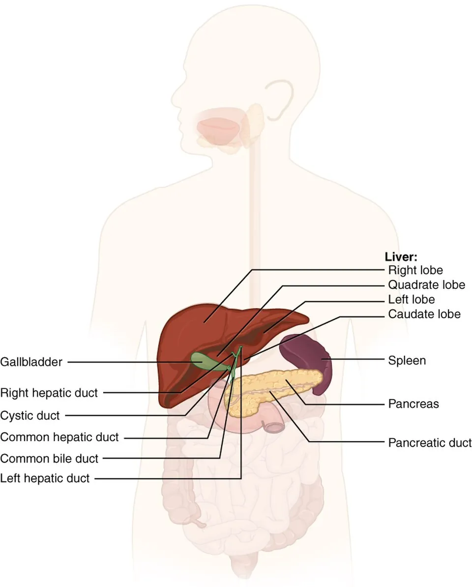
右上腹部、横隔膜の下。約1.5kg で最大の腺。2面:上向きに凸の facies diaphragmatica と、下後方を向く facies visceralis。境は前下方の鋭い margo inferior。
横隔面には lig. falciforme hepatis(鎌状間膜)が付き、その遊離縁に lig. teres hepatis(臍静脈の遺残)が入る。上面で鎌状間膜が左右へ分かれて lig. coronarium となり、その2葉に挟まれた腹膜のない部分が area nuda(無漿膜野)——ここで肝は横隔膜に直接付いている。
臓側面はH字の溝で4葉に分かれる。右の縦溝=fossa vesicae biliaris(前)と sulcus venae cavae(後)、左の縦溝=fissura lig. teretis(前)と fissura lig. venosi(後)、横棒=porta hepatis(肝門)。Hの前上が lobus quadratus(方形葉)、後上が lobus caudatus(尾状葉)。
肝門を通る3つ(門脈三つ組):v. portae hepatis(後)・a. hepatica propria(左前)・ductus hepaticus communis(右前)。「後ろに静脈、前に動脈と胆管、胆管が右」。この3つは lig. hepatoduodenale(小網の遊離縁)に包まれて走る。
形態的な4葉と機能的な区域は一致しない。肝を機能的に分けるのは門脈・肝動脈・胆管の分布で、正中裂(胆嚢窩と下大静脈を結ぶ Cantlie 線)が真の左右の境である。方形葉と尾状葉は形の上では右葉に属するが、機能的には左葉——切除術の計画はこちらに従う(Couinaud の8区域)。
肝には2つの血液が入る。門脈が約70〜75%(酸素は少ないが栄養に富む)、肝動脈が25〜30%。どちらも類洞で混ざる。「入口が2つ、出口は肝静脈1系統で下大静脈へ」——肝門を通らない肝静脈が、無漿膜野の近くで直接注ぐ点が要注意である。
肝臓は「区域」で切る。肝静脈と門脈枝の走行が決める Couinaud の8区域は、外から見える葉の境(鎌状間膜)とは一致しない。外科の右葉・左葉の境はCantlie 線(胆嚢窩と下大静脈を結ぶ線)である。
肝硬変では類洞の抵抗が上がって門脈圧亢進になり、食道静脈瘤・腹壁静脈怒張(メドゥーサの頭)・痔核という門脈大循環吻合が開く。加えて脾腫・腹水。
肝は二重の血流を受ける(門脈70〜75%、肝動脈25〜30%)。だから肝動脈を塞栓しても肝実質は生き残るが、腫瘍は壊死する——肝細胞癌に対する TACE の理論的な根拠である。
肝は右第4〜5肋間の高さまで上がるので、下位肋骨の外傷では肝損傷を必ず考える。
出典:イラスト解剖学 誌面451頁「肝臓のCT画像:模式図を描く」
English version
Core
In the right upper abdomen beneath the diaphragm; about 1.5 kg, the largest gland. Two surfaces: the convex, upward-facing facies diaphragmatica and the down- and backward-facing facies visceralis, separated by the sharp margo inferior.
The diaphragmatic surface carries the lig. falciforme hepatis, with the lig. teres hepatis (the remnant of the umbilical vein) in its free margin. Above, the falciform ligament divides right and left as the lig. coronarium, and the part between its two leaves that has no peritoneum is the area nuda — where the liver is applied directly to the diaphragm.
The visceral surface is divided into four lobes by an H of fissures. The right vertical limb = the fossa vesicae biliaris (front) and sulcus venae cavae (behind); the left = the fissura lig. teretis (front) and fissura lig. venosi (behind); the cross-bar = the porta hepatis. In front of and above the H is the lobus quadratus, behind and above it the lobus caudatus.
Going further
Three structures pass through the porta (the portal triad): v. portae hepatis (behind), a. hepatica propria (front left), ductus hepaticus communis (front right). "Vein behind, artery and duct in front, duct to the right." All three are wrapped in the lig. hepatoduodenale (the free margin of the lesser omentum).
The four morphological lobes do not correspond to the functional segments. The liver is divided functionally by the distribution of portal vein, hepatic artery and bile duct, and the true boundary of right and left is the median plane (Cantlie’s line, from the gallbladder fossa to the inferior vena cava). The quadrate and caudate lobes belong morphologically to the right lobe but functionally to the left — and the planning of resection follows the latter (Couinaud’s eight segments).
The liver receives two blood supplies: the portal vein about 70–75 per cent (oxygen-poor but nutrient-rich) and the hepatic artery 25–30 per cent, mixing in the sinusoids. "Two entrances, one exit — the hepatic veins to the inferior vena cava." Note that the hepatic veins do not pass through the porta and drain directly near the bare area.
In the clinic
The liver is cut by segments. Couinaud’s eight segments, determined by the course of the hepatic veins and portal branches, do not correspond to the lobar boundary visible from outside (the falciform ligament). The surgical boundary of right and left is Cantlie’s line (from the gallbladder fossa to the inferior vena cava).
In cirrhosis the resistance of the sinusoids rises and portal hypertension follows, opening the portosystemic anastomoses — oesophageal varices, distended abdominal wall veins (caput medusae) and haemorrhoids — with splenomegaly and ascites.
The liver has a double blood supply (portal 70–75 per cent, arterial 25–30 per cent). So embolising the hepatic artery leaves the parenchyma alive but necroses a tumour — the rationale of TACE for hepatocellular carcinoma.
The liver rises to the level of the right fourth or fifth intercostal space, so always consider hepatic injury in trauma to the lower ribs.
肝臓の働き — 門脈の出口に置かれた関所
肝の仕事は大きく6つに分けて言う。
| 働き | 中身 |
|---|---|
| ① 消化(外分泌) | 胆汁を絶えず作り、肝管から十二指腸へ送る(1日に約0.5〜1 L)。胆汁酸が脂肪を乳化する |
| ② 代謝 | 糖:グリコーゲンとして蓄え、空腹時は糖新生で血糖を保つ。蛋白:アルブミン・凝固因子を作り、アンモニアを尿素に変える。脂質:リポ蛋白・コレステロールを作る |
| ③ 解毒(関所) | 腸から来た薬物・毒物・アンモニアを、全身に出る前に処理する |
| ④ 貪食 | 類洞の Kupffer 細胞(マクロファージ)が門脈血の細菌や異物を取り除く |
| ⑤ 貯蔵と熱産生 | ビタミン A・D・B12、鉄を蓄える。代謝が盛んなので熱の産生源にもなる |
| ⑥ 胎生期の造血 | 胎児期の中ごろまで、赤血球を作る主な場所である |
この配置の理由は門脈にある。胃・腸・脾・膵から来た血液は、全部いったん門脈に集まって肝を通ってから全身へ出る。吸収された栄養も、腸から入った毒物や細菌も、まず肝が受け取って処理する——肝は腸と全身のあいだに置かれた関所である。
胎児の造血は卵黄嚢 → 肝 → 脾 → 骨髄と場所を移す。大人でも骨髄が働けなくなると、肝と脾が造血を再開して腫れる(髄外造血)。
肝不全の症状は、失われた働きの一覧である。胆汁が出せず黄疸、アルブミンが作れず浮腫と腹水、凝固因子が作れずプロトロンビン時間が延びて出血しやすくなる、尿素に変えられないアンモニアが脳に届いて肝性脳症、糖新生ができず低血糖。肝硬変の重症度を表す Child-Pugh 分類が、ビリルビン・アルブミン・プロトロンビン時間・腹水・脳症の5つで点数をつけるのは、この働きを順に測っているからである。
口から飲んだ薬は肝を一度通るので、効く前に一部が分解される(初回通過効果)。肝硬変ではこれが弱まり、門脈大循環シャントもできるので、薬が効きすぎることがある。
English version
Core
The work of the liver is usually given as six functions.
| Function | Content |
|---|---|
| (1) Digestive (exocrine) | Produces bile continuously and sends it through the hepatic ducts to the duodenum (about 0.5–1 L a day). Bile acids emulsify fat |
| (2) Metabolic | Carbohydrate: stores glycogen and keeps blood glucose by gluconeogenesis when fasting. Protein: makes albumin and clotting factors, turns ammonia into urea. Fat: makes lipoproteins and cholesterol |
| (3) Barrier (detoxification) | Handles drugs, toxins and ammonia from the gut before they reach the body |
| (4) Phagocytic | Kupffer cells (macrophages) in the sinusoids remove bacteria and particles from portal blood |
| (5) Storage and heat | Stores vitamins A, D, B12 and iron; its high metabolic rate makes it a source of body heat |
| (6) Fetal blood formation | The main site of red cell production until about the middle of fetal life |
Going further
The reason for this layout is the portal vein. Blood from the stomach, intestine, spleen and pancreas all collects in the portal vein and passes through the liver before reaching the body. Absorbed nutrients, and toxins or bacteria from the gut, are first received by the liver — a checkpoint between the gut and the rest of the body.
Fetal blood formation moves from yolk sac to liver to spleen to bone marrow. In adults whose marrow fails, the liver and spleen can restart blood formation and enlarge (extramedullary haematopoiesis).
In the clinic
The signs of liver failure are a list of lost functions. No bile excretion gives jaundice; no albumin gives oedema and ascites; no clotting factors gives a long prothrombin time and bleeding; ammonia not turned into urea reaches the brain as hepatic encephalopathy; no gluconeogenesis gives hypoglycaemia. The Child–Pugh score for cirrhosis uses bilirubin, albumin, prothrombin time, ascites and encephalopathy because it measures these functions one by one.
Drugs taken by mouth pass the liver once, and part is broken down before they act (first-pass effect). In cirrhosis this is weaker and portosystemic shunts open, so drugs may act too strongly.
腹部の9区分と、肝の上界・下界Regiones abdominis, topographia hepatis
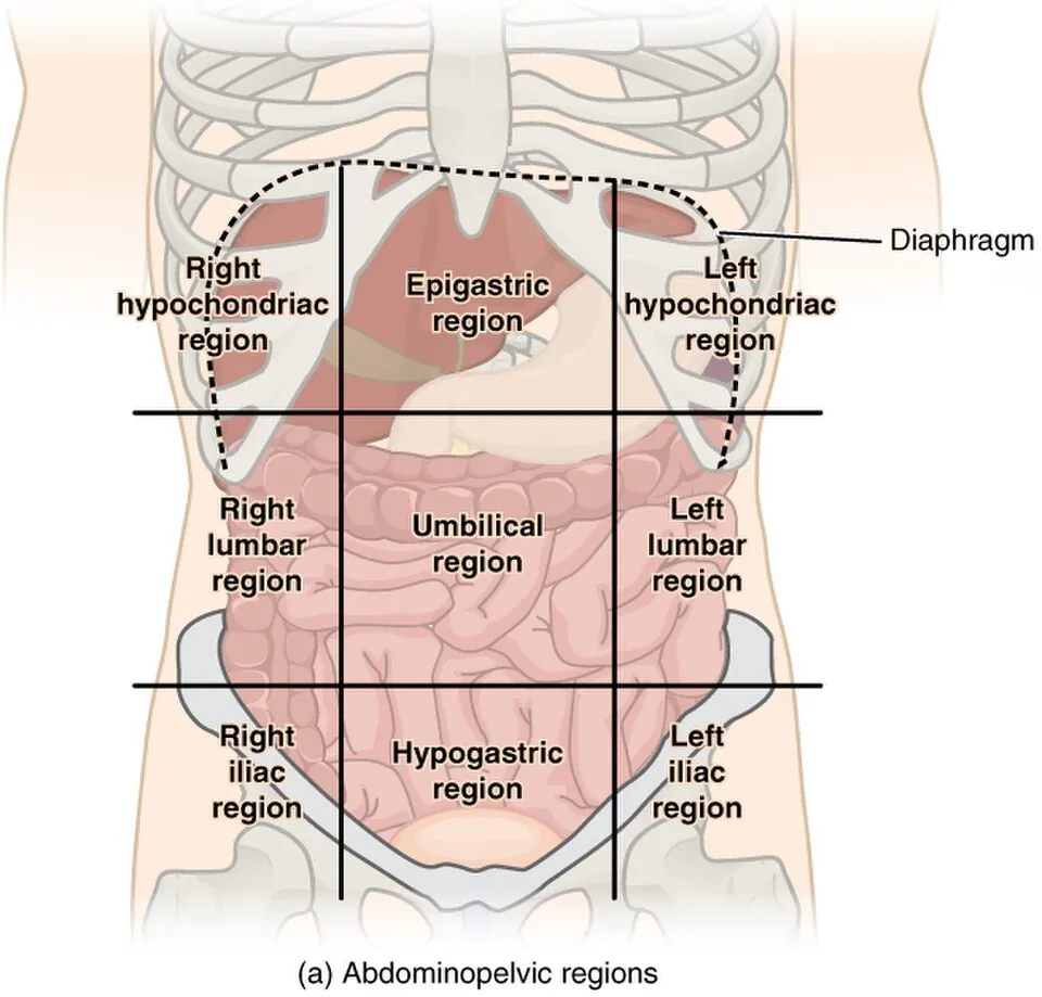
腹部の9区分は、横の線2本と縦の線2本で作る。横は上が左右の肋骨弓の最も低い点(第10肋骨)を結ぶ線、下が左右の腸骨(上前腸骨棘または腸骨稜の結節)を結ぶ線、縦は左右の鎖骨中線を下へ延ばした線である(下の横線の取り方は本によって少し違う)。
| 右 | 中央 | 左 | |
|---|---|---|---|
| 上(epigastrium) | regio hypochondriaca dextra(右下肋部・右季肋部) | regio epigastrica(上胃部・心窩部) | regio hypochondriaca sinistra |
| 中(mesogastrium) | regio lateralis dextra(右側腹部) | regio umbilicalis(臍部) | regio lateralis sinistra |
| 下(hypogastrium) | regio inguinalis dextra(右鼠径部) | regio pubica(恥骨部・下腹部) | regio inguinalis sinistra |
肝の位置(holotopia)は右下肋部・上胃部・左下肋部の一部。上界は右の中腋窩線で第10肋骨から始まり、右鎖骨中線で第4肋間まで上がり、胸骨の左で第5肋間に下がる。下界は同じ点(右中腋窩線・第10肋骨)から肋骨弓に沿って進み、第8・9肋軟骨の合わさる所で弓を離れて上胃部を斜めに横切り、左の第7・8肋軟骨の合わさる所に達して、左の第5〜6肋間付近で上界と合わさる。後ろでは Th9〜Th11 の高さにある。
肝の境界は呼吸で動く。肝は無漿膜野と冠状間膜で横隔膜に付いているので、吸気で横隔膜が下がると一緒に下がる。だから下縁は深く吸わせながら右の肋骨弓の下で触れる——成人では鎖骨中線で肋骨弓をほとんど越えないのが正常で、幼児では肋骨弓の下に1〜2 cm 触れても正常である。
上界が乳頭の高さ近くまで上がることは、右下胸部の外傷で肺と同時に肝を傷つけうることを意味する。
肝の上の打診で濁音(肝濁音界)を確かめ、肝腫大を見積もる。消化管が穿孔して腹腔に空気が入ると、空気は肝の前に上がって肝濁音界が消える——立位の胸部 X 線で横隔膜下に遊離ガスを探すのと同じ理屈で、ベッドサイドでわかる穿孔の徴候である。
English version
Core
The nine abdominal regions are made by two horizontal and two vertical lines. The upper horizontal line joins the lowest points of the costal margins (10th rib); the lower joins the iliac bones (anterior superior iliac spines or the iliac tubercles); the vertical lines continue the right and left midclavicular lines downward (books differ slightly on the lower line).
| Right | Middle | Left | |
|---|---|---|---|
| Upper (epigastrium) | regio hypochondriaca dextra | regio epigastrica | regio hypochondriaca sinistra |
| Middle (mesogastrium) | regio lateralis dextra | regio umbilicalis | regio lateralis sinistra |
| Lower (hypogastrium) | regio inguinalis dextra | regio pubica | regio inguinalis sinistra |
Position of the liver (holotopia): right hypochondrium, epigastrium and part of the left hypochondrium. The upper border starts on the right midaxillary line at the 10th rib, rises to the 4th intercostal space on the right midclavicular line, and falls to the 5th intercostal space left of the sternum. The lower border starts at the same point, follows the costal margin, leaves it where the 8th and 9th costal cartilages meet, crosses the epigastrium obliquely to where the left 7th and 8th cartilages meet, and joins the upper border near the left 5th–6th intercostal space. Posteriorly the liver lies at T9–T11.
Going further
The borders move with breathing. The liver is fixed to the diaphragm by the bare area and coronary ligament, so it descends with the diaphragm on inspiration. That is why the lower edge is felt under the right costal margin while the patient breathes in deeply. In adults it normally hardly passes the costal margin in the midclavicular line; in small children an edge 1–2 cm below the margin is normal.
Because the upper border rises almost to the level of the nipple, an injury to the lower right chest can damage both lung and liver.
In the clinic
Percussion over the liver gives dullness, used to estimate liver size. When the gut perforates and air enters the peritoneal cavity, the air rises in front of the liver and liver dullness disappears — the bedside version of looking for free gas under the diaphragm on an erect chest X-ray.
胆嚢と胆道 — 上流から下流へVesica biliaris et ductus choledochus
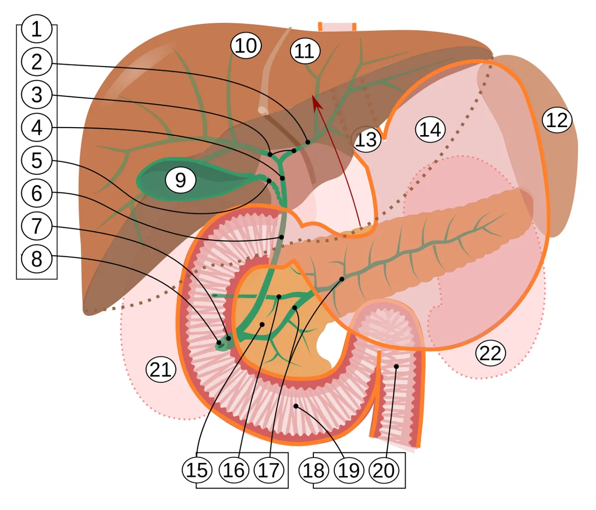
Vesica biliaris (fellea) は肝の臓側面の胆嚢窩に収まる袋(容量30〜50mL)。3部:fundus(底・肝の下縁から突出し、右鎖骨中線と第9肋軟骨の交点で体表に触れる)・corpus・collum(頸・S字に曲がる)。
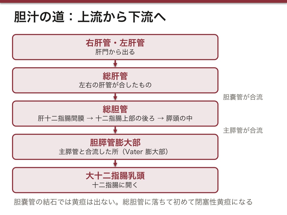
胆汁の道を上流から:Ductus hepaticus dexter + sinister → ductus hepaticus communis →(ductus cysticus が合流)→ ductus choledochus (biliaris) → 主膵管と合して ampulla hepatopancreatica → papilla duodeni major。
Ductus choledochus は3区間を走る:肝十二指腸間膜の中 → 十二指腸上部の後ろ → 膵頭の中。
Calot 三角(胆嚢三角)=ductus cysticus・ductus hepaticus communis・肝の下面で囲まれる三角。この中を胆嚢動脈が走るので、腹腔鏡下胆嚢摘出術ではここを展開して動脈と胆管を確認してから切る(critical view of safety)。解剖の三角がそのまま手術手技の定義になっている。
胆嚢管と胆嚢頸の粘膜には plica spiralis(Heister 弁)というらせんヒダがあり、内腔を保つ。ここに石が嵌頓すると胆嚢だけが腫れて黄疸は出ない(胆汁の本流は通じているため)。総胆管に落ちて初めて閉塞性黄疸になる。石の位置と症状が一対一で対応する。
膵頭癌が閉塞性黄疸で見つかるのは、総胆管の末端が膵頭の中を通るからである。無痛性で腫大した胆嚢を触れる(Courvoisier 徴候)——結石症では慢性炎症で胆嚢が萎縮しているため腫れない、という対比で理解する。
胆嚢底は右鎖骨中線と第9肋軟骨の交点に当たる。ここを押さえながら吸気させると痛みで息が止まる(Murphy 徴候)——解剖の位置がそのまま診察手技になる。
胆嚢炎・胆石の痛みが右肩に放散するのは、胆嚢が横隔膜に接し、横隔神経(C3〜C5)が刺激されるためである(関連痛)。
Calot 三角(胆嚢管・総肝管・肝下縁)の中を胆嚢動脈が走る。腹腔鏡下胆嚢摘出術で最も重要な指標で、ここを誤ると総胆管損傷という重大な合併症になる。
Charcot 3徴(発熱・黄疸・右季肋部痛)は急性胆管炎。ショックと意識障害が加わると Reynolds 5徴で、緊急ドレナージの適応である。
English version
Core
The vesica biliaris (fellea) lies in the gallbladder fossa on the visceral surface (capacity 30–50 mL). Three parts: fundus (projecting beyond the inferior border of the liver, palpable where the right midclavicular line crosses the ninth costal cartilage), corpus, collum (curving in an S).
The path of the bile from above down: ductus hepaticus dexter + sinister → ductus hepaticus communis → (joined by the ductus cysticus) → ductus choledochus (biliaris) → joining the main pancreatic duct as the ampulla hepatopancreatica → papilla duodeni major.
The ductus choledochus runs in three stretches: within the hepatoduodenal ligament → behind the superior part of the duodenum → within the head of the pancreas.
Going further
Calot’s (the cystohepatic) triangle = bounded by the ductus cysticus, the ductus hepaticus communis and the inferior surface of the liver. The cystic artery runs within it, so in laparoscopic cholecystectomy this is opened up and artery and duct identified before anything is divided (the critical view of safety). A triangle of anatomy that has become the definition of a surgical technique.
The mucosa of the cystic duct and neck carries the spiral fold (Heister’s valve), which keeps the lumen open. A stone impacted here distends the gallbladder alone without jaundice (the main stream of bile is still patent). Only when it falls into the common duct does obstructive jaundice follow. The position of the stone corresponds one to one with the symptoms.
Carcinoma of the head of the pancreas is found through obstructive jaundice because the terminal common bile duct runs within the head. A painless, distended, palpable gallbladder (Courvoisier’s sign) — understood by contrast with calculous disease, in which chronic inflammation has shrunk the gallbladder so it does not distend.
In the clinic
The fundus of the gallbladder lies where the right midclavicular line crosses the ninth costal cartilage. Pressing there and asking the patient to breathe in arrests inspiration through pain (Murphy’s sign) — the position in the anatomy becoming directly the technique of examination.
The pain of cholecystitis and gallstones radiates to the right shoulder because the gallbladder touches the diaphragm and the phrenic nerve (C3–C5) is irritated (referred pain).
The cystic artery runs within Calot’s triangle (cystic duct, common hepatic duct, inferior border of the liver). It is the most important landmark in laparoscopic cholecystectomy, and a mistake there gives the serious complication of injury to the common bile duct.
Charcot’s triad (fever, jaundice, right hypochondrial pain) is acute cholangitis. With shock and altered consciousness it becomes Reynolds’ pentad, an indication for emergency drainage.
膵臓 — 後腹膜のC字の相手Pancreas
胃の後ろ、L1〜L2 の高さを横に走る後腹膜臓器(尾部だけが脾腎間膜に入り腹膜内)。長さ約15cm。
4部:caput(頭・十二指腸のC字に抱かれる。左下へ突き出す processus uncinatus が上腸間膜動静脈の後ろへ回る)・collum(頸)・corpus(体)・cauda(尾・脾門に達する)。
導管:Ductus pancreaticus(Wirsung)が全長を貫き、総胆管と合して大乳頭へ。Ductus pancreaticus accessorius(Santorini)は頭部から小乳頭へ。
外分泌部(消化酵素)と内分泌部(膵島・インスリンとグルカゴン)を併せ持つ。
後腹膜にあることが症状を決める。膵炎の痛みが背部へ抜けるのも、膵癌が長く無症状なのも、腹膜を刺激せずに済む位置にあるためである。逆に炎症が起きると後腹膜腔に沿って広がる。
頸部の後ろで上腸間膜静脈と脾静脈が合流して門脈になる。だから膵頭部の腫瘍は門脈へ浸潤しやすく、切除可能性の判断が門脈との関係で決まる。鉤状突起が上腸間膜動脈の背側へ回り込む位置関係も、同じ理由で重要である。
導管が2本ある理由は発生にある。膵は腹側原基と背側原基の2つから作られ、腹側原基が十二指腸の回転に伴って背側へ回り込んで融合する。主膵管は背側の遠位+腹側、副膵管は背側の近位——2本あるのは融合の跡である。融合が起きないと pancreas divisum、腹側原基が輪状に回り込むと anular pancreas(十二指腸狭窄)になる。
膵は後腹膜にあるので、炎症も痛みも背中へ抜ける。急性膵炎の心窩部痛が背部へ放散し、前屈で楽になるのはこのためである。
後腹膜への出血が体表に出るとGrey Turner 徴候(側腹部)・Cullen 徴候(臍周囲)——重症膵炎のサインになる。
膵頭部癌 — 総胆管を締めて無痛性黄疸。鉤状突起は上腸間膜動静脈を抱くので、ここに浸潤すると切除不能と判定される。解剖の位置関係がそのまま治療方針を決める。
膵体尾部癌は黄疸が出ないため発見が遅れる。脾動静脈が膵の上縁・後面を走るので、浸潤すると脾静脈血栓 → 左側門脈圧亢進 → 孤立性胃静脈瘤を起こす。
English version
Core
A retroperitoneal organ running transversely behind the stomach at the level of L1–L2 (only the tail enters the splenorenal ligament and is intraperitoneal). About 15 cm long.
Four parts: caput (embraced by the C of the duodenum; the processus uncinatus projecting to the lower left passes behind the superior mesenteric vessels), collum, corpus, cauda (reaching the splenic hilum).
Ducts: the ductus pancreaticus (of Wirsung) traverses its whole length and joins the common bile duct at the major papilla; the ductus pancreaticus accessorius (of Santorini) runs from the head to the minor papilla.
It combines an exocrine part (digestive enzymes) with an endocrine part (the islets, insulin and glucagon).
Going further
Being retroperitoneal decides the symptoms. That the pain of pancreatitis passes through to the back, and that carcinoma of the pancreas is silent for so long, are both because it lies where it does not irritate the peritoneum. Conversely, once inflammation begins it spreads along the retroperitoneal space.
Behind the neck the superior mesenteric and splenic veins join to form the portal vein. So a tumour of the head invades the portal vein readily, and resectability is decided by its relation to that vein. The uncinate process passing behind the superior mesenteric artery matters for the same reason.
The reason there are two ducts lies in development. The pancreas is built from a ventral and a dorsal primordium, the ventral swinging round behind with the rotation of the duodenum and fusing. The main duct is the distal dorsal plus the ventral, the accessory duct the proximal dorsal — two ducts as the trace of that fusion. Failure of fusion gives pancreas divisum; a ventral primordium that encircles gives an anular pancreas (duodenal stenosis).
In the clinic
The pancreas is retroperitoneal, so both the inflammation and the pain pass through to the back. This is why the epigastric pain of acute pancreatitis radiates to the back and is eased by leaning forward.
Retroperitoneal haemorrhage reaching the surface gives Grey Turner’s sign (flank) and Cullen’s sign (periumbilical) — markers of severe pancreatitis.
Carcinoma of the head — constricts the common bile duct, giving painless jaundice. The uncinate process embraces the superior mesenteric vessels, so invasion there renders it unresectable. The positional anatomy decides the treatment directly.
Carcinoma of the body and tail causes no jaundice and so is found late. The splenic vessels run along the upper border and posterior surface, so invasion gives splenic vein thrombosis → left-sided portal hypertension → isolated gastric varices.
脾臓 — 左季肋部の血液の器官Splen
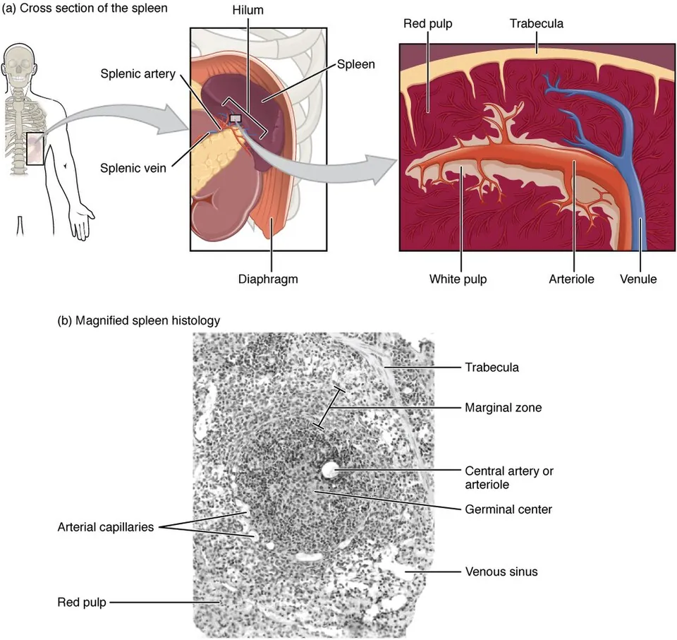
左季肋部、第9〜11肋骨の内側、横隔膜と胃底のあいだ。約150g、11×7×4cm(1・3・5・7・9・11 の法則:1×3×5インチ、7オンス、第9〜11肋骨)。完全に腹膜に包まれる(肝と違い無漿膜野が無い)。
2面:facies diaphragmatica(凸・平滑)と facies visceralis(凹・胃圧痕、腎圧痕、結腸圧痕)。臓側面の中央が hilum splenicum で、脾動静脈と膵尾が入る。
2つの間膜で吊られる:lig. gastrosplenicum(短胃動脈・左胃大網動脈)と lig. splenorenale(脾動静脈・膵尾)。
正常な脾は触れない。肋骨弓の下に触れるのは2〜3倍以上に腫れたときで、左季肋部から右下方(臍の方向)へ向かって腫れる——脾の長軸が第10肋骨に沿うためである。腎腫大との鑑別点になる(腎は下方へ、脾は内下方へ)。
脾は最も損傷しやすい腹部臓器である。①被膜が薄い、②肋骨のすぐ内側にある、③血流が豊富(心拍出量の約5%)。左第9〜11肋骨の骨折を見たら脾損傷を疑うのは、この位置関係そのものである。遅発性破裂(被膜下血腫が数日後に破れる)もある。
脾摘後は莢膜を持つ細菌に弱くなる(肺炎球菌・インフルエンザ桿菌・髄膜炎菌)。脾のマクロファージがオプソニン化された莢膜細菌を血中から除く唯一の場であるためで、脾摘後重症感染症(OPSI)を防ぐためにワクチンを事前に打つ。解剖の機能が予防接種の適応を決めている。
脾は第9〜11肋骨に守られている。左下位肋骨骨折を見たら脾損傷を疑うのが外傷診療の鉄則で、腹腔内出血の原因臓器として最も多い。
正常では触れない。触知できれば約2〜3倍に腫大していることを意味する。腫大は左季肋部から右下方(臍の方向)へ伸びる——腎腫大との鑑別点である。
脾摘後重症感染症(OPSI) — 脾は莢膜を持つ細菌を除去する主要な場所なので、摘出後は肺炎球菌・インフルエンザ桿菌 b 型・髄膜炎菌による電撃的な敗血症の危険が生涯続く。術前のワクチン接種が必須である。
脾動脈は腹腔動脈の枝で、膵の上縁を蛇行する。脾動脈瘤は妊娠中に破裂しやすい。
English version
Core
In the left hypochondrium, deep to the ninth to eleventh ribs, between the diaphragm and the fundus of the stomach. About 150 g, 11 × 7 × 4 cm (the 1-3-5-7-9-11 rule: 1 × 3 × 5 inches, 7 ounces, ribs 9–11).
It is completely invested by peritoneum (unlike the liver it has no bare area).
Two surfaces: facies diaphragmatica (convex, smooth) and facies visceralis (concave, with gastric, renal and colic impressions). At the centre of the visceral surface is the hilum splenicum, entered by the splenic vessels and the tail of the pancreas.
Slung by two ligaments: the lig. gastrosplenicum (short gastric and left gastro-omental vessels) and the lig. splenorenale (splenic vessels and the tail of the pancreas).
Going further
A normal spleen cannot be felt. It is palpable below the costal margin only when enlarged two- or threefold or more, and it enlarges from the left hypochondrium towards the right and downwards (towards the umbilicus) — because its long axis follows the tenth rib. This distinguishes it from renal enlargement (the kidney enlarges downwards, the spleen down and medially).
The spleen is the abdominal organ most readily injured: ① the capsule is thin ② it lies immediately inside the ribs ③ its blood flow is large (about 5 per cent of the cardiac output). Suspecting splenic injury on seeing a fracture of the left ninth to eleventh ribs is that positional relationship itself. Delayed rupture also occurs (a subcapsular haematoma bursting days later).
After splenectomy the patient becomes vulnerable to encapsulated organisms (pneumococcus, Haemophilus influenzae, meningococcus), the splenic macrophages being the only place where opsonised encapsulated bacteria are cleared from the blood. Vaccines are given in advance to prevent overwhelming post-splenectomy infection (OPSI). A function of anatomy deciding the indication for an immunisation.
In the clinic
The spleen is protected by the ninth to eleventh ribs. Suspecting splenic injury on seeing a fracture of the lower left ribs is a rule of trauma care, and it is the commonest source of intraperitoneal haemorrhage.
Normally it cannot be palpated. If it can be felt, it is enlarged two- or threefold. The enlargement extends from the left hypochondrium down and to the right (towards the umbilicus) — the point that distinguishes it from renal enlargement.
Overwhelming post-splenectomy infection (OPSI) — the spleen is the principal site at which encapsulated organisms are removed, so after removal the risk of fulminant sepsis from pneumococcus, H. influenzae type b and meningococcus lasts for life. Preoperative vaccination is essential.
The splenic artery is a branch of the coeliac artery, running tortuously along the upper border of the pancreas. A splenic artery aneurysm is prone to rupture in pregnancy.
腹膜 — 袋と、そのひだPeritoneum
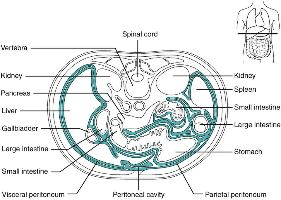
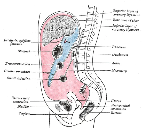
Peritoneum parietale(壁側・腹壁の内面)と peritoneum viscerale(臓側・臓器の表面)が連続した1枚の閉じた袋。そのあいだが cavitas peritonealis(男性では完全に閉鎖、女性では卵管を通じて外界と交通する)。
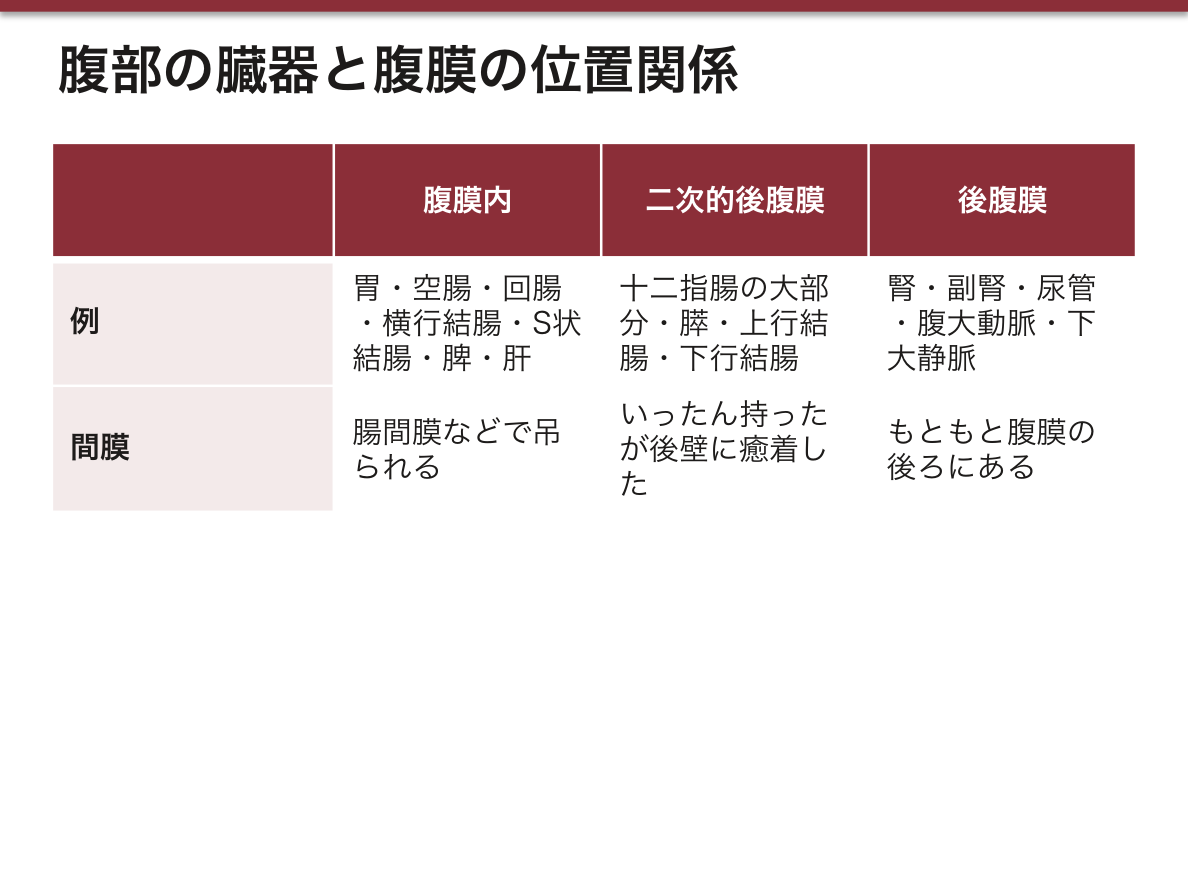
臓器と腹膜の関係には分け方が2通りある。ヨーロッパの教科書は覆われ方で腹膜内(intra)・腹膜間(meso:3面を覆われる)・腹膜後(retro)の3つに分け、肝・胆嚢・上行結腸・下行結腸は meso に入る。日本の教科書は成り立ちで腹膜内・後腹膜・二次的後腹膜に分け、肝は腹膜内として扱うことが多い。両者の対応は次の項目の表にまとめた。
ひだの種類:mesenterium(腸間膜)・mesocolon transversum・mesoappendix・mesosigmoideum・omentum majus / minus・lig. gastrocolicum・lig. falciforme。
Omentum minus(小網)=肝と胃小弯・十二指腸上部を繋ぐ2葉。lig. hepatogastricum と lig. hepatoduodenale(門脈三つ組を含む遊離縁)からなる。Omentum majus(大網)=胃大弯から前掛けのように垂れ、折り返して横行結腸へ付く4葉の膜。
Bursa omentalis(網嚢)は胃の後ろの腹膜腔の袋小路。唯一の入口が foramen omentale (epiploicum)(Winslow 孔)で、その前縁が lig. hepatoduodenale(=門脈三つ組)、後縁が下大静脈、上縁が肝尾状葉、下縁が十二指腸上部。4つの縁を全部言えるようにする——出血時に前縁を指で挟んで肝動脈を止める手技(Pringle 法)は、この孔に指を入れて行う。
大網は「腹腔の警察官」と呼ばれる。炎症の場所へ移動して包み込み、限局化させる。虫垂炎が穿孔しても汎発性腹膜炎にならずに膿瘍で留まることがあるのは、この働きによる。
陥凹は立位・仰臥位で液が溜まる場所:recessus subphrenicus・recessus subhepaticus(Morison 窩・仰臥位で最も低い)・excavatio rectovesicalis(男性)/excavatio rectouterina(Douglas 窩・女性で最も低い)・excavatio vesicouterina。FAST で腹腔内出血を探すのはまさにこの4か所である。
腹膜の神経支配が、腹痛の性質と場所を決める。
臓側腹膜 — 自律神経支配。鈍く、局在のはっきりしない痛みで、正中に感じる(発生の分節に対応:前腸=心窩部、中腸=臍周囲、後腸=下腹部)。
壁側腹膜 — 体性神経支配。鋭く、場所がはっきりした痛み。反跳痛・筋性防御が出る。
虫垂炎の痛みが心窩部・臍周囲から右下腹部へ移動するのは、臓側腹膜の痛み(中腸=臍周囲)から、炎症が壁側腹膜に及んだ体性痛(McBurney 点)へ切り替わるからである。腹膜の解剖がそのまま病歴になる典型例。
Douglas 窩(直腸子宮窩)は立位・坐位で腹腔の最下点。腹水・膿・血液が最初に溜まるので、穿刺・ドレナージの標的になる。
English version
Core
The peritoneum parietale (lining the abdominal wall) and peritoneum viscerale (covering the organs) are one continuous closed sac, with the cavitas peritonealis between them (completely closed in the man; in the woman it communicates with the exterior through the uterine tubes).
There are two ways to classify how organs relate to the peritoneum. European textbooks sort by how much surface is covered: intraperitoneal, mesoperitoneal (covered on three sides) and retroperitoneal. In this system the liver, gallbladder, ascending and descending colon are mesoperitoneal. Japanese textbooks sort by development: intraperitoneal, retroperitoneal and secondarily retroperitoneal, and usually count the liver as intraperitoneal. The next item puts the two systems side by side.
Kinds of fold: mesenterium, mesocolon transversum, mesoappendix, mesosigmoideum, omentum majus / minus, lig. gastrocolicum, lig. falciforme.
Going further
Omentum minus — two layers joining liver to lesser curvature and superior duodenum, consisting of the lig. hepatogastricum and the lig. hepatoduodenale (whose free margin contains the portal triad). Omentum majus — four layers hanging like an apron from the greater curvature and turning back to the transverse colon.
The bursa omentalis is the cul-de-sac of the peritoneal cavity behind the stomach. Its only entrance is the foramen omentale (epiploicum), of Winslow, bounded in front by the lig. hepatoduodenale (= the portal triad), behind by the inferior vena cava, above by the caudate lobe, below by the superior duodenum. Be able to name all four boundaries — the manoeuvre of pinching the anterior boundary between the fingers to arrest the hepatic artery in haemorrhage (Pringle’s manoeuvre) is performed by putting a finger into this foramen.
The greater omentum is called the policeman of the abdomen. It migrates to the site of inflammation, wraps it and localises it. That a perforated appendix may remain an abscess rather than a general peritonitis is due to this.
The recesses are where fluid collects, standing or supine: recessus subphrenicus, recessus subhepaticus (Morison’s pouch, the lowest point when supine), excavatio rectovesicalis (man) / excavatio rectouterina (the pouch of Douglas, the lowest in the woman), excavatio vesicouterina. These four are exactly where FAST looks for intraperitoneal blood.
In the clinic
The innervation of the peritoneum decides the character and the site of abdominal pain.
Visceral peritoneum — autonomic supply. Dull, poorly localised pain felt in the midline (corresponding to the developmental segment: foregut = epigastrium, midgut = periumbilical, hindgut = lower abdomen).
Parietal peritoneum — somatic supply. Sharp, well-localised pain, with rebound tenderness and guarding.
That the pain of appendicitis moves from the epigastrium and umbilicus to the right lower quadrant is the switch from visceral pain (midgut = periumbilical) to the somatic pain of inflammation reaching the parietal peritoneum (McBurney’s point). A model case of the anatomy of the peritoneum being itself the history.
The pouch of Douglas is the lowest point of the peritoneal cavity standing or sitting, so ascites, pus and blood collect there first, and it is the target for aspiration and drainage.
腹膜と臓器の位置関係 — intra / meso / retro と、日本式の分け方
ヨーロッパの教科書は「成人で、臓器の表面のどれだけを腹膜が覆っているか」で4つに分ける。日本の教科書は「発生の途中でどう後ろの壁に付いたか」で分ける。並べると次のようになる。
| ヨーロッパ式 | 覆われ方 | 主な臓器 | 日本の教科書での扱い |
|---|---|---|---|
| Intraperitoneal(腹膜内) | 全面。間膜で吊られ、よく動く | 胃・十二指腸の起始部・空腸・回腸・盲腸・虫垂・横行結腸・S状結腸・直腸の上1/3・脾・卵管 | 腹膜内臓器(同じ) |
| Mesoperitoneal(3面) | 前と両側の3面。後ろは壁に付く | 肝・胆嚢・上行結腸・下行結腸・直腸の中1/3・充満した膀胱・子宮 | 肝・胆嚢・子宮は腹膜内、上行・下行結腸は二次的後腹膜、膀胱は腹膜外 |
| Retroperitoneal(腹膜後) | 前面だけ | 十二指腸の大部分・膵・腎・副腎・尿管 | 腎・副腎・尿管=(一次的)後腹膜、十二指腸・膵=二次的後腹膜 |
| Extraperitoneal(腹膜外) | 覆われない | 直腸の下1/3。空虚な膀胱は腹膜の前下にあり preperitoneal(前腹膜)と呼ぶ | 腹膜外・腹膜下 |
2つの分け方は、違う問いに答えている。ヨーロッパ式は今の形を見ている。肝は無漿膜野と肝門と胆嚢窩を除けば腹膜に包まれるが、後上部で横隔膜にじかに付くので「3面」とされる。日本式は来歴を見ている。肝は腹側間膜(小網・鎌状間膜)の中で育った臓器なので腹膜内、上行結腸はもとは腸間膜を持っていたが後腹壁に癒着したので二次的後腹膜、となる。
どちらが正しいという話ではないので、答えるときは問われている分け方に合わせ、必要なら両方の名前を添える。とくに「肝=meso」「上行・下行結腸=meso」は、日本の教科書で育った人がつまずく所である。
手術の入り方がこの区分で決まる。後腹膜の腎は腹腔を開けずに背中側から到達できる(後腹膜鏡下手術)。二次的後腹膜の上行・下行結腸は、癒着した面(Toldt の癒合筋膜)を剥がして持ち上げる——結腸切除で「授動」と呼ぶ操作は、発生で起きた癒着を逆にたどることである。
動きやすさも同じ区分で読める。間膜を持つ S状結腸・横行結腸・小腸はねじれる(S状結腸軸捻転)が、壁に付いた上行・下行結腸はねじれない。後腹膜の出血は腹膜刺激症状を出しにくく、見逃されやすい。
English version
Core
European textbooks sort organs by how much of the adult organ surface is covered by peritoneum. Japanese textbooks sort them by how the organ became attached to the posterior wall during development. The table compares the two.
| European system | Covering | Main organs | In Japanese textbooks |
|---|---|---|---|
| Intraperitoneal | All sides; hangs on a mesentery and moves freely | Stomach, first part of duodenum, jejunum, ileum, caecum, appendix, transverse colon, sigmoid colon, upper third of rectum, spleen, uterine tube | Intraperitoneal (same) |
| Mesoperitoneal | Front and both sides; the back is fixed to the wall | Liver, gallbladder, ascending and descending colon, middle third of rectum, full bladder, uterus | Liver, gallbladder and uterus: intraperitoneal; ascending/descending colon: secondarily retroperitoneal; bladder: extraperitoneal |
| Retroperitoneal | Front only | Most of the duodenum, pancreas, kidney, adrenal gland, ureter | Kidney, adrenal, ureter: (primary) retroperitoneal; duodenum, pancreas: secondarily retroperitoneal |
| Extraperitoneal | Not covered | Lower third of rectum. The empty bladder lies below and in front of the peritoneum (preperitoneal) | Extraperitoneal / subperitoneal |
Going further
The two systems answer different questions. The European system looks at the present shape. The liver is wrapped in peritoneum except the bare area, the porta and the gallbladder fossa, but its upper back part is fixed directly to the diaphragm, so it is called “three-sided”. The Japanese system looks at history. The liver grew inside the ventral mesentery (lesser omentum, falciform ligament), so it is intraperitoneal. The ascending colon once had a mesentery and later fused to the posterior wall, so it is secondarily retroperitoneal.
Neither is wrong. Answer in the system the question uses, and add the other name when helpful. “Liver = mesoperitoneal” and “ascending/descending colon = mesoperitoneal” are the points where students trained on Japanese books often slip.
In the clinic
The classification decides the surgical approach. The retroperitoneal kidney can be reached from the back without opening the peritoneal cavity. The secondarily retroperitoneal colon is lifted by separating the fused plane (Toldt’s fusion fascia); “mobilisation” in colectomy simply reverses the fusion that happened in the embryo.
Mobility follows the same logic. Organs with a mesentery (sigmoid and transverse colon, small bowel) can twist, as in sigmoid volvulus; the fixed ascending and descending colon cannot. Retroperitoneal bleeding gives few peritoneal signs and is easily missed.
腹膜腔の3階 — 横行結腸間膜と分界線で区切るCavitas peritonealis
腹膜腔は上下に3つの階に分けて考える。
| 階 | 範囲 | 中にあるもの・区画 |
|---|---|---|
| 上階 | 横隔膜 〜 横行結腸と横行結腸間膜 | 肝・胆嚢・胃・脾・十二指腸の上部。区画は肝嚢・胃前嚢・網嚢(次の項目) |
| 中階 | 横行結腸間膜 〜 骨盤の分界線(linea terminalis) | 空腸・回腸・上行/下行結腸。区画は左右の腸間膜洞と左右の結腸傍溝、後腹壁の陥凹 |
| 下階 | 分界線より下(小骨盤) | 骨盤の窩。男性は直腸膀胱窩1つ、女性は膀胱子宮窩と直腸子宮窩の2つ |
英語の教科書は横行結腸間膜を境に supracolic / infracolic compartment(結腸上区・結腸下区)と呼び、骨盤を別に数えることが多い。上階=結腸上区、中階+下階=結腸下区と読み替えればよい。
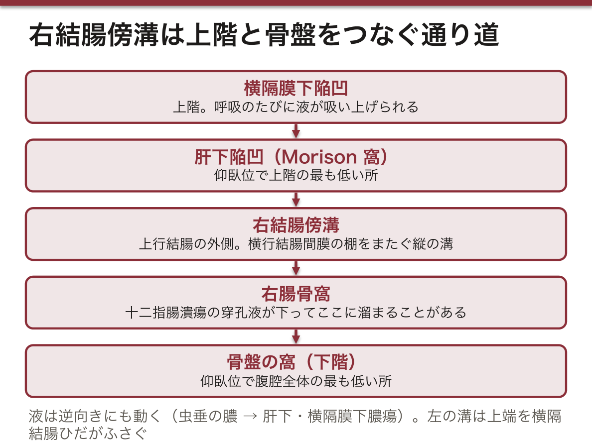
横行結腸間膜は腹腔を横に仕切る「棚」である。棚の上では液体は横隔膜下と肝下へ集まり、棚の下では斜めに走る腸間膜根がさらに左右を分ける。仕切りを越えて上下をつなぐのが、上行結腸と下行結腸の外側にある結腸傍溝である。
したがって腹腔の液体の動きは「棚・斜めの根・2本の縦の溝」の3つで予測できる。仰臥位で最も低いのは上階では肝腎陥凹(Morison 窩)、全体では骨盤の窩である。
膿がどこに溜まるかは、どの階で起きたかと、溝がつながっているかで決まる。虫垂炎の膿は右結腸傍溝を上って肝下・横隔膜下膿瘍になりうるし、上階の液体は同じ溝を下って骨盤へ落ちる。左の溝は上端を横隔結腸ひだがふさぐので、左側の膿は脾の周りへ上がりにくい。
English version
Core
Think of the peritoneal cavity as three storeys.
| Storey | Limits | Contents and spaces |
|---|---|---|
| Upper | Diaphragm to the transverse colon and transverse mesocolon | Liver, gallbladder, stomach, spleen, upper duodenum. Spaces: hepatic bursa, pregastric bursa, omental bursa (next item) |
| Middle | Transverse mesocolon to the pelvic brim (linea terminalis) | Jejunum, ileum, ascending/descending colon. Spaces: right and left mesenteric sinuses, right and left paracolic gutters, recesses of the posterior wall |
| Lower | Below the pelvic brim (lesser pelvis) | Pelvic pouches: one in the male (rectovesical), two in the female (vesicouterine and rectouterine) |
English textbooks usually speak of supracolic and infracolic compartments, divided by the transverse mesocolon, and count the pelvis separately. Upper storey = supracolic; middle + lower storeys = infracolic.
Going further
The transverse mesocolon is a horizontal shelf across the abdomen. Above it, fluid collects under the diaphragm and under the liver; below it, the oblique root of the mesentery divides right from left. The paracolic gutters lateral to the ascending and descending colon cross the shelf and link the storeys.
So fluid movement can be predicted from three things: the shelf, the oblique root and the two vertical gutters. In the supine position the lowest point of the upper storey is the hepatorenal recess (Morison’s pouch); the lowest point overall is the pelvis.
In the clinic
Where pus collects depends on the storey of origin and on the gutters. Pus from appendicitis can climb the right paracolic gutter and form a subhepatic or subphrenic abscess; fluid from the upper storey runs down the same gutter into the pelvis. The left gutter is closed above by the phrenicocolic ligament, so pus on the left rises less easily to the spleen.
上階 — 肝嚢・胃前嚢・網嚢と、横隔膜下・肝下の陥凹Bursa hepatica, bursa pregastrica
上階は3つの嚢に分けて呼ぶ。
Bursa hepatica(肝嚢)— 鎌状間膜より右で、肝右葉を包む区画。横隔膜と肝の間の recessus subphrenicus(横隔膜下陥凹)と、肝の臓側面の下の recessus subhepaticus(肝下陥凹)からなる。肝下陥凹の後ろの奥が右腎との間の肝腎陥凹(Morison 窩)。
Bursa pregastrica(胃前嚢)— 鎌状間膜より左で、肝左葉と胃の前面、脾の周りまでの区画。
Bursa omentalis(網嚢)— 胃と小網の後ろの袋小路。網嚢孔だけで他とつながる(次の項目)。
横隔膜下陥凹は鎌状間膜で右と左に分かれ、冠状間膜が後ろの行き止まりになる。肝嚢・胃前嚢という呼び方は日本の教科書にはほとんど無いが、要は「鎌状間膜の右・左・胃の後ろ」で上階を3つに切っている。
鎌状間膜が仕切りになるのは、これが胎児の腹側間膜の名残で、前腹壁から肝まで正中に張っているからである。だから右の横隔膜下の膿は左へ回りにくく、左右は別の場所として扱われる。
肝下陥凹は網嚢孔を通じて網嚢とつながる。肝の下を指でたどると、肝十二指腸間膜の後ろで網嚢孔に入る——標本ではこの順にたどると迷わない。
横隔膜下膿瘍は上腹部の手術や穿孔のあとに起こり、右に多い。右結腸傍溝が肝下につながり、呼吸で横隔膜が動くたびに液が上へ吸い上げられるためである。肩の痛み(横隔神経)・発熱・同側の胸水で疑う。
English version
Core
The upper storey is described as three bursae.
Bursa hepatica (hepatic bursa) — the space to the right of the falciform ligament around the right lobe. It consists of the recessus subphrenicus between diaphragm and liver and the recessus subhepaticus below the visceral surface. The deep back part of the subhepatic recess, in front of the right kidney, is the hepatorenal recess (Morison’s pouch).
Bursa pregastrica (pregastric bursa) — the space to the left of the falciform ligament, in front of the left lobe and the stomach, reaching around the spleen.
Bursa omentalis (omental bursa) — the blind sac behind the stomach and lesser omentum, connected to the rest only through the omental foramen (next item).
The subphrenic recess is split into right and left by the falciform ligament, and the coronary ligament closes it behind. Japanese books rarely use the terms hepatic and pregastric bursa, but the idea is simple: the upper storey is cut into right of the falciform ligament, left of it, and behind the stomach.
Going further
The falciform ligament acts as a wall because it is the remnant of the embryonic ventral mesentery, stretched in the midline from the anterior wall to the liver. Pus under the right diaphragm therefore does not easily pass to the left, and the two sides are treated as separate spaces.
The subhepatic recess leads into the omental bursa through the omental foramen. Follow the underside of the liver with a finger and you enter the foramen behind the hepatoduodenal ligament; on the specimen this order keeps you oriented.
In the clinic
Subphrenic abscess follows upper abdominal surgery or perforation and is more common on the right. The right paracolic gutter leads to the subhepatic space, and each breath moves the diaphragm and draws fluid upward. Suspect it with shoulder pain (phrenic nerve), fever and an ipsilateral pleural effusion.
網嚢 — 6つの壁と4つの部屋Bursa omentalis
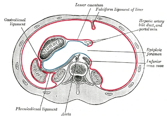
網嚢は胃の後ろの扁平な袋で、右の網嚢孔だけが開いている。壁を6方向で言えるようにする。
| 壁 | 作るもの |
|---|---|
| 上 | 肝尾状葉と横隔膜(を覆う腹膜) |
| 前 | 小網・胃の後面・胃結腸間膜(大網の前2葉) |
| 下 | 横行結腸と横行結腸間膜(大網の前後の葉が合わさる所) |
| 後 | 後腹壁の壁側腹膜と、その奥の膵・腹大動脈・下大静脈・左副腎・左腎上部 |
| 左 | 脾・胃脾間膜・横隔脾ひだ(脾腎間膜) |
| 右 | 網嚢孔(前=肝十二指腸間膜、後=下大静脈、上=尾状葉、下=十二指腸上部) |
中は4つに分ける:vestibulum(網嚢孔のすぐ奥の狭い前庭・膵頭の上)・recessus superior(尾状葉と横隔膜の間)・recessus inferior(胃結腸間膜と横行結腸間膜の間、大網の中へ下がる)・recessus splenicus(脾の方へ伸びる左端)。
網嚢ができるのは、胎生期に胃が回転し、背側胃間膜が左へ袋状に張り出すからである(発生は本章の最後の項目)。だから後壁には背側にあった膵が、左壁には背側胃間膜の中で育った脾が来る。壁の並びは暗記ではなく、胃の回転から再現できる。
膵が網嚢の後壁にあることが臨床を決める。急性膵炎の滲出液や膵仮性嚢胞は網嚢に溜まり、胃を前へ押す。胃後壁の潰瘍が穿孔すると内容物はまず網嚢に入り、網嚢孔を通って右の肝下へ出てくる。
膵を外科的に見るときは、胃結腸間膜を切って網嚢を前から開ける。網嚢孔に指を入れて前縁(肝十二指腸間膜)をつまめば、肝への血流を一時的に止められる(Pringle 法)。
English version
Core
The omental bursa is a flat sac behind the stomach. Its only opening is the omental foramen on the right. Be able to name its walls in six directions.
| Wall | Formed by |
|---|---|
| Superior | Caudate lobe and diaphragm (their peritoneum) |
| Anterior | Lesser omentum, posterior surface of stomach, gastrocolic ligament (front two layers of greater omentum) |
| Inferior | Transverse colon and transverse mesocolon (where the front and back layers of the greater omentum meet) |
| Posterior | Parietal peritoneum of the posterior wall, with pancreas, abdominal aorta, inferior vena cava, left adrenal and upper left kidney behind it |
| Left | Spleen, gastrosplenic ligament, phrenicosplenic (splenorenal) ligament |
| Right | Omental foramen (front: hepatoduodenal ligament; back: IVC; above: caudate lobe; below: first part of duodenum) |
Inside, four parts: the vestibulum (narrow entrance just inside the foramen, above the head of the pancreas), the recessus superior (between caudate lobe and diaphragm), the recessus inferior (between gastrocolic ligament and transverse mesocolon, extending into the greater omentum) and the recessus splenicus (the left end towards the spleen).
Going further
The bursa exists because in the embryo the stomach rotates and the dorsal mesogastrium bulges to the left as a sac (development is in the last item of this chapter). That is why the pancreas, once dorsal, forms the back wall, and the spleen, which grew inside the dorsal mesogastrium, forms the left wall. The order of the walls can be rebuilt from the rotation instead of memorised.
In the clinic
The pancreas lies in the back wall of the bursa, and this shapes clinical findings. Exudate from acute pancreatitis and pancreatic pseudocysts collect in the bursa and push the stomach forward. A perforated ulcer of the posterior gastric wall first empties into the bursa and then appears under the right liver through the foramen.
To see the pancreas at surgery, the gastrocolic ligament is divided and the bursa opened from the front. A finger in the omental foramen can pinch its front edge (hepatoduodenal ligament) and stop inflow to the liver for a while (Pringle manoeuvre).
中階 — 左右の腸間膜洞と、左右の結腸傍溝Sinus mesentericus, sulcus paracolicus
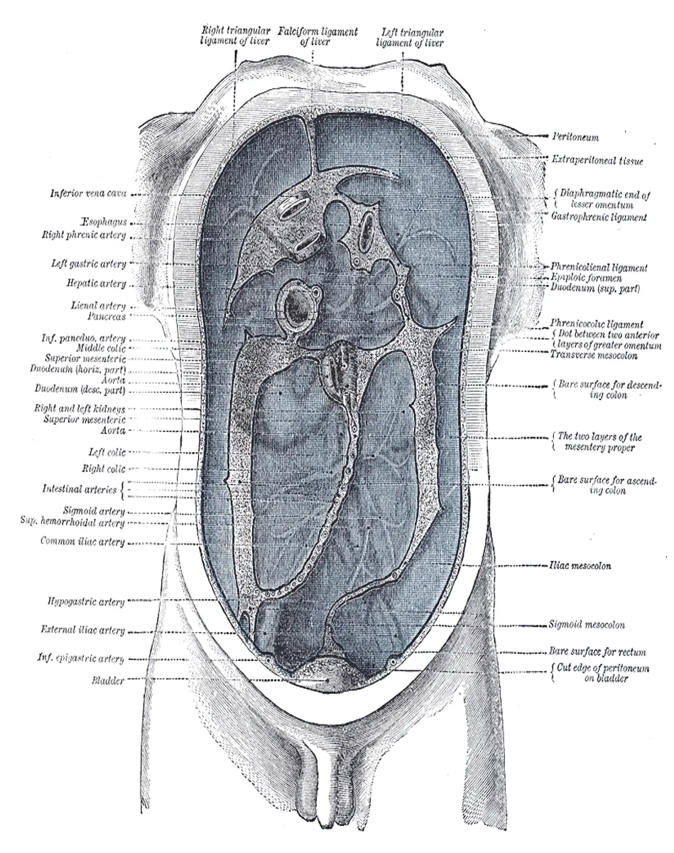
中階は、腸間膜根と上行・下行結腸によって4つの区画に分かれる。
| 区画 | 境界 | 後ろ(腹膜の奥)にあるもの |
|---|---|---|
| Sinus mesentericus dexter(右腸間膜洞) | 左と下=腸間膜根、右=上行結腸、上=横行結腸と横行結腸間膜。三角形で、四方が閉じている | 十二指腸下行部・水平部、右腎、右尿管 |
| Sinus mesentericus sinister(左腸間膜洞) | 右=腸間膜根、左=下行結腸、下方は S状結腸間膜に沿って小骨盤へ開く | 十二指腸上行部、左腎、左尿管 |
| Sulcus paracolicus dexter(右結腸傍溝) | 上行結腸の外側と側腹壁の間 | 上は肝下陥凹へ、下は右腸骨窩から骨盤へ続く |
| Sulcus paracolicus sinister(左結腸傍溝) | 下行結腸の外側と側腹壁の間 | 上端は横隔結腸ひだでふさがれ、下は左腸骨窩から骨盤へ続く |
結腸傍溝は canalis lateralis(外側管)とも呼ばれる。
左右の洞の違いは、腸間膜根が斜めに走ることから出てくる。根は L2 の左から右仙腸関節の前へ下るので、右の洞は根に下をふさがれて閉じた三角になり、左の洞は下が骨盤へ抜ける。
だから右の洞にできた液や膿はそこに閉じ込められやすく、左の洞の液は骨盤へ流れ落ちる。縦の溝(結腸傍溝)は階をまたぐ通路、洞は区切られた部屋、と役割を分けて覚える。
右結腸傍溝は上腹部と骨盤を結ぶ「高速道路」である。十二指腸潰瘍が穿孔すると、流れ出た内容物がこの溝を下って右腸骨窩に溜まり、虫垂炎とまぎらわしい右下腹部痛を起こすことがある(Valentino 症候群)。逆に、骨盤や虫垂の膿が上って肝下膿瘍を作る。腹水や腹腔内出血の超音波検査で、結腸傍溝と骨盤を必ず見るのはこのためである。
English version
Core
The middle storey is divided into four spaces by the root of the mesentery and the ascending and descending colon.
| Space | Boundaries | Behind the peritoneum |
|---|---|---|
| Sinus mesentericus dexter (right mesenteric sinus) | Left and below: root of mesentery; right: ascending colon; above: transverse colon and mesocolon. Triangular and closed on all sides | Descending and horizontal parts of duodenum, right kidney, right ureter |
| Sinus mesentericus sinister (left mesenteric sinus) | Right: root of mesentery; left: descending colon; below it opens into the lesser pelvis along the sigmoid mesocolon | Ascending part of duodenum, left kidney, left ureter |
| Sulcus paracolicus dexter (right paracolic gutter) | Between ascending colon and lateral abdominal wall | Continues up to the subhepatic recess and down through the right iliac fossa to the pelvis |
| Sulcus paracolicus sinister (left paracolic gutter) | Between descending colon and lateral abdominal wall | Closed above by the phrenicocolic ligament; continues down through the left iliac fossa to the pelvis |
The paracolic gutters are also called canales laterales.
Going further
The difference between the two sinuses comes from the oblique course of the mesenteric root. The root runs from the left of L2 down to the front of the right sacroiliac joint. The right sinus is closed below by the root and forms a closed triangle; the left sinus opens downward into the pelvis.
Fluid or pus in the right sinus therefore tends to stay there, while fluid in the left sinus drains into the pelvis. Remember the roles: the vertical gutters are corridors between storeys; the sinuses are closed rooms.
In the clinic
The right paracolic gutter is a highway between the upper abdomen and the pelvis. When a duodenal ulcer perforates, the leaked contents can run down this gutter to the right iliac fossa and cause right lower pain that mimics appendicitis (Valentino syndrome). In the other direction, pus from the pelvis or appendix can climb to form a subhepatic abscess. This is why an ultrasound search for ascites or intra-abdominal bleeding always includes the gutters and the pelvis.
後腹壁の陥凹 — 腸が壁に付く所にできるポケットRecessus duodenalis, recessus ileocaecalis, recessus retrocaecalis, recessus intersigmoideus
腹膜が腸の付着部や血管を乗り越える所には、小さなひだと、その陰のポケット(陥凹)ができる。中階には次の4組がある。
| 陥凹 | 場所と、それを作るひだ |
|---|---|
| Recessus duodenalis superior / inferior | 十二指腸空腸曲の左。上は plica duodenalis superior、下は plica duodenalis inferior が縁を作る(左腸間膜洞の上部) |
| Recessus ileocaecalis superior / inferior | 回腸末端が盲腸に入る所の上と下。上は盲腸へ行く血管を含む plica caecalis vascularis、下は plica ileocaecalis(血管の無いひだ)が作る |
| Recessus retrocaecalis | 盲腸の後ろ。plicae caecales に挟まれる。虫垂が入り込んでいることが多い |
| Recessus intersigmoideus | S状結腸間膜の根が逆V字に付く、その頂点の下。左腹膜の奥で左尿管がこの頂点の後ろを通る |
陥凹は「腸が後腹壁に固定されたときの残り」である。十二指腸空腸曲・回盲部・S状結腸はいずれも、壁に付いた部分と自由に動く部分の境目にあたり、その段差にポケットが残る。
S状結腸間陥凹は手術の目印としても使われる。陥凹の頂点を探れば、その奥に左尿管と左総腸骨動脈の分岐が触れる。
陥凹に小腸が入り込んで抜けなくなると内ヘルニアになる。代表が十二指腸陥凹で起こる傍十二指腸ヘルニアで、腸閉塞の原因になる。
虫垂が盲腸後陥凹にある(盲腸後位)と、前腹壁の圧痛や反跳痛が出にくく、後ろの大腰筋を刺激して股関節を伸ばすと痛む(psoas sign)。虫垂の位置が症状を変える例である。
English version
Core
Where the peritoneum passes over a bowel attachment or a vessel, it makes small folds with pockets (recesses) behind them. There are four groups in the middle storey.
| Recess | Position and the folds that form it |
|---|---|
| Recessus duodenalis superior / inferior | Left of the duodenojejunal flexure, bordered by the plica duodenalis superior and inferior (upper part of the left mesenteric sinus) |
| Recessus ileocaecalis superior / inferior | Above and below the entry of the terminal ileum into the caecum. Above: plica caecalis vascularis carrying vessels to the caecum; below: plica ileocaecalis (a bloodless fold) |
| Recessus retrocaecalis | Behind the caecum, between the plicae caecales. The appendix often lies here |
| Recessus intersigmoideus | Below the apex of the inverted-V root of the sigmoid mesocolon. The left ureter passes behind this apex |
Going further
Recesses are the leftovers of the gut becoming fixed to the posterior wall. The duodenojejunal flexure, the ileocaecal region and the sigmoid colon all lie where a fixed part meets a free part, and a pocket remains at the step.
The intersigmoid recess is also a surgical landmark: feel at its apex and the left ureter and the division of the left common iliac artery lie just behind.
In the clinic
If a loop of small bowel enters a recess and cannot come out, it is an internal hernia. The classic example is the paraduodenal hernia at the duodenal recesses, a cause of bowel obstruction.
When the appendix lies in the retrocaecal recess, tenderness and rebound on the anterior wall may be weak, while stretching the psoas by extending the hip causes pain (psoas sign). The position of the appendix changes the signs.
前腹壁の内面 — 5本のひだと3対の窩Plicae umbilicales, fossae inguinales
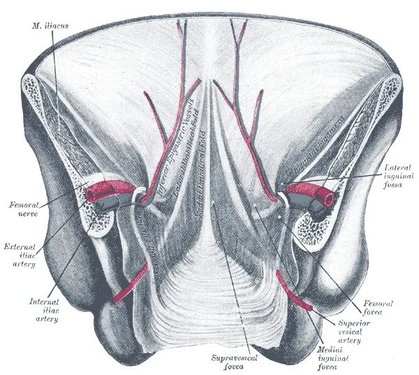
前腹壁を内側から見ると、臍へ向かって5本のひだが立ち、そのあいだに3対の窩ができる。ひだの名前は、中に入っているもので覚える。
| ひだ(内側から) | 中身 | その外側にできる窩 |
|---|---|---|
| Plica umbilicalis mediana(正中臍ひだ・1本) | 尿膜管の遺残=正中臍索 | Fossa supravesicalis(膀胱上窩) |
| Plica umbilicalis medialis(内側臍ひだ・左右) | 閉じた臍動脈=内側臍索 | Fossa inguinalis medialis(内側鼠径窩)— 浅鼠径輪の裏 |
| Plica umbilicalis lateralis(外側臍ひだ・左右) | 下腹壁動静脈 | Fossa inguinalis lateralis(外側鼠径窩)— 深鼠径輪の位置 |
ひだが臍に集まるのは、中身がもともと臍帯の中身だったからである。尿膜管も臍動脈も胎児と胎盤をつないでいた管で、生まれると閉じて索になり、腹膜を持ち上げるひだとして残った。外側臍ひだだけは例外で、今も血液が流れる下腹壁動静脈が作る。
内側臍索は根元の部分だけが開いたまま残り、そこから上膀胱動脈が出る(泌尿器の章の「膀胱の血管」)。
鼠径ヘルニアの区別がこの窩で決まる。外側鼠径窩(下腹壁動静脈の外側)から深鼠径輪に入るのが外鼠径(間接)ヘルニア、内側鼠径窩(下腹壁動静脈の内側・Hesselbach 三角)から腹壁を直接押し出すのが内鼠径(直接)ヘルニア。腹腔鏡でヘルニアを修復するときは、まさにこの5本のひだを内側から見て判断する。
尿膜管が閉じきらないと、臍から尿が漏れる尿膜管瘻や、途中が袋になる尿膜管嚢胞になる(泌尿器の発生)。
English version
Core
Seen from inside, the anterior abdominal wall has five folds converging on the umbilicus, with three pairs of fossae between them. Name each fold by what lies inside it.
| Fold (from medial) | Contents | Fossa lateral to it |
|---|---|---|
| Plica umbilicalis mediana (median, single) | Remnant of the urachus = median umbilical ligament | Fossa supravesicalis |
| Plica umbilicalis medialis (medial, paired) | Obliterated umbilical artery = medial umbilical ligament | Fossa inguinalis medialis — behind the superficial inguinal ring |
| Plica umbilicalis lateralis (lateral, paired) | Inferior epigastric artery and vein | Fossa inguinalis lateralis — at the deep inguinal ring |
Going further
The folds converge on the umbilicus because their contents once belonged to the umbilical cord. The urachus and the umbilical arteries connected the fetus with the placenta; after birth they closed into cords that still lift the peritoneum. The lateral fold is the exception: it is raised by the living inferior epigastric vessels.
Only the root of the medial umbilical ligament stays open, and the superior vesical arteries arise from it (see “Vessels of the bladder” in the urinary chapter).
In the clinic
These fossae separate the two inguinal hernias. A hernia entering the deep ring at the lateral fossa (lateral to the inferior epigastric vessels) is indirect; one pushing the wall straight out at the medial fossa (medial to the vessels, Hesselbach’s triangle) is direct. In laparoscopic hernia repair the surgeon judges exactly these five folds from inside.
If the urachus fails to close, urine may leak from the umbilicus (urachal fistula) or a cyst may form along its course (see urinary development).
腹膜の働きと血管・神経 — 壁側は腹壁の、臓側は臓器のVasa et nervi peritonei
働き:①漿液を出して臓器の表面を滑らかにする ②液や小さな粒子を吸収する ③臓器を間膜・靱帯で吊って固定する ④大網などで炎症を囲い込む ⑤傷ついた所にフィブリンを出して癒着させ、穴をふさぐ。表面積は皮膚とほぼ同じ(約2 m²)と広い。
血管と神経は「壁側は腹壁のもの、臓側は臓器のもの」という1つの規則で決まる。
| 壁側腹膜 | 臓側腹膜 | |
|---|---|---|
| 動脈 | 腹壁の動脈:下位肋間動脈・腰動脈・上/下腹壁動脈・深腸骨回旋動脈・下横隔動脈 | 覆っている臓器の動脈(腹腔動脈・上腸間膜動脈・下腸間膜動脈の枝) |
| 静脈 | 体循環(大静脈系)へ | 門脈系へ |
| 神経 | 体性神経:下位肋間神経(Th7〜12)・腸骨下腹神経など、横隔膜中央部は横隔神経 | 自律神経(内臓求心線維) |
| 痛み | 鋭く、場所がはっきりする | 鈍く、正中にぼんやり感じる |
規則の理由は発生にある。壁側腹膜は体壁(壁側中胚葉)の一部として、臓側腹膜は臓器の壁(臓側中胚葉)の一部として作られる。胸膜でも同じ規則が成り立つ(呼吸器の章「胸膜の血管と神経」)。
吸収は上腹部、とくに横隔膜の腹膜が速い。横隔膜の面にはリンパ管へ直接通じる小孔があり、呼吸のたびに液を吸い込むからである。腹膜炎の患者を半坐位(Fowler 位)にするのは、膿を吸収の遅い骨盤に溜めて、毒素が急に吸収されるのを防ぐためである。
腹膜透析は、広い腹膜と、その下の毛細血管を半透膜として使う治療である。腹腔に透析液を入れて置いておくと、血液中の老廃物と水が腹膜を通って液へ移る。
腹膜が傷つくと出るフィブリンは穴をふさぐ一方で、手術のあとに癒着を残し、何年もたって癒着性腸閉塞の原因になる。腹痛の性質(臓側の鈍い痛みから壁側の鋭い痛みへ)は本章の「腹膜」の項目を参照。
English version
Core
Functions: (1) secretes serous fluid so organ surfaces glide; (2) absorbs fluid and small particles; (3) suspends and fixes organs by mesenteries and ligaments; (4) walls off inflammation, for example with the greater omentum; (5) releases fibrin at injured sites, causing adhesion and sealing holes. Its surface area is about the same as the skin (about 2 m²).
Vessels and nerves follow one rule: the parietal layer takes those of the body wall, the visceral layer those of the organ.
| Parietal peritoneum | Visceral peritoneum | |
|---|---|---|
| Arteries | Body-wall arteries: lower intercostal, lumbar, superior/inferior epigastric, deep circumflex iliac, inferior phrenic | Arteries of the organ it covers (branches of coeliac trunk, SMA, IMA) |
| Veins | To the systemic (caval) veins | To the portal system |
| Nerves | Somatic: lower intercostal nerves (T7–12), iliohypogastric etc.; phrenic nerve for the central diaphragm | Autonomic (visceral afferents) |
| Pain | Sharp and well localised | Dull, felt vaguely in the midline |
Going further
The reason lies in development. The parietal peritoneum forms as part of the body wall (somatic mesoderm); the visceral peritoneum forms as part of the organ wall (splanchnic mesoderm). The same rule works for the pleura (see “Vessels and nerves of the pleura” in the respiratory chapter).
Absorption is fastest in the upper abdomen, especially over the diaphragm, where small openings lead straight into lymphatics and each breath sucks fluid in. Patients with peritonitis are nursed half-sitting (Fowler’s position) so that pus collects in the pelvis, where absorption is slow, and toxins are not absorbed too quickly.
In the clinic
Peritoneal dialysis uses the large peritoneum and the capillaries under it as a semipermeable membrane. Dialysis fluid is left in the abdomen, and waste products and water move from the blood across the peritoneum into the fluid.
The fibrin that seals injuries also leaves adhesions after surgery, which can cause adhesive bowel obstruction years later. For the character of abdominal pain (dull visceral pain turning into sharp parietal pain), see the “Peritoneum” item of this chapter.
肝臓 — 横隔面と臓側面Hepar
体内最大の腺。右季肋部を占め、上面が横隔面 facies diaphragmatica(横隔膜に接して滑らかに凸)、下面が臓側面 facies visceralis(隣の臓器の圧痕で凹凸)。
臓側面にはH 字の溝が刻まれる——右の縦溝が胆嚢と下大静脈、左の縦溝が肝円索と静脈管索、横棒が肝門。この H を読めれば4葉がそのまま決まる。
English version
Core
The largest gland in the body, occupying the right hypochondrium. The upper surface is the facies diaphragmatica (smoothly convex against the diaphragm), the lower the facies visceralis (irregular with the impressions of neighbouring organs).
The visceral surface is engraved with an H — the right vertical limb the gallbladder and inferior vena cava, the left the round and venous ligaments, the cross-bar the porta hepatis. Read this H and the four lobes follow at once.
無漿膜野Area nuda
横隔面の後上部で、腹膜に覆われず横隔膜と直に接している三角形の領域。冠状間膜の上下の葉に挟まれる。
ここでは肝と横隔膜の間に腹膜が無いので、肝の感染が直接横隔膜下へ及ぶ。
English version
Core
The triangular region on the posterosuperior part of the diaphragmatic surface that is not covered by peritoneum and lies directly against the diaphragm, between the upper and lower leaves of the coronary ligament.
Here there is no peritoneum between liver and diaphragm, so hepatic infection can pass directly into the subphrenic space.
肝鎌状間膜Ligamentum falciforme hepatis
肝の横隔面から前腹壁へ張る鎌形の二重腹膜。肝を左右の葉に分ける外見上の境。
下縁の遊離縁に肝円索(臍静脈の遺残)が入る。腹膜の由来は腹側間膜で、消化管でここだけが残ったもの。
English version
Core
The sickle-shaped double fold of peritoneum passing from the diaphragmatic surface of the liver to the anterior abdominal wall — the apparent boundary dividing the liver into right and left lobes. The round ligament (the remnant of the umbilical vein) lies in its free lower margin.
It derives from the ventral mesentery, the only part of it that remains in the gut.
肝右葉Lobus dexter hepatis
鎌状間膜より右の大きな葉。形態学的には全体の約6分の5を占める。
「形態学的な分け方」と「機能的な分け方」がずれる——機能的には胆嚢窩と下大静脈を結ぶ Cantlie 線が境で、方形葉と尾状葉は左側に入る。この食い違いを説明できるようにしておく。
English version
Core
The large lobe to the right of the falciform ligament, morphologically about five sixths of the whole.
The morphological and the functional divisions do not agree — functionally the boundary is Cantlie’s line (from the gallbladder fossa to the inferior vena cava), and the quadrate and caudate lobes fall on the left. Be able to explain this discrepancy.
肝左葉Lobus sinister hepatis
鎌状間膜より左の小さい葉。
English version
Core
The smaller lobe to the left of the falciform ligament.
方形葉Lobus quadratus
臓側面で、肝門より前、胆嚢窩と肝円索裂に挟まれた四角い部分。
English version
Core
The quadrilateral area on the visceral surface in front of the porta, between the gallbladder fossa and the fissure for the round ligament.
尾状葉Lobus caudatus
臓側面で、肝門より後ろ、下大静脈と静脈管索に挟まれた部分。
肝門を中心に「前が方形葉、後ろが尾状葉」。尾状葉は独自に下大静脈へ短い静脈を出すので、Budd-Chiari 症候群でここだけ肥大する。
English version
Core
The area on the visceral surface behind the porta, between the inferior vena cava and the fissure for the ligamentum venosum.
Taking the porta as the centre: quadrate in front, caudate behind.
The caudate lobe sends its own short veins directly to the inferior vena cava, so in Budd–Chiari syndrome it alone hypertrophies.
肝門Porta hepatis
臓側面のH 字の横棒。ここを門脈・固有肝動脈・肝管・神経・リンパ管が出入りする。
標本では方形葉と尾状葉の間の横の溝として示す。
English version
Core
The cross-bar of the H on the visceral surface. The portal vein, proper hepatic artery, hepatic ducts, nerves and lymphatics enter and leave here.
On the specimen, show it as the transverse fissure between the quadrate and caudate lobes.
門脈・固有肝動脈Vena portae, arteria hepatica propria
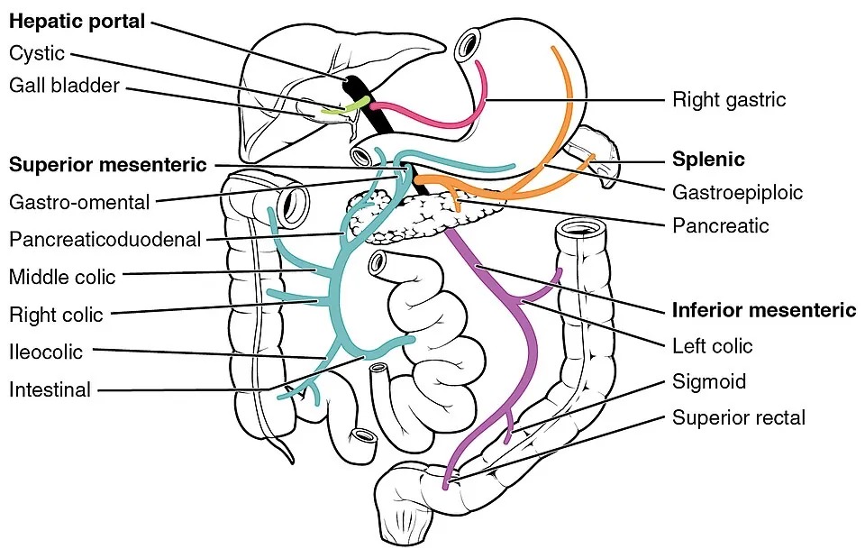
肝十二指腸間膜の中を上る。並びは前右が総胆管、前左が固有肝動脈、その後ろに門脈——この三者の位置関係が定番の設問。
肝への血流は門脈が約7割・動脈が約3割。門脈は脾静脈と上腸間膜静脈が膵頭の後ろで合してできる。
English version
Core
They ascend within the hepatoduodenal ligament. The arrangement is common bile duct anterior and to the right, proper hepatic artery anterior and to the left, and the portal vein behind them — the relation of these three is a standard question.
The liver’s blood is about 70 per cent portal and 30 per cent arterial. The portal vein is formed behind the head of the pancreas by the union of the splenic and superior mesenteric veins.
胆嚢(底・体・頸)Vesica biliaris
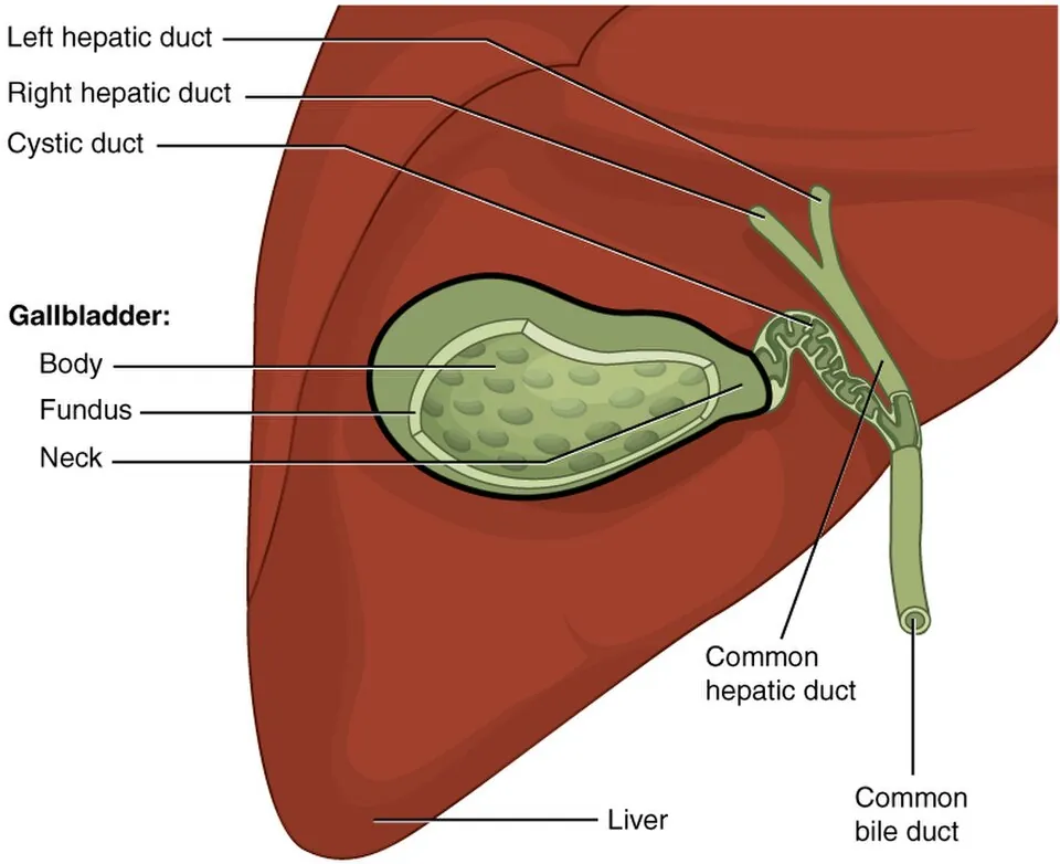
肝の臓側面、胆嚢窩に収まる袋。底 fundus・体 corpus・頸 collum の3部。
底は肝の下縁から前へ出て前腹壁に触れる——その体表投影が右鎖骨中線と第9肋軟骨の交点で、Murphy 徴候をとる場所。頸は S 字に曲がり胆嚢管に続く。
English version
Core
The sac lodged in the gallbladder fossa on the visceral surface of the liver, in three parts — fundus, corpus, collum.
The fundus projects forwards beyond the inferior border of the liver and touches the anterior abdominal wall; its surface marking, where the right midclavicular line crosses the ninth costal cartilage, is where Murphy’s sign is elicited. The neck curves in an S into the cystic duct.
胆嚢管Ductus cysticus
胆嚢頸から出て総肝管に合流する。内腔にらせんヒダ(Heister 弁)があり、閉じずに保つ。
胆嚢管・総肝管・肝下縁で囲まれるCalot 三角に胆嚢動脈が走る。胆嚢摘出でここを確実に出すのが手順の要。
English version
Core
Runs from the neck of the gallbladder to join the common hepatic duct. Its lumen carries a spiral fold (Heister’s valve) which keeps it from closing.
The cystic artery runs in Calot’s triangle (cystic duct, common hepatic duct, inferior border of the liver). Displaying it reliably is the crux of the procedure in cholecystectomy.
総肝管Ductus hepaticus communis
肝門で左右の肝管が合してできる。下って胆嚢管と合し、総胆管になる。
English version
Core
Formed at the porta by the union of the right and left hepatic ducts. It descends and joins the cystic duct to become the common bile duct.
総胆管Ductus choledochus
総肝管+胆嚢管。肝十二指腸間膜を下り、膵頭の後ろを通って大十二指腸乳頭に開く。
膵頭癌がここを圧迫すると閉塞性黄疸になる。無痛性黄疸+腫大した胆嚢=Courvoisier 徴候。
English version
Core
Common hepatic duct + cystic duct. It descends in the hepatoduodenal ligament, passes behind the head of the pancreas and opens at the major duodenal papilla.
Compression by carcinoma of the head of the pancreas gives obstructive jaundice. Painless jaundice with a distended gallbladder = Courvoisier’s sign.
膵頭Caput pancreatis
十二指腸のC字にはめ込まれた右端の部分。左下へ出る突起が鉤状突起で、その前を上腸間膜動静脈が通る。
後面を総胆管が下るので、ここの腫瘍は黄疸で発見される。
English version
Core
The right-hand part set into the C of the duodenum. The process projecting to the lower left is the uncinate process, in front of which pass the superior mesenteric vessels.
The common bile duct descends on its posterior surface, so a tumour here is found through jaundice.
膵体・膵尾Corpus et cauda pancreatis
体は L1 の高さで脊柱を左へ横切り、尾は脾門に達する。膵は後腹膜だが、尾だけは脾腎間膜の中に入って腹腔内になる。
そのため脾摘のときに膵尾を傷つけることがある。
English version
Core
The body crosses the vertebral column to the left at the level of L1, and the tail reaches the splenic hilum.
The pancreas is retroperitoneal, but the tail alone enters the splenorenal ligament and becomes intraperitoneal. This is why the tail may be injured during splenectomy.
脾臓 — 横隔面と臓側面Splen
左季肋部、第9〜11肋骨の裏にある。凸の横隔面と、胃・腎・結腸の圧痕がある臓側面。
正常では肋骨弓下に触れない。触れたら腫大。上縁の切痕が触診の目印になる。
English version
Core
In the left hypochondrium, behind the ninth to eleventh ribs. A convex diaphragmatic surface and a visceral surface bearing gastric, renal and colic impressions.
Normally it cannot be felt below the costal margin; if it can, it is enlarged. The notch on its upper border is the landmark for palpation.
脾門Hilum splenicum
臓側面の中央にある裂で、脾動静脈が出入りする。膵尾がここに接する。
脾は胃脾間膜と脾腎間膜の2枚で吊られ、どちらも脾門に付く。
English version
Core
The fissure at the centre of the visceral surface, through which the splenic vessels pass; the tail of the pancreas touches it.
The spleen is slung by two ligaments, gastrosplenic and splenorenal, and both attach at the hilum.
大網Omentum majus
胃の大弯から前掛けのように垂れ下がり、折り返して横行結腸へ戻る4枚の腹膜。
脂肪とマクロファージに富み、炎症のある場所へ自分から移動して包み込む——「腹腔の警察官」。虫垂炎が限局するのはこの働きによる。
English version
Core
The four layers of peritoneum hanging like an apron from the greater curvature of the stomach and turning back to the transverse colon. Rich in fat and macrophages, it migrates of its own accord to a site of inflammation and wraps it — "the policeman of the abdomen". That appendicitis becomes localised is due to this.
直腸子宮窩(Douglas 窩)Excavatio rectouterina
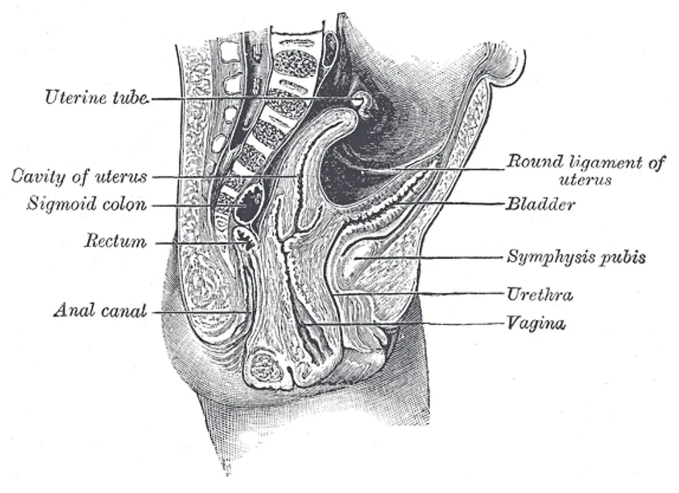
女性で子宮の後ろと直腸の間にできる腹膜のくぼみ。
立位・仰臥位のどちらでも腹腔の最下部なので、腹腔内の液体(血液・膿・腹水)が最初に溜まる。後腟円蓋から穿刺できる。
English version
Core
The peritoneal hollow between the uterus and the rectum in the woman. It is the lowest part of the peritoneal cavity both standing and supine, so intraperitoneal fluid (blood, pus, ascites) collects there first. It can be aspirated through the posterior fornix.
膀胱子宮窩Excavatio vesicouterina
女性で膀胱と子宮の間のくぼみ。Douglas 窩より浅い。
女性の骨盤腹膜は「膀胱 → 子宮 → 直腸」と越えるので、くぼみが2つできる。
English version
Core
The hollow between bladder and uterus in the woman, shallower than the pouch of Douglas.
The pelvic peritoneum of the woman passes over bladder → uterus → rectum, and so forms two hollows.
直腸膀胱窩Excavatio rectovesicalis
男性で膀胱と直腸の間のくぼみ。男性では子宮が無いので、くぼみは1つだけ。ここが男性の腹腔最下部になる。
English version
Core
The hollow between bladder and rectum in the man. With no uterus there is only one hollow, and this is the lowest part of the peritoneal cavity in the male.
発生 — 腹側間膜から肝と小網が、背側間膜から大網と脾ができるMesenterium ventrale et dorsale
原始腸管は背側間膜で後腹壁から吊られ、腹部食道・胃・十二指腸の上部(前腸の部分)だけは腹側間膜でも前腹壁につながる。
肝は第4週に十二指腸の内胚葉から芽(肝憩室)として出て、腹側間膜の中(横中隔)へ伸びて育つ。その結果、腹側間膜は肝で2つに分けられ、胃〜肝の部分が小網、肝〜前腹壁の部分が鎌状間膜になる。鎌状間膜の下縁を走っていた臍静脈が、生後の肝円索である。胆嚢は同じ芽の小さな枝から、膵は背側と腹側の2つの芽からできる(本章の「膵臓」)。
胃は長軸のまわりに右へ90°回り(左側面が前を向く)、背側胃間膜が左へ大きく張り出して胃の後ろに袋を作る——これが網嚢である。張り出した背側胃間膜はさらに下へ垂れて4枚の大網になり、後ろの葉は横行結腸間膜と癒合する。脾は背側胃間膜の中に生じるので、胃脾間膜と脾腎間膜で吊られる。十二指腸と膵は後腹壁に押し付けられ、間膜が癒合して二次的後腹膜になる。
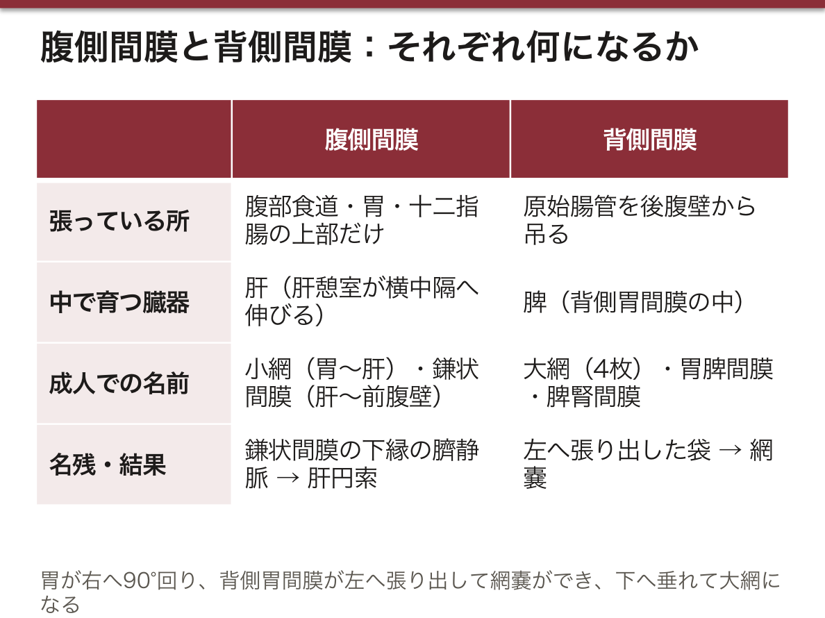
胃の回転が説明することは多い。左の迷走神経が胃の前面に、右が後面に回るのは、胃の左側面が前を向いたからである。小網が網嚢の前にあるのも、大網が4枚なのも、脾動静脈が脾腎間膜の中を走るのも、同じ回転と張り出しの結果である。
この時期の異常には次のものがある。胆道閉鎖症——胆管がふさがり、新生児期を過ぎても黄疸が続き、白っぽい便が出る。早く手術(肝門部と腸をつなぐ)をするほど自分の肝で生きられる見込みが上がる。輪状膵——膵の腹側芽が十二指腸を取り囲み、十二指腸の狭窄を起こす。異所性膵——胃や十二指腸の壁に膵組織が残る。
English version
Core
The primitive gut hangs from the posterior wall by the dorsal mesentery. Only the abdominal oesophagus, stomach and upper duodenum (the foregut part) are also joined to the anterior wall by a ventral mesentery.
The liver appears in week 4 as a bud (hepatic diverticulum) from the endoderm of the duodenum and grows into the ventral mesentery (septum transversum). The liver therefore splits the ventral mesentery in two: the stomach–liver part becomes the lesser omentum, and the liver–anterior wall part becomes the falciform ligament. The umbilical vein that ran in the free edge of the falciform ligament becomes the ligamentum teres after birth. The gallbladder comes from a small branch of the same bud; the pancreas comes from dorsal and ventral buds (see “Pancreas” in this chapter).
The stomach turns 90° to the right around its long axis (its left side comes to face forward), and the dorsal mesogastrium bulges strongly to the left, making a sac behind the stomach — the omental bursa. The bulging dorsal mesogastrium then hangs down as the four-layered greater omentum, and its back layers fuse with the transverse mesocolon. The spleen forms inside the dorsal mesogastrium, so it hangs by the gastrosplenic and splenorenal ligaments. The duodenum and pancreas are pressed against the posterior wall, their mesentery fuses, and they become secondarily retroperitoneal.
Going further
The rotation of the stomach explains a lot. The left vagus comes to lie on the front of the stomach and the right vagus on the back because the left side of the stomach turned forward. The lesser omentum lying in front of the bursa, the greater omentum having four layers, and the splenic vessels running in the splenorenal ligament are all results of the same rotation and bulging.
In the clinic
Anomalies of this stage include biliary atresia — the bile ducts are blocked, jaundice persists beyond the newborn period and stools are pale. The earlier the operation (joining the porta hepatis to the bowel), the better the chance of living on the child’s own liver. Annular pancreas — the ventral pancreatic bud encircles the duodenum and narrows it. Heterotopic pancreas — pancreatic tissue left in the wall of the stomach or duodenum.
まとめ
| 肝臓 — 2つの面と、臓側面のH | 右上腹部、横隔膜の下。約1.5kg で最大の腺。 |
|---|---|
| 肝臓の働き — 門脈の出口に置かれた関所 | 肝の仕事は大きく6つに分けて言う。働き中身① 消化(外分泌)胆汁を絶えず作り、肝管から十二指腸へ送る(1日に約0.5〜1 L)。 |
| 腹部の9区分と、肝の上界・下界 | 腹部の9区分は、横の線2本と縦の線2本で作る。 |
| 胆嚢と胆道 — 上流から下流へ | Vesica biliaris (fellea) は肝の臓側面の胆嚢窩に収まる袋(容量30〜50mL)。 |
| 膵臓 — 後腹膜のC字の相手 | 胃の後ろ、L1〜L2 の高さを横に走る後腹膜臓器(尾部だけが脾腎間膜に入り腹膜内)。 |
| 脾臓 — 左季肋部の血液の器官 | 左季肋部、第9〜11肋骨の内側、横隔膜と胃底のあいだ。 |
| 腹膜 — 袋と、そのひだ | Peritoneum parietale(壁側・腹壁の内面)と peritoneum viscerale(臓側・臓器の表面)が連続した1枚の閉じた袋。 |
| 腹膜と臓器の位置関係 — intra / meso / retro と、日本式の分け方 | ヨーロッパの教科書は「成人で、臓器の表面のどれだけを腹膜が覆っているか」で4つに分ける。 |
| 腹膜腔の3階 — 横行結腸間膜と分界線で区切る | 腹膜腔は上下に3つの階に分けて考える。階範囲中にあるもの・区画上階横隔膜 〜 横行結腸と横行結腸間膜肝・胆嚢・胃・脾・十二指腸の上部。 |
| 上階 — 肝嚢・胃前嚢・網嚢と、横隔膜下・肝下の陥凹 | 上階は3つの嚢に分けて呼ぶ。Bursa hepatica(肝嚢)— 鎌状間膜より右で、肝右葉を包む区画。 |
| 網嚢 — 6つの壁と4つの部屋 | 網嚢は胃の後ろの扁平な袋で、右の網嚢孔だけが開いている。 |
| 中階 — 左右の腸間膜洞と、左右の結腸傍溝 | 中階は、腸間膜根と上行・下行結腸によって4つの区画に分かれる。 |
| 後腹壁の陥凹 — 腸が壁に付く所にできるポケット | 腹膜が腸の付着部や血管を乗り越える所には、小さなひだと、その陰のポケット(陥凹)ができる。 |
| 前腹壁の内面 — 5本のひだと3対の窩 | 前腹壁を内側から見ると、臍へ向かって5本のひだが立ち、そのあいだに3対の窩ができる。 |
| 腹膜の働きと血管・神経 — 壁側は腹壁の、臓側は臓器の | 働き:①漿液を出して臓器の表面を滑らかにする ②液や小さな粒子を吸収する ③臓器を間膜・靱帯で吊って固定する ④大網などで炎症を囲い込む ⑤傷ついた所にフィブリンを出して癒着させ、… |
| 肝臓 — 横隔面と臓側面 | 体内最大の腺。右季肋部を占め、上面が横隔面 facies diaphragmatica(横隔膜に接して滑らかに凸)、下面が臓側面 facies visceralis(隣の臓器の圧痕… |
| 無漿膜野 | 横隔面の後上部で、腹膜に覆われず横隔膜と直に接している三角形の領域。 |
| 肝鎌状間膜 | 肝の横隔面から前腹壁へ張る鎌形の二重腹膜。 |
| 肝右葉 | 鎌状間膜より右の大きな葉。形態学的には全体の約6分の5を占める。 |
| 肝左葉 | 鎌状間膜より左の小さい葉。 |
| 方形葉 | 臓側面で、肝門より前、胆嚢窩と肝円索裂に挟まれた四角い部分。 |
| 尾状葉 | 臓側面で、肝門より後ろ、下大静脈と静脈管索に挟まれた部分。 |
| 肝門 | 臓側面のH 字の横棒。ここを門脈・固有肝動脈・肝管・神経・リンパ管が出入りする。 |
| 門脈・固有肝動脈 | 肝十二指腸間膜の中を上る。並びは前右が総胆管、前左が固有肝動脈、その後ろに門脈——この三者の位置関係が定番の設問。 |
| 胆嚢(底・体・頸) | 肝の臓側面、胆嚢窩に収まる袋。底 fundus・体 corpus・頸 collum の3部。 |
| 胆嚢管 | 胆嚢頸から出て総肝管に合流する。内腔にらせんヒダ(Heister 弁)があり、閉じずに保つ。 |
| 総肝管 | 肝門で左右の肝管が合してできる。下って胆嚢管と合し、総胆管になる。 |
| 総胆管 | 総肝管+胆嚢管。肝十二指腸間膜を下り、膵頭の後ろを通って大十二指腸乳頭に開く。 |
| 膵頭 | 十二指腸のC字にはめ込まれた右端の部分。 |
| 膵体・膵尾 | 体は L1 の高さで脊柱を左へ横切り、尾は脾門に達する。 |
| 脾臓 — 横隔面と臓側面 | 左季肋部、第9〜11肋骨の裏にある。凸の横隔面と、胃・腎・結腸の圧痕がある臓側面。 |
| 脾門 | 臓側面の中央にある裂で、脾動静脈が出入りする。 |
| 大網 | 胃の大弯から前掛けのように垂れ下がり、折り返して横行結腸へ戻る4枚の腹膜。 |
| 直腸子宮窩(Douglas 窩) | 女性で子宮の後ろと直腸の間にできる腹膜のくぼみ。 |
| 膀胱子宮窩 | 女性で膀胱と子宮の間のくぼみ。Douglas 窩より浅い。 |
| 直腸膀胱窩 | 男性で膀胱と直腸の間のくぼみ。男性では子宮が無いので、くぼみは1つだけ。 |
| 発生 — 腹側間膜から肝と小網が、背側間膜から大網と脾ができる | 原始腸管は背側間膜で後腹壁から吊られ、腹部食道・胃・十二指腸の上部(前腸の部分)だけは腹側間膜でも前腹壁につながる。 |
このノートは医学生が授業の内容を自分の言葉でまとめたもので、診療の判断に使うものではありません。誤りに気づいたらお知らせください。