標本では脳底面と矢状断の2つの見え方を使い分ける——中脳の腹側と視床下部は脳底から、四丘体と第三脳室は矢状断から示す。どちらの標本を見ているかで探し方を変えられるようにしておく。
間脳は4つに分かれる:thalamus(視床)・hypothalamus(視床下部)・epithalamus(松果体と手綱)・metathalamus(内外の膝状体)。間脳の構造はすべてこの4つのどれかに属するので、まず「どれの話をしているか」を決めてから探すと速い。
この範囲の芯は「小さい構造ほど臨床が直結する」ことである。黒質・赤核・乳頭体・視交叉は、いずれも数ミリの構造でありながら、パーキンソン病・Wernicke 脳症・両耳側半盲という具体的な所見をそのまま指している。
- 中脳 — 外形
- 視床 — 感覚の関所
- 視床下部 — 脳底で示す構造
- 第三脳室 — 6面の壁
- 大脳脚
- 脚間窩
- 上丘・下丘
- 上丘腕
- 下丘腕
- 中脳水道
- 赤核
- 黒質
- 脳神経(脳表での起始)
- 松果体
- 視床間橋
- 内側膝状体
- 外側膝状体
- 視交叉
- 視索
- 乳頭体
- 漏斗
- 下垂体
- 室間孔(Monro 孔)
- 視索上陥凹
- 漏斗陥凹
- 松果体上陥凹
- 中脳 — 外形Mesencephalon
- 視床 — 感覚の関所Thalamus
- 視床下部 — 脳底で示す構造Hypothalamus
- 第三脳室 — 6面の壁Ventriculus tertius
- 大脳脚Pedunculi cerebri
- 脚間窩Fossa interpeduncularis
- 上丘・下丘Colliculi superiores et inferiores
- 上丘腕Brachium colliculi superioris
- 下丘腕Brachium colliculi inferioris
- 中脳水道Aqueductus mesencephali
- 赤核Nucleus ruber
- 黒質Substantia nigra
- 脳神経(脳表での起始)Nervi craniales
- 松果体Glandula pinealis
- 視床間橋Adhesio interthalamica
- 内側膝状体Corpus geniculatum mediale
- 外側膝状体Corpus geniculatum laterale
- 視交叉Chiasma opticum
- 視索Tractus opticus
- 乳頭体Corpus mamillare
- 漏斗Infundibulum
- 下垂体Hypophysis
- 室間孔(Monro 孔)Foramen interventriculare
- 視索上陥凹Recessus supraopticus
- 漏斗陥凹Recessus infundibuli
- 松果体上陥凹Recessus suprapinealis
- まとめ
中脳 — 外形Mesencephalon
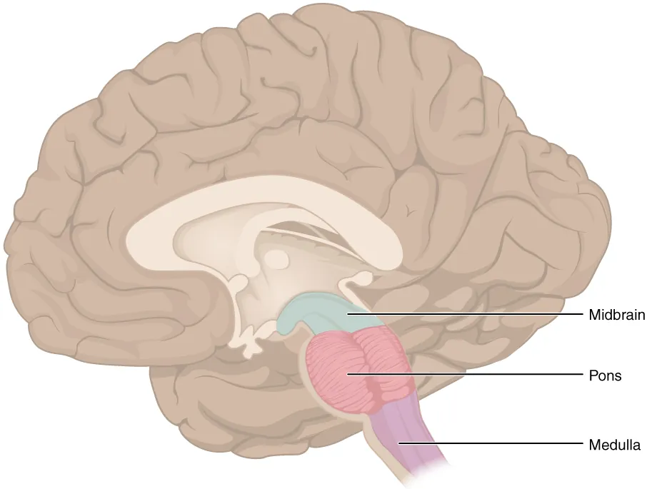
脳幹のいちばん上、橋と間脳のあいだ。長さ約2cm と短いが、通る構造は多い。
腹側:左右の太い柱 pedunculi cerebri(大脳脚)と、そのあいだの fossa interpeduncularis。窩の底は小孔だらけで substantia perforata posterior という。III 動眼神経はこの窩の内側壁から出る。
背側:lamina tecti(四丘体)に colliculi superiores(上丘・視覚反射)と colliculi inferiores(下丘・聴覚中継)が2対。各丘から前外側へ伸びる腕が brachium colliculi superioris / inferioris で、それぞれ外側膝状体・内側膝状体へ向かう。IV 滑車神経は下丘の直下から背側に出る唯一の脳神経。
中心を aqueductus mesencephali が貫き、第三脳室と第四脳室を繋ぐ。
断面は水道を基準に3層に読む。腹側から crus cerebri(大脳脚底、下行路)→ substantia nigra → tegmentum(被蓋)→ 水道 → lamina tecti。黒質より腹側が「通り道」、背側が「働く場所」と分けると迷わない。
Substantia nigra はメラニンを含むため肉眼で黒く見える。緻密部(ドパミン細胞、線条体へ)と網様部(GABA、視床・上丘へ出力)がある。Nucleus ruber は鉄を含み赤みを帯びた大きな核で、上小脳脚を受けて赤核脊髄路を出す。肉眼標本で色が付いて見える2つの核として覚える。
Colliculus superior は目を、inferior は耳を扱う。上丘は視覚性の眼球・頭部の反射(対光反射の中継も)、下丘は聴覚路の必発の中継点で、内側膝状体を経て側頭葉へ行く。
Weber 症候群——中脳の大脳脚の病変。同側の動眼神経麻痺(眼が外下方を向き、眼瞼下垂、散瞳)+反対側の片麻痺。上位の交叉性片麻痺の代表である。
鉤ヘルニア(テント切痕ヘルニア)では、側頭葉の鉤が中脳へ落ち込む。押されるのは同側の動眼神経(→ 同側の散瞳・対光反射消失)と同側の大脳脚(→ 反対側の片麻痺)。「瞳孔は病変側、麻痺は反対側」——中脳の断面図がそのまま所見の順番になる。
Parinaud 症候群——上丘(松果体部腫瘍)の圧迫で上方注視麻痺。中脳背側が「見る」場所であることの裏返しである。
出典:イラスト解剖学 誌面578頁「脳ヘルニア」
English version
Core
The uppermost part of the brainstem, between pons and diencephalon. Only about 2 cm long, but a great deal passes through it.
Ventral: the two thick columns, the pedunculi cerebri, and between them the fossa interpeduncularis. The floor of the fossa is riddled with small holes and is called the substantia perforata posterior. The oculomotor nerve (III) emerges from the medial wall of this fossa.
Dorsal: on the lamina tecti (quadrigeminal plate), two pairs — the colliculi superiores (visual reflexes) and colliculi inferiores (auditory relay). The arms extending anterolaterally from each are the brachium colliculi superioris / inferioris, running to the lateral and medial geniculate bodies respectively. The trochlear nerve (IV) emerges just below the inferior colliculus — the only cranial nerve to leave dorsally.
The aqueductus mesencephali pierces the centre, joining the third to the fourth ventricle.
Going further
Read a section in three layers, taking the aqueduct as the reference. From ventral: crus cerebri (the descending tracts) → substantia nigra → tegmentum → aqueduct → lamina tecti. Divide it as "ventral to the substantia nigra is the thoroughfare, dorsal to it is where the work is done" and you will not get lost.
The substantia nigra looks black to the naked eye because it contains melanin. It has a pars compacta (dopaminergic cells, to the striatum) and a pars reticulata (GABA, output to thalamus and superior colliculus).
The nucleus ruber is a large nucleus tinged red by its iron content, receiving the superior cerebellar peduncle and giving off the rubrospinal tract. Learn them as the two nuclei visible by their colour on a gross specimen.
The superior colliculus deals with the eye, the inferior with the ear. The superior handles visual reflexes of eye and head (including the relay of the light reflex); the inferior is an obligatory relay of the auditory pathway, passing through the medial geniculate body to the temporal lobe.
In the clinic
Weber’s syndrome — a lesion of the cerebral peduncle of the midbrain. Ipsilateral oculomotor palsy (the eye turned down and out, ptosis, a dilated pupil) plus contralateral hemiplegia. The representative crossed hemiplegia of the upper brainstem.
In uncal (transtentorial) herniation the uncus of the temporal lobe descends onto the midbrain. What is compressed is the ipsilateral oculomotor nerve (→ ipsilateral dilated pupil with loss of the light reflex) and the ipsilateral cerebral peduncle (→ contralateral hemiplegia). "The pupil is on the side of the lesion, the paralysis on the opposite side" — the cross-section of the midbrain is itself the order of the findings.
Parinaud’s syndrome — compression of the superior colliculi (a pineal region tumour) gives palsy of upward gaze. The converse of the dorsal midbrain being the place for looking.
視床 — 感覚の関所Thalamus
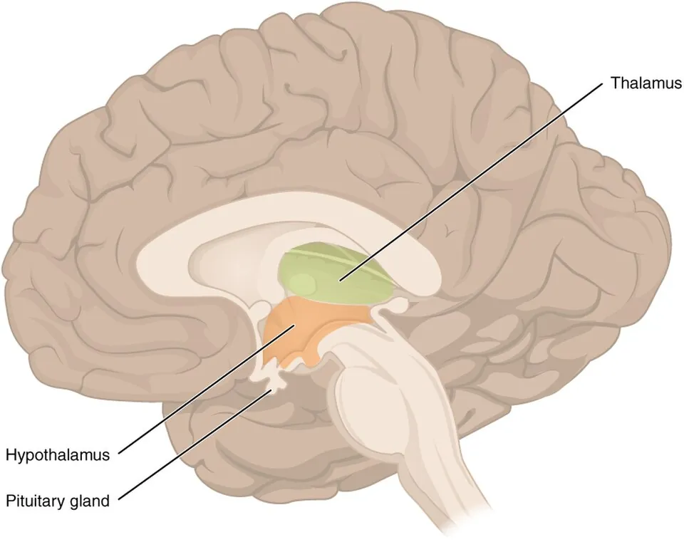
間脳の主部。卵形の灰白質の塊が左右に1つずつあり、多くの人で adhesio interthalamica(視床間橋)で繋がる。第三脳室の外側壁を作る。
肉眼で示す構造は2つ。前端の膨らみ tuberculum anterius thalami(室間孔の後ろの縁)と、後端の膨らみ pulvinar(枕)。
内部は lamina medullaris interna という Y 字の白質板で前核群・内側核群・外側核群に分けられる。嗅覚以外のすべての感覚が、大脳皮質へ行く前にここで乗り換える。
中継核の対応を押さえる。
| 核 | 受ける | 送る |
|---|---|---|
| VPL(後外側腹側核) | 体幹・四肢の感覚(内側毛帯・脊髄視床路) | 中心後回 |
| VPM(後内側腹側核) | 顔面の感覚(三叉神経毛帯)・味覚 | 中心後回 |
| Corpus geniculatum laterale | 視覚(視索) | 後頭葉(視放線) |
| Corpus geniculatum mediale | 聴覚(下丘から) | 側頭葉(聴放線) |
| VA / VL | 大脳基底核・小脳 | 運動野 |
膝状体は間脳の後下面にある小さな2つの隆起(metathalamus)で、標本では pulvinar の下外側を探す。外側=視覚、内側=聴覚(「外は見る、内は聴く」)。
Epithalamus は背側で、glandula pinealis(松果体)と手綱(habenula)。松果体はメラトニンを出し、成人では石灰化するため単純X線・CTで正中の目印になる。
視床は感覚の関所なので、傷めば反対側半身の全感覚が落ちる。
視床出血——高血圧性脳出血の好発部位(被殻に次ぐ)。反対側の感覚障害が運動麻痺より目立つのが特徴で、内下方への共同偏視(鼻先を見つめる眼)が出れば視床を強く疑う。第三脳室へ穿破すると急性水頭症になる。
視床痛(Déjerine–Roussy 症候群)——発症から数週〜数か月遅れて、感覚が戻る過程で耐えがたい自発痛が現れる。触れただけで激痛になる(allodynia)。中枢性疼痛の典型で、通常の鎮痛薬が効かない。
English version
Core
The main part of the diencephalon: an ovoid mass of grey matter on each side, joined in most people by the adhesio interthalamica. It forms the lateral wall of the third ventricle.
Two structures are shown grossly: the swelling at the front, the tuberculum anterius thalami (the posterior margin of the interventricular foramen), and the swelling behind, the pulvinar.
Internally the Y-shaped white lamina medullaris interna divides it into anterior, medial and lateral nuclear groups.
Every sensation except smell relays here before reaching the cerebral cortex.
Going further
Fix the relay nuclei.
| Nucleus | Receives | Sends to |
|---|---|---|
| VPL | Sensation from trunk and limbs (medial lemniscus, spinothalamic) | Postcentral gyrus |
| VPM | Sensation from the face (trigeminal lemniscus), taste | Postcentral gyrus |
| Corpus geniculatum laterale | Vision (optic tract) | Occipital lobe (optic radiation) |
| Corpus geniculatum mediale | Hearing (from the inferior colliculus) | Temporal lobe (acoustic radiation) |
| VA / VL | Basal ganglia, cerebellum | Motor cortex |
The geniculate bodies are the two small elevations on the posteroinferior surface of the diencephalon (the metathalamus); on the specimen look for them below and lateral to the pulvinar. Lateral = vision, medial = hearing ("the outer one looks, the inner one listens").
The epithalamus is dorsal — the glandula pinealis and the habenula. The pineal secretes melatonin and is commonly calcified in the adult, making it a midline marker on plain films and CT.
In the clinic
The thalamus is the gate of sensation, so damage to it removes all sensation from the opposite half of the body.
Thalamic haemorrhage — a site of predilection for hypertensive intracerebral haemorrhage (second to the putamen). Characteristically the contralateral sensory disturbance is more conspicuous than the motor weakness, and a conjugate deviation downwards and inwards (eyes gazing at the tip of the nose) strongly suggests the thalamus. Rupture into the third ventricle causes acute hydrocephalus.
Thalamic pain (Déjerine–Roussy syndrome) — weeks to months after the onset, as sensation returns, an unbearable spontaneous pain appears. The lightest touch becomes agonising (allodynia). The archetype of central pain, and unresponsive to ordinary analgesics.
視床下部 — 脳底で示す構造Hypothalamus
視床の下、sulcus hypothalamicus より腹側。第三脳室の下壁と下部の外側壁を作る。
脳底から見える構造を前から後ろへ順に示せるようにする。
- Chiasma opticum(視交叉)
- Tractus opticus(視索・後外側へ走る)
- Tuber cinereum(灰白隆起)
- Infundibulum(漏斗)→ hypophysis(下垂体)
- Corpora mamillaria(乳頭体・左右2つの小球)
この5つが前→後に一列に並ぶ。乳頭体の後ろはもう脚間窩(中脳)である。
下垂体は2つの由来からできている。前葉(腺性下垂体)は口窩の上皮 Rathke 嚢から、後葉(神経性下垂体)は間脳の底が下へ伸びたもの。だから後葉だけが漏斗を通じて脳と神経で直接つながる——視索上核・室傍核の軸索がそのまま後葉まで届き、ADH とオキシトシンを放出する。前葉には神経がなく、下垂体門脈系という血管で視床下部ホルモンを受ける。
乳頭体は Papez 回路の一員である(海馬 → 脳弓 → 乳頭体 → 視床前核 → 帯状回 → 海馬)。Wernicke 脳症でここが選択的に傷むので、記憶障害・作話が出る(Korsakoff)。解剖の小球1つが臨床症状に直結する典型例。
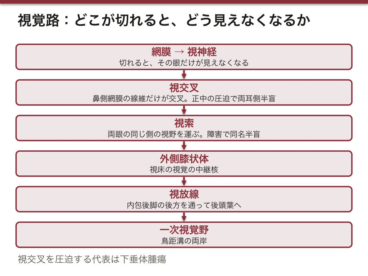
視交叉の位置は視野障害の型を決める。交叉の中央(鼻側網膜からの線維)が圧迫されると両耳側半盲——下垂体腺腫が上へ伸びたときの古典的所見である。
視交叉のすぐ下に下垂体がある——この位置関係が、視野の異常を作る。
下垂体腺腫が上へ伸びると視交叉の中央(鼻側網膜からの交叉線維)を圧迫し、両耳側半盲になる。「外側が見えなくなる」のは、外側の視野を担うのが鼻側網膜=交叉する線維だからである。
尿崩症——視索上核・室傍核から下垂体後葉へ届く軸索が断たれると ADH が出なくなり、多尿・多飲・低比重尿。下垂体茎より上の障害ほど重い(後葉だけの障害では軸索の断端から漏れ続けるため軽い)。
視床下部はほかに体温・摂食・概日リズムの中枢でもある。視交叉上核が体内時計の本体で、ここが光を受けて全身のリズムを合わせる。
English version
Core
Below the thalamus, ventral to the sulcus hypothalamicus. It forms the floor and the lower lateral wall of the third ventricle.
Be able to show, in order from front to back, the structures visible from the base of the brain.
- Chiasma opticum
- Tractus opticus (running posterolaterally)
- Tuber cinereum
- Infundibulum → hypophysis
- Corpora mamillaria (two small spheres)
These five lie in a row from front to back. Behind the mamillary bodies is already the interpeduncular fossa of the midbrain.
Going further
The pituitary is built from two origins. The anterior lobe (adenohypophysis) from the epithelium of the stomodeum (Rathke’s pouch), the posterior lobe (neurohypophysis) from a downgrowth of the floor of the diencephalon. So only the posterior lobe is connected to the brain by nerve through the infundibulum — the axons of the supraoptic and paraventricular nuclei reach it directly and release ADH and oxytocin. The anterior lobe has no nerves and receives the hypothalamic hormones through a vascular route, the hypophyseal portal system.
The mamillary body is a member of the Papez circuit (hippocampus → fornix → mamillary body → anterior thalamic nucleus → cingulate gyrus → hippocampus). In Wernicke’s encephalopathy it is damaged selectively, giving amnesia and confabulation (Korsakoff). A model case of one small sphere of anatomy leading straight to a clinical symptom.
The position of the chiasm decides the type of visual field defect. Compression of its centre (the fibres from the nasal retinae) gives bitemporal hemianopia — the classic finding when a pituitary adenoma extends upwards.
In the clinic
The pituitary lies immediately beneath the optic chiasm — and this relationship creates the field defect. When a pituitary adenoma extends upwards it compresses the centre of the chiasm (the crossing fibres from the nasal retinae), giving bitemporal hemianopia. It is the outer field that is lost, because the outer field is served by the nasal retina — the fibres that cross.
Diabetes insipidus — when the axons running from the supraoptic and paraventricular nuclei to the posterior lobe are interrupted, ADH ceases: polyuria, polydipsia, dilute urine. The higher above the stalk the lesion, the more severe (a lesion of the posterior lobe alone is mild, because hormone continues to leak from the cut ends of the axons).
The hypothalamus is also the centre for temperature, feeding and circadian rhythm. The suprachiasmatic nucleus is the body clock itself, receiving light and setting the rhythm of the whole body.
第三脳室 — 6面の壁Ventriculus tertius
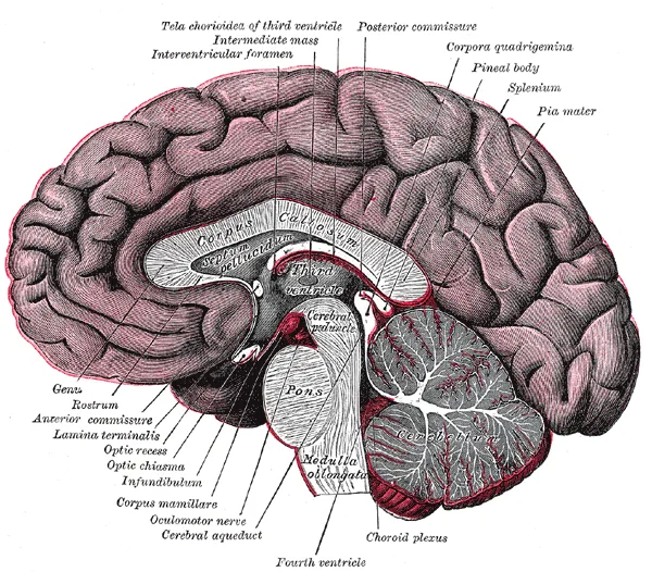
左右の間脳のあいだの、正中の狭い裂隙。矢状断で見せるのが最も分かりやすい。
| 壁 | 作るもの |
|---|---|
| 外側壁 | 視床(上部)と視床下部(下部)。境が sulcus hypothalamicus |
| 前壁 | Lamina terminalis と前交連 |
| 後壁 | 後交連・松果体・松果体上陥凹 |
| 底 | 視交叉・灰白隆起・漏斗・乳頭体・脚間窩 |
| 天井 | Tela choroidea と脈絡叢 |
Foramen interventriculare(Monro 孔)で前上方の左右の側脳室と繋がり、後下方は中脳水道で第四脳室へ続く。
陥凹 recessus は4つ。前下方から recessus supraopticus(視交叉の上)・recessus infundibuli(漏斗の中へ)、後方に recessus pinealis(松果体の茎の中)・recessus suprapinealis(松果体の上)。矢状断の輪郭を追うと、この4つが出っ張りとして順に現れる。
Lamina terminalis は終脳の前端そのものである。神経管の前端(前神経孔)が閉じた跡で、ここより前が終脳、後ろが間脳という発生上の境界になる。第三脳室に嚢胞(コロイド嚢胞)ができると Monro 孔を塞ぎ、体位で変動する頭痛と両側側脳室の拡大を起こす。
第三脳室の天井、Monro 孔のすぐそばにできるのがコロイド嚢胞である。左右の Monro 孔を同時に塞ぐと、両側の側脳室だけが急に拡大する。体位で変わる激烈な頭痛と、突然死を起こしうる急性閉塞性水頭症の典型例として知られる。
底には視交叉・灰白隆起・漏斗・乳頭体が並ぶ。だから第三脳室底の腫瘍(頭蓋咽頭腫・胚細胞腫)は、視野障害+内分泌障害+水頭症を同時に起こす。「1つの腔の底に、見る道とホルモンの道が並んでいる」ことの帰結である。
English version
Core
The narrow median cleft between the two halves of the diencephalon. It is clearest shown on a sagittal section.
| Wall | Formed by |
|---|---|
| Lateral | Thalamus (above) and hypothalamus (below), separated by the sulcus hypothalamicus |
| Anterior | Lamina terminalis and the anterior commissure |
| Posterior | Posterior commissure, pineal gland, suprapineal recess |
| Floor | Optic chiasm, tuber cinereum, infundibulum, mamillary bodies, interpeduncular fossa |
| Roof | Tela choroidea and choroid plexus |
It communicates anterosuperiorly with the two lateral ventricles through the foramen interventriculare (of Monro), and continues posteroinferiorly into the fourth ventricle through the cerebral aqueduct.
Going further
There are four recesses. Anteroinferiorly the recessus supraopticus (above the chiasm) and the recessus infundibuli (down into the infundibulum); posteriorly the recessus pinealis (into the stalk of the pineal) and the recessus suprapinealis (above the pineal). Follow the outline on a sagittal section and the four appear in turn as projections.
The lamina terminalis is the anterior end of the telencephalon itself — the trace of the closure of the anterior neuropore, and the developmental boundary: in front of it is telencephalon, behind it diencephalon.
A cyst in the third ventricle (a colloid cyst) blocks the foramen of Monro, causing headache that varies with posture and dilatation of both lateral ventricles.
In the clinic
A colloid cyst arises in the roof of the third ventricle, right beside the foramen of Monro. Blocking both foramina at once, the lateral ventricles alone dilate suddenly. It is known as the classic cause of a violent posture-dependent headache and of acute obstructive hydrocephalus that can kill suddenly.
Along the floor lie the optic chiasm, tuber cinereum, infundibulum and mamillary bodies. So a tumour of the floor of the third ventricle (craniopharyngioma, germinoma) causes visual field loss, endocrine failure and hydrocephalus together — the consequence of the pathway for seeing and the pathway for hormones lying side by side on the floor of one cavity.
大脳脚Pedunculi cerebri
中脳の腹側にある左右一対の太い柱。下行する皮質脊髄路・皮質核路・皮質橋路が束になって通る。
脳底面の標本では、橋の上に八の字に開く2本の柱として見える。その間が脚間窩。
English version
Core
The pair of thick columns on the ventral aspect of the midbrain. The descending corticospinal, corticonuclear and corticopontine tracts pass through them in a bundle.
On a specimen of the base of the brain they appear as two columns diverging above the pons; between them is the interpeduncular fossa.
脚間窩Fossa interpeduncularis
左右の大脳脚に挟まれたくぼみ。底は小孔だらけで後有孔質 substantia perforata posterior と呼ばれ、中心枝がここから入る。
動眼神経(III)はこの窩の内側壁から出る。脚間槽が満たすのもここ。
English version
Core
The hollow between the two cerebral peduncles. Its floor is riddled with small holes and is called the substantia perforata posterior, through which the central branches enter.
The oculomotor nerve (III) emerges from the medial wall of this fossa. The interpeduncular cistern fills it.
上丘・下丘Colliculi superiores et inferiores
中脳の背側にある4つのふくらみ=四丘体 corpora quadrigemina。
上丘は視覚の反射(物を目で追う・対光反射の経路)、下丘は聴覚の中継。「上が目、下が耳」——この対応を必ず押さえる。
English version
Core
The four elevations on the dorsal midbrain = the corpora quadrigemina. The superior colliculus handles visual reflexes (following an object with the eyes, part of the pathway of the light reflex); the inferior colliculus is the auditory relay.
"Upper is the eye, lower is the ear" — fix this correspondence without fail.
上丘腕Brachium colliculi superioris
上丘から外側膝状体へ向かう線維の隆起。視覚の道すじ。
English version
Core
The ridge of fibres running from the superior colliculus to the lateral geniculate body. The visual route.
下丘腕Brachium colliculi inferioris
下丘から内側膝状体へ向かう線維の隆起。聴覚の道すじ。
丘と膝状体を結ぶ腕——上丘腕→外側膝状体(視覚)、下丘腕→内側膝状体(聴覚)。ここで内外がたすき掛けになるので取り違えやすい。
English version
Core
The ridge of fibres running from the inferior colliculus to the medial geniculate body. The auditory route.
The arms joining colliculus to geniculate body — superior brachium → lateral geniculate (vision), inferior brachium → medial geniculate (hearing). Medial and lateral swap over here, so they are easily confused.
中脳水道Aqueductus mesencephali
中脳を貫く細い管で、第三脳室と第四脳室をつなぐ。周囲は中心灰白質。
脳室系でいちばん細く、詰まりやすい。ここが閉塞すると上流だけが拡がる非交通性水頭症になる。
English version
Core
The narrow canal piercing the midbrain and joining the third to the fourth ventricle, surrounded by the central grey matter.
It is the narrowest part of the ventricular system and the readiest to block. Occlusion here gives a non-communicating hydrocephalus in which only what lies upstream dilates.
赤核Nucleus ruber
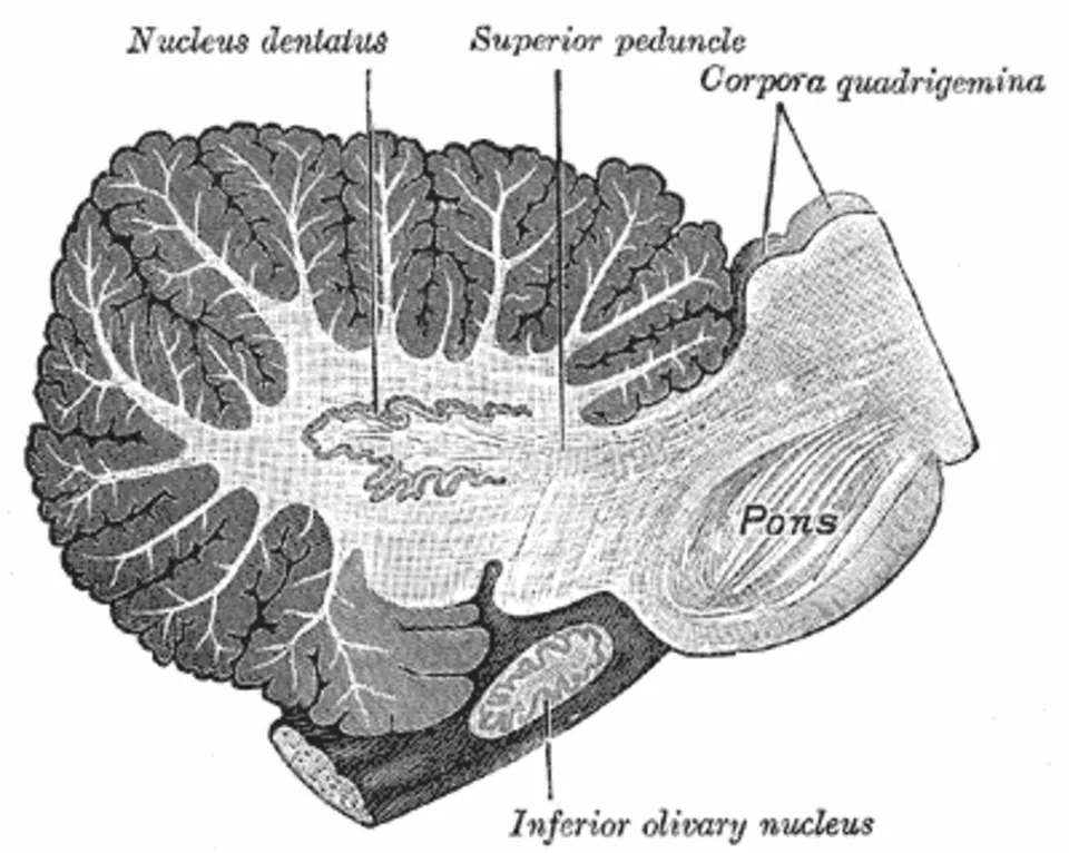
中脳被蓋にある核。生の標本では鉄と血管に富むため赤みを帯びて見えるのが名前の由来。
上小脳脚からの入力を受け、赤核脊髄路を出す。断面でしか見えないので、中脳の水平断標本で示す。
English version
Core
A nucleus in the tegmentum of the midbrain. In a fresh specimen it looks reddish from its content of iron and vessels — hence the name. It receives the superior cerebellar peduncle and gives off the rubrospinal tract.
It can only be seen on section, so it is shown on a horizontal section of the midbrain.
黒質Substantia nigra
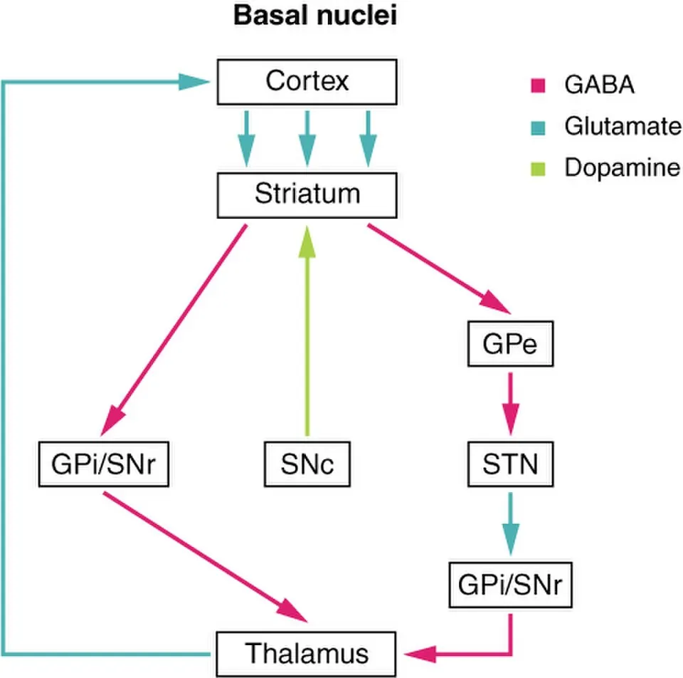
大脳脚と被蓋の境にある層。神経メラニンを含むため黒く見える。
緻密部のドパミン細胞が線条体へ投射する。ここの脱落が Parkinson 病で、標本でも黒質の色が薄くなる。標本では「中脳断面で黒い帯」として指す。
English version
Core
The layer at the boundary between the cerebral peduncle and the tegmentum. It looks black because it contains neuromelanin. The dopaminergic cells of the pars compacta project to the striatum.
Their loss is Parkinson’s disease, and the substantia nigra is visibly paler even on a gross specimen. On a specimen, point to it as "the black band on a section of the midbrain".
脳神経(脳表での起始)Nervi craniales
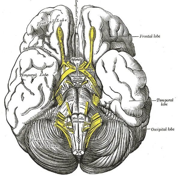
脳底面の標本で、12対がどこから出るかを順に指せるようにする。
I 嗅神経=嗅球/II 視神経=視交叉/III 動眼=脚間窩/IV 滑車=唯一の背側から(下丘の下)/V 三叉=橋の外側/VI 外転=橋延髄溝の内側/VII 顔面・VIII 内耳=小脳橋角/IX・X・XI=後外側溝/XII 舌下=前外側溝。
IV が背側から出る唯一の脳神経という点は必ず押さえる。
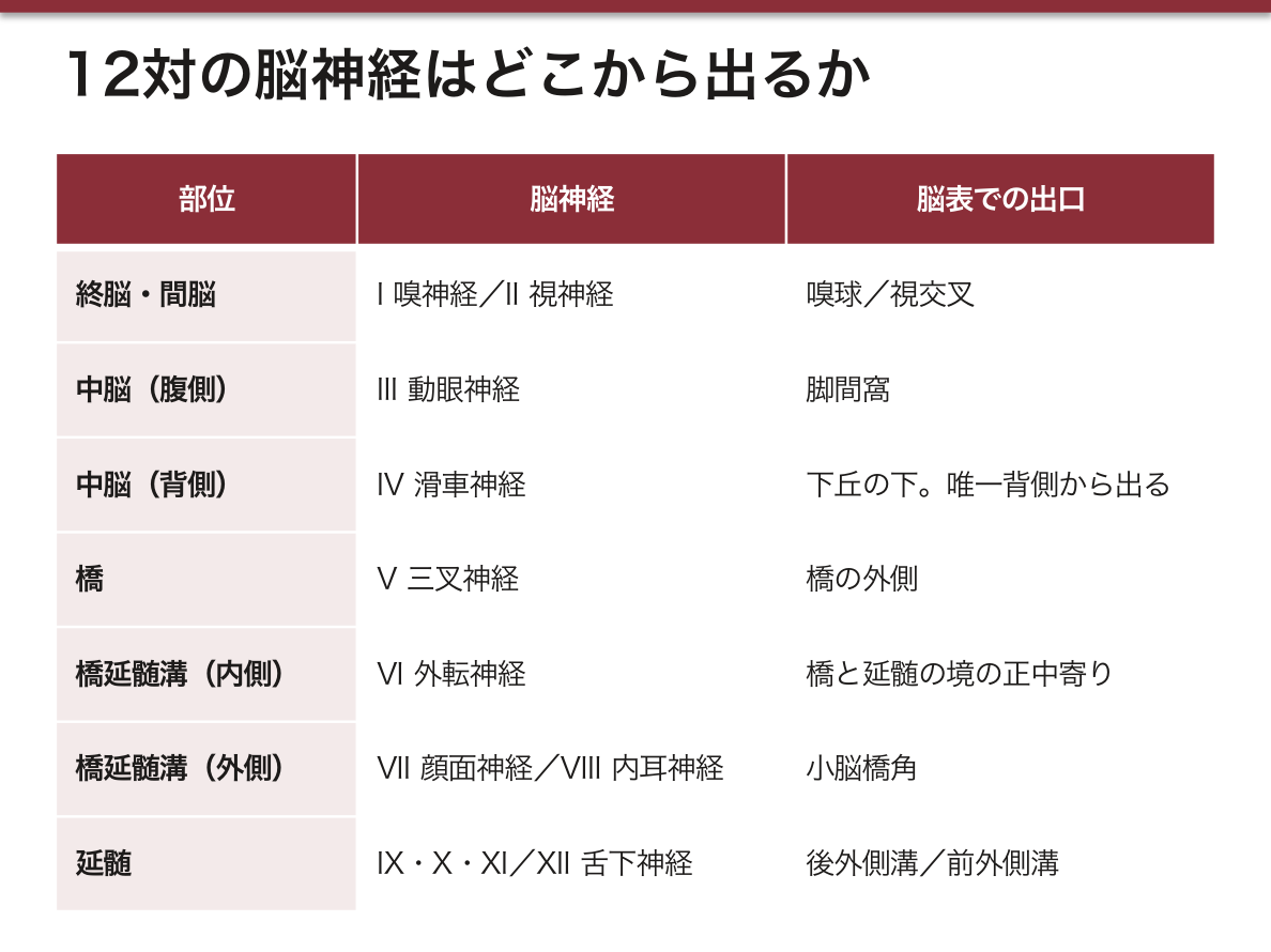
English version
Core
Be able to point out, in order, where all twelve pairs emerge on a specimen of the base of the brain.
I olfactory = olfactory bulb / II optic = optic chiasm / III oculomotor = interpeduncular fossa / IV trochlear = the only one from the dorsal surface (below the inferior colliculus) / V trigeminal = lateral aspect of the pons / VI abducens = medial part of the pontomedullary sulcus / VII facial and VIII vestibulocochlear = the cerebellopontine angle / IX, X, XI = posterolateral sulcus / XII hypoglossal = anterolateral sulcus.
That IV is the only cranial nerve leaving dorsally must be known without fail.
松果体Glandula pinealis
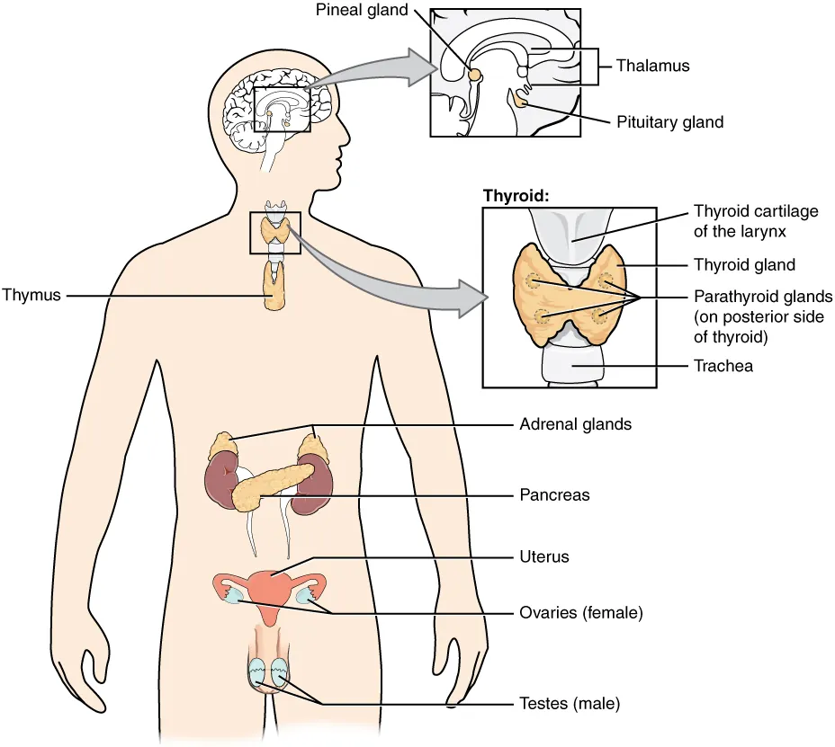
第三脳室の後壁から後方へ突き出す小さな腺。四丘体(上丘)のすぐ上に乗る。
メラトニンを出して概日リズムを司る。成人では石灰化することが多く、頭部X線・CTで正中の目印になる。
English version
Core
The small gland projecting backwards from the posterior wall of the third ventricle, sitting just above the quadrigeminal plate (superior colliculi). It secretes melatonin and governs the circadian rhythm.
It is often calcified in the adult and so serves as a midline marker on skull radiographs and CT.
視床間橋Adhesio interthalamica
第三脳室を横切って左右の視床をつなぐ灰白質の柱。神経の連絡路というより癒着で、約2割の人では存在しない。
正中矢状断の標本で、第三脳室の真ん中を渡る橋として示す。
English version
Core
The column of grey matter crossing the third ventricle and joining the two thalami. It is an adhesion rather than a pathway, and is absent in about 20 per cent of people.
On a median sagittal specimen, show it as the bridge crossing the middle of the third ventricle.
内側膝状体Corpus geniculatum mediale
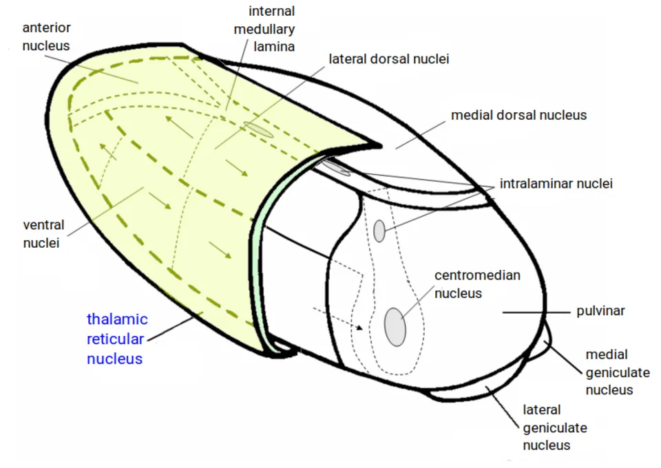
視床後部(視床枕の下外側)のふくらみ。聴覚の中継核で、下丘腕を受けて聴放線を出す。
English version
Core
The elevation of the posterior thalamus (below and lateral to the pulvinar). The relay nucleus of hearing, receiving the inferior brachium and giving off the acoustic radiation.
外側膝状体Corpus geniculatum laterale
その外側のふくらみ。視覚の中継核で、視索を受けて視放線を出す。
内側が耳、外側が目。丘の側(上丘=目、下丘=耳)と逆になるので、必ず一組で確認する。
English version
Core
The elevation lateral to it. The relay nucleus of vision, receiving the optic tract and giving off the optic radiation.
Medial is the ear, lateral is the eye — the reverse of the colliculi (superior = eye, inferior = ear), so always check the two together.
視交叉Chiasma opticum
脳底面、トルコ鞍のすぐ上で左右の視神経が交わるX字。鼻側網膜からの線維だけが交叉し、耳側は交叉しない。
そのためここの正中が圧迫されると両耳側半盲になる(下垂体腫瘍)。脳底面の標本では最も目立つ正中の目印で、位置決めの基準に使える。
English version
Core
The X on the base of the brain, just above the sella turcica, where the two optic nerves meet. Only the fibres from the nasal retinae cross; the temporal fibres do not. So compression of its centre gives bitemporal hemianopia (pituitary tumour).
It is the most conspicuous midline landmark on a specimen of the base of the brain and can be used as the reference for orientation.
視索Tractus opticus
視交叉から後外側へ、大脳脚を巻くように走って外側膝状体へ至る束。
交叉を過ぎているので、1本の視索は「両眼の同じ側の視野」を運ぶ。ここの障害は同名半盲。
English version
Core
The bundle running posterolaterally from the chiasm, winding around the cerebral peduncle to the lateral geniculate body. Being past the decussation, one optic tract carries the same side of the visual field from both eyes. A lesion here gives a homonymous hemianopia.
乳頭体Corpus mamillare
脳底面で脚間窩の前にある、左右一対の小さな球。視床下部の一部。
脳弓を受けて視床前核へ送る(Papez 回路)。Wernicke 脳症で最初に壊れる場所で、記憶障害と結びつく。
English version
Core
The pair of small spheres on the base of the brain in front of the interpeduncular fossa; part of the hypothalamus. It receives the fornix and sends to the anterior thalamic nucleus (the Papez circuit).
It is the first place destroyed in Wernicke’s encephalopathy, and is thus tied to memory disturbance.
漏斗Infundibulum
視床下部から下へ伸びて下垂体をぶら下げる茎。鞍隔膜の孔を通る。
視床下部から下垂体後葉への軸索と、前葉への下垂体門脈がここを通る。
English version
Core
The stalk descending from the hypothalamus and suspending the pituitary, passing through the opening in the diaphragma sellae.
Through it run the axons from the hypothalamus to the posterior lobe and the hypophyseal portal vessels to the anterior lobe.
下垂体Hypophysis
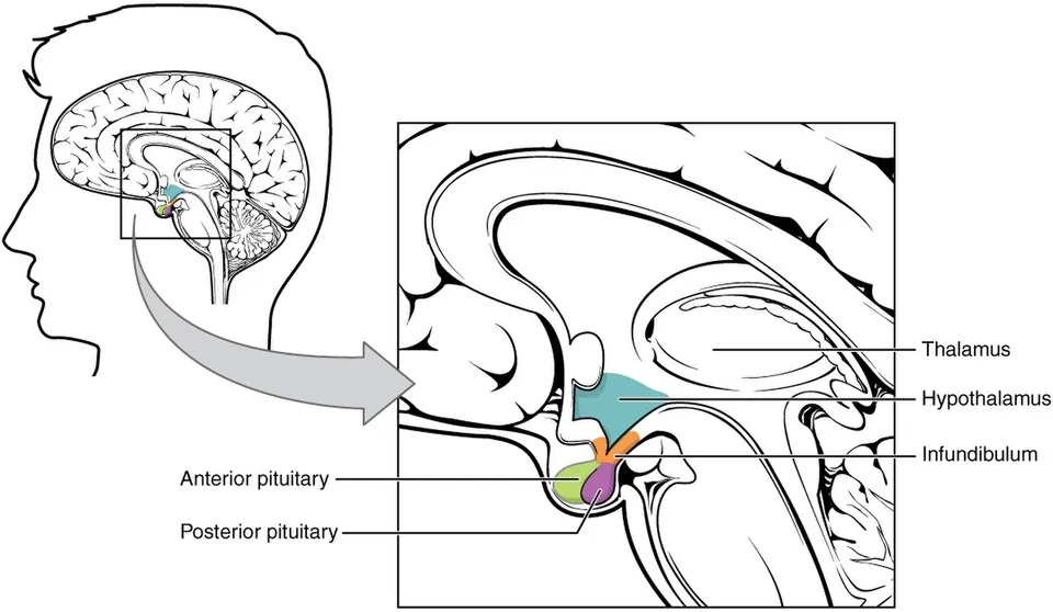
トルコ鞍の下垂体窩に収まる。由来の違う2つからできている——前葉(腺性)は口窩の上皮(Rathke 嚢)から、後葉(神経性)は間脳の下方への伸びから。
「同じ器官に見えて出自が違う」ことが要点である。上に視交叉が乗るので、腫大すると両耳側半盲が出る。
English version
Core
Housed in the hypophyseal fossa of the sella turcica. Built of two parts of different origin — the anterior lobe (glandular) from the epithelium of the stomodeum (Rathke’s pouch), the posterior lobe (neural) from a downgrowth of the diencephalon. That what looks like one organ has two origins is the point.
The optic chiasm lies on top of it, so enlargement gives bitemporal hemianopia.
室間孔(Monro 孔)Foramen interventriculare
側脳室と第三脳室をつなぐ左右一対の孔。前方を脳弓柱、後方を視床前端が囲む。
髄液が側脳室から出る唯一の出口なので、片側が詰まれば片側の側脳室だけが拡がる。
English version
Core
The pair of openings joining the lateral ventricles to the third, bounded in front by the column of the fornix and behind by the anterior end of the thalamus.
It is the only exit for CSF from a lateral ventricle, so if one side blocks, only that lateral ventricle dilates.
視索上陥凹Recessus supraopticus
第三脳室が視交叉の上へ張り出したくぼみ。
English version
Core
The recess by which the third ventricle projects above the optic chiasm.
漏斗陥凹Recessus infundibuli
第三脳室が漏斗の中へ下向きに落ち込むくぼみ。
視索上陥凹と並んで前下方の2つ。正中矢状断で、視交叉と漏斗を目印に探す。
English version
Core
The recess by which the third ventricle descends into the infundibulum. With the supraoptic recess these are the two anteroinferior ones. On a median sagittal section, find them using the chiasm and the infundibulum as landmarks.
松果体上陥凹Recessus suprapinealis
第三脳室が松果体の上へ後方に張り出すくぼみ。
3つの陥凹は「前下に2つ(視交叉・漏斗)、後上に1つ(松果体)」で位置を整理する。
English version
Core
The recess by which the third ventricle projects backwards above the pineal gland.
Organise the three recesses by position: two anteroinferior (chiasm, infundibulum) and one posterosuperior (pineal).
まとめ
| 中脳 — 外形 | 脳幹のいちばん上、橋と間脳のあいだ。長さ約2cm と短いが、通る構造は多い。 |
|---|---|
| 視床 — 感覚の関所 | 間脳の主部。卵形の灰白質の塊が左右に1つずつあり、多くの人で adhesio interthalamica(視床間橋)で繋がる。 |
| 視床下部 — 脳底で示す構造 | 視床の下、sulcus hypothalamicus より腹側。 |
| 第三脳室 — 6面の壁 | 左右の間脳のあいだの、正中の狭い裂隙。矢状断で見せるのが最も分かりやすい。 |
| 大脳脚 | 中脳の腹側にある左右一対の太い柱。下行する皮質脊髄路・皮質核路・皮質橋路が束になって通る。 |
| 脚間窩 | 左右の大脳脚に挟まれたくぼみ。底は小孔だらけで後有孔質 substantia perforata posterior と呼ばれ、中心枝がここから入る。 |
| 上丘・下丘 | 中脳の背側にある4つのふくらみ=四丘体 corpora quadrigemina。 |
| 上丘腕 | 上丘から外側膝状体へ向かう線維の隆起。視覚の道すじ。 |
| 下丘腕 | 下丘から内側膝状体へ向かう線維の隆起。聴覚の道すじ。 |
| 中脳水道 | 中脳を貫く細い管で、第三脳室と第四脳室をつなぐ。 |
| 赤核 | 中脳被蓋にある核。生の標本では鉄と血管に富むため赤みを帯びて見えるのが名前の由来。 |
| 黒質 | 大脳脚と被蓋の境にある層。神経メラニンを含むため黒く見える。 |
| 脳神経(脳表での起始) | 脳底面の標本で、12対がどこから出るかを順に指せるようにする。 |
| 松果体 | 第三脳室の後壁から後方へ突き出す小さな腺。 |
| 視床間橋 | 第三脳室を横切って左右の視床をつなぐ灰白質の柱。 |
| 内側膝状体 | 視床後部(視床枕の下外側)のふくらみ。聴覚の中継核で、下丘腕を受けて聴放線を出す。 |
| 外側膝状体 | その外側のふくらみ。視覚の中継核で、視索を受けて視放線を出す。 |
| 視交叉 | 脳底面、トルコ鞍のすぐ上で左右の視神経が交わるX字。 |
| 視索 | 視交叉から後外側へ、大脳脚を巻くように走って外側膝状体へ至る束。 |
| 乳頭体 | 脳底面で脚間窩の前にある、左右一対の小さな球。 |
| 漏斗 | 視床下部から下へ伸びて下垂体をぶら下げる茎。 |
| 下垂体 | トルコ鞍の下垂体窩に収まる。由来の違う2つからできている——前葉(腺性)は口窩の上皮(Rathke 嚢)から、後葉(神経性)は間脳の下方への伸びから。 |
| 室間孔(Monro 孔) | 側脳室と第三脳室をつなぐ左右一対の孔。前方を脳弓柱、後方を視床前端が囲む。 |
| 視索上陥凹 | 第三脳室が視交叉の上へ張り出したくぼみ。 |
| 漏斗陥凹 | 第三脳室が漏斗の中へ下向きに落ち込むくぼみ。 |
| 松果体上陥凹 | 第三脳室が松果体の上へ後方に張り出すくぼみ。 |
このノートは医学生が授業の内容を自分の言葉でまとめたもので、診療の判断に使うものではありません。誤りに気づいたらお知らせください。