覚える構造がいちばん多い範囲である。
攻め方は「面を決めてから探す」。上外側面(溝と回)→ 内側面(脳梁・帯状回・鳥距溝)→ 下面(嗅球・海馬傍回)→ 水平断(基底核と内包)→ 側脳室。面を宣言してから指すと、見ている位置を見失わない。
この範囲の芯は「1つのC字」である。側脳室・尾状核・脳弓・海馬はすべて、終脳が前から後ろへ、そして下前方へ弧を描いて育った跡をなぞっている。この弧を1本思い浮かべられれば、4つの構造の走行を別々に暗記しなくてよい。
- 大脳の溝と回 — 上外側面
- 大脳の内側面と下面
- 皮質中枢 — 回の名前と機能の対応
- 大脳の白質 — 3種類の線維と内包
- 大脳基底核 — 灰白質の塊
- 側脳室 — C字の4部
- 大脳縦裂
- 大脳横裂
- 中心溝(Rolando 溝)
- 外側溝(Sylvius 溝)
- 中心前溝
- 上前頭溝
- 下前頭溝
- 中心後溝
- 頭頂間溝
- 上側頭溝
- 下側頭溝
- 脳梁溝
- 帯状溝
- 頭頂後頭溝
- 後頭側頭溝
- 嗅溝
- 中心前回
- 上前頭回
- 中前頭回
- 下前頭回
- 中心後回
- 上側頭回
- 中側頭回
- 下側頭回
- 島
- 楔前部
- 楔部
- 帯状回
- 嗅球
- 嗅索
- 脳梁
- 脳梁嘴
- 脳梁膝
- 脳梁幹
- 脳梁膨大
- 脳弓
- 透明中隔
- 側脳室中心部
- 側脳室前角
- 側脳室後角
- 尾状核頭
- 尾状核体
- レンズ核
- 前障
- 外包
- 内包
- 内包前脚
- 内包膝
- 内包後脚
- 大脳の溝と回 — 上外側面Sulci et gyri cerebri
- 大脳の内側面と下面Facies medialis et inferior hemispherii
- 皮質中枢 — 回の名前と機能の対応Centra corticalia
- 大脳の白質 — 3種類の線維と内包Substantia alba hemispherii
- 大脳基底核 — 灰白質の塊Nuclei basales
- 側脳室 — C字の4部Ventriculus lateralis
- 大脳縦裂Fissura longitudinalis cerebri
- 大脳横裂Fissura transversa cerebri
- 中心溝(Rolando 溝)Sulcus centralis cerebri
- 外側溝(Sylvius 溝)Sulcus lateralis cerebri
- 中心前溝Sulcus precentralis cerebri
- 上前頭溝Sulcus frontalis superior cerebri
- 下前頭溝Sulcus frontalis inferior cerebri
- 中心後溝Sulcus postcentralis cerebri
- 頭頂間溝Sulcus intraparietalis
- 上側頭溝Sulcus temporalis superior cerebri
- 下側頭溝Sulcus temporalis inferior cerebri
- 脳梁溝Sulcus corporis callosi
- 帯状溝Sulcus cinguli
- 頭頂後頭溝Sulcus parietooccipitalis cerebri
- 後頭側頭溝Sulcus occipitotemporalis cerebri
- 嗅溝Sulcus olfactorius cerebri
- 中心前回Gyrus precentralis
- 上前頭回Gyrus frontalis superior
- 中前頭回Gyrus frontalis medius
- 下前頭回Gyrus frontalis inferior
- 中心後回Gyrus postcentralis
- 上側頭回Gyrus temporalis superior
- 中側頭回Gyrus temporalis medius
- 下側頭回Gyrus temporalis inferior
- 島Insula
- 楔前部Precuneus
- 楔部Cuneus
- 帯状回Gyrus cinguli
- 嗅球Bulbus olfactorius
- 嗅索Tractus olfactorius
- 脳梁Corpus callosum
- 脳梁嘴Rostrum corporis callosi
- 脳梁膝Genu corporis callosi
- 脳梁幹Truncus corporis callosi
- 脳梁膨大Splenium corporis callosi
- 脳弓Fornix
- 透明中隔Septum pellucidum
- 側脳室中心部Pars centralis ventriculi lateralis
- 側脳室前角Cornu anterius ventriculi lateralis
- 側脳室後角Cornu posterius ventriculi lateralis
- 尾状核頭Caput nuclei caudati
- 尾状核体Corpus nuclei caudati
- レンズ核Nucleus lentiformis
- 前障Claustrum
- 外包Capsula externa
- 内包Capsula interna
- 内包前脚Crus anterius capsulae internae
- 内包膝Genu capsulae internae
- 内包後脚Crus posterius capsulae internae
- まとめ
大脳の溝と回 — 上外側面Sulci et gyri cerebri
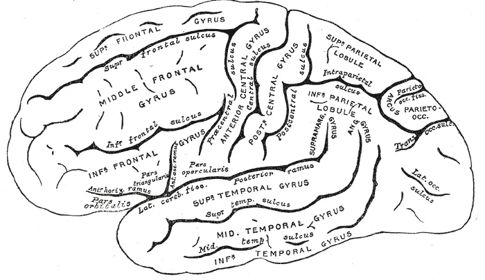
標本では「まず2本の基準溝を見つけてから、残りを相対位置で言う」のが確実である。
基準① Sulcus centralis(中心溝、Rolando)— 上縁からやや斜め前下方へ走る。前が前頭葉、後ろが頭頂葉。基準② Sulcus lateralis(外側溝、Sylvius)— 前下方から後上方へ。下が側頭葉。
中心溝の前が gyrus precentralis(一次運動野)、後ろが gyrus postcentralis(一次体性感覚野)。この2つを挟む溝が sulcus precentralis と sulcus postcentralis である。
前頭葉は sulcus frontalis superior / inferior で gyrus frontalis superior・medius・inferior の3本に、側頭葉は sulcus temporalis superior / inferior で gyrus temporalis superior・medius・inferior の3本に分かれる。頭頂葉は sulcus intraparietalis で lobulus parietalis superior / inferior に分かれる。
Insula(島)は外側溝の底に隠れており、側頭葉を下へ引かないと見えない。
中心溝の見分け方(標本で迷ったとき)。①上縁からほぼ途切れず斜めに下る唯一の深い溝、②その前後の回(中心前回・中心後回)が他より太く平行に走る、③中心前回のほうがやや太い。前頭・側頭の溝は3本ずつに分かれるので、「1本で走り切る溝」は中心溝だけと考えてよい。
回の太さには意味がある。中心前回が太いのは、そこに巨大な Betz 細胞を含む第V層が発達しているためで、組織学の「運動野は顆粒層が乏しく錐体細胞が大きい」(agranular cortex)という所見と対応する。逆に中心後回は顆粒層が厚い(granular cortex)。肉眼所見と組織像が同じことを別の言い方で述べていると分かると、両科目の勉強が1回で済む。
中大脳動脈(MCA)は外側溝の中を走る。だから外側溝の周りの回=MCA 領域である。
MCA 領域の梗塞——反対側の片麻痺と感覚障害が、顔・上肢に強く下肢に軽い。中心前回・中心後回の外側下方が顔と手を担うためで、ホムンクルスの並びがそのまま症状の重みになる。
優位半球(多くは左)なら失語、劣位半球なら半側空間無視が加わる。共同偏視は病巣側を向く(前頭眼野が働かなくなるため)。
標本で中心溝を見つけられることは、そのまま「どこが動かなくなるか」を言えることに直結する。
English version
Core
On a specimen the sure method is to find two reference sulci first and then give everything else by its position relative to them.
- Reference ① Sulcus centralis (of Rolando) — running obliquely downwards and forwards from the superior margin. In front is the frontal lobe, behind the parietal.
- Reference ② Sulcus lateralis (of Sylvius) — from anteroinferior to posterosuperior. Below it is the temporal lobe.
In front of the central sulcus is the gyrus precentralis (primary motor area), behind it the gyrus postcentralis (primary somatosensory area); the sulci bounding these are the sulcus precentralis and sulcus postcentralis.
The frontal lobe is divided by the sulcus frontalis superior / inferior into three gyri (superior, middle, inferior), the temporal lobe by the sulcus temporalis superior / inferior into three. The parietal lobe is divided by the sulcus intraparietalis into superior and inferior parietal lobules.
The insula lies hidden at the floor of the lateral sulcus and cannot be seen without drawing the temporal lobe downwards.
Going further
How to recognise the central sulcus when lost on a specimen. ① It is the only deep sulcus running obliquely down from the superior margin almost without interruption. ② The gyri in front of and behind it (precentral, postcentral) are thicker than the rest and run parallel. ③ The precentral gyrus is slightly the thicker. The frontal and temporal sulci each split into three, so "the sulcus that runs the whole way as one" can be taken to be the central sulcus.
The thickness of the gyri has a meaning. The precentral gyrus is thick because layer V, containing the giant Betz cells, is developed there, which corresponds to the histological finding that the motor cortex is poor in granular layers with large pyramidal cells (agranular cortex). The postcentral gyrus, conversely, has a thick granular layer (granular cortex). Realise that the gross appearance and the histology are the same statement in two languages, and the two subjects are studied at once.
In the clinic
The middle cerebral artery (MCA) runs within the lateral sulcus. So the gyri around that sulcus are the MCA territory.
Infarction in the MCA territory — contralateral hemiparesis and sensory loss, severe in the face and arm and slight in the leg. Because the lower lateral parts of the precentral and postcentral gyri serve the face and hand, the order of the homunculus is directly the weighting of the symptoms. Aphasia is added if the dominant hemisphere (usually the left) is involved, hemispatial neglect if the non-dominant. Conjugate deviation is towards the side of the lesion (the frontal eye field having ceased to work).
Being able to find the central sulcus on a specimen is directly being able to say what will stop moving.
大脳の内側面と下面Facies medialis et inferior hemispherii
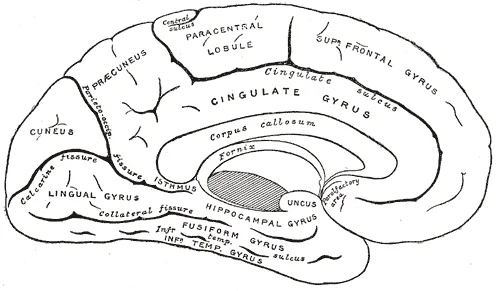
内側面は矢状断でしか見えない。中央に corpus callosum が弓なりに横たわり、その上を sulcus corporis callosi、さらに上を sulcus cinguli が走る。2つの溝に挟まれた回が gyrus cinguli(帯状回)。
後方に2本の重要な溝:sulcus parietooccipitalis(頭頂葉と後頭葉の境)と sulcus calcarinus(鳥距溝)。2本が作る V 字の前が cuneus(楔部)、その前が precuneus(楔前部)。鳥距溝の両岸が一次視覚野である。
下面(底面):前頭葉底部に sulcus olfactorius があり、内側の gyrus rectus と外側の眼窩回を分ける。溝の中に bulbus olfactorius と tractus olfactorius が乗り、後方で trigonum olfactorium に広がる。側頭葉底部は sulcus collateralis と sulcus occipitotemporalis で gyrus parahippocampalis・gyrus occipitotemporalis medialis / lateralis に分かれ、海馬傍回の前端が鉤状に曲がったのが uncus。
Uncus は臨床で最も重要な脳回である。テント切痕のすぐ内側にあるため、頭蓋内圧が上がるとここから中脳へ向かって落ち込む(鉤ヘルニア)。押されるのは同側の動眼神経(→ 同側の散瞳・眼瞼下垂)と大脳脚(→ 対側の片麻痺)。「瞳孔は病変側、麻痺は反対側」が典型像である。
鳥距溝の上下で視野が分かれる。上唇(cuneus 側)が下視野、下唇(舌状回側)が上視野を担う。後頭極が黄斑部で、後大脳動脈が閉塞しても中大脳動脈からの側副で黄斑が残ることがある(黄斑回避)。
帯状回・海馬傍回・鉤は輪をなす(大脳辺縁葉)。Broca が「大脳半球の縁を回る回」と呼んだもので、Papez 回路の外側の枠にあたる。
前大脳動脈(ACA)は大脳鎌に沿って内側面を走る。内側面には中心傍小葉=下肢の運動感覚野があるので、ACA 領域の梗塞では下肢に強い麻痺が出る。MCA が「顔と手」、ACA が「足」——内側面の解剖がそのまま鑑別になる。
両側の内側前頭葉が落ちると無動性無言・尿失禁・把握反射など前頭葉徴候が出る。
下面では鉤 uncus がテント切痕のすぐ内側にある。頭蓋内圧が上がるとここから中脳へ落ち込むのが鉤ヘルニアで、同側の散瞳が最初のサインになる(中脳の項)。
出典:イラスト解剖学 誌面578頁「脳ヘルニア」
English version
Core
The medial surface can only be seen on a sagittal section. In the middle the corpus callosum lies as an arch, with the sulcus corporis callosi above it and the sulcus cinguli above that. The gyrus between the two sulci is the gyrus cinguli.
Behind are two important sulci: the sulcus parietooccipitalis (the boundary of parietal and occipital lobes) and the sulcus calcarinus. In front of the V they make is the cuneus, and in front of that the precuneus. Both banks of the calcarine sulcus are the primary visual cortex.
Inferior surface: on the base of the frontal lobe the sulcus olfactorius separates the medial gyrus rectus from the lateral orbital gyri. The bulbus olfactorius and tractus olfactorius lie in that sulcus, spreading behind into the trigonum olfactorium. The base of the temporal lobe is divided by the sulcus collateralis and sulcus occipitotemporalis into the gyrus parahippocampalis and the medial and lateral occipitotemporal gyri, and the hooked anterior end of the parahippocampal gyrus is the uncus.
Going further
The uncus is the clinically most important gyrus of all. Lying just inside the tentorial notch, it descends towards the midbrain when the intracranial pressure rises (uncal herniation). What is compressed is the ipsilateral oculomotor nerve (→ ipsilateral dilated pupil, ptosis) and the cerebral peduncle (→ contralateral hemiplegia). "Pupil on the side of the lesion, paralysis on the opposite side" is the typical picture.
The visual field divides above and below the calcarine sulcus. The upper lip (the cuneal side) serves the lower field, the lower lip (the lingual side) the upper field. The occipital pole is the macula, and even when the posterior cerebral artery occludes, collateral supply from the middle cerebral artery may spare the macula (macular sparing).
The cingulate gyrus, parahippocampal gyrus and uncus form a ring (the limbic lobe) — what Broca called the gyri encircling the margin of the hemisphere, the outer frame of the Papez circuit.
In the clinic
The anterior cerebral artery (ACA) runs on the medial surface along the falx. The medial surface carries the paracentral lobule = the motor and sensory area of the leg, so an ACA infarct gives weakness predominantly in the leg. MCA is "face and hand", ACA is "foot" — the anatomy of the medial surface is itself the differential.
Bilateral medial frontal damage gives frontal signs — akinetic mutism, incontinence of urine, grasp reflex.
On the inferior surface, the uncus lies just inside the tentorial notch. When the intracranial pressure rises it descends onto the midbrain — uncal herniation — and an ipsilateral dilated pupil is the first sign.
皮質中枢 — 回の名前と機能の対応Centra corticalia
皮質の中枢(cortical centers)は、回の名前と機能を一対一で言えるようにする。
| 中枢 | 場所 | Brodmann |
|---|---|---|
| 一次運動野 | Gyrus precentralis | 4 |
| 一次体性感覚野 | Gyrus postcentralis | 3, 1, 2 |
| 一次視覚野 | Sulcus calcarinus の両岸 | 17 |
| 一次聴覚野 | Gyri temporales transversi(Heschl 回・上側頭回の上面) | 41, 42 |
| 嗅覚野 | Uncus・海馬傍回前部 | — |
| 味覚野 | 中心後回の下端・島 | 43 |
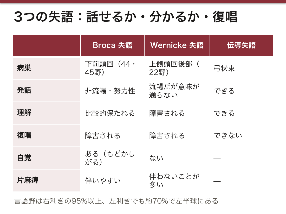
| Broca(運動性言語) | 下前頭回の弁蓋部・三角部 | 44, 45 |
| Wernicke(感覚性言語) | 上側頭回後部 | 22 |
言語野は約95%で左半球(右利きではほぼ全例、左利きでも多数)。
Homunculus は「面積=精密さ」である。中心前回・中心後回とも、内側面に下肢、円蓋部に体幹、外側下方に上肢・顔面が並ぶ。手と口唇が不釣り合いに大きいのは、そこに必要な運動と感覚の分解能が高いためで、筋の大きさとは無関係である。
下肢が内側面にあることが、前大脳動脈の閉塞で下肢優位の麻痺が出る理由。中大脳動脈は外側面を養うので上肢・顔面優位になる。血管支配と homunculus を重ねると、麻痺の分布から責任血管が言える。
Broca と Wernicke を繋ぐのが fasciculus arcuatus(弓状束)。ここだけ切れると、理解も発話もできるのに復唱だけができない(伝導失語)。
言語野は片側にしかない。右利きの 95%以上、左利きでも約 70%で左半球にある。
Broca 野(下前頭回・44/45野)——運動性失語。言いたいことは分かっているのに言葉が出ない。発話は非流暢で努力性、復唱も障害される。本人は誤りを自覚していて、もどかしがる。中心前回に近いので右片麻痺を伴いやすい。
Wernicke 野(上側頭回後部・22野)——感覚性失語。流暢にしゃべるが意味が通らず、聞いても理解できない。本人に自覚がないのが Broca との決定的な違いである。運動野から遠いので麻痺は伴わないことが多い。
両者を結ぶ弓状束だけが切れると伝導失語(理解も発話もできるが復唱だけができない)。「話せるか・分かるか・復唱できるか」の3つを順に確かめるのが失語の診かたである。
出典:イラスト解剖学 誌面117頁「脳の各部名称とはたらき(ぜんぶわかる人体解剖図)」
English version
Core
For the cortical centres, be able to give gyrus and function one to one.
| Centre | Place | Brodmann |
|---|---|---|
| Primary motor | Gyrus precentralis | 4 |
| Primary somatosensory | Gyrus postcentralis | 3, 1, 2 |
| Primary visual | Both banks of the sulcus calcarinus | 17 |
| Primary auditory | Gyri temporales transversi (Heschl’s gyri, on the upper surface of the superior temporal gyrus) | 41, 42 |
| Olfactory | Uncus, anterior parahippocampal gyrus | — |
| Taste | Lower end of the postcentral gyrus, insula | 43 |
| Broca (motor speech) | Pars opercularis and triangularis of the inferior frontal gyrus | 44, 45 |
| Wernicke (sensory speech) | Posterior superior temporal gyrus | 22 |
The speech areas are in the left hemisphere in about 95 per cent (in virtually all right-handers and in most left-handers).
Going further
In the homunculus, area = precision. In both precentral and postcentral gyri the leg lies on the medial surface, the trunk at the vertex, and the arm and face laterally and below. The hand and the lips are disproportionately large because the resolution of movement and sensation required there is high — it has nothing to do with the size of the muscles.
That the leg lies on the medial surface is why occlusion of the anterior cerebral artery gives leg-predominant weakness. The middle cerebral artery supplies the lateral surface, hence arm and face predominance. Superimpose the vascular territories on the homunculus and the responsible vessel can be named from the distribution of the weakness.
Broca and Wernicke are joined by the fasciculus arcuatus. Cut that alone and the patient can understand and can speak but cannot repeat (conduction aphasia).
In the clinic
The speech areas exist on one side only — the left in over 95 per cent of right-handers and about 70 per cent of left-handers.
Broca’s area (inferior frontal gyrus, areas 44/45) — motor aphasia. The patient knows what they want to say but the words will not come. Speech is non-fluent and effortful, and repetition is impaired. The patient is aware of the errors and is frustrated by them. Being close to the precentral gyrus, a right hemiparesis often accompanies it.
Wernicke’s area (posterior superior temporal gyrus, area 22) — sensory aphasia. Speech is fluent but makes no sense, and what is heard is not understood. That the patient is unaware of it is the decisive difference from Broca’s. Being far from the motor cortex, weakness is often absent.
Interruption of the arcuate fasciculus alone gives conduction aphasia (comprehension and speech intact, repetition alone impaired). Checking in order "can they speak, can they understand, can they repeat" is how aphasia is examined.
大脳の白質 — 3種類の線維と内包Substantia alba hemispherii
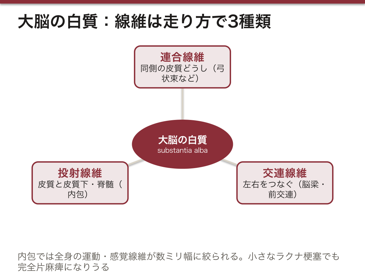
白質の線維は走り方で3種類。
- 連合線維(同側の皮質どうし)— 弓状束・上縦束・鉤状束など
- 交連線維(左右をつなぐ)— Corpus callosum・前交連・脳弓交連
- 投射線維(皮質と皮質下・脊髄)— Capsula interna
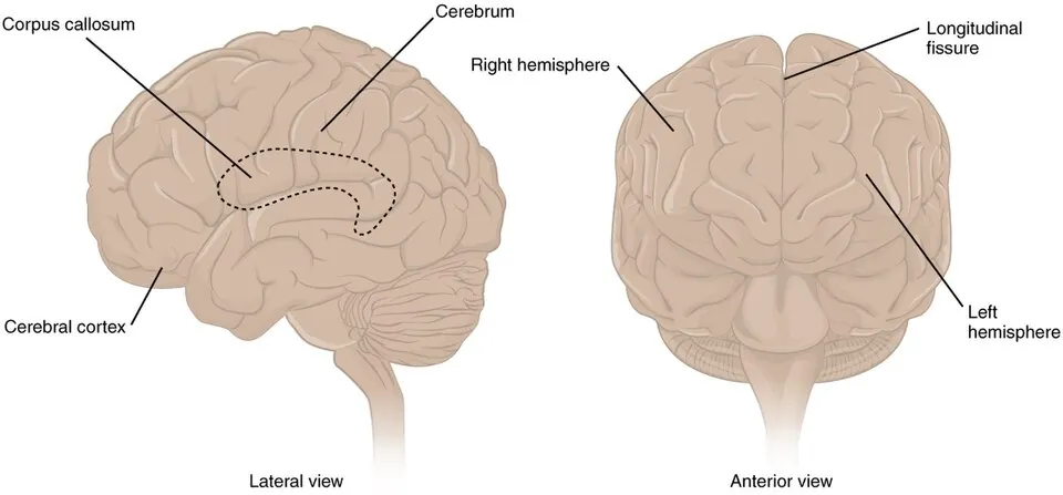
Corpus callosum は前から rostrum → genu → truncus → splenium の4部。膝から前へ回る線維が forceps minor、膨大から後ろへ回るのが forceps major。
Fornix(脳弓)は海馬から乳頭体への線維束。Septum pellucidum は脳梁と脳弓のあいだに張る薄い膜で、左右の側脳室前角を隔てる。
Capsula interna は水平断で「くの字」に見える。内側が視床と尾状核、外側が nucleus lentiformis(被殻+淡蒼球)。3部に分ける:
| 部位 | 挟むもの | 通る線維 |
|---|---|---|
| Crus anterius | 尾状核頭と被殻 | 前視床脚・前頭橋路 |
| Genu | くの字の角 | 皮質核路(顔面・舌の運動) |
| Crus posterius | 視床と被殻 | 皮質脊髄路・視床皮質路・視放線・聴放線 |
内包は面積のわりに被害が大きい。ここでは全身の運動・感覚線維が数ミリの幅に絞られているので、小さなラクナ梗塞1つで完全片麻痺が起こりうる。皮質の同じ量の損傷では部分的な麻痺にしかならない。
外側にはさらに capsula externa(被殻と claustrum のあいだ)と capsula extrema(前障と島皮質のあいだ)がある。「内包・外包・前障」を内側から外側へ順に指せるようにする。
内包は運動と感覚の線維が最も密に束ねられる場所である。だからごく小さな病変で全身が動かなくなる。
ラクナ梗塞——内包後脚を養うのはレンズ核線条体動脈などの穿通枝で、高血圧で傷みやすい終末動脈である。ここが詰まると純粋運動性片麻痺(感覚も高次機能も保たれ、反対側の顔・上肢・下肢だけが均等に麻痺する)になる。皮質症状(失語・失認)が無いことが皮質梗塞との鑑別点である。
被殻出血は内包を圧迫して同じ像を作り、共同偏視は病巣側を向く。
脳梁が切れると離断症候群——左手だけが命令に従わない(左手の失行)、左視野に出した物の名前が言えない、といった「左右の脳が話し合えない」症状が出る。
出典:イラスト解剖学 誌面613頁「貫通枝(穿通枝)をちょっと詳しく」
English version
Core
The fibres of the white matter are of three kinds by their course.
- Association fibres (cortex to cortex on the same side) — arcuate, superior longitudinal, uncinate fasciculi and others.
- Commissural fibres (joining the two sides) — corpus callosum, anterior commissure, fornix.
- Projection fibres (cortex to subcortex and cord) — capsula interna.
The corpus callosum has four parts from the front: rostrum → genu → truncus → splenium. Fibres curving forwards from the genu form the forceps minor, those curving backwards from the splenium the forceps major.
The fornix is the bundle from hippocampus to mamillary body. The septum pellucidum is the thin membrane stretched between corpus callosum and fornix, separating the anterior horns of the two lateral ventricles.
Going further
On a horizontal section the capsula interna appears as a bent V. Medial to it are the thalamus and caudate nucleus, lateral the nucleus lentiformis (putamen + globus pallidus). It divides into three:
| Part | Between | Fibres passing |
|---|---|---|
| Crus anterius | Head of caudate and putamen | Anterior thalamic radiation, frontopontine tract |
| Genu | The angle of the V | Corticonuclear tract (movement of face and tongue) |
| Crus posterius | Thalamus and putamen | Corticospinal tract, thalamocortical fibres, optic and acoustic radiations |
The internal capsule does damage out of all proportion to its area. The motor and sensory fibres of the whole body are compressed into a few millimetres here, so a single small lacunar infarct can cause a complete hemiplegia. The same volume of damage in the cortex would give only a partial weakness.
Lateral to it are also the capsula externa (between putamen and claustrum) and the capsula extrema (between claustrum and insular cortex). Be able to point from medial to lateral: internal capsule, external capsule, claustrum.
In the clinic
The internal capsule is where the motor and sensory fibres are packed most densely. So the smallest lesion can stop the whole body moving.
Lacunar infarction — the posterior limb is supplied by perforating vessels such as the lenticulostriate arteries, end arteries readily damaged by hypertension. Occlusion here gives a pure motor hemiparesis (sensation and higher function preserved, with the contralateral face, arm and leg weak to an equal degree). The absence of cortical signs (aphasia, agnosia) distinguishes it from a cortical infarct.
Putaminal haemorrhage compresses the internal capsule and produces the same picture, with conjugate deviation towards the side of the lesion.
When the corpus callosum is divided, a disconnection syndrome follows — the left hand alone will not obey a command (left-hand apraxia), an object shown in the left visual field cannot be named: the symptoms of the two hemispheres being unable to confer.
大脳基底核 — 灰白質の塊Nuclei basales
半球の深部にある灰白質の集まり。押さえるのは4つ。
- Nucleus caudatus(尾状核)— caput(側脳室前角の外側壁)・corpus・cauda。側脳室に沿ってC字に曲がる
- Nucleus lentiformis(レンズ核)— 外側の被殻 putamen と内側の淡蒼球 globus pallidus
- Claustrum(前障)— 被殻の外側の薄い灰白質の板
- Corpus amygdaloideum(扁桃体)— 側頭葉前端、側脳室下角の先端
Striatum(線条体)=尾状核+被殻、lentiform nucleus=被殻+淡蒼球。被殻が両方に属するため名前が重なる。
尾状核と被殻はもともと1つだった。発生の途中で内包の線維が貫いて2つに分かれ、残った灰白質の橋が縞に見えるので striatum(縞のある体) という。肉眼所見の名前がそのまま発生の履歴になっている。
回路は直接路と間接路の2本。直接路(線条体 → 淡蒼球内節 → 視床)は視床の抑制を外して運動を出す、間接路(線条体 → 淡蒼球外節 → 視床下核 → 淡蒼球内節)は運動を抑える。黒質緻密部のドパミンは直接路を促進し間接路を抑制する——つまり両方の経路で「運動を出す」向きに働く。だからドパミンが減ると動けなくなる(パーキンソン病)。
逆に間接路が壊れると動きが止まらない。視床下核の障害でヘミバリスム、線条体の変性でハンチントン病の舞踏運動。「足りないと動けない、抑えが壊れると止まらない」という一組で覚える。
Parkinson 病——黒質緻密部のドパミン神経が脱落し、線条体への入力が減る。直接路が働かず間接路が優位になるため、動きが出にくくなる(寡動)。安静時振戦・筋強剛(歯車様)・無動・姿勢反射障害の4徴。安静時に震え、動かすと止まるのが小脳性の企図振戦と正反対である。
Huntington 病——線条体(とくに尾状核)の変性。間接路が先に落ちるので動きが止められなくなる(舞踏運動)。CT で尾状核頭の萎縮により側脳室前角が外側へ張り出す。
ヘミバリスム——視床下核の病変。反対側の上下肢が投げ出すように大きく動く。
被殻は高血圧性脳出血の最好発部位である。基底核の位置を覚えることが、そのまま出血部位の推定になる。
English version
Core
The collections of grey matter deep in the hemisphere. Four are to be known.
- Nucleus caudatus — caput (the lateral wall of the anterior horn), corpus, cauda. Curves in a C along the lateral ventricle.
- Nucleus lentiformis — the lateral putamen and the medial globus pallidus.
- Claustrum — the thin plate of grey matter lateral to the putamen.
- Corpus amygdaloideum — the anterior end of the temporal lobe, at the tip of the inferior horn.
Striatum = caudate + putamen; lentiform nucleus = putamen + globus pallidus. The putamen belongs to both, which is why the names overlap.
Going further
The caudate and the putamen were originally one. During development the fibres of the internal capsule grew through and divided them in two, and the remaining bridges of grey matter look striped — hence striatum, the striped body. The name of a gross appearance is itself the record of the development.
The circuitry is two paths, direct and indirect. The direct path (striatum → internal pallidum → thalamus) releases the thalamus from inhibition and produces movement; the indirect path (striatum → external pallidum → subthalamic nucleus → internal pallidum) suppresses it. Dopamine from the pars compacta of the substantia nigra facilitates the direct path and inhibits the indirect — that is, it acts in the direction of producing movement through both.
So when dopamine falls, movement cannot be produced (Parkinson’s disease). Conversely, when the indirect path is destroyed, movement cannot be stopped: hemiballismus from a lesion of the subthalamic nucleus, the chorea of Huntington’s disease from degeneration of the striatum. Learn them as a pair: too little and you cannot move; the brake broken and you cannot stop.
In the clinic
Parkinson’s disease — the dopaminergic neurones of the pars compacta are lost and the input to the striatum falls. The direct path fails and the indirect predominates, so movement is hard to produce (bradykinesia). The tetrad is resting tremor, rigidity (cogwheel), akinesia and postural instability. Trembling at rest and stopping on movement is the exact opposite of the intention tremor of cerebellar disease.
Huntington’s disease — degeneration of the striatum (especially the caudate). The indirect path fails first, so movement cannot be stopped (chorea). On CT, atrophy of the head of the caudate lets the anterior horns bulge laterally.
Hemiballismus — a lesion of the subthalamic nucleus. The contralateral limbs make large flinging movements.
The putamen is the commonest site of hypertensive intracerebral haemorrhage. Learning the position of the basal nuclei is itself learning to infer the site of a haemorrhage.
側脳室 — C字の4部Ventriculus lateralis
各半球に1つずつ。半球の形に沿ってC字に曲がる。4部に分ける。
| 部 | ある葉 | 目印 |
|---|---|---|
| Cornu anterius(前角) | 前頭葉 | 外側壁=尾状核頭、内側壁=透明中隔 |
| Pars centralis(中心部) | 頭頂葉 | 床=視床と尾状核体 |
| Cornu posterius(後角) | 後頭葉 | 内側壁に鳥距溝の隆起(calcar avis) |
| Cornu inferius(下角) | 側頭葉 | 床=hippocampus、先端に扁桃体 |
Foramen interventriculare(Monro 孔)で第三脳室に開く。位置は脳弓の柱と視床前結節のあいだ。
脈絡叢は前角と後角には無い。中心部から下角にかけて連続して存在し、Monro 孔で第三脳室の脈絡叢に続く。
側脳室・海馬・脳弓・尾状核はすべて同じC字を描く。終脳が胎生期に前から後ろ、そして下前方へ弧を描いて成長したためで、1つの弧を覚えれば4つの構造の走行が同時に説明できる。標本では下角に指を入れて、床にある海馬をなぞって示すとよい。
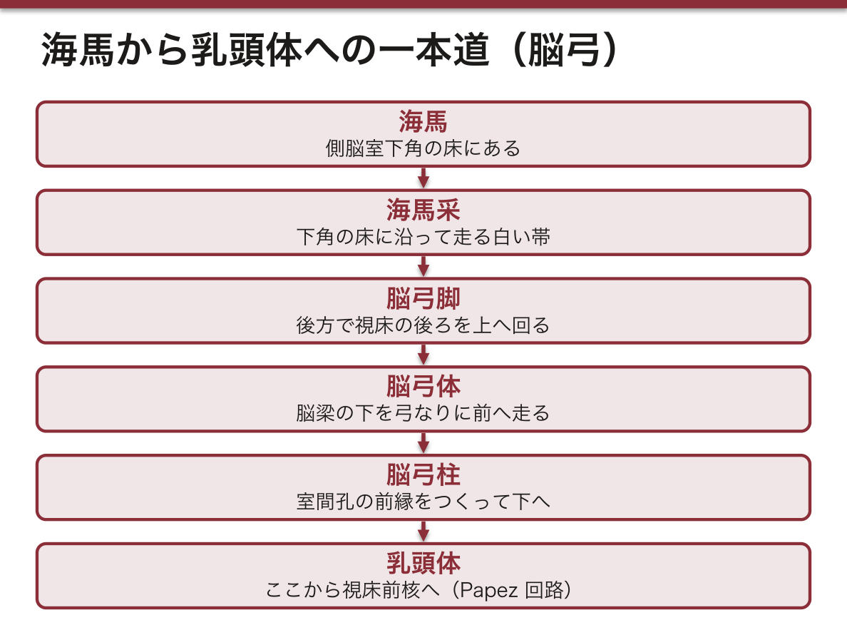
下角の床に沿って走る白い帯が fimbria hippocampi で、これが後方で crus fornicis となり、脳弓へ続く。海馬 → 采 → 脳弓脚 → 脳弓体 → 脳弓柱 → 乳頭体という一本の道である。
脳室拡大は「詰まって広がった」のか「脳が縮んで広がった」のかを分ける。
閉塞性水頭症——詰まった点より上流だけが拡大する。中脳水道の閉塞なら側脳室+第三脳室が拡大し、第四脳室は正常。脳溝は狭くなる(圧で押しつぶされるため)。
脳萎縮による代償性拡大(ex vacuo)——脳室も脳溝も一緒に広がる。圧は上がっていない。
正常圧水頭症(NPH)——認知症・歩行障害・尿失禁の3徴。脳室は拡大しているのに髄液圧は正常(200mmH2O 以下)。髄液を抜くと歩行が改善する(タップテスト)ので、治せる認知症として必ず鑑別に挙げる。
出典:イラスト解剖学 誌面587頁「水頭症って?」
English version
Core
One in each hemisphere, curving in a C to follow the shape of the hemisphere. It divides into four parts.
| Part | Lobe | Landmark |
|---|---|---|
| Cornu anterius | Frontal | Lateral wall = head of caudate; medial wall = septum pellucidum |
| Pars centralis | Parietal | Floor = thalamus and body of caudate |
| Cornu posterius | Occipital | The calcar avis on the medial wall (the impression of the calcarine sulcus) |
| Cornu inferius | Temporal | Floor = hippocampus; the amygdala at its tip |
It opens into the third ventricle at the foramen interventriculare (of Monro), between the column of the fornix and the anterior tubercle of the thalamus.
Going further
There is no choroid plexus in the anterior or the posterior horn. It runs continuously from the central part into the inferior horn and continues through the foramen of Monro into the plexus of the third ventricle.
The lateral ventricle, hippocampus, fornix and caudate nucleus all describe the same C, because in fetal life the telencephalon grew forwards, backwards, then down and forwards in an arc. Learn one arc and the course of all four structures is explained at once.
On a specimen, put a finger into the inferior horn and trace the hippocampus along its floor. The white band running along that floor is the fimbria hippocampi, which behind becomes the crus fornicis and continues into the fornix. Hippocampus → fimbria → crus → body → column → mamillary body is a single road.
In the clinic
With ventricular dilatation, separate "widened because blocked" from "widened because the brain shrank".
Obstructive hydrocephalus — only what lies upstream of the block dilates. With aqueduct stenosis the lateral and third ventricles dilate and the fourth is normal. The sulci are narrowed (crushed by the pressure).
Compensatory (ex vacuo) dilatation from atrophy — ventricles and sulci widen together. The pressure is not raised.
Normal pressure hydrocephalus (NPH) — the triad of dementia, gait disturbance and urinary incontinence. The ventricles are dilated yet the CSF pressure is normal (below 200 mmH₂O). Removing CSF improves the gait (the tap test), so it must always be raised as a treatable dementia.
大脳縦裂Fissura longitudinalis cerebri
左右の大脳半球を隔てる正中の深い裂。中に大脳鎌が垂れ下がる。
底では脳梁でつながっているので、裂は脳梁までしか届かない。
English version
Core
The deep median cleft separating the two cerebral hemispheres, with the falx cerebri hanging in it.
At its floor the two sides are joined by the corpus callosum, so the fissure reaches only as far as the callosum.
大脳横裂Fissura transversa cerebri
大脳と小脳の間の水平な裂。ここに小脳テントが入る。
縦裂と対にして「縦=左右を分ける、横=大脳と小脳を分ける」。
English version
Core
The horizontal cleft between cerebrum and cerebellum, into which the tentorium cerebelli passes.
Learn it as a pair with the longitudinal fissure: longitudinal separates right from left, transverse separates cerebrum from cerebellum.
中心溝(Rolando 溝)Sulcus centralis cerebri
上外側面を斜め前下がりに走る溝。前頭葉と頭頂葉の境。
この溝を決めないと他の回が読めないので、標本ではまずここを探す。前の回が運動、後ろの回が感覚。
English version
Core
The sulcus running obliquely down and forwards on the superolateral surface, the boundary of frontal and parietal lobes.
Without settling this sulcus the other gyri cannot be read, so on a specimen look for it first. The gyrus in front is motor, the one behind sensory.
外側溝(Sylvius 溝)Sulcus lateralis cerebri
半球の外側面を前下から後上へ走る、最も深く目立つ溝。側頭葉を前頭葉・頭頂葉から分ける。
奥を開くと島が現れる。中を中大脳動脈が走る。
English version
Core
The deepest and most conspicuous sulcus of the lateral surface, running from anteroinferior to posterosuperior, separating the temporal lobe from the frontal and parietal lobes.
Open its depth and the insula appears. The middle cerebral artery runs within it.
中心前溝Sulcus precentralis cerebri
中心溝の前を、ほぼ平行に走る溝。中心溝との間が中心前回。
English version
Core
The sulcus running almost parallel in front of the central sulcus. Between the two is the precentral gyrus.
上前頭溝Sulcus frontalis superior cerebri
中心前溝から前へ伸びる2本の溝のうち上のほう。上前頭回と中前頭回の境。
English version
Core
The upper of the two sulci extending forwards from the precentral sulcus. The boundary between the superior and middle frontal gyri.
下前頭溝Sulcus frontalis inferior cerebri
同じく前へ伸びる下のほうの溝。中前頭回と下前頭回の境。
前頭葉は「2本の横溝で3段に分かれる」——この作りが分かれば回の名前は自動で決まる。
English version
Core
The lower of those sulci. The boundary between the middle and inferior frontal gyri.
The frontal lobe is divided into three tiers by two horizontal sulci — grasp that construction and the names of the gyri follow automatically.
中心後溝Sulcus postcentralis cerebri
中心溝の後ろを平行に走る溝。中心溝との間が中心後回。
English version
Core
The sulcus running parallel behind the central sulcus. Between the two is the postcentral gyrus.
頭頂間溝Sulcus intraparietalis
中心後溝から後方へ水平に走り、頭頂葉を上頭頂小葉と下頭頂小葉に分ける。
English version
Core
Running horizontally backwards from the postcentral sulcus, dividing the parietal lobe into superior and inferior parietal lobules.
上側頭溝Sulcus temporalis superior cerebri
外側溝と平行に走る、側頭葉の上の横溝。上側頭回と中側頭回の境。
English version
Core
The upper horizontal sulcus of the temporal lobe, running parallel to the lateral sulcus. The boundary between the superior and middle temporal gyri.
下側頭溝Sulcus temporalis inferior cerebri
その下の横溝。中側頭回と下側頭回の境。
側頭葉も前頭葉と同じ「2本の横溝で3段」の作り。
English version
Core
The horizontal sulcus below it. The boundary between the middle and inferior temporal gyri.
The temporal lobe has the same construction as the frontal — three tiers made by two horizontal sulci.
脳梁溝Sulcus corporis callosi
内側面で、脳梁の背面に沿って走る溝。脳梁と帯状回の境。
English version
Core
On the medial surface, the sulcus running along the dorsal surface of the corpus callosum. The boundary between corpus callosum and cingulate gyrus.
帯状溝Sulcus cinguli
脳梁溝のさらに外側を、脳梁を取り巻くように弧を描く溝。帯状回の外側の境。
内側面は「脳梁 → 脳梁溝 → 帯状回 → 帯状溝」の順に外へ読む。
English version
Core
The sulcus lateral to the callosal sulcus, arching around the corpus callosum. The outer boundary of the cingulate gyrus.
Read the medial surface outwards in the order: corpus callosum → callosal sulcus → cingulate gyrus → cingulate sulcus.
頭頂後頭溝Sulcus parietooccipitalis cerebri
内側面を深く走り、頭頂葉と後頭葉を分ける。鳥距溝と合してY字を作る。
この溝と鳥距溝に挟まれるのが楔部、前が楔前部。
English version
Core
Running deeply on the medial surface and separating the parietal from the occipital lobe. It joins the calcarine sulcus to form a Y.
Between this sulcus and the calcarine is the cuneus; in front of it, the precuneus.
後頭側頭溝Sulcus occipitotemporalis cerebri
半球の下面を前後に走る溝。外側が下側頭回、内側が紡錘状回。
English version
Core
The anteroposterior sulcus on the inferior surface of the hemisphere. Lateral to it is the inferior temporal gyrus, medial the fusiform gyrus.
嗅溝Sulcus olfactorius cerebri
前頭葉下面の内側寄りを前後に走る溝。ここに嗅球と嗅索が乗るので、標本では嗅索を持ち上げて示す。内側が直回。
English version
Core
The anteroposterior sulcus on the medial part of the inferior surface of the frontal lobe. The olfactory bulb and tract lie in it, so on a specimen lift the tract to show it. Medial to it is the gyrus rectus.
中心前回Gyrus precentralis
中心溝のすぐ前の回。一次運動野(Brodmann 4 野)で、ここから皮質脊髄路が出る。
体の各部が逆さまに並ぶ運動のホムンクルス。手と口の領域が不釣り合いに広い。
English version
Core
The gyrus immediately in front of the central sulcus. The primary motor area (Brodmann 4), from which the corticospinal tract arises.
The parts of the body are laid out upside down as the motor homunculus, with the areas for hand and mouth disproportionately large.
上前頭回Gyrus frontalis superior
上前頭溝より上の回。内側面へ回り込む。
English version
Core
The gyrus above the superior frontal sulcus, turning onto the medial surface.
中前頭回Gyrus frontalis medius
上前頭溝と下前頭溝に挟まれた回。後端に前頭眼野がある。
English version
Core
The gyrus between the superior and inferior frontal sulci. The frontal eye field lies at its posterior end.
下前頭回Gyrus frontalis inferior
下前頭溝より下の回。優位半球(多くは左)では Broca の運動性言語野(44・45 野)。
障害で運動性失語——言葉は分かるが話せない。
English version
Core
The gyrus below the inferior frontal sulcus. In the dominant hemisphere (usually the left) it is Broca’s motor speech area (areas 44, 45).
A lesion gives motor aphasia — the patient understands but cannot speak.
中心後回Gyrus postcentralis
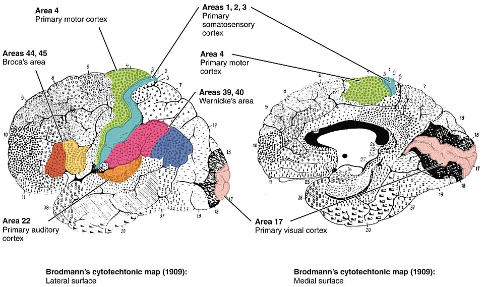
中心溝のすぐ後ろの回。一次体性感覚野(3・1・2 野)。
中心溝を挟んで前が運動、後ろが感覚——必ず押さえる対。
English version
Core
The gyrus immediately behind the central sulcus. The primary somatosensory area (areas 3, 1, 2).
Motor in front of the central sulcus, sensory behind — a pair to know without fail.
上側頭回Gyrus temporalis superior
外側溝のすぐ下の回。上面に横側頭回(Heschl 回)=一次聴覚野が隠れている。
後部はWernicke の感覚性言語野。障害で感覚性失語——流暢に話すが意味が通らない。
English version
Core
The gyrus immediately below the lateral sulcus. On its upper surface, hidden, are the transverse temporal gyri (of Heschl) = the primary auditory area. Its posterior part is Wernicke’s sensory speech area.
A lesion gives sensory aphasia — fluent speech that makes no sense.
中側頭回Gyrus temporalis medius
上側頭溝と下側頭溝に挟まれた回。
English version
Core
The gyrus between the superior and inferior temporal sulci.
下側頭回Gyrus temporalis inferior
下側頭溝より下の回。下面へ回り込む。
English version
Core
The gyrus below the inferior temporal sulcus, turning onto the inferior surface.
島Insula
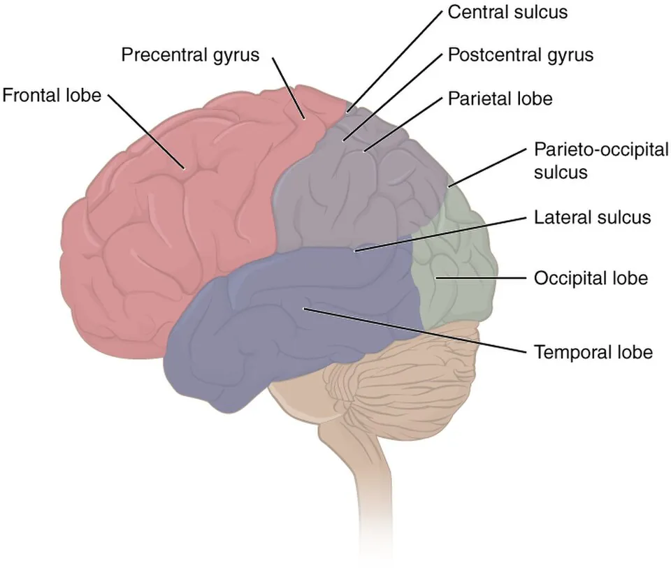
外側溝の底に埋もれた皮質。周りの前頭・頭頂・側頭の弁蓋に覆われて、外からは見えない。
標本では外側溝を指で開いて示す。味覚と内臓感覚に関わる。
English version
Core
The cortex buried at the floor of the lateral sulcus, covered by the frontal, parietal and temporal opercula and invisible from outside.
On a specimen, open the lateral sulcus with a finger to show it. It is concerned with taste and visceral sensation.
楔前部Precuneus
内側面で、辺縁枝と頭頂後頭溝に挟まれた四角い領域。楔部の前にある。
English version
Core
The quadrilateral area on the medial surface between the marginal branch and the parieto-occipital sulcus, in front of the cuneus.
楔部Cuneus
内側面で、頭頂後頭溝と鳥距溝に挟まれたくさび形。一次視覚野の一部(上唇)。
楔前部と並べて「頭頂後頭溝の前が楔前部、後ろが楔部」。
English version
Core
The wedge on the medial surface between the parieto-occipital and calcarine sulci. Part of the primary visual cortex (the upper lip).
Set beside the precuneus: in front of the parieto-occipital sulcus is the precuneus, behind it the cuneus.
帯状回Gyrus cinguli
脳梁をぐるりと取り巻く回。脳梁溝と帯状溝に挟まれる。
大脳辺縁系の中心的な回で、後方は脳梁膨大の下を回って海馬傍回へ続く。
English version
Core
The gyrus encircling the corpus callosum, between the callosal and cingulate sulci. The central gyrus of the limbic system, continuing behind beneath the splenium into the parahippocampal gyrus.
嗅球Bulbus olfactorius
前頭葉下面、嗅溝の中に乗る棍棒状のふくらみ。篩板を貫いてきた嗅細胞の軸索を受ける。
嗅神経(I)の実体はここで、脳の一部が前へ伸びたもの。
English version
Core
The club-shaped swelling lying in the olfactory sulcus on the inferior surface of the frontal lobe, receiving the axons of the olfactory cells that have pierced the cribriform plate.
This is the substance of the olfactory nerve (I) — a part of the brain extended forwards.
嗅索Tractus olfactorius
嗅球から後方へ伸びる細い索。後端で内側・外側の嗅条に分かれ、その間が前有孔質。
脳底面の標本では最も前方の細長い構造で、位置決めの目印になる。
English version
Core
The slender tract running backwards from the olfactory bulb, dividing at its posterior end into medial and lateral striae with the anterior perforated substance between them.
On a specimen of the base of the brain it is the most anterior elongated structure and serves as a landmark for orientation.
脳梁Corpus callosum
左右の半球をつなぐ最大の交連線維(約2億本)。正中矢状断で、大きな弓形の白い板として見える。
前から嘴・膝・幹・膨大の4部。標本ではこの4部を順に指す。
English version
Core
The largest commissure joining the two hemispheres (about 200 million fibres). On a median sagittal section it appears as a great arched white plate. Four parts from the front: rostrum, genu, trunk, splenium.
On a specimen, point to these four in order.
脳梁嘴Rostrum corporis callosi
脳梁の最前下部。膝から後下方へ細く伸びて終板に続く。
English version
Core
The most anteroinferior part of the corpus callosum, tapering back and down from the genu to continue into the lamina terminalis.
脳梁膝Genu corporis callosi
脳梁の前端が下へ折れ曲がる部分。ここから出る線維が前頭葉へ放散して小鉗子 forceps minor をつくる。
English version
Core
The part where the anterior end of the corpus callosum bends downwards. The fibres leaving it radiate into the frontal lobe as the forceps minor.
脳梁幹Truncus corporis callosi
膝と膨大の間の長い水平部。側脳室の天井になる。
English version
Core
The long horizontal part between genu and splenium. It forms the roof of the lateral ventricle.
脳梁膨大Splenium corporis callosi
脳梁の後端の厚い膨らみ。ここから出る線維が後頭葉へ放散して大鉗子 forceps major をつくる。
すぐ下に松果体と大大脳静脈槽がある。
English version
Core
The thick swelling at the posterior end of the corpus callosum. The fibres leaving it radiate into the occipital lobe as the forceps major.
Immediately beneath it lie the pineal gland and the cistern of the great cerebral vein.
脳弓Fornix
脳梁の下を弓なりに走る白質束。海馬 → 乳頭体をつなぐ、辺縁系の主要な出力路(Papez 回路)。
前方の脳弓柱が室間孔の前縁をつくる。
English version
Core
The bundle of white matter arching beneath the corpus callosum, joining hippocampus to mamillary body: the principal output of the limbic system (the Papez circuit).
The column of the fornix in front forms the anterior margin of the interventricular foramen.
透明中隔Septum pellucidum
脳梁と脳弓の間に張る、薄く透明な2枚の膜。左右の側脳室前角を隔てる。
2枚の間に腔が残ることがある(透明中隔腔)。
English version
Core
The two thin, translucent membranes stretched between corpus callosum and fornix, separating the anterior horns of the two lateral ventricles.
A cavity may persist between the two leaves (cavum septi pellucidi).
側脳室中心部Pars centralis ventriculi lateralis
頭頂葉の中を前後に走る部分。天井は脳梁、床は視床と尾状核体。
側脳室はC字形で、中心部+前角・後角・下角の4部からなる。
English version
Core
The part running anteroposteriorly within the parietal lobe. The roof is the corpus callosum, the floor the thalamus and the body of the caudate nucleus.
The lateral ventricle is C-shaped, consisting of the central part plus the anterior, posterior and inferior horns.
側脳室前角Cornu anterius ventriculi lateralis
前頭葉へ伸びる部分。室間孔より前。内側壁が透明中隔、外側壁が尾状核頭のふくらみ。
English version
Core
The part extending into the frontal lobe, in front of the interventricular foramen. The medial wall is the septum pellucidum, the lateral wall the bulge of the head of the caudate nucleus.
側脳室後角Cornu posterius ventriculi lateralis
後頭葉へ伸びる部分。内側壁に鳥距溝が押し込んだ鳥距のふくらみがある。左右で長さが違うことが多い。
English version
Core
The part extending into the occipital lobe. On its medial wall is the calcar avis, the impression pushed in by the calcarine sulcus. Its length often differs between the two sides.
尾状核頭Caput nuclei caudati
尾状核の最も太い前端で、側脳室前角の外側壁に膨らむ。
Huntington 病でここが萎縮し、前角が拡がって見える。
English version
Core
The thickest, anterior end of the caudate nucleus, bulging into the lateral wall of the anterior horn.
In Huntington’s disease it atrophies and the anterior horn appears widened.
尾状核体Corpus nuclei caudati
頭から後方へ細くなりながら、視床の外側を側脳室中心部の床に沿って走る部分。
尾状核は側脳室に寄り添ってC字に曲がる——この形が分かると水平断が読める。
English version
Core
The part running backwards from the head, narrowing as it goes, lateral to the thalamus and along the floor of the central part of the ventricle.
The caudate nucleus curves in a C hugging the lateral ventricle — grasp this shape and the horizontal section can be read.
レンズ核Nucleus lentiformis
内包の外側にある、水平断でくさび(レンズ)形に見える灰白質。外側の被殻と内側の淡蒼球からなる。
被殻+尾状核=線条体という括りとは切り口が違うので、混同しないこと。
English version
Core
The grey matter lateral to the internal capsule, wedge- (lens-) shaped on a horizontal section, consisting of the lateral putamen and the medial globus pallidus.
It is cut differently from the grouping putamen + caudate = striatum, so do not confuse the two.
前障Claustrum
レンズ核(被殻)と島皮質の間にはさまれた薄い灰白質の板。
English version
Core
The thin plate of grey matter caught between the lentiform nucleus (putamen) and the insular cortex.
外包Capsula externa
被殻と前障の間の白質の薄い層。
水平断では外から島 → 最外包 → 前障 → 外包 → 被殻の順に並ぶ。この層の順序をそのまま言えるようにする。
English version
Core
The thin layer of white matter between putamen and claustrum.
On a horizontal section the order from outside in is insula → extreme capsule → claustrum → external capsule → putamen. Be able to state that order as it stands.
内包Capsula interna
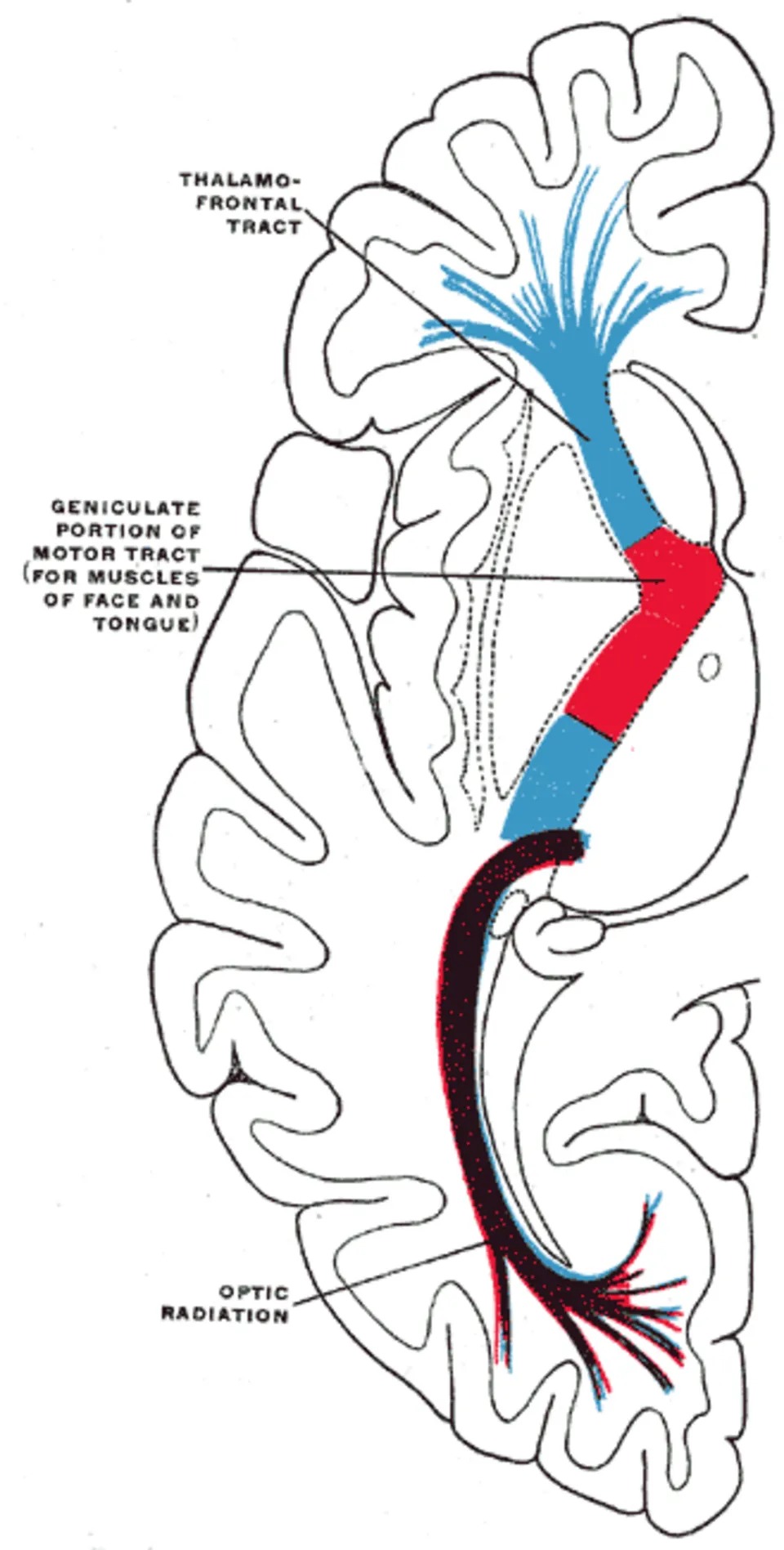
尾状核・視床とレンズ核の間を通る白質。水平断で外向きのV字(への字)に見える。
大脳皮質へ出入りするほぼ全ての線維がここに集まるので、小さな出血でも重い麻痺が出る。前脚・膝・後脚の3部。
English version
Core
The white matter passing between the caudate nucleus and thalamus on one side and the lentiform nucleus on the other, appearing on a horizontal section as a V opening laterally.
Nearly all the fibres entering and leaving the cerebral cortex gather here, so even a small haemorrhage causes a severe paralysis. Three parts: anterior limb, genu, posterior limb.
内包前脚Crus anterius capsulae internae
尾状核頭とレンズ核の間。前頭橋路と視床前放線が通る。
English version
Core
Between the head of the caudate nucleus and the lentiform nucleus. The frontopontine tract and the anterior thalamic radiation pass through it.
内包膝Genu capsulae internae
V字の折れ曲がり。皮質核路(顔・舌へ)が通る。
English version
Core
The bend of the V. The corticonuclear tract (to face and tongue) passes through it.
内包後脚Crus posterius capsulae internae
視床とレンズ核の間。皮質脊髄路が通り、その後方に視放線・聴放線が続く。
「前脚=前頭橋路、膝=顔、後脚=手足」——この並びが、内包出血でどこが麻痺するかを決める。
English version
Core
Between the thalamus and the lentiform nucleus. The corticospinal tract passes through it, with the optic and acoustic radiations behind.
"Anterior limb = frontopontine, genu = face, posterior limb = limbs" — this order decides what is paralysed in a capsular haemorrhage.
まとめ
| 大脳の溝と回 — 上外側面 | 標本では「まず2本の基準溝を見つけてから、残りを相対位置で言う」のが確実である。 |
|---|---|
| 大脳の内側面と下面 | 内側面は矢状断でしか見えない。中央に corpus callosum が弓なりに横たわり、その上を sulcus corporis callosi、さらに上を sulcus cin… |
| 皮質中枢 — 回の名前と機能の対応 | 皮質の中枢(cortical centers)は、回の名前と機能を一対一で言えるようにする。 |
| 大脳の白質 — 3種類の線維と内包 | 白質の線維は走り方で3種類。連合線維(同側の皮質どうし)— 弓状束・上縦束・鉤状束など交連線維(左右をつなぐ)— Corpus callosum・前交連・脳弓交連投射線維(皮質と皮… |
| 大脳基底核 — 灰白質の塊 | 半球の深部にある灰白質の集まり。押さえるのは4つ。 |
| 側脳室 — C字の4部 | 各半球に1つずつ。半球の形に沿ってC字に曲がる。 |
| 大脳縦裂 | 左右の大脳半球を隔てる正中の深い裂。中に大脳鎌が垂れ下がる。 |
| 大脳横裂 | 大脳と小脳の間の水平な裂。ここに小脳テントが入る。 |
| 中心溝(Rolando 溝) | 上外側面を斜め前下がりに走る溝。前頭葉と頭頂葉の境。 |
| 外側溝(Sylvius 溝) | 半球の外側面を前下から後上へ走る、最も深く目立つ溝。 |
| 中心前溝 | 中心溝の前を、ほぼ平行に走る溝。中心溝との間が中心前回。 |
| 上前頭溝 | 中心前溝から前へ伸びる2本の溝のうち上のほう。 |
| 下前頭溝 | 同じく前へ伸びる下のほうの溝。中前頭回と下前頭回の境。 |
| 中心後溝 | 中心溝の後ろを平行に走る溝。中心溝との間が中心後回。 |
| 頭頂間溝 | 中心後溝から後方へ水平に走り、頭頂葉を上頭頂小葉と下頭頂小葉に分ける。 |
| 上側頭溝 | 外側溝と平行に走る、側頭葉の上の横溝。上側頭回と中側頭回の境。 |
| 下側頭溝 | その下の横溝。中側頭回と下側頭回の境。 |
| 脳梁溝 | 内側面で、脳梁の背面に沿って走る溝。脳梁と帯状回の境。 |
| 帯状溝 | 脳梁溝のさらに外側を、脳梁を取り巻くように弧を描く溝。 |
| 頭頂後頭溝 | 内側面を深く走り、頭頂葉と後頭葉を分ける。 |
| 後頭側頭溝 | 半球の下面を前後に走る溝。外側が下側頭回、内側が紡錘状回。 |
| 嗅溝 | 前頭葉下面の内側寄りを前後に走る溝。ここに嗅球と嗅索が乗るので、標本では嗅索を持ち上げて示す。 |
| 中心前回 | 中心溝のすぐ前の回。一次運動野(Brodmann 4 野)で、ここから皮質脊髄路が出る。 |
| 上前頭回 | 上前頭溝より上の回。内側面へ回り込む。 |
| 中前頭回 | 上前頭溝と下前頭溝に挟まれた回。後端に前頭眼野がある。 |
| 下前頭回 | 下前頭溝より下の回。優位半球(多くは左)では Broca の運動性言語野(44・45 野)。 |
| 中心後回 | 中心溝のすぐ後ろの回。一次体性感覚野(3・1・2 野)。 |
| 上側頭回 | 外側溝のすぐ下の回。上面に横側頭回(Heschl 回)=一次聴覚野が隠れている。 |
| 中側頭回 | 上側頭溝と下側頭溝に挟まれた回。 |
| 下側頭回 | 下側頭溝より下の回。下面へ回り込む。 |
| 島 | 外側溝の底に埋もれた皮質。周りの前頭・頭頂・側頭の弁蓋に覆われて、外からは見えない。 |
| 楔前部 | 内側面で、辺縁枝と頭頂後頭溝に挟まれた四角い領域。 |
| 楔部 | 内側面で、頭頂後頭溝と鳥距溝に挟まれたくさび形。 |
| 帯状回 | 脳梁をぐるりと取り巻く回。脳梁溝と帯状溝に挟まれる。 |
| 嗅球 | 前頭葉下面、嗅溝の中に乗る棍棒状のふくらみ。 |
| 嗅索 | 嗅球から後方へ伸びる細い索。後端で内側・外側の嗅条に分かれ、その間が前有孔質。 |
| 脳梁 | 左右の半球をつなぐ最大の交連線維(約2億本)。 |
| 脳梁嘴 | 脳梁の最前下部。膝から後下方へ細く伸びて終板に続く。 |
| 脳梁膝 | 脳梁の前端が下へ折れ曲がる部分。ここから出る線維が前頭葉へ放散して小鉗子 forceps minor をつくる。 |
| 脳梁幹 | 膝と膨大の間の長い水平部。側脳室の天井になる。 |
| 脳梁膨大 | 脳梁の後端の厚い膨らみ。ここから出る線維が後頭葉へ放散して大鉗子 forceps major をつくる。 |
| 脳弓 | 脳梁の下を弓なりに走る白質束。海馬 → 乳頭体をつなぐ、辺縁系の主要な出力路(Papez 回路)。 |
| 透明中隔 | 脳梁と脳弓の間に張る、薄く透明な2枚の膜。 |
| 側脳室中心部 | 頭頂葉の中を前後に走る部分。天井は脳梁、床は視床と尾状核体。 |
| 側脳室前角 | 前頭葉へ伸びる部分。室間孔より前。 |
| 側脳室後角 | 後頭葉へ伸びる部分。内側壁に鳥距溝が押し込んだ鳥距のふくらみがある。 |
| 尾状核頭 | 尾状核の最も太い前端で、側脳室前角の外側壁に膨らむ。 |
| 尾状核体 | 頭から後方へ細くなりながら、視床の外側を側脳室中心部の床に沿って走る部分。 |
| レンズ核 | 内包の外側にある、水平断でくさび(レンズ)形に見える灰白質。 |
| 前障 | レンズ核(被殻)と島皮質の間にはさまれた薄い灰白質の板。 |
| 外包 | 被殻と前障の間の白質の薄い層。水平断では外から島 → 最外包 → 前障 → 外包 → 被殻の順に並ぶ。 |
| 内包 | 尾状核・視床とレンズ核の間を通る白質。水平断で外向きのV字(への字)に見える。 |
| 内包前脚 | 尾状核頭とレンズ核の間。前頭橋路と視床前放線が通る。 |
| 内包膝 | V字の折れ曲がり。皮質核路(顔・舌へ)が通る。 |
| 内包後脚 | 視床とレンズ核の間。皮質脊髄路が通り、その後方に視放線・聴放線が続く。 |
このノートは医学生が授業の内容を自分の言葉でまとめたもので、診療の判断に使うものではありません。誤りに気づいたらお知らせください。