泌尿器は「後腹膜の器官」であるという一点が、位置・被膜・手術アプローチ・痛みの出方をすべて決める。
標本では腎の外形(極・面・縁・門)→ 割面(皮質・髄質・洞)→ 尿路(腎杯・腎盂・尿管・膀胱)の順に進める。腎門の中の3つ(前から静脈・動脈・腎盂)は必ず押さえる。
この範囲の芯は「3か所の狭窄」である。尿管には生まれつき狭い場所が3つあり、結石はそこで止まり、痛みはその高さの皮膚分節へ放散する。石の位置と痛む場所の対応は、解剖から完全に予測できる。
- 腎臓 — 外形と被膜
- 腎の割面 — 皮質・髄質・腎杯
- ネフロン — 割面の縞と、管の通り道
- 尿管 — 3つの狭窄
- 膀胱と尿道
- 膀胱の血管と神経 — 臍動脈の名残から来る動脈
- 腎の外側縁・内側縁
- 腎門
- 腎洞
- 腎皮質
- 腎錐体
- 腎乳頭
- 腎柱
- 腎盂
- 尿管
- 膀胱尖
- 膀胱体
- 膀胱底
- 発生 — 前腎・中腎・後腎、そして尿管芽
- 腎臓 — 外形と被膜Ren
- 腎の割面 — 皮質・髄質・腎杯Structura interna renis
- ネフロン — 割面の縞と、管の通り道Nephronum
- 尿管 — 3つの狭窄Ureter
- 膀胱と尿道Vesica urinaria et urethra
- 膀胱の血管と神経 — 臍動脈の名残から来る動脈Arteriae vesicales
- 腎の外側縁・内側縁Margo lateralis, medialis renis
- 腎門Hilum renale
- 腎洞Sinus renalis
- 腎皮質Cortex renalis
- 腎錐体Pyramis renalis
- 腎乳頭Papilla renalis
- 腎柱Columnae renales
- 腎盂Pelvis renis
- 尿管Ureter
- 膀胱尖Apex vesicae
- 膀胱体Corpus vesicae
- 膀胱底Fundus vesicae
- 発生 — 前腎・中腎・後腎、そして尿管芽Pronephros, mesonephros, metanephros
- まとめ
腎臓 — 外形と被膜Ren
Th12〜L3 の後腹膜、脊柱の両側。右腎は肝のぶん約1〜2cm 低い。長さ約11cm、150g。
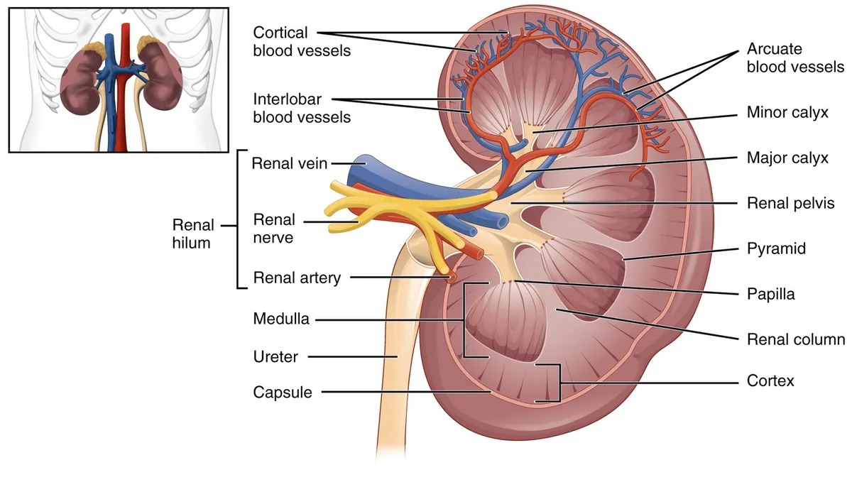
2つの極(extremitas superior / inferior)、2面(facies anterior / posterior)、2縁(凸の margo lateralis と凹の margo medialis)。内側縁の中央の切れ込みが hilum renale で、そこから中へ広がる空間が sinus renalis。
被膜は外から3層:fascia renalis(Gerota 筋膜)→ capsula adiposa(脂肪被膜)→ capsula fibrosa(線維被膜・腎実質に密着し、正常では容易に剥がれる)。さらに外側に傍腎脂肪体。
腎門の中身は前から順に:v. renalis → a. renalis → pelvis renalis(VAP)。
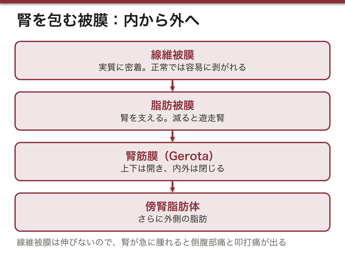
脂肪被膜が腎を支えている。急激な体重減少で脂肪が減ると腎が下垂し(遊走腎)、尿管が折れて水腎症を起こすことがある。Gerota 筋膜は上下では開いているが内外では閉じているので、腎周囲膿瘍は下方へ広がり、正中を越えにくい。
右腎静脈は短く、左腎静脈は長い(下大静脈が右寄りにあるため)。左腎静脈は大動脈と上腸間膜動脈のあいだを通るので、ここで挟まれると nutcracker 症候群(血尿・左側腹部痛)になる。また左腎静脈には左副腎静脈と左精巣/卵巣静脈が注ぐ——だから精索静脈瘤は左に多い(左は腎静脈へ直角に注ぎ圧が高く、右は下大静脈へ斜めに注ぐ)。左右差が症状の左右差を作る典型例。
線維被膜は伸びない。急性腎盂腎炎や腎梗塞で腎が急に腫れると被膜が張って側腹部の鈍痛と叩打痛が出る。慢性に萎縮した腎では被膜が実質に癒着して剥がれにくくなる。
右腎は肝のぶん低い。だから腎生検・腎瘻は右のほうが到達しやすい。
肋骨脊柱角(CVA)の叩打痛は腎盂腎炎・尿路結石の基本手技である。第12肋骨と脊柱がなす角——そこが腎の位置だから叩く。
腎は後腹膜にあり、脂肪被膜(Gerota 筋膜)に包まれる。この膜が感染や出血を腎周囲に閉じ込めるので、腎周囲膿瘍は症状が乏しいまま大きくなる。
腎茎の並びは前から後ろへ「静脈・動脈・腎盂(VAP)」。左腎静脈は腹大動脈と上腸間膜動脈に挟まれるので、圧迫されるとナットクラッカー症候群(血尿・左側腹部痛、男性では左精索静脈瘤)。成人で左の精索静脈瘤が突然できたら、左腎腫瘍を疑う。
腎の上に副腎が乗るが、別の被膜に包まれ、血流も別——腎を下げても副腎はついてこない。
English version
Core
Retroperitoneal, at Th12–L3, on either side of the vertebral column. The right kidney is 1–2 cm lower, because of the liver. About 11 cm long, 150 g.
Two poles (extremitas superior / inferior), two surfaces (anterior / posterior), two borders (the convex margo lateralis and the concave margo medialis). The notch in the middle of the medial border is the hilum renale, and the space opening from it into the interior is the sinus renalis.
Three coverings from outside in: fascia renalis (Gerota’s) → capsula adiposa → capsula fibrosa (adherent to the parenchyma; in health it strips easily), with pararenal fat outside that.
The contents of the hilum from front to back: v. renalis → a. renalis → pelvis renalis (VAP).
Going further
The fat capsule supports the kidney. Rapid weight loss reduces it, the kidney descends (a floating kidney), and the ureter may kink and cause hydronephrosis.
Gerota’s fascia is open above and below but closed medially and laterally, so a perinephric abscess spreads downwards and does not readily cross the midline.
The right renal vein is short and the left long (the inferior vena cava lying to the right). The left renal vein passes between the aorta and the superior mesenteric artery, and if it is caught there gives the nutcracker syndrome (haematuria, left flank pain). The left adrenal vein and the left testicular or ovarian vein also drain into the left renal vein — which is why varicocele is commoner on the left (entering the renal vein at a right angle and so at higher pressure, whereas the right enters the vena cava obliquely). A model case of an asymmetry of anatomy creating an asymmetry of disease.
The fibrous capsule does not stretch. When the kidney swells suddenly in acute pyelonephritis or infarction the capsule is stretched and gives flank pain and tenderness on percussion. In a chronically shrunken kidney the capsule adheres and strips with difficulty.
In the clinic
The right kidney is lower because of the liver, so renal biopsy and nephrostomy are easier on the right.
Percussion tenderness at the costovertebral angle is the basic manoeuvre for pyelonephritis and ureteric stone — the angle between the twelfth rib and the spine is where the kidney is, and that is why it is struck.
The kidney is retroperitoneal and enclosed in the fat capsule within Gerota’s fascia. That membrane confines infection and haemorrhage around the kidney, so a perinephric abscess grows large with few symptoms.
The order of the pedicle from front to back is vein, artery, pelvis (VAP). The left renal vein is caught between the aorta and the superior mesenteric artery, giving the nutcracker syndrome when compressed (haematuria, left flank pain, and in the man a left varicocele). A left varicocele appearing suddenly in an adult raises the suspicion of a left renal tumour.
The adrenal gland sits on the kidney but is enclosed in a separate fascia with a separate blood supply — lower the kidney and the adrenal does not follow.
腎の割面 — 皮質・髄質・腎杯Structura interna renis
前頭断で見ると外から順に:
- Cortex renalis(皮質)— 外側の顆粒状の層。糸球体がある
- Pyramides renales(腎錐体)— 髄質。放射状の縞。8〜18個
- Papillae renales(腎乳頭)— 錐体の先端。小腎杯に突き出す
- Columnae renales(腎柱・Bertin 柱)— 錐体のあいだへ入り込む皮質
尿の集まる道:Calyx renalis minor(7〜14個・乳頭を包む)→ calyx renalis major(2〜3個)→ pelvis renalis(腎盂)→ ureter。すべて sinus renalis の中にあり、周囲は脂肪で満たされる。
1つの錐体と、それを覆う皮質を合わせて lobus renalis(腎葉)。
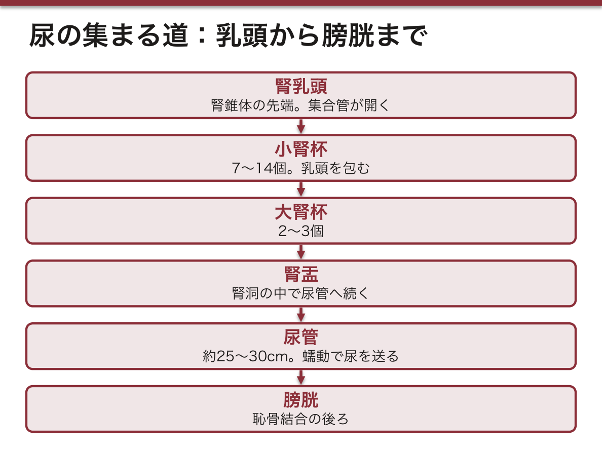
皮質と髄質の縞の向きが機能を表す。皮質が顆粒状に見えるのは糸球体(球)があるから、髄質が放射状に見えるのは集合管とヘンレ係蹄(直行する管)が並んでいるから。肉眼所見が、そのまま組織像の要約になっている。
腎乳頭は虚血に最も弱い。髄質は対向流機構を保つためにもともと酸素分圧が低く、血流も乏しい。だから鎮痛薬乱用・鎌状赤血球症・糖尿病で腎乳頭壊死が起こる。
腎盂腎杯の形は感染と結石の性質を決める。複数の小腎杯を持つ袋状の構造なので、閉塞すると尿が停滞しやすい。珊瑚状結石が腎盂腎杯の形をそのまま鋳型にして育つのは、この空間の形による。
腎動脈は終動脈である。区域間で吻合が無いので、1本の区域動脈が詰まればその区域が完全に梗塞する(楔状の梗塞巣)。逆に、この独立性があるから部分切除が可能になる。
皮質と髄質では酸素の余裕が違う。髄質は対向流を保つために血流が少なく、もともと低酸素である。だから虚血や薬剤で最初に傷むのは髄質(急性尿細管壊死)。
腎乳頭壊死 — 糖尿病・鎮痛薬乱用・鎌状赤血球症・尿路閉塞で起こる。壊死した乳頭が剥がれて尿管を詰まらせる。
腎盂腎炎は上行性感染で、集合管が乳頭に開くところから逆流して皮質へ広がるため、病変は楔形(くさび形)になる。小児の膀胱尿管逆流は腎瘢痕を残し、将来の高血圧・腎不全につながる。
腎細胞癌は皮質から、腎盂癌(尿路上皮癌)は腎盂から発生する。組織が違えば治療も違う——腎細胞癌は化学療法が効きにくく、尿路上皮癌は膀胱にも多発する。
English version
Core
On coronal section, from outside in:
- Cortex renalis — the outer granular layer, containing the glomeruli.
- Pyramides renales — the medulla; radially striated; 8–18 of them.
- Papillae renales — the tips of the pyramids, projecting into the minor calyces.
- Columnae renales (of Bertin) — cortex extending down between the pyramids.
The path of the urine: minor calyx (7–14, cupping a papilla) → major calyx (2–3) → pelvis renalis → ureter. All lie within the renal sinus, packed round with fat.
One pyramid with the cortex covering it is a lobus renalis.
Going further
The direction of the striations expresses the function. The cortex looks granular because of the glomeruli (spheres); the medulla looks radially striped because the collecting ducts and loops of Henle (straight tubules) are ranged in it. The gross appearance is a summary of the histology.
The renal papilla is the most vulnerable to ischaemia. The medulla has a low oxygen tension and little blood flow in order to preserve the countercurrent mechanism, so papillary necrosis occurs in analgesic abuse, sickle cell disease and diabetes.
The shape of the pelvicalyceal system decides the character of infection and of stones. Being a sac with several minor calyces, urine stagnates when it is obstructed. A staghorn calculus grows as a cast of that space precisely because of its shape.
The renal arteries are end arteries. There is no anastomosis between segments, so occlusion of one segmental artery infarcts that segment completely (a wedge-shaped infarct). Conversely, it is that independence which makes partial nephrectomy possible.
In the clinic
Cortex and medulla differ in their margin of oxygen. The medulla has little blood flow in order to preserve the countercurrent and is inherently hypoxic, so what is damaged first by ischaemia or by drugs is the medulla (acute tubular necrosis).
Papillary necrosis — in diabetes, analgesic abuse, sickle cell disease and urinary obstruction. The sloughed papilla obstructs the ureter.
Pyelonephritis is an ascending infection, refluxing from where the collecting ducts open on the papilla and spreading to the cortex, so the lesion is wedge-shaped. Vesicoureteric reflux in children leaves renal scars and leads to hypertension and renal failure later.
Renal cell carcinoma arises from the cortex, urothelial carcinoma of the pelvis from the pelvis. Different tissue means different treatment — renal cell carcinoma responds poorly to chemotherapy, and urothelial carcinoma is multiple in the bladder as well.
ネフロン — 割面の縞と、管の通り道Nephronum
腎の働きの単位がネフロンで、1つの腎に約100万個ある。上流から次の順に並ぶ。
| 部分 | 中身 | 腎のどこにあるか |
|---|---|---|
| corpusculum renis(腎小体) | 糸球体(輸入細動脈 → 毛細血管の房 → 輸出細動脈)と、それを包む糸球体包(Bowman 嚢) | 皮質(曲部) |
| tubulus proximalis(近位尿細管) | 曲部 pars convoluta → 直部 pars recta | 曲部は皮質、直部は髄放線から髄質へ |
| ansa nephroni(Henle ループ) | 下行脚と上行脚(細い部分と太い部分) | 髄質(錐体) |
| tubulus distalis(遠位尿細管) | 直部 → 曲部。自分の糸球体の血管極に戻って接する(緻密斑) | 曲部は皮質 |
| 結合部(pars intercalata)→ 集合管 | いくつものネフロンの尿を集めて下る | 髄放線 → 錐体 |
| ductus papillaris(乳頭管) | 集合管が合わさった太い管。乳頭の小孔(篩状野)から小腎杯へ開く | 腎乳頭 |
皮質は、腎小体と曲がった管が詰まった曲部(pars convoluta)と、まっすぐな管が束になって皮質へ差し込む放線部(pars radiata=髄放線)に分かれる。古い名で近位部・遠位部を portio nephroni proximalis / distalis と呼ぶ本もある。細胞と働きは組織学の「腎の組織」で扱う。
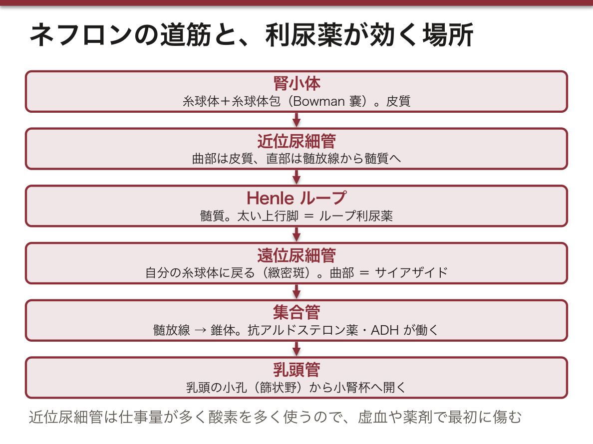
肉眼で見える割面の模様は、ネフロンの並び方そのものである。皮質が細かい粒に見えるのは腎小体が散らばるから、錐体が縦の縞に見えるのはループと集合管が乳頭へ向かって平行に走るから、皮質に差し込む縞(髄放線)はまっすぐな管の束である。
腎小体が皮質の深い所にある傍髄質ネフロンは、ループが乳頭の近くまで長く下りて、尿を濃くする働きの中心になる。浅い所の皮質ネフロンはループが短い。遠位尿細管が自分の糸球体に戻ってくるのは、流れてきた尿の塩分を見て、その糸球体の血流とレニン分泌を調節するためである(傍糸球体装置)。
利尿薬はネフロンの部位で分類される。ループ利尿薬はHenle ループの太い上行脚、サイアザイドは遠位尿細管の曲部、抗アルドステロン薬は集合管に効く。抗利尿ホルモン(ADH)は集合管に水チャネルを出させて尿を濃くする。
病気も部位で分かれる。糸球体腎炎は腎小体の病気で血尿・蛋白尿、虚血や薬剤による急性尿細管壊死は、仕事量が多く酸素を多く使う近位尿細管から傷む。ネフロンは生まれた後に新しく作られないので、失われた数は戻らない。
English version
Core
The working unit of the kidney is the nephron; each kidney has about one million. From upstream they are arranged as follows.
| Part | Content | Where in the kidney |
|---|---|---|
| corpusculum renis (renal corpuscle) | Glomerulus (afferent arteriole → tuft of capillaries → efferent arteriole) and the glomerular (Bowman’s) capsule around it | Cortex (convoluted part) |
| tubulus proximalis (proximal tubule) | Convoluted part (pars convoluta) → straight part (pars recta) | Convoluted part in cortex; straight part in medullary ray, then medulla |
| ansa nephroni (loop of Henle) | Descending and ascending limbs (thin and thick parts) | Medulla (pyramid) |
| tubulus distalis (distal tubule) | Straight part → convoluted part; returns to touch its own glomerulus at the vascular pole (macula densa) | Convoluted part in cortex |
| Connecting part (pars intercalata) → collecting duct | Collects urine from many nephrons and runs down | Medullary ray → pyramid |
| ductus papillaris (papillary duct) | Thick duct formed by collecting ducts; opens by small pores (area cribrosa) on the papilla into a minor calyx | Renal papilla |
The cortex is divided into the convoluted part (pars convoluta), packed with corpuscles and coiled tubules, and the radiate part (pars radiata = medullary rays), bundles of straight tubules reaching into the cortex. Some books use the older names portio nephroni proximalis / distalis. Cells and functions are covered in histology, “Kidney”.
Going further
The pattern seen on the cut surface is the arrangement of nephrons itself. The cortex looks granular because corpuscles are scattered in it; the pyramids look striped because loops and collecting ducts run in parallel toward the papilla; the stripes reaching into the cortex (medullary rays) are bundles of straight tubules.
Juxtamedullary nephrons, whose corpuscles lie deep in the cortex, have long loops reaching near the papilla and do most of the work of concentrating urine. Superficial cortical nephrons have short loops. The distal tubule returns to its own glomerulus so that it can sense the salt in the passing fluid and adjust that glomerulus’s blood flow and renin release (juxtaglomerular apparatus).
In the clinic
Diuretics are classified by nephron segment. Loop diuretics act on the thick ascending limb of Henle, thiazides on the distal convoluted tubule, and aldosterone antagonists on the collecting duct. Antidiuretic hormone (ADH) makes the collecting duct insert water channels and concentrates the urine.
Diseases also divide by segment. Glomerulonephritis is a disease of the corpuscle with haematuria and proteinuria; acute tubular necrosis from ischaemia or drugs first hits the proximal tubule, which does the most work and uses the most oxygen. Nephrons are not made after birth, so lost ones do not come back.
尿管 — 3つの狭窄Ureter
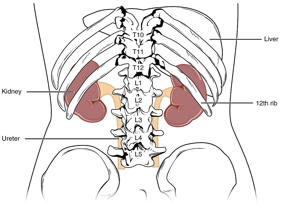
腎盂から膀胱まで約25〜30cm の筋性の管。後腹膜を下り、大腰筋の前面を通り、総腸骨動脈の分岐部を越えて骨盤へ入る。2部:pars abdominalis と pars pelvica。
3つの生理的狭窄 — 結石が止まる場所である。
| 狭窄 | 位置 |
|---|---|
| 第1 | 腎盂尿管移行部(UPJ) |
| 第2 | 総腸骨動脈との交叉部(骨盤入口) |
| 第3 | 膀胱壁を貫く部(UVJ)— 最も狭い |
壁は移行上皮+2〜3層の平滑筋で、蠕動によって尿を送る(重力ではない)。
「水は橋の下を流れる」(water under the bridge)。骨盤内で尿管は、男性では精管の後ろ(下)を、女性では子宮動脈の後ろ(下)を通る。子宮全摘で尿管を損傷するのは、子宮動脈を結紮する場所がこの交叉のすぐ近く(子宮頸の外側約2cm)だからである。1つの語呂で、2つの術中合併症が予測できる。
疝痛の放散部位が石の高さを教える。上部(UPJ)=側腹部から腰、中部(交叉部)=下腹部から鼠径部、下部(UVJ)=陰嚢・陰唇と大腿内側、頻尿・排尿時痛。尿管の神経支配が Th11〜L2 の分節に対応しているためで、痛みは尿管そのものではなく、その分節の皮膚に感じられる。
膀胱壁の中を斜めに貫くことが逆流を防ぐ。膀胱が充満すると壁内部が押しつぶされて弁のように閉じる(受動的な弁)。この斜走が短いと膀胱尿管逆流症となり、小児の反復性腎盂腎炎の原因になる。
3つの生理的狭窄が、そのまま結石の嵌頓部位である。①腎盂尿管移行部②総腸骨動脈との交叉部(骨盤縁)③膀胱壁内部。膀胱壁内部が最も細い。
尿管結石の疝痛は、詰まった高さで放散先が変わる。上部=側腹部から鼠径部、下部=陰嚢/大陰唇と大腿内側。尿管の分節神経支配(Th11〜L2)がそのまま関連痛の地図になる。
尿管は総腸骨動脈の分岐部を前から越えて骨盤へ入る。この交叉は骨盤内手術での損傷部位として最重要である。
女性では子宮動脈が尿管の前を横切る(water under the bridge)。子宮全摘で子宮動脈を結紮するときに尿管を巻き込むのが古典的な合併症で、この位置関係を知らないと避けられない。
English version
Core
A muscular tube of 25–30 cm from renal pelvis to bladder, descending retroperitoneally over the front of psoas major, crossing the bifurcation of the common iliac artery and entering the pelvis. Two parts: pars abdominalis and pars pelvica.
Three physiological constrictions — where stones lodge.
| Constriction | Position |
|---|---|
| First | The pelviureteric junction (UPJ) |
| Second | The crossing of the common iliac artery (the pelvic brim) |
| Third | The intramural part in the bladder wall (UVJ) — the narrowest |
The wall is transitional epithelium with two or three layers of smooth muscle, and the urine is driven by peristalsis, not by gravity.
Going further
"Water under the bridge." In the pelvis the ureter passes behind (below) the ductus deferens in the man and behind (below) the uterine artery in the woman. The ureter is injured at hysterectomy because the uterine artery is ligated close to that crossing (about 2 cm lateral to the cervix). One mnemonic predicts two operative complications.
Where the colic radiates tells you the level of the stone. Upper (UPJ) = flank and loin; middle (the crossing) = lower abdomen and groin; lower (UVJ) = scrotum or labium and the medial thigh, with frequency and pain on voiding. The segmental innervation of the ureter (Th11–L2) corresponds, and the pain is felt not in the ureter but in the skin of that segment.
That it pierces the bladder wall obliquely prevents reflux. As the bladder fills, the intramural part is crushed and closes like a valve (a passive valve). If that oblique course is short, vesicoureteric reflux results and causes recurrent pyelonephritis in children.
In the clinic
The three physiological constrictions are directly the sites at which stones impact. ① the pelviureteric junction ② the crossing of the common iliac artery (the pelvic brim) ③ the intramural part in the bladder wall. The intramural part is the narrowest.
The radiation of ureteric colic changes with the level of the obstruction: upper = flank to groin; lower = scrotum or labium majus and the medial thigh. The segmental supply of the ureter (Th11–L2) is itself the map of the referred pain.
The ureter crosses in front of the bifurcation of the common iliac artery to enter the pelvis. This crossing is the most important site of injury in pelvic surgery.
In the woman the uterine artery crosses in front of the ureter (water under the bridge). Catching the ureter while ligating the uterine artery at hysterectomy is the classic complication, and it cannot be avoided without knowing this relationship.
膀胱と尿道Vesica urinaria et urethra
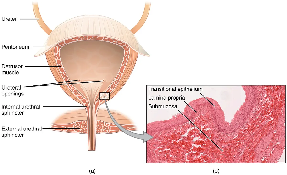
Vesica urinaria:恥骨結合の後ろの骨盤腔内。Apex vesicae(前上・正中臍索が続く)・corpus・fundus(後下)・cervix(頸・尿道へ)。壁の筋が m. detrusor vesicae(排尿筋)。
内面はヒダに富むが、trigonum vesicae(膀胱三角)だけは常に平滑である。三角の3つの角は2つの ostium ureteris と ostium urethrae internum。左右の尿管口を結ぶ隆起が plica interureterica。
尿道は男女で大きく異なる。Urethra feminina:約4cm、まっすぐ。Urethra masculina:約18〜20cm、pars prostatica → pars membranacea → pars spongiosa の3部で、末端が fossa navicularis、開口が ostium urethrae externum。
膀胱三角が平滑なのは由来が違うから。膀胱の大部分は尿生殖洞(内胚葉)由来だが、三角部は中腎管(中胚葉)が取り込まれてできる。粘膜が下層に密着しているのでヒダを作らない。ここが最も感受性が高く、膀胱炎の症状(尿意切迫)と癌の好発部位になるのも、由来の違いによる。
膀胱は充満すると腹腔内へ上がる。空虚時は骨盤内に収まるが、充満すると恥骨結合の上へ出て腹壁と腹膜のあいだに接する。だから恥骨上穿刺は腹膜を通らずにできる——超音波で膀胱の膨らみを確認するのはこのためである。小児では常に腹腔内に近い位置にある。
男性尿道の3部が、カテーテル挿入の抵抗の場所を決める。最も狭く動かないのが膜様部(尿生殖隔膜を貫く)で、外尿道括約筋がある。骨盤骨折ではこの膜様部が断裂しやすい。球部の損傷(騎乗型外傷)では尿が会陰へ漏出する。
女性尿道が短くまっすぐであることが、女性に膀胱炎が多い理由。外尿道口が腟前庭にあり会陰に近いことも重なる。解剖の差が、疾患の性差の大半を説明する。
膀胱三角は粘膜が平滑で伸びないため、炎症も腫瘍もここに目立つ。膀胱鏡ではまず三角部と尿管口を観察する。
男性の尿道は長く、2つの弯曲と3つの狭窄を持つ。カテーテル挿入時は陰茎を上前方へ引いて弯曲を伸ばす。膜性部が最も細く、骨盤骨折で断裂しやすい。外尿道口からの出血・会陰部の血腫・前立腺の浮動があればカテーテルを入れてはいけない。
女性の尿道は約4cm と短く、腟前庭に開く。女性に尿路感染が圧倒的に多い解剖学的な理由である。
排尿は仙髄(S2〜S4)の副交感神経が排尿筋を収縮させ、陰部神経の外尿道括約筋がゆるむことで起こる。橋の排尿中枢より上の障害では過活動膀胱、仙髄より下(馬尾・末梢)では尿閉になる。
English version
Core
Vesica urinaria: in the pelvis behind the pubic symphysis. Apex vesicae (anterosuperior, continued as the median umbilical ligament), corpus, fundus (posteroinferior), cervix (the neck, into the urethra). The muscle of the wall is the m. detrusor vesicae.
The interior is much folded, but the trigonum vesicae alone is always smooth. Its three angles are the two ostia ureteris and the ostium urethrae internum, with the plica interureterica joining the ureteric orifices.
The urethra differs greatly between the sexes. Urethra feminina: about 4 cm, straight. Urethra masculina: 18–20 cm, in three parts — pars prostatica → pars membranacea → pars spongiosa — ending in the fossa navicularis and the external urethral orifice.
Going further
The trigone is smooth because its origin differs. Most of the bladder derives from the urogenital sinus (endoderm), but the trigone is formed by incorporation of the mesonephric ducts (mesoderm). Its mucosa is bound to the layer beneath and forms no folds. That it is the most sensitive area (urgency in cystitis) and a site of predilection for carcinoma also follows from the difference in origin.
When full, the bladder rises into the abdomen. Empty it lies within the pelvis, but full it passes above the symphysis and comes into contact with the abdominal wall between wall and peritoneum. So suprapubic puncture can be made without traversing the peritoneum — which is why the distended bladder is confirmed on ultrasound. In children it lies close to the abdomen at all times.
The three parts of the male urethra decide where resistance is met on catheterisation. The narrowest and least mobile is the membranous part (piercing the urogenital diaphragm), where the external sphincter lies. Pelvic fracture ruptures this membranous part readily. Injury to the bulb (a straddle injury) leaks urine into the perineum.
That the female urethra is short and straight is why cystitis is common in women, with the external orifice in the vestibule and close to the perineum. A difference in anatomy explains most of the sex difference in disease.
In the clinic
The mucosa of the trigone is smooth and does not stretch, so both inflammation and tumour are conspicuous there. At cystoscopy the trigone and ureteric orifices are examined first.
The male urethra is long, with two curves and three narrowings. On catheterisation, draw the penis up and forwards to straighten the curve. The membranous part is the narrowest and ruptures readily in pelvic fracture. With blood at the meatus, a perineal haematoma or a high-riding prostate, do not pass a catheter.
The female urethra is about 4 cm long and opens into the vestibule — the anatomical reason why urinary infection is overwhelmingly commoner in women.
Micturition occurs when the sacral (S2–S4) parasympathetic contracts the detrusor and the external sphincter (pudendal nerve) relaxes. A lesion above the pontine micturition centre gives an overactive bladder; below the sacral cord (cauda equina, peripheral) gives retention.
膀胱の血管と神経 — 臍動脈の名残から来る動脈Arteriae vesicales
膀胱の動脈はすべて内腸骨動脈の枝から来る。
| 動脈 | 起こり | 主に養う所 |
|---|---|---|
| a. vesicalis superior(上膀胱動脈) | 臍動脈の開いたまま残った根元(2〜3本) | 膀胱の上部と体 |
| a. vesicalis inferior(下膀胱動脈) | 内腸骨動脈(主に男性)。女性では腟動脈の枝が代わる | 膀胱底・膀胱三角・頸、男性では前立腺・精嚢・尿管下部も |
| 補助の枝 | 閉鎖動脈・下殿動脈・中直腸動脈、女性では子宮動脈 | 周辺部 |
静脈は膀胱の下部を取り巻く膀胱静脈叢に集まり、男性では前立腺静脈叢、女性では腟静脈叢とつながって内腸骨静脈へ注ぐ。リンパは外腸骨・内腸骨リンパ節へ。神経は下下腹神経叢から来る膀胱神経叢で、副交感(骨盤内臓神経 S2〜S4)が排尿筋を縮め、交感(Th11〜L2)が排尿筋をゆるめて膀胱頸を締める。
上膀胱動脈が臍動脈から出るのは、胎児の臍動脈が膀胱の両わきを通って臍へ向かっていたからである。生まれると臍動脈の先は閉じて内側臍索(前腹壁の内側臍ひだの芯)になり、膀胱へ枝を出していた根元だけが血管として残る。
膀胱静脈叢と前立腺静脈叢には弁が無く、脊柱の周りの静脈叢とつながる。腹圧がかかると血液が脊柱の方へ逆流できるので、前立腺癌や膀胱癌は腰椎・骨盤の骨に転移しやすい。
膀胱全摘では、両側の血管の束(上膀胱動脈の束と下膀胱動脈の束)を順に処理する。膀胱腫瘍や放射線性膀胱炎で出血が止まらないときは、内腸骨動脈の枝を塞ぐ(塞栓術)。骨盤骨折の大量出血も、多くは骨盤の静脈叢と内腸骨動脈の枝からである。
English version
Core
All arteries of the bladder come from branches of the internal iliac artery.
| Artery | Origin | Main area supplied |
|---|---|---|
| a. vesicalis superior | The still-open root of the umbilical artery (2–3 branches) | Upper part and body |
| a. vesicalis inferior | Internal iliac artery (mainly in males); in females branches of the vaginal artery take its place | Fundus, trigone and neck; in males also prostate, seminal vesicles and lower ureter |
| Additional branches | Obturator, inferior gluteal, middle rectal arteries; uterine artery in females | Surrounding parts |
Veins collect in the vesical venous plexus around the lower bladder, which joins the prostatic plexus in males and the vaginal plexus in females and drains to the internal iliac vein. Lymph goes to the external and internal iliac nodes. Nerves come through the vesical plexus from the inferior hypogastric plexus: parasympathetic fibres (pelvic splanchnic nerves, S2–S4) contract the detrusor; sympathetic fibres (T11–L2) relax the detrusor and close the bladder neck.
Going further
The superior vesical artery comes from the umbilical artery because in the fetus the umbilical arteries ran along both sides of the bladder to the umbilicus. After birth the far part closes to become the medial umbilical ligament (the core of the medial umbilical fold on the anterior wall), and only the root, which gave branches to the bladder, remains a vessel.
The vesical and prostatic plexuses have no valves and connect with the venous plexus around the spine. When abdominal pressure rises, blood can flow back toward the spine, so prostate and bladder cancer often spread to the lumbar spine and pelvic bones.
In the clinic
In radical cystectomy, the vascular bundles on each side (superior and inferior vesical pedicles) are divided in turn. When bleeding from a bladder tumour or radiation cystitis will not stop, branches of the internal iliac artery are blocked (embolisation). Heavy bleeding in pelvic fracture also comes mostly from pelvic venous plexuses and internal iliac branches.
腎の外側縁・内側縁Margo lateralis, medialis renis
外側縁は凸、内側縁は凹で、凹の中央が腎門。ソラマメ形のどちら向きかを決める基準。
English version
Core
The lateral border is convex and the medial concave, with the hilum at the middle of the concavity. The reference for deciding which way the bean is facing.
腎門Hilum renale
内側縁のくぼみ。前から順に「腎静脈・腎動脈・腎盂」が並ぶ。
この前後の並びが定番の設問。頭文字で VAP と覚える。
English version
Core
The depression in the medial border. From the front: renal vein, renal artery, renal pelvis. This anteroposterior order is a standard question — remember it as VAP.
腎洞Sinus renalis
腎門から内部へ続く空所。腎杯・腎盂・血管・脂肪が詰まる。
English version
Core
The space continuing from the hilum into the interior, filled by the calyces, pelvis, vessels and fat.
腎皮質Cortex renalis
被膜のすぐ内側の外層。腎小体(糸球体+Bowman 嚢)と曲尿細管があり、断面では点々として見える。
English version
Core
The outer layer immediately beneath the capsule, containing the renal corpuscles (glomerulus and Bowman’s capsule) and the convoluted tubules; on section it appears stippled.
腎錐体Pyramis renalis
髄質を作る円錐で、底が皮質側、尖が腎洞側。8〜18個ある。
中を直尿細管と集合管が走るので、断面で縞に見えるのが皮質との違い。
English version
Core
The cone forming the medulla, its base towards the cortex and its apex towards the sinus; 8–18 of them.
The straight tubules and collecting ducts run within it, so it appears striped on section — the difference from the cortex.
腎乳頭Papilla renalis
腎錐体の尖端。篩状野という多数の小孔があり、そこから尿が小腎杯へ滴る。
尿が腎実質を離れる、まさにその点。鎮痛薬腎症ではここが壊死する。
English version
Core
The apex of the pyramid, bearing the cribriform area with its many small openings, from which urine drips into a minor calyx. The very point at which the urine leaves the renal parenchyma.
It is what necroses in analgesic nephropathy.
腎柱Columnae renales
錐体と錐体の間に入り込んだ皮質。ここを弓状動脈へ向かう葉間動脈が通る。
English version
Core
Cortex extending down between the pyramids. The interlobar arteries pass through it towards the arcuate arteries.
腎盂Pelvis renis
大腎杯が合してできる漏斗。腎門を出て尿管に続く。
流れは腎乳頭 → 小腎杯 → 大腎杯 → 腎盂 → 尿管。この順を言えることが要る。
English version
Core
The funnel formed by the union of the major calyces, leaving the hilum to become the ureter.
The flow is papilla → minor calyx → major calyx → pelvis → ureter, and being able to give that order is what is required.
尿管Ureter
腎盂から膀胱まで約25〜30cm、後腹膜を下る。
3か所の生理的狭窄——①腎盂尿管移行部 ②総腸骨動脈との交差部 ③膀胱壁内部。結石が嵌まるのはこの3か所。骨盤では女性で子宮動脈が、男性で精管が尿管の上を越える(water under the bridge)。
English version
Core
25–30 cm from renal pelvis to bladder, descending retroperitoneally. Three physiological constrictions — ① the pelviureteric junction ② the crossing of the common iliac artery ③ the intramural part. Stones impact at these three.
In the pelvis the uterine artery in the woman and the ductus deferens in the man cross above the ureter (water under the bridge).
膀胱尖Apex vesicae
膀胱の前上端。ここから正中臍索(尿膜管の遺残)が臍へ向かう。
English version
Core
The anterosuperior end of the bladder, from which the median umbilical ligament (the remnant of the urachus) runs to the umbilicus.
膀胱体Corpus vesicae
尖と底の間の大部分。空虚時は骨盤内に収まるが、充満すると恥骨結合を越えて腹腔へ上がるので、そこで恥骨上穿刺ができる。
English version
Core
The large part between apex and fundus. Empty it lies within the pelvis, but full it rises above the pubic symphysis into the abdomen — which is where suprapubic puncture is made.
膀胱底Fundus vesicae
後下方を向く三角形の面。内面に膀胱三角——2つの尿管口と内尿道口を結ぶ三角——があり、ここだけ粘膜が平滑で伸びない。
由来が中腎管系で他と違うためで、感染・腫瘍の好発部位。
English version
Core
The triangular surface facing down and back. Its inner aspect carries the trigone — bounded by the two ureteric orifices and the internal urethral orifice — where alone the mucosa is smooth and does not stretch.
Its origin from the mesonephric system differs from the rest, and it is a site of predilection for infection and tumour.
発生 — 前腎・中腎・後腎、そして尿管芽Pronephros, mesonephros, metanephros
泌尿器は中間中胚葉(腎形成索・尿生殖堤)と総排泄腔から作られる。腎は頭から尾へ3回作り直される。
| 腎 | 時期と場所 | 運命 |
|---|---|---|
| 前腎 | 第4週の初め、頸部。7〜10個の細胞の塊 | 働かないまま第4週の終わりに消える |
| 中腎 | 第4週から、上胸部〜L3 の高さ。S字の細管と糸球体を持ち、中腎管(Wolff 管)で総排泄腔へ注ぐ | 胎生初期に短く働き、第2か月の終わりまでにほぼ消える。男性では一部の細管と中腎管が生殖路に使われる |
| 後腎(永久腎) | 第5週、仙骨部 | 成人の腎になる |
後腎は2つの出身を持つ。中腎管の下端から出る尿管芽が伸びて枝分かれし、尿管・腎盂・大小腎杯・集合管になる。尿管芽の先にかぶさる後腎組織(造後腎組織)からはネフロン(糸球体包〜遠位尿細管)ができ、遠位尿細管が集合管につながる。腎はその後、仙骨部から腰部へ上昇し、腎門が内側を向く。血液も、骨盤の動脈から順に高い位置の大動脈の枝へ乗り換え、最後は L2 の高さの腎動脈になる。
膀胱は、総排泄腔が尿直腸中隔で前の尿生殖洞と後ろの肛門直腸管に分けられ、その尿生殖洞の上部からできる。膀胱の頂は尿膜管で臍とつながり、生後は正中臍索になる。膀胱三角は取り込まれた中腎管(中胚葉)の部分である。
集合系とネフロンは別々の場所で作られ、あとでつながる。この接続や枝分かれの段階のつまずきが、多くの異常を説明する。
腎が上昇の途中で止まれば骨盤腎、左右の下極が癒合すると馬蹄腎になり、下腸間膜動脈の根元に引っかかってそれ以上上がれない。古い高さの動脈が残れば副腎動脈(過剰な腎動脈)になる。尿管芽が早く2つに割れると重複尿管で、完全重複では上の腎からの尿管が膀胱の下方・内側に開く——女児ではこれが括約筋より下(尿道や腟)に開き、普通に排尿しているのに下着がいつもぬれる。
両側の腎無形成では胎児の尿が出ず、羊水過少から肺の低形成と特有の顔貌が起こる(Potter シークエンス)。尿膜管が閉じきらないと、臍から尿が漏れる尿膜管瘻、途中が袋になる尿膜管嚢胞、臍側だけ開く尿膜管洞になる。下大静脈の発生の異常で尿管がその後ろを回ると(下大静脈後尿管)、右の水腎症の原因になる。
English version
Core
The urinary system comes from the intermediate mesoderm (nephrogenic cord, urogenital ridge) and the cloaca. The kidney is built three times, from head to tail.
| Kidney | Time and place | Fate |
|---|---|---|
| Pronephros | Start of week 4, neck region; 7–10 cell groups | Never works; gone by the end of week 4 |
| Mesonephros | From week 4, upper thoracic to L3 level; S-shaped tubules with glomeruli, draining by the mesonephric (Wolffian) duct into the cloaca | Works briefly in early fetal life; mostly gone by the end of month 2. In males some tubules and the duct are reused for the genital tract |
| Metanephros (permanent kidney) | Week 5, sacral region | Becomes the adult kidney |
The metanephros has two sources. The ureteric bud grows from the lower end of the mesonephric duct and branches to form the ureter, pelvis, major and minor calyces and collecting ducts. The metanephric tissue (blastema) capping the bud forms the nephrons (capsule to distal tubule), and each distal tubule joins a collecting duct. The kidney then ascends from the sacral to the lumbar region and its hilum turns medially. Its blood supply also moves step by step from pelvic arteries to higher aortic branches, ending as the renal artery at L2.
The bladder forms after the urorectal septum divides the cloaca into the urogenital sinus in front and the anorectal canal behind; the upper urogenital sinus becomes the bladder. The apex is joined to the umbilicus by the urachus, which becomes the median umbilical ligament after birth. The trigone is the incorporated part of the mesonephric ducts (mesoderm).
Going further
The collecting system and the nephrons are made in different places and joined later. Trouble at the joining or branching steps explains many anomalies.
If ascent stops on the way, the kidney stays in the pelvis; if the lower poles fuse, a horseshoe kidney forms and catches on the root of the inferior mesenteric artery so it cannot rise further. If an artery from an old level persists, it becomes an accessory renal artery. If the ureteric bud splits early, the ureter is duplicated. In complete duplication the ureter from the upper kidney opens lower and more medially in the bladder; in girls it may open below the sphincter (urethra or vagina), so the child voids normally yet is always wet.
In the clinic
In bilateral renal agenesis the fetus makes no urine; oligohydramnios leads to lung hypoplasia and a typical face (Potter sequence). If the urachus fails to close, urine leaks from the umbilicus (urachal fistula), a cyst forms along it (urachal cyst), or only the umbilical end stays open (urachal sinus). If a fault in the development of the inferior vena cava makes the ureter pass behind it (retrocaval ureter), right hydronephrosis can result.
まとめ
| 腎臓 — 外形と被膜 | Th12〜L3 の後腹膜、脊柱の両側。右腎は肝のぶん約1〜2cm 低い。 |
|---|---|
| 腎の割面 — 皮質・髄質・腎杯 | 前頭断で見ると外から順に:Cortex renalis(皮質)— 外側の顆粒状の層。 |
| ネフロン — 割面の縞と、管の通り道 | 腎の働きの単位がネフロンで、1つの腎に約100万個ある。 |
| 尿管 — 3つの狭窄 | 腎盂から膀胱まで約25〜30cm の筋性の管。 |
| 膀胱と尿道 | Vesica urinaria:恥骨結合の後ろの骨盤腔内。 |
| 膀胱の血管と神経 — 臍動脈の名残から来る動脈 | 膀胱の動脈はすべて内腸骨動脈の枝から来る。 |
| 腎の外側縁・内側縁 | 外側縁は凸、内側縁は凹で、凹の中央が腎門。 |
| 腎門 | 内側縁のくぼみ。前から順に「腎静脈・腎動脈・腎盂」が並ぶ。 |
| 腎洞 | 腎門から内部へ続く空所。腎杯・腎盂・血管・脂肪が詰まる。 |
| 腎皮質 | 被膜のすぐ内側の外層。腎小体(糸球体+Bowman 嚢)と曲尿細管があり、断面では点々として見える。 |
| 腎錐体 | 髄質を作る円錐で、底が皮質側、尖が腎洞側。 |
| 腎乳頭 | 腎錐体の尖端。篩状野という多数の小孔があり、そこから尿が小腎杯へ滴る。 |
| 腎柱 | 錐体と錐体の間に入り込んだ皮質。ここを弓状動脈へ向かう葉間動脈が通る。 |
| 腎盂 | 大腎杯が合してできる漏斗。腎門を出て尿管に続く。 |
| 尿管 | 腎盂から膀胱まで約25〜30cm、後腹膜を下る。 |
| 膀胱尖 | 膀胱の前上端。ここから正中臍索(尿膜管の遺残)が臍へ向かう。 |
| 膀胱体 | 尖と底の間の大部分。空虚時は骨盤内に収まるが、充満すると恥骨結合を越えて腹腔へ上がるので、そこで恥骨上穿刺ができる。 |
| 膀胱底 | 後下方を向く三角形の面。内面に膀胱三角——2つの尿管口と内尿道口を結ぶ三角——があり、ここだけ粘膜が平滑で伸びない。 |
| 発生 — 前腎・中腎・後腎、そして尿管芽 | 泌尿器は中間中胚葉(腎形成索・尿生殖堤)と総排泄腔から作られる。 |
このノートは医学生が授業の内容を自分の言葉でまとめたもので、診療の判断に使うものではありません。誤りに気づいたらお知らせください。