血圧は臨床で最も多く測られる物理量である。この章では「圧力とは何か」から「血圧計が何を測っているか」までを、流体の物理で一本につなぐ。
骨組みは4つ。①圧力と静水圧(体位で血圧が変わる理由)、②流量・連続の式・ベルヌーイの定理(狭窄部で何が起きるか)、③ポアズイユの法則(なぜ血管の太さが血流を支配するか)、④測定法(直接法と間接法、コロトコフ音とオシロメトリック法)。最後に、気体が液体に溶ける量を決めるヘンリーの法則から減圧症を見る。
要の式は Q = πr4ΔP/(8ηl)。半径が4乗で効くことが、動脈硬化・血管拡張薬・自律神経による血圧調節のすべてを説明する。
- 圧力と単位 — なぜ血圧は mmHg で測るのか
- 静水圧 — 立つと足の血圧は頭より高い
- 流量・連続の式・ベルヌーイの定理 — 狭いところは速く、圧は低い
- ポアズイユの法則 — 半径が4乗で効く
- 血圧の測り方 — 直接法・聴診法・オシロメトリック法
- 血管壁の弾性 — 動脈が硬くなると何が起きるか
- 気体の分圧とヘンリーの法則 — 減圧症はなぜ起こる
圧力と単位 — なぜ血圧は mmHg で測るのか
圧力は面に垂直にはたらく単位面積あたりの力である:P = F/A、単位 Pa(N/m2)。
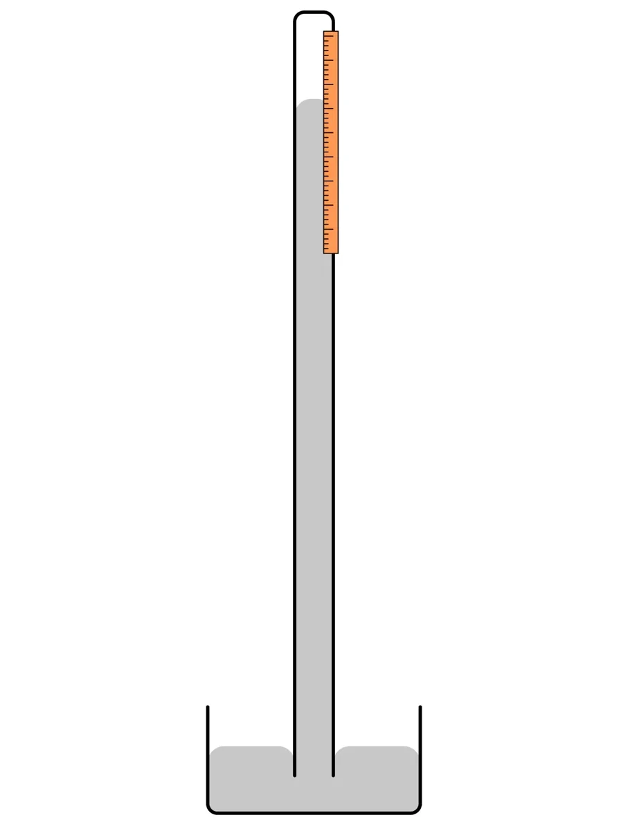
大気圧は、上空の空気の重さが地表を押す圧力。トリチェリは一端を閉じたガラス管に水銀を満たして水銀槽に逆さに立て、水銀柱が約 760 mm で止まることを示した。大気圧が水銀柱の重さとつり合っているからである(P = ρHggh)。
760 mmHg = 1 気圧 ≈ 101.3 kPa、1 mmHg ≈ 133 Pa。
血圧は大気圧を基準にした値(ゲージ圧)である。「120 mmHg」は大気圧より 120 mmHg 高いという意味。
mmHg が残っているのは歴史的な理由——水銀柱で直接測っていたから——だけでなく、数字の大きさが扱いやすいからでもある。120 mmHg を SI で書くと約16 kPa、大気圧からの絶対圧なら約117 kPa になり、変化が見えにくい。呼吸器や中心静脈圧では、より小さな圧を見るため cmH2O(1 cmH2O ≈ 0.74 mmHg)も使う。水銀の密度は水の約13.6倍なので、水で同じ圧を測ると柱は13.6倍高くなる。
中心静脈圧や人工呼吸の気道内圧は cmH2O、動脈圧や眼圧は mmHg と、臨床では単位が混在する。値を比べるときは単位をそろえる習慣をつける。
English version
Core
Pressure is force per unit area acting perpendicular to a surface: P = F/A, unit Pa (N/m2).
Atmospheric pressure is the push of the weight of the air above on the ground. Torricelli filled a glass tube closed at one end with mercury, stood it upside down in a dish of mercury and showed that the column stops at about 760 mm, because atmospheric pressure balances the weight of the column (P = ρHggh).
760 mmHg = 1 atm ≈ 101.3 kPa; 1 mmHg ≈ 133 Pa.
Blood pressure is measured relative to atmospheric pressure (gauge pressure). “120 mmHg” means 120 mmHg above atmospheric pressure.
Going further
mmHg remains not only for history — it was measured directly with mercury columns — but also because the numbers are convenient. 120 mmHg is about 16 kPa in SI, or about 117 kPa as absolute pressure, which makes changes hard to see. For breathing and central venous pressure, cmH2O (1 cmH2O ≈ 0.74 mmHg) is used for smaller pressures. Mercury is about 13.6 times denser than water, so a water column for the same pressure is 13.6 times taller.
In the clinic
Clinical practice mixes units: central venous pressure and airway pressure in ventilation in cmH2O, arterial and intraocular pressure in mmHg. Make it a habit to use the same unit when comparing values.
静水圧 — 立つと足の血圧は頭より高い
静止した液体の中では、深さ h の点の圧力は上の液柱の重さだけ増える。
P = P0 + ρgh
圧力は深さだけで決まり、容器の形や広さにはよらない(2 m の深さなら湖でもプールでも同じ)。水中では約10 m ごとに1気圧ずつ増える。
血管の中も同じで、立っている人では心臓より低い場所ほど血圧が高く、高い場所ほど低い。
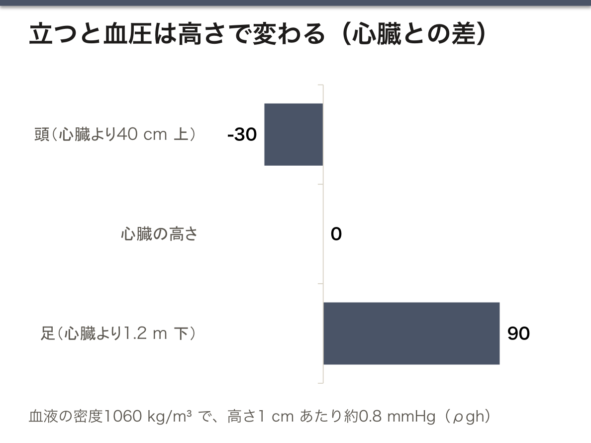
血液の密度を約1060 kg/m3 とすると、ρgh は高さ1 cm あたり約0.8 mmHg。立位で足が心臓より1.2 m 下にあれば、足の動脈圧は心臓の高さより約90 mmHg 高い。頭が40 cm 上なら約30 mmHg 低い。
心臓が送り出す圧(駆動圧)は姿勢で変わらないが、各部位の血管壁にかかる圧は静水圧のぶん変わる。血圧を測るとき腕を心臓の高さに置くのは、この静水圧の差を消すためである。
腕を心臓より下げて測ると血圧は高く、上げて測ると低く出る(10 cm で約8 mmHg)。手首式血圧計の値がぶれやすいのは、手首の高さがずれやすいからである。立位で下肢の静脈圧が高くなることは、下肢静脈瘤や足のむくみの原因になる。急に立ち上がると血液が下肢にたまり、脳の血圧が下がって起立性低血圧のめまいが起こる。
English version
Core
In a liquid at rest, the pressure at depth h increases by the weight of the column above.
P = P0 + ρgh
Pressure depends only on depth, not on the shape or size of the container (at 2 m deep it is the same in a lake or a pool). In water it rises by about 1 atm every 10 m.
Blood vessels are the same: in a standing person, blood pressure is higher in places below the heart and lower in places above it.
Going further
With blood density about 1060 kg/m3, ρgh is about 0.8 mmHg per centimeter of height. Standing, if the feet are 1.2 m below the heart, arterial pressure in the feet is about 90 mmHg higher than at heart level. If the head is 40 cm above, it is about 30 mmHg lower.
The driving pressure from the heart does not change with posture, but the pressure on vessel walls in each region changes by the hydrostatic amount. The arm is held at heart level for blood pressure measurement to cancel this hydrostatic difference.
In the clinic
Measured with the arm below the heart, blood pressure reads high; with the arm above, low (about 8 mmHg per 10 cm). Wrist monitors vary easily because the wrist height shifts easily. High venous pressure in the legs when standing contributes to varicose veins and swollen feet. Standing up quickly lets blood pool in the legs, pressure in the brain falls, and the dizziness of orthostatic hypotension follows.
流量・連続の式・ベルヌーイの定理 — 狭いところは速く、圧は低い
流量 Q は単位時間に流れる体積で、断面積 A と平均流速 v から Q = A·v(m3/s)。心臓では心拍出量として表し、安静の成人で約5 L/min(4.8〜5.6 L/min)。
管がつながっていれば、どの断面でも流量は同じ(連続の式 A1v1 = A2v2)。細いところほど速く流れる。
ベルヌーイの定理(エネルギー保存則から導かれる):粘性を無視すると、流線に沿って
P + ½ρv2 + ρgh = 一定
同じ高さなら、流れが速いところほど(側壁にかかる)圧は低い。
狭窄部で何が起きるか。連続の式で流速が上がり、ベルヌーイの定理でその分だけ圧が下がる。したがって血圧は狭窄の最も狭いところで最も低い(「最も高い」は誤り)。圧のエネルギーが運動エネルギーに変わったのである。狭窄を抜けて広がると速さは落ちるが、乱流でエネルギーが失われるので、圧は元の値まで戻らない。
体の循環全体では、大動脈の断面積は小さいが毛細血管は数が膨大なので総断面積は大動脈の数百倍以上ある。だから毛細血管では流れが非常に遅くなり、物質交換の時間が稼がれる。
狭窄部の速い流れはドプラ法で測れる。簡易ベルヌーイ式 ΔP ≈ 4v2(v:m/s、ΔP:mmHg)を使うと、心エコーで測った最高流速から弁の前後の圧較差を推定できる。大動脈弁狭窄で 4 m/s なら約64 mmHg の較差である。
English version
Core
Flow rate Q is the volume passing per unit time; from the cross-section A and the mean velocity v, Q = A·v (m3/s). For the heart it is the cardiac output, about 5 L/min (4.8–5.6 L/min) in an adult at rest.
In a connected tube, the flow rate is the same at every cross-section (continuity equation A1v1 = A2v2). Flow is faster where the tube is narrower.
Bernoulli’s principle (derived from conservation of energy): ignoring viscosity, along a streamline
P + ½ρv2 + ρgh = constant
At the same height, the faster the flow, the lower the (side-wall) pressure.
Going further
What happens at a narrowing? The continuity equation makes the flow faster, and Bernoulli’s principle lowers the pressure by the same amount. So blood pressure is lowest at the narrowest point of a stenosis (“highest” is wrong). Pressure energy has turned into kinetic energy. After the narrowing the flow slows again, but energy is lost to turbulence, so the pressure does not fully recover.
In the whole circulation, the aorta has a small cross-section, but capillaries are so many that their total cross-section is hundreds of times larger. So flow in capillaries is very slow, which gives time for exchange.
In the clinic
The fast flow at a narrowing can be measured by Doppler. Using the simplified Bernoulli equation ΔP ≈ 4v2 (v in m/s, ΔP in mmHg), echocardiography estimates the pressure gradient across a valve from the peak velocity. In aortic stenosis, 4 m/s means a gradient of about 64 mmHg.
ポアズイユの法則 — 半径が4乗で効く
粘性のある液体が管を層流で流れるとき、流量は
Q = πr4ΔP/(8ηl)
(r:半径、ΔP:管の両端の圧の差、η:粘度、l:長さ)。粘性のある流れには必ず圧の低下がともなう。
オームの法則(I = ΔV/R)と同じ形に書くと Q = ΔP/X、流れの抵抗は
X = 8ηl/(πr4)
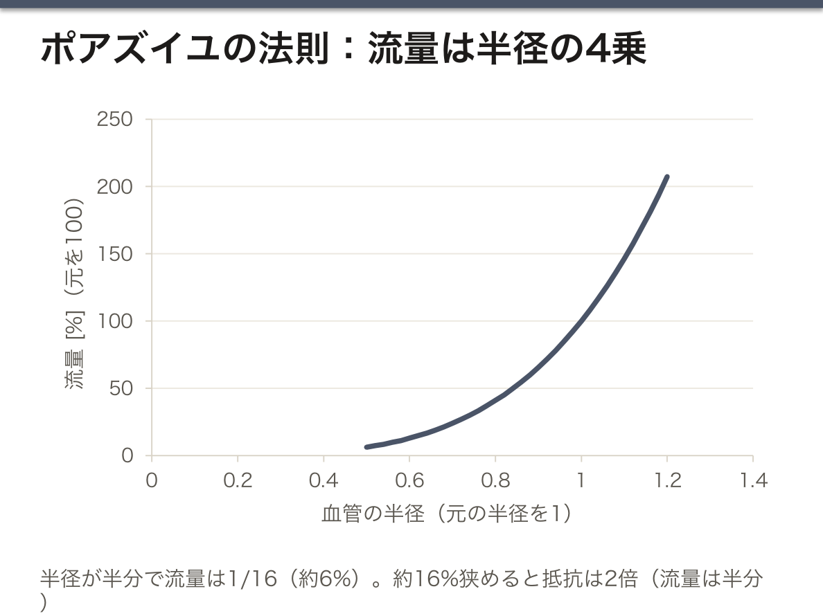
長さが2倍なら抵抗は2倍、粘度が高ければ抵抗は大きい、半径が2倍なら流量は16倍。
なぜ4乗なのか。半径を大きくすると、①流れる断面積が r2 で増え、②壁から遠い中心部の速い流れの割合が増えて平均流速も r2 で増える。2つの効果の積で r4 になる。
この4乗のため、体は血管の太さを少し変えるだけで血流を大きく調節できる。半径を約16%狭めるだけで抵抗は2倍になる。全身の血管抵抗を決めているのは主に細動脈の平滑筋の収縮である。
体全体では 平均血圧 ≈ 心拍出量 × 全末梢血管抵抗。血圧は心臓が押し出す量と、血管がどれだけ狭いかの積で決まる。平均血圧は拡張期血圧+脈圧/3 で近似できる(120/80 なら約93 mmHg)。
動脈硬化で内腔が半分になると、同じ圧では血流は1/16に落ちる——狭心症や間欠性跛行が「運動したときだけ」症状を出すのは、需要が増えたときに狭い血管が流量を増やせないからである。降圧薬のカルシウム拮抗薬や ACE 阻害薬・ARB は、細動脈を広げて抵抗を下げることで血圧を下げる。
English version
Core
When a viscous liquid flows through a tube in laminar flow, the flow rate is
Q = πr4ΔP/(8ηl)
(r: radius, ΔP: pressure difference between the ends, η: viscosity, l: length). Viscous flow always comes with a fall in pressure.
Written in the same form as Ohm’s law (I = ΔV/R), Q = ΔP/X, and the resistance to flow is
X = 8ηl/(πr4)
Twice the length gives twice the resistance, higher viscosity gives more resistance, and twice the radius gives 16 times the flow.
Going further
Why the fourth power? A larger radius (1) increases the area for flow by r2, and (2) increases the share of fast flow in the center far from the wall, so the mean velocity also rises by r2. The product of the two gives r4.
Because of this fourth power, the body can control blood flow strongly by changing vessel width only a little. Narrowing the radius by about 16% doubles the resistance. The resistance of the whole body is set mainly by contraction of smooth muscle in the arterioles.
For the whole body, mean arterial pressure ≈ cardiac output × total peripheral resistance. Blood pressure is the product of how much the heart pushes out and how narrow the vessels are. Mean pressure is approximately diastolic + pulse pressure/3 (about 93 mmHg for 120/80).
In the clinic
If atherosclerosis halves the lumen, flow at the same pressure falls to 1/16 — angina and intermittent claudication cause symptoms “only on exertion” because the narrow vessel cannot raise flow when demand increases. Antihypertensive drugs such as calcium channel blockers and ACE inhibitors or ARBs lower blood pressure by widening arterioles and lowering resistance.
血圧の測り方 — 直接法・聴診法・オシロメトリック法
収縮期血圧は心室が収縮して血液を送り出すときの最高の圧(上の値)、拡張期血圧は心臓が弛緩しているときの最低の圧(下の値)。差を脈圧という。
| 方法 | 原理 | 特徴 |
|---|---|---|
| 直接法 | 動脈にカテーテルを入れて圧センサにつなぐ | 最も正確(基準)。1拍ごとの変化をリアルタイムで見られる。侵襲的 |
| 聴診法 | カフで動脈を圧迫し、ゆるめながら聴診器でコロトコフ音を聞く | 非侵襲。手技と聴力に依存 |
| オシロメトリック法 | カフ内圧にのる脈ごとの小さな振動をセンサで捉え、計算で推定 | 自動血圧計の方式。不整脈で誤差が出やすい |
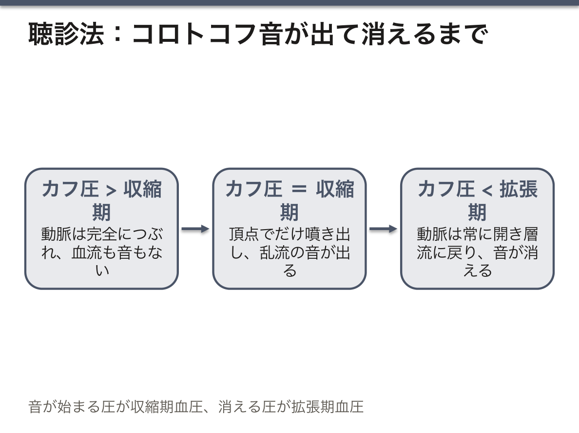
聴診法の物理。カフ圧が収縮期血圧より高いと動脈は完全につぶれ、血流も音もない。カフ圧を下げて収縮期血圧と等しくなると、収縮期の頂点でだけ血液が狭い隙間を噴き出し、乱流が音を生む(コロトコフ音の始まり=収縮期血圧)。カフ圧が拡張期血圧より下がると動脈は常に開き、流れは層流に戻って音が消える(=拡張期血圧)。
オシロメトリック法では、カフ内の振動が最大になる圧が平均血圧にほぼ一致する。収縮期・拡張期の値は、振動の大きさの変化から機種ごとの計算法で推定している。上腕式が最も信頼でき、手首式は高さの影響を受けやすく、指先式は精度が低い。
正しく測る条件:5分以上の安静、会話しない、背もたれのある椅子で足を組まず、カフの中心を心臓の高さに。腕周に対して細すぎるカフは高く出る(カフのゴム袋の幅は腕周の約40%が目安)。日本の高血圧治療ガイドラインでは診察室血圧 140/90 mmHg 以上、家庭血圧 135/85 mmHg 以上を高血圧とし、正常血圧は 120/80 mmHg 未満。分類の区切りは国やガイドラインで少し違う。
English version
Core
Systolic pressure is the highest pressure when the ventricle contracts and ejects blood (the upper number); diastolic pressure is the lowest pressure while the heart relaxes (the lower number). The difference is the pulse pressure.
| Method | Principle | Features |
|---|---|---|
| Direct | A catheter in an artery connected to a pressure sensor | Most accurate (the reference). Shows beat-to-beat changes in real time. Invasive |
| Auscultatory | A cuff compresses the artery; while releasing it, Korotkoff sounds are heard with a stethoscope | Non-invasive. Depends on technique and hearing |
| Oscillometric | A sensor picks up the small pulse oscillations in cuff pressure and the values are calculated | Used in automatic monitors. Errors are likely with arrhythmias |
Going further
The physics of the auscultatory method. With cuff pressure above systolic pressure, the artery is completely closed: no flow, no sound. As cuff pressure falls to systolic pressure, blood spurts through a narrow gap only at the peak of systole, and turbulence makes sound (the first Korotkoff sound = systolic pressure). When cuff pressure falls below diastolic pressure, the artery stays open, flow becomes laminar again and the sound disappears (= diastolic pressure).
In the oscillometric method, the cuff pressure with the largest oscillation is close to the mean arterial pressure. Systolic and diastolic values are estimated from how the oscillation size changes, by each maker’s own calculation. Upper-arm monitors are the most reliable; wrist monitors are sensitive to height; finger monitors are inaccurate.
In the clinic
Conditions for correct measurement: rest for at least 5 minutes, no talking, sit in a chair with a back and legs uncrossed, and the center of the cuff at heart level. A cuff too narrow for the arm reads high (the bladder width should be about 40% of arm circumference). Japanese hypertension guidelines define hypertension as office pressure of 140/90 mmHg or more, or home pressure of 135/85 mmHg or more, and normal pressure as below 120/80 mmHg. The cut-offs differ a little between countries and guidelines.
血管壁の弾性 — 動脈が硬くなると何が起きるか
大動脈などの弾性動脈は、収縮期に送り出された血液の一部を壁を膨らませてため、拡張期に縮んで押し出す。これにより心臓の間欠的な拍出が、末梢では途切れない流れに変わる(ふいご効果、Windkessel 効果)。
動脈が硬くなる(弾性が失われる)と、ためる働きが弱まり、収縮期血圧は上がり拡張期血圧は下がる——脈圧が開く。
円筒形の血管の壁にかかる張力は ラプラスの法則 T = P·r(T:単位長さあたりの壁張力)で、半径が大きいほど同じ圧でも壁の張力は大きい。
なぜ硬い動脈で収縮期血圧が上がるのか。同じ量の血液を押し込むとき、柔らかい壁なら膨らんで受け止めるが、硬い壁は膨らまないので圧が急に上がる。拡張期には、ためていた血液が少ないので圧が早く落ちる。ばねの硬さ(ヤング率)が、そのまま血圧の波形に現れている。
動脈が硬いと、脈の波が壁を伝わる速さ(脈波伝播速度)も上がる。波の速さは壁の弾性率の平方根にほぼ比例するので、脈波伝播速度は動脈硬化の指標になる。
ラプラスの法則には悪循環が隠れている:血管が膨らむ→半径が大きくなる→壁の張力が増す→さらに膨らむ。
高齢者に多い収縮期高血圧(拡張期は正常か低いのに収縮期だけ高い)は、大動脈の弾性低下の表れである。日本では足首と上腕の脈波伝播速度(baPWV)が動脈硬化の検査に使われる。大動脈瘤(aneurysma aortae)は径が大きいほど壁張力が増して破裂しやすくなるので、径が一定の大きさ(腹部でおよそ5〜5.5 cm)を超えると手術を考える。
English version
Core
Elastic arteries such as the aorta store part of the blood ejected in systole by stretching their walls, and push it on by recoiling in diastole. This turns the intermittent output of the heart into continuous flow in the periphery (the Windkessel effect).
When arteries stiffen (lose elasticity), this storage weakens, and systolic pressure rises while diastolic pressure falls — the pulse pressure widens.
The tension in the wall of a cylindrical vessel follows Laplace’s law T = P·r (T: wall tension per unit length): the larger the radius, the greater the wall tension at the same pressure.
Going further
Why does a stiff artery raise systolic pressure? When the same volume of blood is pushed in, a soft wall stretches to take it, but a stiff wall does not, so pressure rises sharply. In diastole, less blood has been stored, so pressure falls quickly. The stiffness of the “spring” (its Young’s modulus) appears directly in the blood pressure wave.
In stiff arteries, the speed at which the pulse wave travels along the wall (pulse wave velocity) also rises. Wave speed is roughly proportional to the square root of the wall’s elastic modulus, so pulse wave velocity is an index of arterial stiffness.
Laplace’s law hides a vicious circle: the vessel bulges → the radius grows → wall tension increases → it bulges more.
In the clinic
Isolated systolic hypertension, common in older people (only systolic pressure is high while diastolic is normal or low), reflects loss of aortic elasticity. In Japan, the brachial–ankle pulse wave velocity (baPWV) is used to test arterial stiffness. Aortic aneurysm (aneurysma aortae) becomes more likely to rupture as its diameter grows and wall tension rises, so surgery is considered once the diameter passes a certain size (about 5–5.5 cm in the abdomen).
気体の分圧とヘンリーの法則 — 減圧症はなぜ起こる
ドルトンの法則:混合気体の全圧は、各成分の分圧の和である。乾燥空気の酸素は約21%なので、海面での酸素分圧は 0.21 × 760 ≈ 160 mmHg。
ヘンリーの法則:液体に溶ける気体の量は、その気体の分圧に比例する。分圧が高いほど多く溶け、低いほど少なく溶ける。溶けやすさ(溶解度)は気体によって違い、CO2 は O2 の約20倍溶けやすい。
減圧症(潜水病)の物理。潜ると水深10 m ごとに1気圧ずつ圧が増え、吸う空気の窒素分圧も上がる。ヘンリーの法則に従い、血液と組織に窒素が多く溶け込む。そこから急に浮上すると圧が下がり、溶けていられなくなった窒素が気泡になって出てくる——炭酸飲料の栓を開けたときと同じである。ゆっくり浮上すれば、余分な窒素は血液から肺へ運ばれて吐き出される。
気泡は血管を詰まらせて虚血を起こし、さらに凝固系・補体・血小板を活性化して血管透過性を高め、組織を傷める。
減圧症の症状は関節痛(ベンズ)、皮膚のかゆみや大理石様の斑、重症では神経症状や呼吸困難。治療は高気圧酸素療法で、再び圧を上げて気泡を小さくし、窒素の排出を促す。麻酔では吸入麻酔薬・酸素・亜酸化窒素などの混合比を流量計と気化器で管理する。亜酸化窒素は溶けやすく、投与をやめると肺胞へ一気に出てきて酸素を薄めるため、拡散性低酸素を防ぐのに終了後しばらく高濃度の酸素を吸わせる。
English version
Core
Dalton’s law: the total pressure of a gas mixture is the sum of the partial pressures of its parts. Dry air is about 21% oxygen, so the oxygen partial pressure at sea level is 0.21 × 760 ≈ 160 mmHg.
Henry’s law: the amount of a gas dissolved in a liquid is proportional to its partial pressure. The higher the partial pressure, the more dissolves; the lower, the less. Solubility differs between gases; CO2 is about twenty times more soluble than O2.
Going further
The physics of decompression sickness. Diving, pressure rises by 1 atm every 10 m of depth, and so does the nitrogen partial pressure of the inhaled air. Following Henry’s law, more nitrogen dissolves in blood and tissue. If the diver rises too quickly, pressure falls and nitrogen that can no longer stay dissolved comes out as bubbles — like opening a carbonated drink. With a slow ascent, the extra nitrogen is carried from the blood to the lungs and breathed out.
The bubbles block vessels and cause ischemia, and they also activate clotting, complement and platelets, raise vascular permeability and damage tissue.
In the clinic
Symptoms of decompression sickness are joint pain (the bends), itchy or marbled skin, and in severe cases neurological symptoms or breathing difficulty. Treatment is hyperbaric oxygen therapy, which raises the pressure again to shrink the bubbles and helps remove nitrogen. In anesthesia, the mixture of inhaled anesthetic, oxygen and nitrous oxide is controlled with flowmeters and vaporizers. Nitrous oxide is very soluble; when it is stopped it floods into the alveoli and dilutes oxygen, so high-concentration oxygen is given for a while afterward to prevent diffusion hypoxia.
まとめ
| 圧力と単位 — なぜ血圧は mmHg で測るのか | 圧力は面に垂直にはたらく単位面積あたりの力である:P = F/A、単位 Pa(N/m2)。 |
|---|---|
| 静水圧 — 立つと足の血圧は頭より高い | 静止した液体の中では、深さ h の点の圧力は上の液柱の重さだけ増える。 |
| 流量・連続の式・ベルヌーイの定理 — 狭いところは速く、圧は低い | 流量 Q は単位時間に流れる体積で、断面積 A と平均流速 v から Q = A·v(m3/s)。 |
| ポアズイユの法則 — 半径が4乗で効く | 粘性のある液体が管を層流で流れるとき、流量はQ = πr4ΔP/(8ηl)(r:半径、ΔP:管の両端の圧の差、η:粘度、l:長さ)。 |
| 血圧の測り方 — 直接法・聴診法・オシロメトリック法 | 収縮期血圧は心室が収縮して血液を送り出すときの最高の圧(上の値)、拡張期血圧は心臓が弛緩しているときの最低の圧(下の値)。 |
| 血管壁の弾性 — 動脈が硬くなると何が起きるか | 大動脈などの弾性動脈は、収縮期に送り出された血液の一部を壁を膨らませてため、拡張期に縮んで押し出す。 |
| 気体の分圧とヘンリーの法則 — 減圧症はなぜ起こる | ドルトンの法則:混合気体の全圧は、各成分の分圧の和である。 |
このノートは医学生が授業の内容を自分の言葉でまとめたもので、診療の判断に使うものではありません。誤りに気づいたらお知らせください。