この章は「何であるか」ではなく「標本の上でどう見分けるか」に寄せて書いてある。
脊髄を読む順番は毎回同じでよい。①深い裂のあるほうが前 → ②灰白質の角の形で高さを決める → ③白質の索を3つに分ける → ④膜と隙間を外から数える。この4手順で、切片でも模型でも同じように答えられる。
この範囲の要は「脊髄と脊柱の長さが合っていない」ことである。円錐が L1 で終わること・馬尾があること・分節と椎骨番号がずれること・腰椎穿刺が安全であることは、すべてこの1点から出てくる。
- 脊髄 — 外形
- 脊髄円錐と馬尾
- 脊髄の溝 — 4本で前後左右が決まる
- 灰白質 — 蝶の形の読み方
- 白質 — 3本の索
- 脊髄分節 — 1対の神経根が出る単位
- 脊髄膜 — 3枚と、そのあいだの3つの隙間
- 灰白質の核 — どの核が、どの路の起点か
- Rexed 層 — 灰白質を I〜X で呼ぶ
- 反射弓 — 脊髄が単独で閉じる回路
- 神経系の分類と、ニューロンの型
- 神経系の発生 — 神経板から神経管と神経堤へ
- 脳胞 — 3つから5つへ、そして感覚器とのつながり
- 脊髄 — 外形Medulla spinalis
- 脊髄円錐と馬尾Conus medullaris et cauda equina
- 脊髄の溝 — 4本で前後左右が決まるSulci medullae spinalis
- 灰白質 — 蝶の形の読み方Substantia grisea
- 白質 — 3本の索Substantia alba
- 脊髄分節 — 1対の神経根が出る単位Segmentum medullae spinalis
- 脊髄膜 — 3枚と、そのあいだの3つの隙間Meninges medullae spinalis
- 灰白質の核 — どの核が、どの路の起点かNuclei medullae spinalis
- Rexed 層 — 灰白質を I〜X で呼ぶLaminae spinales
- 反射弓 — 脊髄が単独で閉じる回路Arcus reflexus
- 神経系の分類と、ニューロンの型Systema nervosum
- 神経系の発生 — 神経板から神経管と神経堤へTubus neuralis
- 脳胞 — 3つから5つへ、そして感覚器とのつながりVesiculae encephali
- まとめ
脊髄 — 外形Medulla spinalis
大後頭孔(foramen magnum)で延髄に続き、成人では L1〜L2 の高さで 円錐 conus medullaris となって終わる。長さ約42〜45cm、直径1cm ほどの円柱。
太さは一様ではなく、2つの膨大 intumescentia がある。頸膨大(C4〜Th1)は上肢へ、腰仙膨大(L1〜S3)は下肢へ行く神経細胞が集まるため太い。膨大の位置=四肢の神経支配の位置である。
表面の溝で前後が決まる。前正中に深い裂 fissura mediana anterior、後正中に浅い溝 sulcus medianus posterior。標本を渡されたら、まず「深い裂のあるほうが前」と決めてから他を探す。
なぜ L1 で終わるのか。胎生3か月までは脊髄と脊柱の長さが等しく、神経根は真横に出ている。その後脊柱のほうが速く伸びるため、脊髄が相対的に上へ引き上げられたように見える(ascensus medullae spinalis)。新生児では L3、成人で L1〜L2。「子どもと大人で下端の高さが違う」ことを骨格標本の上で示せるようにしておく。
この不一致の帰結が2つある。①下位の神経根は椎孔まで長く下る=馬尾。②脊髄分節と椎骨番号がずれる(頸部で約1、胸上部で約2、胸下部で約3)。脊髄の高さを椎骨から言うときは必ずこの補正が要る。
腰椎穿刺は L3/L4 または L4/L5 で行う。成人の脊髄は L1〜L2 で終わるので、それより下なら針は馬尾の中に入るだけで、脊髄そのものを刺さない。目印は両側腸骨稜を結ぶ線(Jacoby 線)= L4 棘突起。新生児では脊髄下端が L3 まであるため、より低い高さ(L4/L5 以下)を選ぶ。
English version
Core
Continuous with the medulla oblongata at the foramen magnum, ending in the adult at L1–L2 as the conus medullaris. A cylinder about 42–45 cm long and 1 cm across.
The thickness is not uniform: there are two enlargements (intumescentiae). The cervical enlargement (C4–Th1) is thick because the neurones for the upper limb gather there, the lumbosacral (L1–S3) for the lower limb. The position of an enlargement is the position of the limb it supplies.
The grooves on the surface settle front from back. A deep fissura mediana anterior in front, a shallow sulcus medianus posterior behind. Handed a specimen, settle first that the side with the deep fissure is anterior, and only then look for the rest.
Going further
Why does it end at L1? Until the third fetal month the cord and the column are the same length and the roots leave horizontally. After that the column grows faster, so the cord appears to be drawn upwards (ascensus medullae spinalis). In the newborn it ends at L3, in the adult at L1–L2. Be able to show on a skeleton that the lower end differs between a child and an adult.
Two consequences follow from that mismatch. ① The lower roots must descend a long way to their foramina — the cauda equina. ② The cord segments do not match the vertebral numbers (by about 1 in the neck, 2 in the upper thorax, 3 in the lower thorax). Whenever a cord level is given from a vertebra, that correction is required.
In the clinic
Lumbar puncture is done at L3/L4 or L4/L5. The adult cord ends at L1–L2, so below that the needle only enters the cauda equina and does not touch the cord itself. The landmark is the line joining the iliac crests (Jacoby’s line) = the spinous process of L4.
In the newborn the cord reaches L3, so a lower level (L4/L5 or below) is chosen.
脊髄円錐と馬尾Conus medullaris et cauda equina
Conus medullaris — 脊髄の下端の円錐状の部分(L1〜L2)。ここから細い糸が下へ伸びる。
Filum terminale — 円錐の先から出る終糸。神経組織ではなく軟膜の続きである。内終糸(S2 まで、硬膜嚢の中)と外終糸(尾骨まで、硬膜に包まれる)に分かれ、脊髄を下端で係留する。
Cauda equina — 円錐より下の硬膜嚢の中を、腰・仙・尾神経の根が束になって下る。馬の尾に似るのでこの名。ここには脊髄は無く、根だけがある。
押さえるべきは「conus はどこで終わり、そこから下は何か」である。標本で答えるときは、①円錐の先端を指し、②そこから伸びる1本の細い糸を filum terminale、③その周りに広がる根の束を cauda equina、と3つを順に示す。
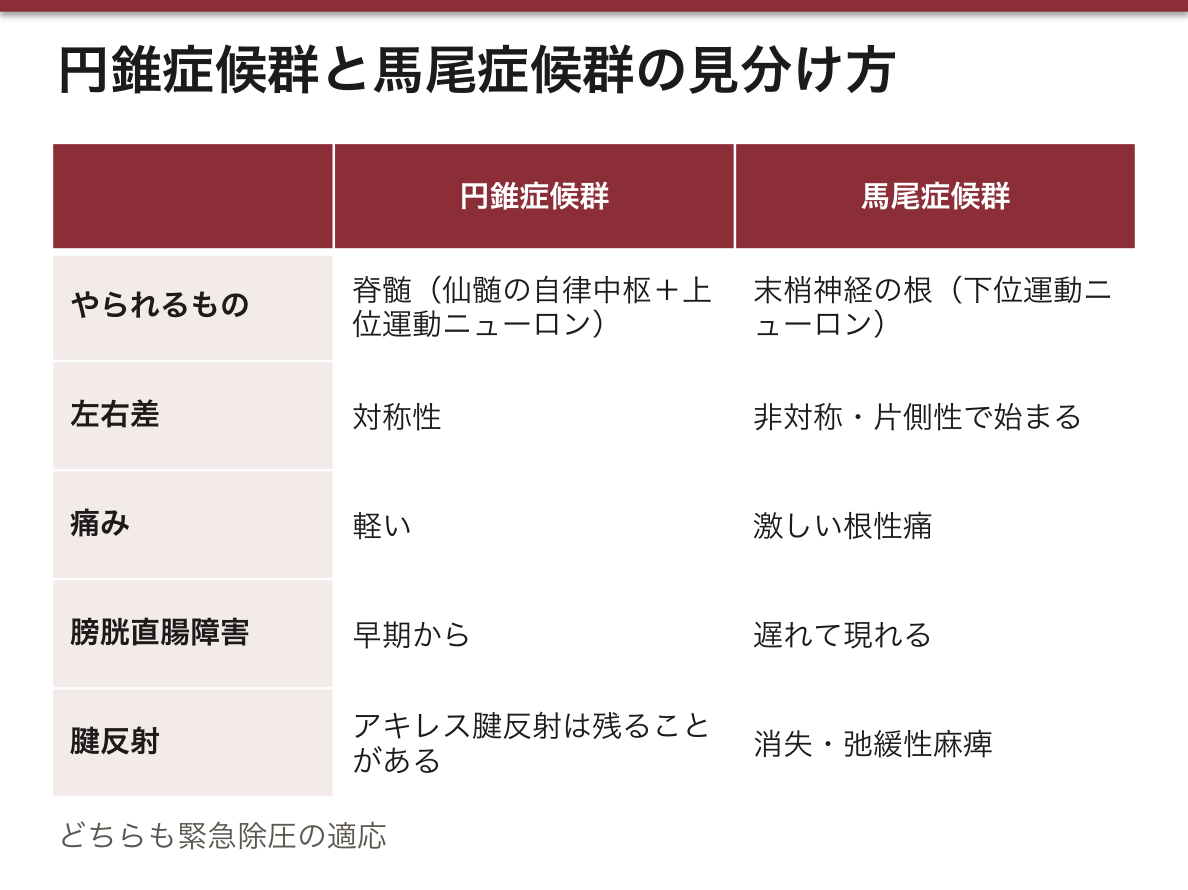
円錐と馬尾は障害の型が違う。円錐は脊髄(上位運動ニューロン+仙髄の自律中枢)、馬尾は末梢神経の根(下位運動ニューロン)である。だから円錐症候群は早期から対称性の膀胱直腸障害が出て、痛みは軽く、腱反射は亢進しうる。馬尾症候群は非対称・強い神経根痛・弛緩性麻痺・腱反射消失となる。
円錐症候群と馬尾症候群は、同じ高さの病変でも型が違う。円錐は脊髄そのもの(仙髄の自律中枢+上位運動ニューロン)、馬尾は末梢神経の根(下位運動ニューロン)だからである。
円錐症候群 — 早期から、対称性の膀胱直腸障害。会陰部の鞍状知覚障害。痛みは軽い。アキレス腱反射は残ることがあり、球海綿体反射は消える。
馬尾症候群 — 非対称・片側性で始まる。激しい根性痛が前面に出る。膀胱直腸障害は遅れて現れ、腱反射は消失、弛緩性麻痺になる。
「痛みが強く、左右差があれば馬尾。痛みが乏しく、対称で、最初から尿が出なければ円錐」と対にして覚える。どちらも緊急除圧の適応である。
出典:イラスト解剖学 誌面690頁「脊髄損傷を考える」
English version
Core
- Conus medullaris — the conical lower end of the cord (L1–L2). A fine thread runs down from it.
- Filum terminale — from the tip of the conus. It is not nervous tissue but a continuation of the pia mater. It divides into an internal part (to S2, within the dural sac) and an external part (to the coccyx, wrapped in dura), anchoring the cord at its lower end.
- Cauda equina — below the conus, the lumbar, sacral and coccygeal roots descend in a bundle within the dural sac, resembling a horse’s tail. There is no cord here, only roots.
Going further
What matters is "where does the conus end, and what is below it". Answering on the specimen, show three things in order: ① the tip of the conus ② the single fine thread running from it — the filum terminale ③ the spread of roots around it — the cauda equina.
The conus and the cauda differ in the type of lesion they give. The conus is cord (upper motor neurone plus the sacral autonomic centres); the cauda is peripheral nerve root (lower motor neurone). So the conus syndrome gives symmetrical bladder and bowel disturbance early, with little pain, and the tendon reflexes may be brisk. The cauda equina syndrome is asymmetrical, with severe radicular pain, flaccid paralysis and absent reflexes.
In the clinic
Conus and cauda equina syndromes differ in type even at the same level, because the conus is cord itself (sacral autonomic centres plus upper motor neurone) and the cauda is peripheral root (lower motor neurone).
Conus syndrome — symmetrical bladder and bowel disturbance from early on. Saddle anaesthesia of the perineum. Pain is slight. The ankle jerk may be preserved; the bulbocavernosus reflex is lost.
Cauda equina syndrome — begins asymmetrically, on one side. Severe radicular pain dominates. Bladder and bowel disturbance appears later; the reflexes are lost and the paralysis is flaccid.
Remember them as a pair: severe pain with asymmetry = cauda; little pain, symmetrical, and retention from the start = conus. Both are indications for emergency decompression.
脊髄の溝 — 4本で前後左右が決まるSulci medullae spinalis
脊髄の表面で押さえる溝は4つ。この4本を順に言えれば、脊髄の向きと根の出どころが同時に説明できる。
| ラテン語 | 位置 | そこから出入りするもの |
|---|---|---|
| Fissura mediana anterior | 前正中・深い裂 | —(前脊髄動脈が走る) |
| Sulcus medianus posterior | 後正中・浅い溝 | —(後正中中隔が入り込む) |
| Sulcus anterolateralis | 前外側 | 前根 radix anterior(運動・出る) |
| Sulcus posterolateralis | 後外側 | 後根 radix posterior(感覚・入る) |
覚え方は「前は出る、後ろは入る」。前根は運動線維が出ていき、後根は感覚線維が入ってくる。
頸髄と上部胸髄にはもう1本、sulcus intermedius posterior がある。後索を fasciculus gracilis(内側・下半身)と fasciculus cuneatus(外側・上半身)に分ける溝で、Th6 より上にしか無い——cuneatus 自体が上肢からの線維なので、それより下では存在しないからである。
Bell–Magendie の法則(前根=運動、後根=感覚)は、この溝の解剖そのものである。後根には途中に ganglion spinale(脊髄神経節)という膨らみがあり、前根には無い。標本で根を追って膨らみを見つけたら、それが後根である。
前正中裂を走るのが前脊髄動脈である。この1本が詰まると前脊髄動脈症候群になり、溝の解剖がそのまま症状の分布になる。
前2/3(前角・外側皮質脊髄路・外側脊髄視床路)が落ちて、損傷高位以下の運動麻痺と温痛覚障害が両側に出る。いっぽう後索は後脊髄動脈が養うので生き残り、深部感覚(触覚・振動覚・位置覚)だけが保たれる。この「動かない・熱さが分からない・でも位置は分かる」という解離が決め手になる。
後根が入る後外側溝の側では、1本の後根=1つのデルマトームが対応する。帯状疱疹が帯状に、片側だけ出るのは、水痘帯状疱疹ウイルスが後根神経節に潜んでいるからである。
出典:イラスト解剖学 誌面750頁「前脊髄動脈症候群・ブラウンセカール症候群」
English version
Core
Four sulci are to be known on the surface of the cord. Say these four in order and you have explained at once the orientation of the cord and where the roots come from.
| Latin | Position | What enters or leaves |
|---|---|---|
| Fissura mediana anterior | Anterior midline, deep | — (the anterior spinal artery runs in it) |
| Sulcus medianus posterior | Posterior midline, shallow | — (the posterior median septum runs in) |
| Sulcus anterolateralis | Anterolateral | Radix anterior (motor, leaving) |
| Sulcus posterolateralis | Posterolateral | Radix posterior (sensory, entering) |
Remember: in front it leaves, behind it enters.
Going further
In the cervical and upper thoracic cord there is one more, the sulcus intermedius posterior, dividing the posterior funiculus into fasciculus gracilis (medial, lower body) and fasciculus cuneatus (lateral, upper body). It exists only above Th6 — because the cuneatus itself carries fibres from the upper limb and does not exist below that.
The Bell–Magendie law (anterior root motor, posterior root sensory) is this anatomy of the sulci itself. The posterior root carries a swelling along its course, the ganglion spinale; the anterior root has none. Follow a root on the specimen and find a swelling, and it is a posterior root.
In the clinic
The anterior spinal artery runs in the anterior median fissure. When this single vessel occludes, the anterior spinal artery syndrome results, and the anatomy of the sulcus becomes the distribution of the symptoms: the anterior two thirds (anterior horn, lateral corticospinal tract, lateral spinothalamic tract) fail, giving bilateral motor paralysis and loss of pain and temperature below the level. The posterior columns are supplied by the posterior spinal arteries and survive, so deep sensation (touch, vibration, position) alone is preserved. The dissociation — cannot move, cannot feel heat, but knows where the limb is — is decisive.
On the side of the posterolateral sulcus where the posterior root enters, one posterior root corresponds to one dermatome. Shingles appears in a band on one side because varicella zoster virus lies latent in the posterior root ganglion.
灰白質 — 蝶の形の読み方Substantia grisea
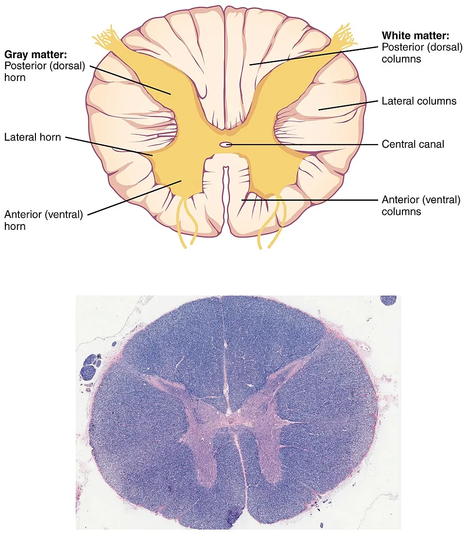
横断面の中央にある H字(蝶)状の部分。神経細胞体の集まりで、外側の白質より暗く見える。左右は commissura grisea で繋がり、その中心に canalis centralis が開く。
3つの角 cornu。
- Cornu anterius(前角)— 運動ニューロン(α・γ)。太く丸い。上肢・下肢を動かす膨大部でとくに大きい
- Cornu posterius(後角)— 感覚の中継。細く尖り、後外側溝に届く
- Cornu laterale(側角)— 自律神経。C8〜L2(交感神経)と S2〜S4(副交感神経)にしか無い
角の形は高さを教える。「この切片は何髄か」の読み方:
| 高さ | 見え方 |
|---|---|
| 頸髄 | 全体が大きく楕円。前角が太い(上肢)。白質が多い |
| 胸髄 | 小さく円い。前角が細い。側角があるのが決め手 |
| 腰髄 | 灰白質が大きく前角が太い(下肢)。白質は相対的に少ない |
| 仙髄 | 断面が小さく、ほとんどが灰白質 |
白質の量は上に行くほど増える——上行路は登るにつれ線維を足し、下行路は降りるにつれ線維を落とすためである。だから「断面が大きくて白い=上のほう」と読める。
側角の上端は教科書によって Th1 と書かれるが、C8 から始まるとする資料もある(C8〜L2)。どちらでも Horner 症候群の局在(毛様脊髄中枢 C8〜Th2)は変わらない。
側角が Th1〜L2 にしか無いことが、Horner 症候群の局在を決める。眼交感神経は視床下部から下って C8〜Th2 の側角(毛様脊髄中枢)で乗り換えるので、この高さより上の脊髄病変・肺尖部腫瘍(Pancoast)・頸部の病変で縮瞳・眼瞼下垂・発汗低下が起きる。
English version
Core
The H-shaped (butterfly) region at the centre of a transverse section: collections of nerve cell bodies, darker than the white matter outside. The two sides are joined by the commissura grisea, with the canalis centralis opening at its centre.
Three horns (cornua).
- Cornu anterius — motor neurones (α and γ). Thick and rounded; especially large at the enlargements that move the limbs.
- Cornu posterius — relay of sensation. Slender and pointed, reaching the posterolateral sulcus.
- Cornu laterale — autonomic. Present only at C8–L2 (sympathetic) and S2–S4 (parasympathetic).
Going further
The shape of the horns tells you the level. How to read which level a section comes from:
| Level | Appearance |
|---|---|
| Cervical | Large and oval overall. Thick anterior horn (upper limb). Much white matter |
| Thoracic | Small and round. Slender anterior horn. The lateral horn is the decisive feature |
| Lumbar | Large grey matter with a thick anterior horn (lower limb). Relatively little white matter |
| Sacral | Small in section, and almost all grey matter |
The amount of white matter increases the higher you go — ascending tracts add fibres as they climb and descending tracts shed them as they descend. So "large section, much white" reads as "high up".
The upper limit of the lateral horn is given as Th1 in some textbooks, but other sources state that it begins at C8 (C8–L2). Either way the localisation of Horner’s syndrome (the ciliospinal centre, C8–Th2) is unchanged.
In the clinic
That the lateral horn exists only at Th1–L2 decides the localisation of Horner’s syndrome. The ocular sympathetic fibres descend from the hypothalamus and relay in the lateral horn at C8–Th2 (the ciliospinal centre), so a cord lesion above that level, an apical lung tumour (Pancoast) or a lesion in the neck gives miosis, ptosis and reduced sweating.
白質 — 3本の索Substantia alba
灰白質の外側を囲む有髄線維の層。溝と角によって3つの索 funiculus に分けられる。
- Funiculus anterior — 前正中裂と前外側溝のあいだ
- Funiculus lateralis — 前外側溝と後外側溝のあいだ(いちばん広い)
- Funiculus posterior — 後外側溝と後正中溝のあいだ
索の中に上行路と下行路が束(tractus / fasciculus)として走る。後索は上行路だけ(触覚・深部感覚)、側索と前索は上下が混在する。
主要な路の位置を、索ごとに1つずつ押さえる。
| 索 | 主な路 | 向き | 運ぶもの |
|---|---|---|---|
| 後索 | Fasciculus gracilis / cuneatus | 上行 | 識別性触覚・振動覚・位置覚(同側) |
| 側索 | Tractus corticospinalis lateralis | 下行 | 随意運動(延髄で交叉済み) |
| 側索 | Tractus spinothalamicus lateralis | 上行 | 痛覚・温度覚(脊髄内で交叉済み=対側) |
| 側索 | Tractus spinocerebellaris ant./post. | 上行 | 無意識性深部感覚(小脳へ) |
| 前索 | Tractus corticospinalis anterior | 下行 | 体幹の運動(交叉せず下り、分節で交叉) |
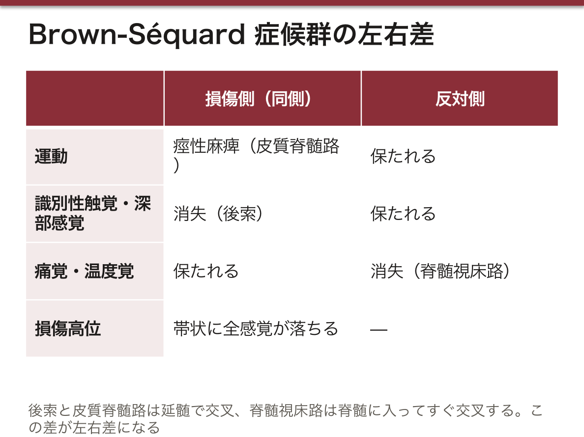
交叉する場所が症状の左右を決める。後索と皮質脊髄路は延髄で交叉するので、脊髄では同側を走る。脊髄視床路は入った分節(1〜2節上)で交叉するので、脊髄ではすでに対側を走っている。この差だけで Brown-Séquard の所見が導ける。
Brown-Séquard 症候群(脊髄半側切断)は、上の交叉の位置がそのまま出る。同側に痙性麻痺(皮質脊髄路)と深部感覚障害(後索)、対側に痛覚・温度覚の脱失(脊髄視床路)。境界の1〜2分節だけ同側の全感覚が落ちる。「触れるのに痛みが分からない側と、痛みは分かるのに動かない側が逆になる」と言えれば、機序を理解している。
English version
Core
The layer of myelinated fibres surrounding the grey matter, divided by the sulci and the horns into three funiculi.
- Funiculus anterior — between the anterior median fissure and the anterolateral sulcus.
- Funiculus lateralis — between the anterolateral and posterolateral sulci (the widest).
- Funiculus posterior — between the posterolateral and posterior median sulci.
Within the funiculi the ascending and descending tracts run as bundles (tractus, fasciculus). The posterior funiculus carries ascending fibres only (touch and deep sensation); the lateral and anterior funiculi mix ascending and descending.
Going further
Fix one main tract per funiculus.
| Funiculus | Tract | Direction | Carries |
|---|---|---|---|
| Posterior | Fasciculus gracilis / cuneatus | Ascending | Discriminative touch, vibration, position (ipsilateral) |
| Lateral | Tractus corticospinalis lateralis | Descending | Voluntary movement (already decussated in the medulla) |
| Lateral | Tractus spinothalamicus lateralis | Ascending | Pain and temperature (decussated within the cord = contralateral) |
| Lateral | Tractus spinocerebellaris ant./post. | Ascending | Unconscious proprioception (to the cerebellum) |
| Anterior | Tractus corticospinalis anterior | Descending | Movement of the trunk (descends uncrossed, decussating at its segment) |
Where a tract crosses decides the side of the symptoms. The posterior columns and the corticospinal tract cross in the medulla, so within the cord they run ipsilaterally. The spinothalamic tract crosses at the segment it enters (one or two above), so within the cord it already runs contralaterally. From that difference alone the findings of Brown-Séquard can be derived.
In the clinic
Brown-Séquard syndrome (hemisection of the cord) is exactly the positions of those decussations. Ipsilateral spastic paralysis (corticospinal) and loss of deep sensation (posterior columns); contralateral loss of pain and temperature (spinothalamic). At the boundary, one or two segments lose all modalities ipsilaterally.
If you can say "the side that cannot feel pain and the side that cannot move are opposite", you have understood the mechanism.
脊髄分節 — 1対の神経根が出る単位Segmentum medullae spinalis
1対の脊髄神経が出る範囲を1分節とする。全部で 31 分節:頸 8・胸 12・腰 5・仙 5・尾 1。
頸神経だけ 8 対あるのに頸椎は 7 個である。C1〜C7 は同じ番号の椎骨の上から出て、C8 は C7 と Th1 のあいだから出る。以降は同じ番号の椎骨の下から出る。
1分節から出るものは決まっている:前根(運動)+後根(感覚)→ 合流して脊髄神経 → すぐ前枝と後枝に分かれる。
Nervus spinalis の枝は4つ。脊髄神経はここまで言えると強い。
- Ramus anterior — 太い。体幹の前外側と四肢。頸・腰・仙で神経叢を作る(胸神経だけは叢を作らず肋間神経になる)
- Ramus posterior — 細い。背部の固有筋と背部の皮膚。分節性を保つ
- Ramus meningeus(反回神経 / 洞椎骨神経)— 椎孔を逆戻りして硬膜・後縦靱帯・椎間板の外層を支配する
- Ramus communicans — 交感神経幹との連絡(白枝は Th1〜L2 のみ、灰白枝は全分節)
Ramus meningeus があるから椎間板は痛む。髄核そのものに神経は無いが、線維輪の外層と後縦靱帯にはこの枝が分布している。椎間板性腰痛の解剖学的な根拠がここにある。
分節が分かれば、損傷の高さが呼吸で決まる。安静時吸気の 2/3 は横隔膜(C3〜C5 支配)による。
C1〜C3 — 横隔膜が完全に麻痺し呼吸停止。C5 より上 — 肋間筋・頸部の吸気筋が落ちて呼吸困難。C4〜C8 — 四肢麻痺。Th1〜L3 — 対麻痺+腹筋など呼気筋の麻痺。L4 以下 — 両下肢麻痺と膀胱直腸の弛緩性麻痺。
徴候は2種類に分けて述べる。髄節徴候=障害された髄節そのもののニューロン障害(その髄節が支配する筋の萎縮・腱反射の消失)、索路徴候=神経路が断たれたことによるその高さより下の障害(感覚障害・痙性麻痺・腱反射亢進・病的反射)。「切れた高さは弛緩、その下は痙性」と読む。
出典:イラスト解剖学 誌面690頁「脊髄損傷を考える」
English version
Core
The extent from which one pair of spinal nerves emerges is one segment. There are 31: 8 cervical, 12 thoracic, 5 lumbar, 5 sacral, 1 coccygeal.
There are 8 cervical nerves but only 7 cervical vertebrae. C1–C7 leave above the vertebra of the same number, and C8 leaves between C7 and Th1; below that each leaves beneath the vertebra of the same number.
What leaves a segment is fixed: anterior root (motor) + posterior root (sensory) → joining as the spinal nerve → dividing at once into anterior and posterior rami.
Going further
The nervus spinalis has four branches. Know this far and the spinal nerve is well covered.
- Ramus anterior — thick. The anterolateral trunk and the limbs. Forms plexuses in the cervical, lumbar and sacral regions (the thoracic nerves alone form no plexus and become the intercostal nerves).
- Ramus posterior — thin. The intrinsic muscles of the back and the skin of the back. Retains its segmental arrangement.
- Ramus meningeus (recurrent, sinuvertebral nerve) — turns back through the foramen to supply the dura, the posterior longitudinal ligament and the outer layer of the intervertebral disc.
- Ramus communicans — connection with the sympathetic trunk (white only at Th1–L2, grey at every segment).
The intervertebral disc hurts because of the ramus meningeus. The nucleus pulposus itself has no nerves, but the outer annulus and the posterior longitudinal ligament are supplied by this branch. Here is the anatomical basis of discogenic back pain.
In the clinic
Know the segments and the level of an injury is settled by the breathing. Two thirds of quiet inspiration is diaphragmatic (C3–C5).
- C1–C3 — the diaphragm is wholly paralysed: respiratory arrest.
- Above C5 — the intercostals and the accessory inspiratory muscles of the neck fail: respiratory difficulty.
- C4–C8 — tetraplegia.
- Th1–L3 — paraplegia with paralysis of the expiratory muscles such as the abdominals.
- Below L4 — paralysis of both legs with flaccid bladder and bowel.
State the signs in two kinds. Segmental signs = damage to the neurones of the injured segment itself (wasting and loss of reflex in the muscles it supplies); tract signs = the consequences below that level of the severed pathways (sensory loss, spastic paralysis, brisk reflexes, pathological reflexes). Read it as "flaccid at the level of the cut, spastic below it".
脊髄膜 — 3枚と、そのあいだの3つの隙間Meninges medullae spinalis
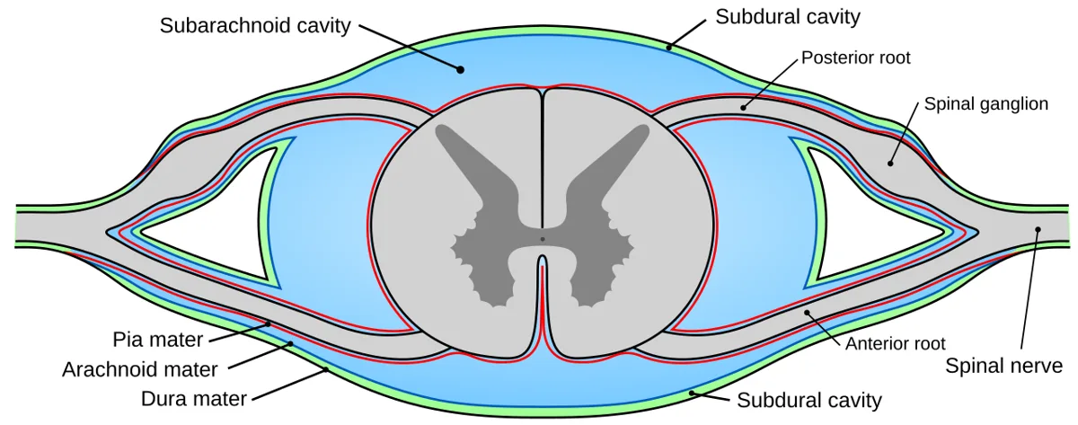
外から 硬膜 dura mater → クモ膜 arachnoidea mater → 軟膜 pia mater の3枚。脳と違い、脊髄硬膜は1枚だけである(脳硬膜の骨内膜層に当たるものが椎骨の骨膜として別にあるため)。
| 膜 | 特徴 |
|---|---|
| Dura mater spinalis | 厚く丈夫な袋(硬膜嚢)。S2 の高さで終わる |
| Arachnoidea mater spinalis | 薄く血管が無い。硬膜の内面に接する |
| Pia mater spinalis | 脊髄の表面に密着。lig. denticulatum を出して硬膜に固定する |
隙間は3つ。それぞれの中身を言えるようにする。
| 隙間 | どこ | 中身 |
|---|---|---|
| Cavitas (spatium) epiduralis | 硬膜と椎骨骨膜のあいだ | 脂肪と内椎骨静脈叢(弁が無い) |
| Spatium subdurale | 硬膜とクモ膜のあいだ | ごく薄い液の層(生理的には隙間ではない) |
| Spatium subarachnoideum | クモ膜と軟膜のあいだ | 脳脊髄液 liquor cerebrospinalis |
脊髄は L1〜L2 で終わるのに硬膜嚢は S2 まである。この差=cisterna lumbalis(腰槽)で、中身は髄液と馬尾だけ。腰椎穿刺が安全に行えるのはこの空間があるからである。
Lig. denticulatum は軟膜が左右21対の歯状に張り出して硬膜に付く靱帯で、脊髄を髄液の中で吊っている。前根と後根のあいだを走るので、手術中に前根と後根を見分ける目印になる。
硬膜外麻酔と脊髄くも膜下麻酔の違いは、針を止める膜が1枚違うだけである。硬膜外は epidural に留めて脂肪と静脈叢の層に薬を置き、脊髄くも膜下は硬膜とクモ膜を貫いて髄液の中に入れる。後者は髄液が返ってくることで確認できる。
硬膜外の内椎骨静脈叢には弁が無く、胸腹腔と交通する。腹圧が上がると血液が逆流しうるため、前立腺癌・乳癌が肺を介さず椎体へ直接転移する経路(Batson 静脈叢)になる。
English version
Core
Three membranes from outside in: dura mater → arachnoidea mater → pia mater. Unlike the brain, the spinal dura is a single layer (what corresponds to the endosteal layer of the cranial dura exists separately as the periosteum of the vertebrae).
| Membrane | Feature |
|---|---|
| Dura mater spinalis | A thick, tough sac (the dural sac). Ends at the level of S2 |
| Arachnoidea mater spinalis | Thin and avascular. Applied to the inner surface of the dura |
| Pia mater spinalis | Adherent to the surface of the cord. Sends out the lig. denticulatum to fix it to the dura |
Going further
There are three spaces; be able to say what is in each.
| Space | Where | Contents |
|---|---|---|
| Cavitas (spatium) epiduralis | Between dura and vertebral periosteum | Fat and the internal vertebral venous plexus (valveless) |
| Spatium subdurale | Between dura and arachnoid | A very thin film of fluid (physiologically not a space) |
| Spatium subarachnoideum | Between arachnoid and pia | Cerebrospinal fluid |
The cord ends at L1–L2 but the dural sac continues to S2. That difference is the cisterna lumbalis, containing only CSF and the cauda equina. Lumbar puncture is safe because this space exists.
The lig. denticulatum is the pia projecting as 21 pairs of tooth-like processes attaching to the dura, suspending the cord within the CSF. It runs between the anterior and posterior roots and so is the landmark for telling them apart at operation.
In the clinic
Epidural and spinal anaesthesia differ by one membrane in where the needle is stopped. The epidural stops in the epidural space and places the drug in the layer of fat and venous plexus; the spinal pierces dura and arachnoid to enter the CSF. The latter is confirmed by CSF returning.
The internal vertebral venous plexus of the epidural space has no valves and communicates with the thorax and abdomen. A rise in abdominal pressure can reverse the flow, making it the route (Batson’s plexus) by which prostate and breast cancer metastasise directly to the vertebrae without passing through the lung.
灰白質の核 — どの核が、どの路の起点かNuclei medullae spinalis
同じ働きの神経細胞体の集まりを核 nucleus という。それぞれ決まった形・大きさ・位置を持つ。脊髄で名前を覚えるのは5つでよい。
- 前角の運動核 nuclei motorii cornu anterioris — 軸索が前根 → 脊髄神経 → 骨格筋へ。すべての下行路が最後に乗り換える場所(=下位運動ニューロン)
- Nucleus proprius(後角)— 痛覚・温度覚・触覚の2次ニューロン。軸索は脊髄で交叉して反対側の脊髄視床路になる
- Nucleus intermediolateralis(側角)— 交感神経細胞。C8〜L2。軸索は前根 → 脊髄神経 → ramus communicans albus → 交感幹
- Nuclei parasympathici sacrales(側角)— 副交感神経細胞。S2〜S4。軸索は前根 → 脊髄神経 → ramus ventralis → 骨盤の自律神経叢
- Substantia gelatinosa(後角の周辺部)— 痛覚・温度覚・触覚の路の入口。中心管のまわりのものは substantia gelatinosa centralis
核の名前を覚える意味は「路の起点が言えること」に尽きる。核を聞かれたら、必ず「どの路になるか」までを続けて答える。
無意識の深部感覚(→ 小脳)の2つの核は、対になっている。
- Nucleus thoracicus posterior — 後角の底の近く。後脊髄小脳路 tractus spinocerebellaris posterior の起点
- Nucleus intermediomedialis — substantia gelatinosa centralis の隣。前脊髄小脳路 tractus spinocerebellaris anterior の起点
前角の運動核は「柱」として並んでいる。特定の筋を支配する運動ニューロンは前角の中で縦の列を作り、その列自体を核と呼ぶ(脳幹の運動核と同じ作り)。ほとんどの筋(分節間筋)は複数の分節にまたがる複数の核から支配を受ける。1分節の中だけに運動ニューロンが収まる筋を単分節筋(指標筋)という——これが「この筋の麻痺=この分節の障害」と言える根拠になる。
Formatio reticularis spinalis(フォルマーティオ・レティクラーリス・スピナーリス)— 後柱の外側で白質に食い込む灰白質。上は脳幹へ続く。感覚の感受性・呼吸・心拍・血圧・運動系・意識状態に関わる。解剖学的な境界がはっきりしないのは、脳の各所のニューロンを含むためである。
Fasciculi proprii — 灰白質のすぐ外側で隣接分節をつなぐ薄い白質の層。脊髄反射が分節をまたげる理由がこれ。
核の分布を知っていると、病巣が「どの高さの、どの角か」で言えるようになる。
- ALS — 前角の運動核(下位運動ニューロン)+皮質脊髄路(上位)。感覚は保たれるのは、後角の核も後索も無事だから
- 脊髄空洞症 — 中心管の周囲が広がり、交叉する脊髄視床路の線維が最初に切れる。両側性・宙吊り型の解離性感覚障害(痛覚・温度覚だけ脱失し、後索の触覚・深部感覚は残る)
- Horner 症候群 — 眼交感神経は C8〜Th2 の側角(毛様脊髄中枢)で乗り換える。この高さの病変や肺尖部腫瘍(Pancoast)で縮瞳・眼瞼下垂・発汗低下
- 脊髄円錐症候群 — S2〜S4 の副交感核が同時にやられるため、早期から膀胱直腸障害が出る。馬尾症候群と分ける決め手がここにある
English version
Core
A collection of nerve cell bodies with the same function is a nucleus, each with its own shape, size and position. Five names suffice in the spinal cord.
- Nuclei motorii cornu anterioris — axons to anterior root → spinal nerve → skeletal muscle. The final relay of every descending tract (= the lower motor neurone).
- Nucleus proprius (posterior horn) — the second neurone of pain, temperature and touch. Its axons decussate within the cord and become the contralateral spinothalamic tract.
- Nucleus intermediolateralis (lateral horn) — sympathetic cells, C8–L2. Axons → anterior root → spinal nerve → ramus communicans albus → sympathetic trunk.
- Nuclei parasympathici sacrales (lateral horn) — parasympathetic cells, S2–S4. Axons → anterior root → spinal nerve → ramus ventralis → the pelvic autonomic plexuses.
- Substantia gelatinosa (the periphery of the posterior horn) — the entrance of the pathways for pain, temperature and touch. That around the central canal is the substantia gelatinosa centralis.
The whole point of learning the names is being able to say where a tract begins. Asked for a nucleus, always go on to say which tract it becomes.
Going further
The two nuclei for unconscious proprioception (to the cerebellum) form a pair.
- Nucleus thoracicus posterior — near the base of the posterior horn. The origin of the tractus spinocerebellaris posterior.
- Nucleus intermediomedialis — beside the substantia gelatinosa centralis. The origin of the tractus spinocerebellaris anterior.
The motor nuclei of the anterior horn are arranged as columns. The motor neurones supplying a particular muscle form a longitudinal column within the horn, and that column is what is called a nucleus (the same arrangement as the motor nuclei of the brainstem). Most muscles (intersegmental muscles) are supplied by several nuclei spanning several segments. A muscle whose motor neurones lie within a single segment is a monosegmental (indicator) muscle — the basis for saying "paralysis of this muscle = a lesion of this segment".
Formatio reticularis spinalis — grey matter encroaching on the white lateral to the posterior column, continuous above with the brainstem. Concerned with the sensitivity of sensation, respiration, heart rate, blood pressure, the motor system and the state of consciousness. Its boundaries are indistinct anatomically because it contains neurones belonging to many parts of the brain.
Fasciculi proprii — a thin layer of white matter just outside the grey, joining adjacent segments. This is why spinal reflexes can span segments.
In the clinic
Knowing the distribution of the nuclei lets a lesion be stated as "which horn, at which level".
- ALS — the motor nuclei of the anterior horn (lower motor neurone) plus the corticospinal tract (upper). Sensation is preserved because neither the posterior horn nuclei nor the posterior columns are touched.
- Syringomyelia — the region around the central canal widens and the decussating spinothalamic fibres are cut first. Bilateral, suspended, dissociated sensory loss (pain and temperature lost, touch and deep sensation of the posterior columns preserved).
- Horner’s syndrome — the ocular sympathetic relays in the lateral horn at C8–Th2 (the ciliospinal centre). A lesion at this level or an apical lung tumour (Pancoast) gives miosis, ptosis and reduced sweating.
- Conus medullaris syndrome — the parasympathetic nuclei of S2–S4 are involved together, so bladder and bowel disturbance appears early. This is the decisive point against a cauda equina syndrome.
Rexed 層 — 灰白質を I〜X で呼ぶLaminae spinales
灰白質は「角」で分けるだけでなく、後ろから前へ I〜X の層 lamina に分けられる(Rexed 層)。角が「見た目の区分」なのに対し、層は核の住所である。「この核は第何層か」を言えるようにしておく。
- 後角 cornu posterius — I〜VI
- 側角 cornu laterale — VII
- 前角 cornu anterius — VIII〜IX
- 中心管の周囲 substantia intermedia centralis — X
層番号は表面から深部へ、後ろから前へという一方向に振られている。だから「marginalis(縁)= I」「motorii(前角の底)= IX」は位置から思い出せる。
核と層の対応。
- I — Nucleus marginalis:後角の表面を覆う。触・温・痛を受けて上位へ
- II — Substantia gelatinosa:外側脊髄視床路の2次ニューロン
- III〜IV — Nucleus proprius:前脊髄視床路の2次ニューロン
- V — 外側部が formatio reticularis spinalis:頸髄と上部胸髄にだけある
- VI — 核を持たない:反射弓の介在ニューロン
- VII — 自律の階:nucleus intermediolateralis(交感・(C8)Th1〜L2)、nuclei parasympathici sacrales(副交感・S2〜S4)、nucleus dorsalis = nucleus thoracicus posterior(Clarke 核・固有覚・C8〜L3)
- VIII — 核を持たない:運動インパルスの調節
- IX — Nuclei motorii(5群):下位運動ニューロン。軸索は前根へ
- X — Nucleus intermediomedialis:固有覚に関わり、線維が交叉する場所
この表は nucleus intermediomedialis を X に置く資料に従った(一般の教科書は VII に置くことが多い)。
層は病巣の局在をそのまま説明する。
- 脊髄空洞症 — 壊れるのは X 層(中心管の周囲)。ここを交叉する脊髄視床路の線維だけが切れるので、両側・分節性に温痛覚だけが落ちる。後索は無事なので触覚と深部感覚は残る(解離性感覚障害)
- ALS — IX 層(下位運動ニューロン)+側索の皮質脊髄路(上位)。感覚の層(I〜IV)に手をつけないので感覚は保たれる
- ポリオ — ウイルスが IX 層だけを選択的に壊す。純粋な弛緩性麻痺になるのはこのため
- 自律の高さ — VII 層。交感が Th1〜L2、副交感が S2〜S4。脊髄損傷で排尿・排便・性機能がどう変わるかは、損傷高がこの2つの帯のどちらより上かで決まる
English version
Core
The grey matter is divided not only into horns but into laminae I–X from behind forwards (Rexed’s laminae). Where the horns are a division by appearance, the laminae are the address of a nucleus. Be able to say which lamina a given nucleus is in.
- Cornu posterius — I–VI
- Cornu laterale — VII
- Cornu anterius — VIII–IX
- Around the central canal, substantia intermedia centralis — X
The numbering runs one way only, from surface to depth, from back to front. So "marginalis (the margin) = I" and "motorii (the base of the anterior horn) = IX" can be recalled from position alone.
Going further
The correspondence of nucleus to lamina.
- I — Nucleus marginalis: covers the surface of the posterior horn. Receives touch, temperature and pain for transmission upwards.
- II — Substantia gelatinosa: the second neurone of the lateral spinothalamic tract.
- III–IV — Nucleus proprius: the second neurone of the anterior spinothalamic tract.
- V — its lateral part is the formatio reticularis spinalis: present only in the cervical and upper thoracic cord.
- VI — contains no nucleus: the interneurones of the reflex arc.
- VII — the autonomic storey: nucleus intermediolateralis (sympathetic, (C8)Th1–L2), nuclei parasympathici sacrales (S2–S4), nucleus dorsalis = nucleus thoracicus posterior (Clarke’s column, proprioception, C8–L3).
- VIII — contains no nucleus: modulation of motor impulses.
- IX — Nuclei motorii (five groups): the lower motor neurones. Axons to the anterior root.
- X — Nucleus intermediomedialis: concerned with proprioception, and the place where fibres decussate.
This list follows sources that place the nucleus intermediomedialis in X (general textbooks usually place it in VII).
In the clinic
The laminae explain the localisation of a lesion directly.
- Syringomyelia — what is destroyed is lamina X (around the central canal). Only the spinothalamic fibres decussating there are cut, so temperature and pain alone are lost, bilaterally and segmentally. The posterior columns are intact, so touch and deep sensation remain (dissociated sensory loss).
- ALS — lamina IX (lower motor neurone) plus the corticospinal tract in the lateral funiculus (upper). It does not touch the sensory laminae (I–IV), so sensation is preserved.
- Poliomyelitis — the virus destroys lamina IX selectively. Hence a pure flaccid paralysis.
- The autonomic levels — lamina VII. Sympathetic Th1–L2, parasympathetic S2–S4. How micturition, defaecation and sexual function change after a cord injury is decided by which of these two bands the level lies above.
反射弓 — 脊髄が単独で閉じる回路Arcus reflexus
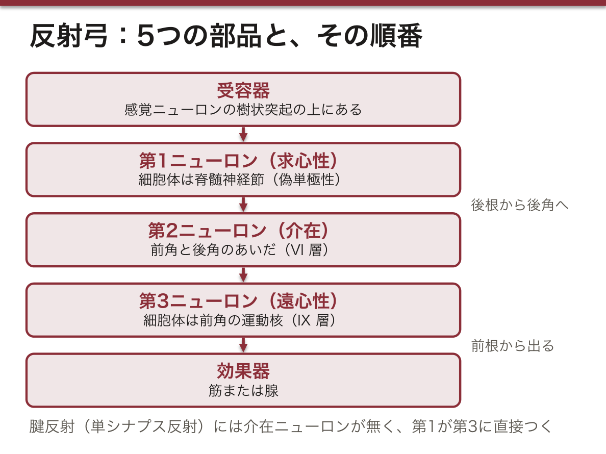
脳を通さずに刺激から反応までを閉じる最小の回路。脊髄が反射中枢として働く。構成は 5つ、順番が決まっている。
- 受容器 receptor — 感覚ニューロンの樹状突起の上にある
- 第1ニューロン(求心性・感覚性) — 細胞体は ganglion spinale(偽単極性)。軸索は後根 radix sensoria から後角へ入る
- 第2ニューロン(介在ニューロン) — 前角と後角のあいだ(VI 層)
- 第3ニューロン(遠心性・運動性) — 細胞体は前角の nuclei motorii(IX 層)。軸索は前根 radix motoria から出る
- 効果器 effector — 筋または腺
入るのは後ろ、出るのは前。溝でいえば sulcus posterolateralis から入り、sulcus anterolateralis から出る。
介在ニューロンが要るかどうかで2種類に分かれる。
- 単シナプス反射 — 感覚ニューロンが運動ニューロンに直接つく。介在ニューロンが無い。腱反射(膝蓋腱反射など)がこれ。潜時が短い
- 多シナプス反射 — あいだに介在ニューロンが入る。屈曲反射・表在反射がこれ
反射弓は分節に閉じているのに対し、cellulae disseminatae の突起は隣の分節へ伸び、fasciculi proprii(灰白質を薄く取り巻く白質)をつくって分節間を連絡する。だから複数の分節にまたがる反応も脊髄だけで起こせる。
「順番に5つ」「それぞれの細胞体はどこか」まで言えるようにしておく。数を先に言ってから並べること。
反射は「高さの物差し」として使う。反射弓のどこか1か所でも切れれば、その反射だけが消える。
- 上腕二頭筋反射 C5〜C6/上腕三頭筋反射 C7/膝蓋腱反射 L2〜L4/アキレス腱反射 S1〜S2
- 反射消失(低下)=下位運動ニューロンまたは求心路の障害(末梢神経障害、神経根症、前角の病変)
- 反射亢進+病的反射=上位運動ニューロンの障害。下行路が介在ニューロンへの抑制を失うため
- 脊髄ショックでは、損傷直後は上位障害でも一時的に反射が消える。数日〜数週で亢進に転じる。急性期に「反射が無いから末梢性」と決めつけないこと
整形外科では、しびれの訴えに対してデルマトーム(感覚)・筋力・反射の3点で高さを詰める。反射はこの3点目にあたる。
English version
Core
The smallest circuit closing from stimulus to response without passing through the brain; the cord acts as the reflex centre. It has five parts, in a fixed order.
- Receptor — on the dendrite of the sensory neurone.
- First neurone (afferent, sensory) — cell body in the ganglion spinale (pseudounipolar). The axon enters the posterior horn through the radix sensoria.
- Second neurone (interneurone) — between anterior and posterior horns (lamina VI).
- Third neurone (efferent, motor) — cell body in the nuclei motorii of the anterior horn (lamina IX). The axon leaves through the radix motoria.
- Effector — muscle or gland.
It enters behind and leaves in front — in terms of the sulci, in at the sulcus posterolateralis and out at the sulcus anterolateralis.
Going further
They divide in two by whether an interneurone is required.
- Monosynaptic reflex — the sensory neurone contacts the motor neurone directly, with no interneurone. The tendon reflexes (the knee jerk and others). Short latency.
- Polysynaptic reflex — an interneurone is interposed. The flexor and superficial reflexes.
Where the reflex arc is closed within a segment, the processes of the cellulae disseminatae extend to neighbouring segments and form the fasciculi proprii (the thin sheet of white matter surrounding the grey). So responses spanning several segments can also be produced by the cord alone.
Be able to name the five in order and say where the cell body of each lies. State the number first, then list them.
In the clinic
Reflexes are used as a measure of level. Break the arc anywhere and that reflex alone is lost.
- Biceps jerk C5–C6 / triceps jerk C7 / knee jerk L2–L4 / ankle jerk S1–S2.
- Absent or reduced reflex = a lesion of the lower motor neurone or of the afferent path (peripheral neuropathy, radiculopathy, anterior horn disease).
- Brisk reflexes with pathological reflexes = an upper motor neurone lesion, the descending tracts having lost their inhibition of the interneurones.
In spinal shock the reflexes are temporarily absent immediately after injury even in an upper motor neurone lesion, turning brisk over days to weeks. Do not conclude "no reflexes, therefore peripheral" in the acute phase.
In orthopaedics, a complaint of numbness is localised by three things — dermatome (sensation), power, and reflex. The reflex is the third of these.
神経系の分類と、ニューロンの型Systema nervosum
神経系を学ぶ入口。「形態的に」と「機能的に」を必ず両方言う。
形態的 morphological
- Systema nervosum centrale — encephalon(脳)+ medulla spinalis(脊髄)
- Systema nervosum periphericum — nervi craniales・nervi spinales・ganglia
機能的 functional
- Systema nervosum animalium(体性)
- Systema nervosum autonomicum = vegetativum(自律)
働きは2つ。全身の細胞・組織・器官の機能を調節し協調させること、そして内外の刺激を受け取り、伝え、応答すること。
ニューロンの型 — 形態で4つ、機能で3つ。「どこにあるか」とセットで覚える。
- Unipolar(単極性)— 胎生期のみ。成体には無い
- Bipolar(双極性)— 感覚器。網膜、嗅球、平衡覚・聴覚の器官
- Pseudounipolar(偽単極性)— 脊髄神経節・脳神経節
- Multipolar(多極性)— 中枢神経系
機能では 感覚性(求心性)・運動性(遠心性)・介在ニューロンの3つ。介在ニューロンが脊髄反射を担う。
細胞体と突起の住み分けが、灰白質と白質の定義そのものになる。細胞体 → 灰白質と神経節/突起 → 白質と末梢神経。この一文から、脊髄の内部構造は全部ほどける。
グリアの各論(星状膠細胞・稀突起膠細胞・上衣細胞・ミクログリア/Schwann 細胞・衛星細胞)は組織学の「神経組織」で扱う。解剖では「髄鞘をつくるのは中枢が oligodendrocytus、末梢が Schwann 細胞」まで言えれば足りる。
型と場所の対応が、そのまま病気の分布になる。
- 帯状疱疹 — 潜伏するのは脊髄神経節の偽単極性細胞。再活性化すると1本の後根の支配域=1本のデルマトームに沿って皮疹が出る。分布が帯状になる理由がここにある
- 多発性硬化症と Guillain-Barré 症候群 — 髄鞘をつくる細胞が違う。中枢=oligodendrocytus(MS)/末梢=Schwann 細胞(GBS)。同じ「脱髄」でも舞台が別
- 網膜色素変性症 — 障害されるのは網膜の視細胞。網膜が双極性ニューロンを持つ数少ない場所であることが、感覚器を「中枢の出張所」として扱う根拠になる
- 自律神経の障害 — 機能的分類の autonomicum 側。起立性低血圧・神経因性膀胱・発汗異常は、体性の運動感覚が正常でも単独で起こりうる
English version
Core
The entrance to the nervous system. Always give both "morphologically" and "functionally".
Morphological
- Systema nervosum centrale — encephalon + medulla spinalis.
- Systema nervosum periphericum — nervi craniales, nervi spinales, ganglia.
Functional
- Systema nervosum animalium (somatic)
- Systema nervosum autonomicum = vegetativum (autonomic)
It does two things: regulates and coordinates the function of the cells, tissues and organs of the whole body, and receives, conducts and responds to stimuli from within and without.
Going further
Types of neurone — four by form, three by function. Learn each with where it is found.
- Unipolar — only in embryonic life; none in the adult.
- Bipolar — the sense organs: retina, olfactory bulb, the organs of balance and hearing.
- Pseudounipolar — the spinal and cranial nerve ganglia.
- Multipolar — the central nervous system.
By function, sensory (afferent), motor (efferent) and interneurone. The interneurones carry the spinal reflexes.
The separation of cell bodies from processes is the very definition of grey and white matter. Cell bodies → grey matter and ganglia; processes → white matter and peripheral nerves. From that one sentence the whole internal structure of the cord unravels.
The details of the glia (astrocyte, oligodendrocyte, ependymal cell, microglia / Schwann cell, satellite cell) are dealt with under nervous tissue in histology. In anatomy it is enough to say that myelin is made by the oligodendrocytus centrally and the Schwann cell peripherally.
In the clinic
The correspondence of type to place is directly the distribution of the disease.
- Shingles — what lies latent is the pseudounipolar cell of the spinal ganglion. On reactivation the rash follows the territory of one posterior root = one dermatome. Here is why the distribution is a band.
- Multiple sclerosis and Guillain–Barré syndrome — the cell that makes myelin differs. Central = oligodendrocyte (MS); peripheral = Schwann cell (GBS). The same "demyelination" on a different stage.
- Retinitis pigmentosa — what is damaged is the photoreceptor of the retina. That the retina is one of the few places with bipolar neurones is the basis for treating a sense organ as an outpost of the central nervous system.
- Autonomic disorders — the autonomicum side of the functional classification. Orthostatic hypotension, neurogenic bladder and abnormal sweating can occur alone with normal somatic movement and sensation.
神経系の発生 — 神経板から神経管と神経堤へTubus neuralis
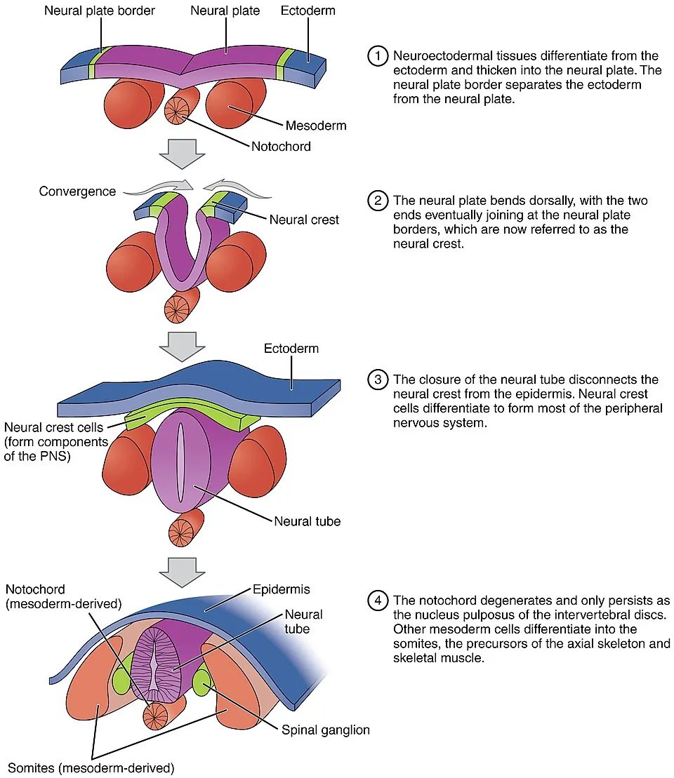
第3週、脊索に誘導されて背側の外胚葉が厚くなり神経板になる。縁が持ち上がって神経ひだ、中央がへこんで神経溝となり、ひだが背側で合わさって神経管ができる。閉鎖は頸の高さ(第22日ごろ)から頭と尾の両方向へ進み、頭側の神経孔は第25日ごろ、尾側の神経孔は第27〜28日ごろに閉じる。
ひだの縁の細胞は管に入らず神経堤として抜け出し、全身へ移動する。
| 神経堤からできるもの | |
|---|---|
| 感覚の神経節 | 脊髄神経節・脳神経の感覚神経節(偽単極性ニューロン) |
| 自律神経 | 交感神経幹の神経節・副交感神経節・副腎髄質 |
| 支える細胞 | Schwann 細胞・神経節の衛星細胞、軟膜とくも膜の一部 |
| その他 | 皮膚の色素細胞、顔面の骨と結合組織の大部分 |
神経管の壁は内から脳室帯・外套層(→ 灰白質)・辺縁層(→ 白質)。外套層は境界溝を境に背側の翼板(感覚 → 後角)と腹側の基板(運動 → 前角)に分かれ、Th1〜L2 ではそのあいだに側角(自律神経)ができる。基板のニューロンの軸索が前根として出て、神経堤由来の脊髄神経節の突起が後根として入り、合わさって脊髄神経になる。
デルマトームがある理由は体節にある。体節は椎板(→ 椎骨)・筋板(→ 筋)・皮板(→ 真皮)に分かれ、それぞれ同じ高さの脊髄神経を連れたまま移動する。だから皮膚も筋も、元の体節の番号で神経に支配される。
脊髄の上昇(新生児で L3、成人で L1〜2)は、胎生3か月以降に脊柱の方が速く伸びるために起こる(本章の「脊髄円錐と馬尾」)。
神経管閉鎖障害は閉じる時期(妊娠4週、本人が妊娠に気づく前)の異常である。尾側が閉じないと二分脊椎——椎弓だけの欠損で皮膚に毛の束やくぼみがある潜在性二分脊椎、髄膜が袋状に出る髄膜瘤、脊髄ごと出る脊髄髄膜瘤(下肢麻痺・膀胱直腸障害、Chiari II 型奇形と水頭症を合併)。頭側が閉じないと無脳症、頭蓋の欠損から脳が出ると脳瘤。
予防には葉酸が効く。妊娠を計画する女性は、妊娠の1か月以上前から1日400 μg をとるよう勧められている——閉鎖が終わる4週には間に合わないと遅いからである。母体血清や羊水のα-フェトプロテイン(AFP)が高いことが、開いた神経管閉鎖障害の手がかりになる。
English version
Core
In week 3 the notochord induces the dorsal ectoderm to thicken into the neural plate. Its edges rise as neural folds and its centre sinks as the neural groove; the folds meet dorsally to form the neural tube. Closure starts at neck level (about day 22) and runs both ways; the cranial neuropore closes around day 25 and the caudal neuropore around days 27–28.
Cells at the edges of the folds do not join the tube; they leave as the neural crest and migrate through the body.
| Neural crest derivatives | |
|---|---|
| Sensory ganglia | Spinal ganglia and sensory ganglia of cranial nerves (pseudounipolar neurons) |
| Autonomic | Sympathetic chain ganglia, parasympathetic ganglia, adrenal medulla |
| Supporting cells | Schwann cells, satellite cells of ganglia, parts of pia and arachnoid |
| Others | Pigment cells of the skin, most bones and connective tissue of the face |
The wall of the neural tube has, from inside out, the ventricular zone, mantle layer (→ grey matter) and marginal layer (→ white matter). The sulcus limitans divides the mantle layer into the dorsal alar plate (sensory → posterior horn) and the ventral basal plate (motor → anterior horn); from T1 to L2 a lateral horn (autonomic) forms between them. Axons of basal-plate neurons leave as the anterior root; processes of neural-crest spinal ganglia enter as the posterior root; together they form the spinal nerve.
Going further
Dermatomes exist because of somites. Each somite splits into sclerotome (→ vertebra), myotome (→ muscle) and dermatome (→ dermis), and each part migrates while keeping the spinal nerve of its own level. So skin and muscles are supplied according to the number of their original somite.
The “ascent” of the spinal cord (L3 in newborns, L1–2 in adults) happens because the vertebral column grows faster from month 3 (see “Conus medullaris and cauda equina” in this chapter).
In the clinic
Neural tube defects arise when closure fails, in week 4 of pregnancy — often before the woman knows she is pregnant. Failure at the caudal end gives spina bifida: spina bifida occulta (only the arch is missing; a tuft of hair or dimple on the skin), meningocele (meninges bulge as a sac) and myelomeningocele (cord included; leg paralysis, bladder and bowel dysfunction, with Chiari II malformation and hydrocephalus). Failure at the cranial end gives anencephaly; brain protruding through a skull defect is an encephalocele.
Folic acid prevents them. Women planning pregnancy are advised to take 400 μg a day from at least one month before conception, because starting after the tube closes in week 4 is too late. Raised alpha-fetoprotein (AFP) in maternal serum or amniotic fluid points to an open neural tube defect.
脳胞 — 3つから5つへ、そして感覚器とのつながりVesiculae encephali
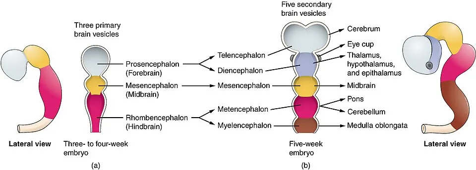
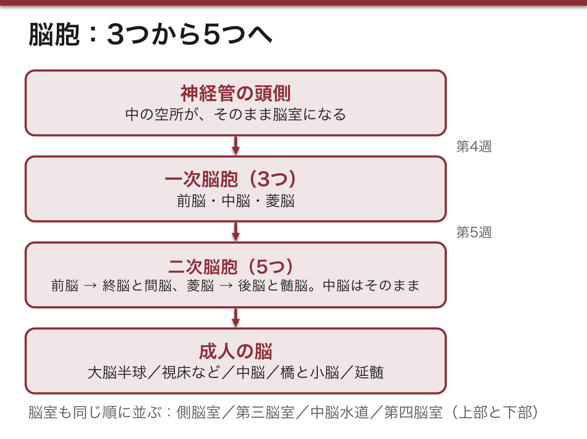
神経管の頭側は第4週に3つにふくらみ(一次脳胞)、第5週に5つになる(二次脳胞)。中の空所がそのまま脳室になる。
| 一次脳胞 | 二次脳胞 | 成人の脳 | 脳室 |
|---|---|---|---|
| prosencephalon(前脳) | telencephalon(終脳) | 大脳半球(皮質・基底核・白質) | 側脳室 |
| diencephalon(間脳) | 視床・視床下部・視床上部(松果体)・視床後部。網膜と視神経、下垂体後葉もここから | 第三脳室 | |
| mesencephalon(中脳) | mesencephalon(中脳) | 中脳蓋・被蓋・大脳脚 | 中脳水道 |
| rhombencephalon(菱脳) | metencephalon(後脳) | 橋と小脳 | 第四脳室の上部 |
| myelencephalon(髄脳) | 延髄 | 第四脳室の下部 |
菱脳の上端の細い所(菱脳峡)からは上髄帆と上小脳脚ができる。脳は成長しながら3か所で曲がる(中脳屈・橋屈・頸屈)。下垂体は2つの出身で、前葉は口の天井の外胚葉(Rathke 嚢)、後葉は間脳の床(漏斗)から下りてくる。
橋屈が菱形窩の形を決める。橋の所で神経管が背側へ折れると、管の壁が本を開くように左右へ開き、屋根は薄く引き伸ばされて第四脳室の脈絡組織になる。開いた結果、基板(運動)が内側に、翼板(感覚)が外側に並ぶ——菱形窩で運動核が正中寄り、感覚核が外側にある理由である(延髄・橋の章の「菱形窩」)。
感覚器は脳の一部として、または脳と組になって作られる。眼は間脳の壁がふくらんだ眼胞から始まり、眼杯が網膜に、そのつけ根(眼茎)が視神経になる。水晶体は、眼胞に誘導された表面の外胚葉(水晶体板)から作られる。鼻の嗅板からは嗅上皮のニューロンが、耳の耳板からは膜迷路ができ、それぞれ終脳と菱脳に線維を送る。感覚器に双極性ニューロンが多いのは、こうした成り立ちと関係する。
視神経は「末梢の神経」ではなく、間脳から伸びた中枢神経の伝導路である。だから髄膜に包まれ、髄鞘は稀突起膠細胞が作り、切れると再生しない。頭蓋内圧が上がると、視神経のまわりのくも膜下腔に圧が伝わって視神経乳頭がむくむ(うっ血乳頭)。多発性硬化症で視神経炎が多いのも、中枢の髄鞘だからである。
Rathke 嚢の名残からは頭蓋咽頭腫ができる(小児に多く、石灰化し、視交叉を下から押して両耳側半盲を起こす)。脳室の中で最も細い中脳水道は、ふさがると上流の側脳室と第三脳室だけが広がる閉塞性水頭症の原因になる。
English version
Core
In week 4 the cranial end of the neural tube swells into three parts (primary vesicles), and in week 5 into five (secondary vesicles). The cavity inside each becomes a ventricle.
| Primary vesicle | Secondary vesicle | Adult brain | Cavity |
|---|---|---|---|
| prosencephalon (forebrain) | telencephalon | Cerebral hemispheres (cortex, basal nuclei, white matter) | Lateral ventricles |
| diencephalon | Thalamus, hypothalamus, epithalamus (pineal), metathalamus; also retina and optic nerve, posterior pituitary | Third ventricle | |
| mesencephalon (midbrain) | mesencephalon | Tectum, tegmentum, cerebral peduncles | Cerebral aqueduct |
| rhombencephalon (hindbrain) | metencephalon | Pons and cerebellum | Upper fourth ventricle |
| myelencephalon | Medulla oblongata | Lower fourth ventricle |
The narrow top of the hindbrain (isthmus rhombencephali) forms the superior medullary velum and superior cerebellar peduncles. The growing brain bends at three places (mesencephalic, pontine and cervical flexures). The pituitary has two origins: the anterior lobe from the ectoderm of the roof of the mouth (Rathke’s pouch), the posterior lobe from the floor of the diencephalon (infundibulum).
Going further
The pontine flexure shapes the rhomboid fossa. When the tube bends dorsally at the pons, its walls open sideways like a book, and the roof is stretched thin into the tela choroidea of the fourth ventricle. As a result the basal plate (motor) lies medially and the alar plate (sensory) laterally — which is why motor nuclei lie near the midline and sensory nuclei laterally in the rhomboid fossa (see “Rhomboid fossa” in the medulla–pons chapter).
Sense organs are built as part of the brain or in partnership with it. The eye begins as the optic vesicle, a bulge of the diencephalic wall: the optic cup becomes the retina and its stalk the optic nerve. The lens forms from surface ectoderm (lens placode) induced by the optic vesicle. The olfactory placode of the nose gives the neurons of the olfactory epithelium, and the otic placode of the ear gives the membranous labyrinth; they send fibres to the telencephalon and the hindbrain. The many bipolar neurons in sense organs reflect these origins.
In the clinic
The optic nerve is not a “peripheral nerve” but a CNS tract grown out from the diencephalon. So it is wrapped in meninges, its myelin is made by oligodendrocytes, and it does not regenerate when cut. When intracranial pressure rises, the pressure passes along the subarachnoid space around the nerve and the optic disc swells (papilloedema). Optic neuritis is common in multiple sclerosis for the same reason: the myelin is central.
Remnants of Rathke’s pouch give rise to craniopharyngioma (common in children, calcified, pressing the optic chiasm from below and causing bitemporal hemianopia). The cerebral aqueduct is the narrowest ventricular passage; if it is blocked, only the lateral and third ventricles upstream enlarge (obstructive hydrocephalus).
まとめ
| 脊髄 — 外形 | 大後頭孔(foramen magnum)で延髄に続き、成人では L1〜L2 の高さで 円錐 conus medullaris となって終わる。 |
|---|---|
| 脊髄円錐と馬尾 | Conus medullaris — 脊髄の下端の円錐状の部分(L1〜L2)。 |
| 脊髄の溝 — 4本で前後左右が決まる | 脊髄の表面で押さえる溝は4つ。この4本を順に言えれば、脊髄の向きと根の出どころが同時に説明できる。 |
| 灰白質 — 蝶の形の読み方 | 横断面の中央にある H字(蝶)状の部分。 |
| 白質 — 3本の索 | 灰白質の外側を囲む有髄線維の層。溝と角によって3つの索 funiculus に分けられる。 |
| 脊髄分節 — 1対の神経根が出る単位 | 1対の脊髄神経が出る範囲を1分節とする。 |
| 脊髄膜 — 3枚と、そのあいだの3つの隙間 | 外から 硬膜 dura mater → クモ膜 arachnoidea mater → 軟膜 pia mater の3枚。 |
| 灰白質の核 — どの核が、どの路の起点か | 同じ働きの神経細胞体の集まりを核 nucleus という。 |
| Rexed 層 — 灰白質を I〜X で呼ぶ | 灰白質は「角」で分けるだけでなく、後ろから前へ I〜X の層 lamina に分けられる(Rexed 層)。 |
| 反射弓 — 脊髄が単独で閉じる回路 | 脳を通さずに刺激から反応までを閉じる最小の回路。 |
| 神経系の分類と、ニューロンの型 | 神経系を学ぶ入口。「形態的に」と「機能的に」を必ず両方言う。 |
| 神経系の発生 — 神経板から神経管と神経堤へ | 第3週、脊索に誘導されて背側の外胚葉が厚くなり神経板になる。 |
| 脳胞 — 3つから5つへ、そして感覚器とのつながり | 神経管の頭側は第4週に3つにふくらみ(一次脳胞)、第5週に5つになる(二次脳胞)。 |
このノートは医学生が授業の内容を自分の言葉でまとめたもので、診療の判断に使うものではありません。誤りに気づいたらお知らせください。