多細胞の体が1つの個体として働けるのは、細胞が互いに結びつき、信号をやりとりしているからである。この章では、その2つを順に見る。前半は細胞のまわりを埋める細胞外基質と、細胞どうしをつなぐ細胞間結合。後半は、信号がどう届き、どの受容体に受け取られ、細胞の中でどう増幅されて応答に変わるかである。
受容体は数が多く見えるが、型は4つしかない。Gタンパク質共役受容体、酵素活性をもつ受容体(受容体チロシンキナーゼなど)、イオンチャネル型受容体、細胞内受容体である。どの型かは信号分子が膜を通れるかどうかでほぼ決まる。それぞれの型に、受容体が壊れたときの病気と、受容体を狙う薬が対応しているので、型と病気・薬をセットで覚えると忘れにくい。
- 細胞外基質 — 細胞が自分で編んで、自分を支える足場
- 細胞間結合 — 塞ぐ・通す・つかむの3つの仕事
- シグナルの届き方 — 距離と経路で分ける
- 受容体と二次メッセンジャー — 膜を通れるかで受け手が決まる
- Gタンパク質共役受容体 — 7回膜貫通と、自分で切れるスイッチ
- 受容体チロシンキナーゼ — 二量体になって、互いをリン酸化する
- イオンチャネル型受容体 — 受容体そのものが門になる
- 細胞内受容体 — ホルモンが転写因子を直接動かす
細胞外基質 — 細胞が自分で編んで、自分を支える足場Matrix extracellularis
細胞外基質(ECM)は、細胞が分泌したタンパク質と多糖が細胞のまわりに編みあげた網である。組織に強さと弾力を与えるだけでなく、細胞の移動・分化・信号のやりとりにもかかわる。量は組織でまったく違い、軟骨・骨・腱では細胞よりECMが主役で、上皮や神経組織ではほとんど無い。
主な成分は次のとおりである。コラーゲンは体の全タンパク質の4分の1以上を占め、引っぱりに強い。28種類が知られ、40を超える遺伝子にコードされる。エラスチンは弾性線維をつくり、伸びて戻る性質を与える。グリコサミノグリカンは二糖の繰り返しからなる長い多糖で、多くは芯のタンパク質についたプロテオグリカンとして水を抱えこむ。フィブロネクチンとラミニンは細胞とECMを結びつける糖タンパク質で、ラミニンは上皮の下の基底板に多い。細胞の側でECMをつかむのは、膜を貫く接着分子のインテグリンである。細胞膜の外側の面を覆う糖の層は糖衣(グリコカリックス)と呼ばれ、接着や細胞どうしの認識にかかわる。
コラーゲンのアミノ酸配列はGly-X-Yの繰り返しで、3本の鎖がより合わさって三重らせんをつくる。なぜ3つ目ごとに必ずグリシンなのか。三重らせんでは、3残基に1つの位置がらせんの中心軸を向き、3本の鎖がそこで密に接する。そこに入れる側鎖は、水素原子1個だけのグリシンしかない。もっと大きな側鎖が入ると、その場所でらせんが膨らみ、全体がゆがむ。
骨形成不全症の重い型の多くは、I型コラーゲンのグリシンがほかのアミノ酸に置き換わる変異で起こる。I型コラーゲンは2本のα1鎖と1本のα2鎖からなるので、α1鎖の片方の対立遺伝子に異常があると、できる分子の4分の3に異常な鎖が入りこむ。異常な鎖が正常な鎖の働きまで台無しにする優性阻害(dominant negative)の典型例である。逆に、片方の遺伝子からまったく鎖ができない変異では、正常な分子が半分に減るだけなので症状は軽い。
English version
Core
The extracellular matrix (ECM) is a network of proteins and polysaccharides that cells secrete and weave around themselves. It gives tissues strength and elasticity, and it also takes part in cell migration, differentiation and communication. The amount differs greatly between tissues. In cartilage, bone and tendon the ECM is the main part; in epithelium and nervous tissue there is almost none.
The main components are these. Collagens make up more than a quarter of all body protein and resist pulling forces. Twenty-eight types are known, encoded by more than 40 genes. Elastin forms elastic fibres that stretch and spring back. Glycosaminoglycans are long polysaccharides made of repeating disaccharides; most are attached to a core protein as proteoglycans and hold water. Fibronectin and laminin are glycoproteins that link cells to the ECM; laminin is common in the basal lamina under epithelia. On the cell side, the ECM is gripped by integrins, transmembrane adhesion molecules. The sugar layer covering the outer face of the membrane is the glycocalyx; it takes part in adhesion and cell–cell recognition.
Going further
The amino acid sequence of collagen is a repeat of Gly-X-Y, and three chains wind together into a triple helix. Why must every third residue be glycine? In the triple helix, one position in every three faces the central axis, where the three chains touch closely. The only side chain that fits there is glycine’s, a single hydrogen atom. A larger side chain makes the helix bulge at that point and distorts the whole molecule.
In the clinic
Many severe forms of osteogenesis imperfecta are caused by variants that replace a glycine in type I collagen with another amino acid. Type I collagen is made of two α1 chains and one α2 chain. So if one allele for the α1 chain is abnormal, three quarters of the molecules contain an abnormal chain. This is a classic dominant negative effect: the abnormal chain spoils the normal chains too. In contrast, a variant that makes no chain at all from one allele only halves the number of normal molecules, so the disease is milder.
細胞間結合 — 塞ぐ・通す・つかむの3つの仕事Desmosoma
多くの組織では、細胞どうしが専用のタンパク質の複合体で直接つながっている。仕事で分けると塞ぐ結合、通す結合、つかむ(固定する)結合の3つになる。
| 結合 | 仕事 | 主な分子 | つながる相手 |
|---|---|---|---|
| 密着結合 | 隣の上皮細胞との隙間を塞ぎ、溶けた分子が上皮をすり抜けるのを防ぐ | クローディン・オクルディン | 隣の細胞 |
| ギャップ結合 | イオンや小さな分子を隣の細胞の細胞質へ直接通す | コネキシン | 隣の細胞 |
| 接着結合 | 細胞どうしをつかむ | カドヘリン | 細胞内のアクチン線維 |
| デスモソーム | 細胞どうしを強くつかむ(タンパク質の厚い板がある) | カドヘリン(デスモグレインなど) | 細胞内の中間径フィラメント |
| ヘミデスモソーム | 上皮細胞を下の基底板につなぎとめる | インテグリンなど | 中間径フィラメントと基底板 |
つかむ結合が細胞内の線維とつながっていることに意味がある。デスモソームは隣の細胞の中間径フィラメント同士を結ぶので、引っぱる力が1つの細胞に集中せず、シート全体に分散する。皮膚や心筋のように絶えず力がかかる組織にデスモソームが多いのはこのためである。ギャップ結合は逆に「通す」結合で、心筋が一斉に収縮できるのは、電気的な興奮がギャップ結合を通って隣の細胞へすぐに伝わるからである。
自己免疫性の水疱症は、どの結合が攻撃されるかで水疱の性質が決まる。尋常性天疱瘡ではデスモソームのデスモグレインに対する抗体ができ、表皮の細胞どうしが離れて表皮の中に裂け目ができる。屋根が薄いので破れやすい弛緩性の水疱になり、こすると表皮がずれる(Nikolsky現象)。水疱性類天疱瘡ではヘミデスモソームの成分(BP180など)が標的になり、表皮の下で裂ける。屋根が表皮全体なので張りのある緊満性の水疱になる。結合の位置を知っていれば、水疱の見た目から病気の見当がつく。
English version
Core
In many tissues, cells are linked directly by special protein complexes. By job, there are three groups: junctions that seal, junctions that let things through, and junctions that anchor.
| Junction | Job | Main molecules | Linked to |
|---|---|---|---|
| Tight junction | Seals the gap between epithelial cells and stops dissolved molecules from slipping through the sheet | Claudins, occludin | Neighbouring cell |
| Gap junction | Lets ions and small molecules pass directly into the neighbour’s cytoplasm | Connexins | Neighbouring cell |
| Adherens junction | Holds cells together | Cadherins | Actin filaments inside the cell |
| Desmosome | Holds cells together strongly (has a thick protein plaque) | Cadherins (such as desmogleins) | Intermediate filaments inside the cell |
| Hemidesmosome | Anchors an epithelial cell to the basal lamina below | Integrins and others | Intermediate filaments and basal lamina |
Going further
It matters that anchoring junctions are linked to fibres inside the cell. Desmosomes connect the intermediate filaments of neighbouring cells, so a pulling force does not fall on one cell but spreads across the whole sheet. This is why desmosomes are common in tissues under constant force, such as skin and heart muscle. Gap junctions do the opposite job: they let things through. Heart muscle contracts all at once because electrical excitation passes quickly to the next cell through gap junctions.
In the clinic
In autoimmune blistering diseases, which junction is attacked decides the type of blister. In pemphigus vulgaris, antibodies form against desmoglein in desmosomes. Epidermal cells separate and a split forms inside the epidermis. The roof is thin, so the blisters are flaccid and break easily, and rubbing makes the epidermis slide (Nikolsky sign). In bullous pemphigoid, parts of the hemidesmosome (such as BP180) are the target, and the split forms below the epidermis. The roof is the full epidermis, so the blisters are tense. If you know where the junctions are, the look of the blister points to the disease.
シグナルの届き方 — 距離と経路で分ける
細胞外の信号分子を使った情報のやりとりは、6つの段階からなる。①信号分子の合成、②放出、③標的細胞への運搬、④特異的な受容体による検出、⑤細胞の応答、⑥信号の除去である。最後の「除去」を忘れやすいが、信号が消えなければ細胞は次の信号を受けとれない。
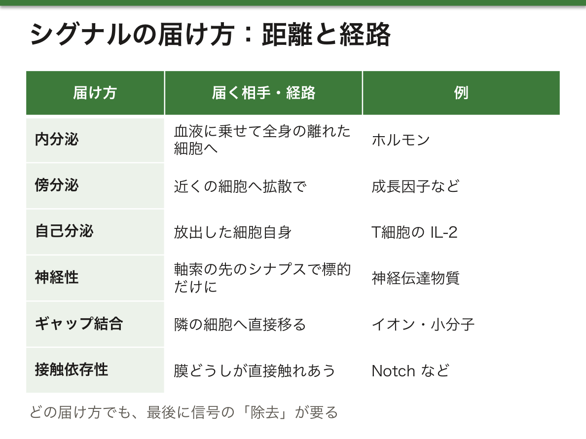
届け方は距離と経路で分けられる。内分泌は血液に乗せて全身の離れた細胞に届ける(ホルモン)。傍分泌は近くの細胞に拡散で届ける(成長因子など)。自己分泌は放出した細胞自身に効く。ギャップ結合を通した信号は、イオンや小分子が隣の細胞へ直接移る。神経性の信号では、神経細胞が軸索を長く伸ばし、標的のすぐそば(シナプス)で神経伝達物質を放出する。接触依存性(ジャクスタクリン)の信号では、送る細胞の膜にある信号分子と、受ける細胞の膜の受容体が直接触れあう。
自己分泌は一見むだに見えるが、増幅のスイッチとして働く。たとえば抗原で刺激されたT細胞は、自分の増殖を促す成長因子(IL-2)をつくり、それを自分で受けとってさらに増える。同じ刺激を受けたT細胞が集団で同時にこれを行うので、反応が一気に大きくなる。
神経性の信号は、内分泌と同じく遠くへ届くが、宛先が1つに絞られている点が違う。ホルモンは全身に配られ、受容体をもつ細胞がすべて反応する。神経伝達物質はシナプスの狭い隙間に放出されるので、標的の細胞だけが反応し、しかも速い。なぜ両方が必要なのかは、「全身にゆっくり一斉に」と「1か所にすばやく正確に」という要求が違うからだと考えるとわかりやすい。接触依存性の信号(Notchなど)は、隣りあう細胞が別々の運命を選ぶ発生の場面で重要になる。
T細胞の自己分泌ループは薬の標的にもなっている。臓器移植で使うカルシニューリン阻害薬(シクロスポリン、タクロリムス)は、T細胞がIL-2の遺伝子を転写するのを抑え、この増幅を止めることで拒絶反応を防ぐ。
English version
Core
Communication by extracellular signal molecules has six steps: (1) synthesis of the signal molecule, (2) release, (3) transport to the target cell, (4) detection by a specific receptor, (5) the cell’s response, and (6) removal of the signal. Removal is easy to forget, but if a signal never disappears, the cell cannot receive the next one.
Delivery is classified by distance and route. Endocrine signals travel in the blood to distant cells (hormones). Paracrine signals diffuse to nearby cells (such as growth factors). Autocrine signals act on the cell that released them. Signals through gap junctions move ions and small molecules directly into the neighbour. In neuronal signalling, a neuron extends a long axon and releases a neurotransmitter right next to its target (the synapse). In contact-dependent (juxtacrine) signalling, a signal molecule on the sending cell’s membrane touches a receptor on the receiving cell’s membrane.
Going further
Autocrine signalling looks wasteful, but it works as an amplifying switch. For example, a T cell stimulated by antigen makes a growth factor (IL-2) that drives its own proliferation, receives it itself, and multiplies further. Many T cells that met the same antigen do this at the same time, so the response grows very quickly.
Neuronal signals reach far, like endocrine signals, but the address is limited to one target. Hormones are sent around the whole body, and every cell with the receptor responds. Neurotransmitters are released into the narrow synaptic gap, so only the target cell responds, and fast. Why are both needed? Because “slowly, everywhere at once” and “quickly and precisely, in one place” are different needs. Contact-dependent signals (such as Notch) matter in development, when neighbouring cells choose different fates.
In the clinic
The T-cell autocrine loop is also a drug target. Calcineurin inhibitors used in organ transplantation (ciclosporin, tacrolimus) block transcription of the IL-2 gene in T cells. They stop this amplification and so prevent rejection.
受容体と二次メッセンジャー — 膜を通れるかで受け手が決まる
信号分子(リガンド、一次メッセンジャー)は、受容体の形の合う部位に、複数の弱い非共有結合ではまりこむ。水に溶けやすい信号分子(ペプチドホルモン、神経伝達物質など)は膜を通れないので細胞膜の受容体に、脂に溶けやすい信号分子(ステロイドホルモンなど)は膜を通り抜けて細胞内の受容体に結合する。
受容体の親和性が高いと、リガンドが低い濃度でもほとんどの受容体が埋まる。親和性が低いと、高い濃度が必要になる。リガンドに長くさらされると細胞は脱感作する。受容体ごと細胞内に取りこむ(受容体を介したエンドサイトーシス)、あるいはアレスチンなどのタンパク質が受容体に結合して信号を止める、という形で起こる。
膜の受容体が活性化すると、多くはエフェクター(たいてい酵素)が二次メッセンジャーをつくり、信号を細胞の中へ運ぶ。水溶性で細胞質を動くもの(cAMP・cGMP・IP3・Ca2+)、脂溶性で膜にとどまるもの(DAG・PIP3)、気体で膜も細胞質も通るもの(NO・CO)の3種類がある。
同じリガンドが、細胞によって違う応答を起こす。アドレナリンは心筋では収縮を強め、腸の平滑筋では逆に緩める。なぜか。応答を決めるのはリガンドではなく、受け手の細胞がもつ受容体の種類と、その下につながる酵素や標的タンパク質だからである。信号は「何をせよ」という命令ではなく「合図」にすぎず、合図を何に変えるかは細胞の側が決めている。逆に、別々のリガンドが同じ応答を起こすこともある。
二次メッセンジャーを挟む意味は増幅にある。1つの受容体が多数のGタンパク質を活性化し、1つの酵素が多数のcAMPをつくり、cAMPに活性化された1つのキナーゼが多数の標的をリン酸化する。各段階で数が掛け算で増えるので、ごく少量のホルモンが細胞全体を動かせる。
気体の二次メッセンジャーは身近な薬につながる。ニトログリセリンは体内でNOを生じ、平滑筋の中でcGMPを増やして血管を広げ、狭心症の発作を止める。cGMPを分解する酵素(PDE5)を阻害するシルデナフィルは、同じ経路のブレーキを外す薬である。この2つを一緒に使うと血圧が下がりすぎるので、併用は禁忌とされる。
English version
Core
A signal molecule (ligand, first messenger) fits into a matching site on its receptor through several weak non-covalent bonds. Water-soluble signals (peptide hormones, neurotransmitters) cannot cross the membrane, so they bind cell-surface receptors. Fat-soluble signals (such as steroid hormones) pass through the membrane and bind intracellular receptors.
If a receptor has high affinity, most receptors are filled even at a low ligand concentration. With low affinity, a high concentration is needed. Long exposure to a ligand makes the cell desensitised. This happens when the receptor is taken into the cell (receptor-mediated endocytosis), or when a protein such as arrestin binds the receptor and stops the signal.
When a surface receptor is activated, an effector (usually an enzyme) often makes a second messenger that carries the signal into the cell. There are three kinds: water-soluble ones that move through the cytoplasm (cAMP, cGMP, IP3, Ca2+), fat-soluble ones that stay in the membrane (DAG, PIP3), and gases that pass through both (NO, CO).
Going further
The same ligand can cause different responses in different cells. Adrenaline makes heart muscle contract more strongly but relaxes smooth muscle in the gut. Why? The response is decided not by the ligand but by the receptor type in the receiving cell and the enzymes and target proteins connected below it. A signal is not an order (“do this”); it is only a cue, and the cell decides what to turn it into. The reverse also happens: different ligands can cause the same response.
The point of a second messenger is amplification. One receptor activates many G proteins, one enzyme makes many cAMP molecules, and one kinase activated by cAMP phosphorylates many targets. The numbers multiply at each step, so a tiny amount of hormone can move the whole cell.
In the clinic
Gaseous second messengers lead to familiar drugs. Nitroglycerin releases NO in the body, raises cGMP in smooth muscle, widens blood vessels and stops an angina attack. Sildenafil blocks the enzyme that breaks down cGMP (PDE5), removing the brake on the same pathway. Together they can lower blood pressure too much, so using them together is contraindicated.
Gタンパク質共役受容体 — 7回膜貫通と、自分で切れるスイッチ
Gタンパク質共役受容体(GPCR)は、膜を7回貫くαヘリックスをもつ受容体で、細胞質側に三量体Gタンパク質(α・β・γの3つのサブユニット)が結びつく。ヒトのゲノムには約800のGPCR遺伝子があり、その約半分は嗅覚の受容体である。視覚・味覚の受容体、多くの神経伝達物質の受容体、代謝を調節するホルモンの受容体の大半がこの型で、アドレナリン、グルカゴン、セロトニン、バソプレシン、ACTHなどを受けとる。
休んでいるGαはGDPを結合している。リガンドが受容体に結合すると受容体の形が変わり、GαがGDPを放してGTPを取りこむ。GTPを結合したGαはβγから離れ、標的のエフェクターを動かす。
| Gα | エフェクター | 結果 |
|---|---|---|
| Gs | アデニル酸シクラーゼを活性化 | cAMP↑ → プロテインキナーゼA |
| Gi | アデニル酸シクラーゼを抑制 | cAMP↓ |
| Gq | ホスホリパーゼCβを活性化 | IP3(Ca2+放出)とDAG(プロテインキナーゼC) |
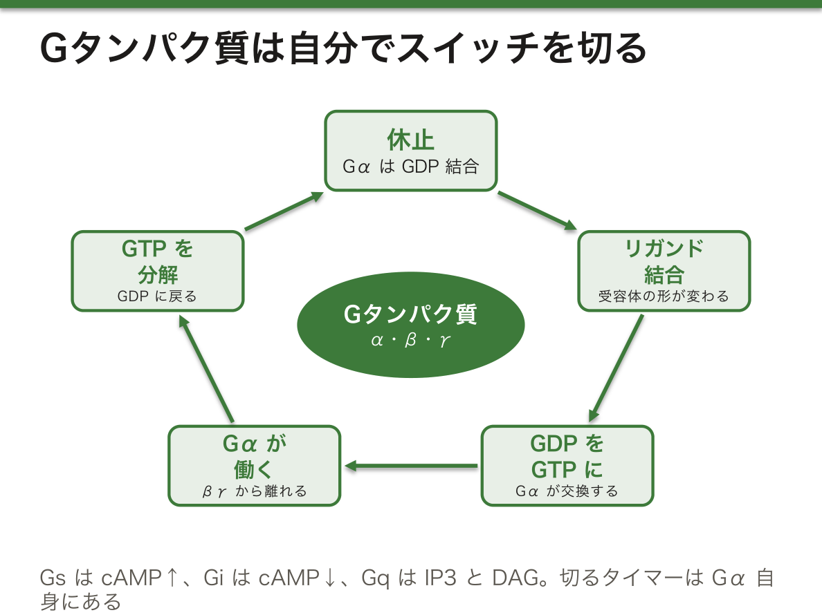
なぜGαは「自分でGTPを分解する」性質をもつのか。GαはGTPを加水分解する酵素(GTPase)でもあり、しばらくするとGTPをGDPに戻して自動的に元の休止状態にもどる。つまり、スイッチを入れるのは受容体だが、切るタイマーはGタンパク質自身に組みこまれている。信号の「除去」がここに作りこまれているおかげで、リガンドが去れば応答は短時間で止まる。このタイマーが壊れると、リガンドが無くても信号が出続ける。
コレラ毒素はGsαをADPリボシル化し、GTPaseの働きを止める。Gsαは「入」のまま固定され、アデニル酸シクラーゼがcAMPを作り続ける。腸の上皮では高いcAMPがCl–チャネルのCFTRを開きっぱなしにし、Cl–に続いて水が大量に腸管内へ出ていく。これが米のとぎ汁様の激しい下痢と脱水の仕組みである。治療の中心が抗菌薬より経口補水なのは、毒素で止まらない糖とNa+の共輸送を使って水を取りもどせるからである。
逆にGPCRを薬で動かす例が、2型糖尿病のGLP-1受容体作動薬(セマグルチドなど)である。β細胞のGLP-1受容体はGsにつながり、cAMPを介してインスリン分泌を強めるが、その効果は血糖が高いときに強く出るので、単独では低血糖を起こしにくい。
English version
Core
A G protein-coupled receptor (GPCR) has seven α-helices that cross the membrane, and a trimeric G protein (α, β and γ subunits) binds it on the cytoplasmic side. The human genome has about 800 GPCR genes, and about half are smell receptors. Most receptors for vision and taste, many neurotransmitter receptors, and most receptors for hormones that control metabolism are of this type. They receive adrenaline, glucagon, serotonin, vasopressin, ACTH and many others.
At rest, Gα holds GDP. When a ligand binds, the receptor changes shape, and Gα releases GDP and takes up GTP. Gα with GTP leaves βγ and acts on a target effector.
| Gα | Effector | Result |
|---|---|---|
| Gs | Activates adenylyl cyclase | cAMP↑ → protein kinase A |
| Gi | Inhibits adenylyl cyclase | cAMP↓ |
| Gq | Activates phospholipase Cβ | IP3 (Ca2+ release) and DAG (protein kinase C) |
Going further
Why does Gα break down its own GTP? Gα is also an enzyme that hydrolyses GTP (GTPase). After a while it turns GTP back into GDP and returns to rest automatically. So the receptor turns the switch on, but the timer that turns it off is built into the G protein itself. Because signal “removal” is built in here, the response stops soon after the ligand leaves. If this timer breaks, the signal continues even with no ligand.
In the clinic
Cholera toxin ADP-ribosylates Gsα and blocks its GTPase activity. Gsα is locked “on”, and adenylyl cyclase keeps making cAMP. In intestinal epithelium, high cAMP holds the CFTR chloride channel open; Cl– leaves into the gut and a large amount of water follows. This is the mechanism of the massive “rice-water” diarrhoea and dehydration. Oral rehydration is the core of treatment, more than antibiotics, because sugar–Na+ cotransport is not stopped by the toxin and can pull water back in.
Drugs can also switch GPCRs on. GLP-1 receptor agonists (such as semaglutide) for type 2 diabetes are an example. The GLP-1 receptor on β cells is coupled to Gs and increases insulin secretion through cAMP. The effect is strong when blood glucose is high, so on their own these drugs rarely cause hypoglycaemia.
受容体チロシンキナーゼ — 二量体になって、互いをリン酸化するAchondroplasia
受容体チロシンキナーゼ(RTK)は、膜を1回だけ貫く受容体で、細胞質側の部分そのものがチロシンキナーゼ(チロシン残基にリン酸をつける酵素)になっている。インスリン、上皮成長因子(EGF)、線維芽細胞成長因子(FGF)、神経栄養因子など、主に成長と代謝にかかわる信号を受けとる。
リガンドが結合すると受容体が2つ寄りあって二量体になり、互いのチロシンをリン酸化しあう(自己リン酸化)。リン酸化されたチロシンは、SH2ドメインをもつ細胞内のタンパク質の「足場」になる。そこから主に3つの経路が始まる。Ras–MAPキナーゼ経路(増殖・分化)、PI3キナーゼ–Akt経路(生存・代謝)、ホスホリパーゼCγによるIP3/DAG経路である。
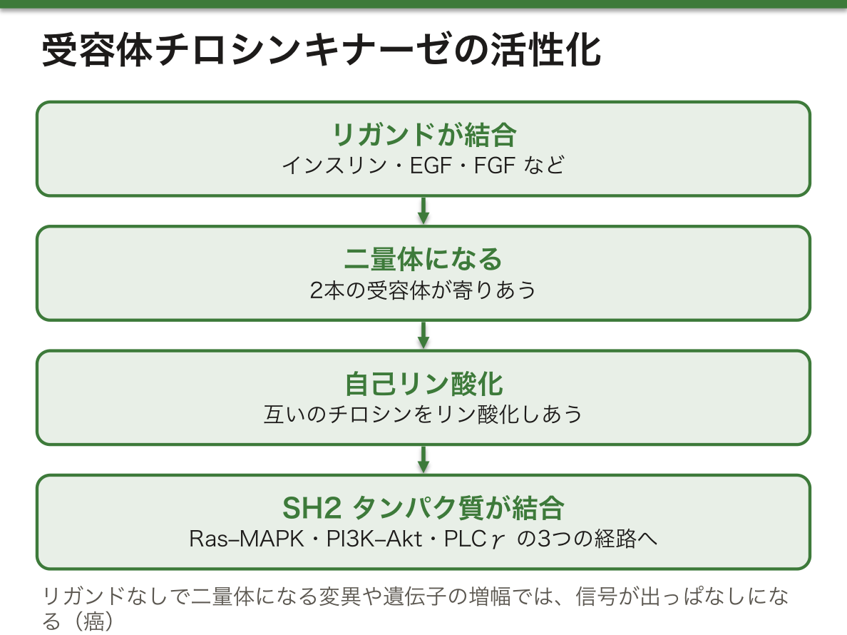
なぜ「二量体になる」ことが活性化の鍵なのか。1本の受容体では、細胞質側のキナーゼの手が届く相手がいない。リガンドが2本の受容体を橋渡しして近づけると、はじめて相手のチロシンをリン酸化できる。だから、リガンドが無くても受容体どうしがくっついてしまう変異や、受容体の数が異常に増えて勝手に出会ってしまう状態(遺伝子の増幅)は、信号が出っぱなしになる。癌でRTKの増幅や活性化型の変異がよく見られるのはこのためである。
軟骨無形成症は四肢が短くなる低身長の代表的な病気で、ほとんど(約98%)がFGFR3の膜貫通部分にある同じ変異(p.Gly380Arg)による。FGFR3は成長板の軟骨細胞に「増えすぎるな」とブレーキをかける受容体である。変異で受容体が常に活性化される機能獲得型になるため、ブレーキが踏まれっぱなしになり、軟骨細胞の増殖が抑えられて長管骨が伸びない。「受容体が活性化しすぎて成長が止まる」という一見逆説的な関係は、FGFR3がブレーキ役だと知れば納得できる。治療薬のボソリチド(CNPの類似体)は、FGFR3の下流のMAPキナーゼ経路を抑えて、ブレーキを弱める。
English version
Core
A receptor tyrosine kinase (RTK) crosses the membrane only once, and its cytoplasmic part is itself a tyrosine kinase (an enzyme that adds phosphate to tyrosine residues). It receives signals mainly for growth and metabolism: insulin, epidermal growth factor (EGF), fibroblast growth factor (FGF), neurotrophins and others.
When a ligand binds, two receptors come together as a dimer and phosphorylate each other’s tyrosines (autophosphorylation). The phosphorylated tyrosines become docking sites for proteins with an SH2 domain. Three main pathways start from there: the Ras–MAP kinase pathway (growth and differentiation), the PI3 kinase–Akt pathway (survival and metabolism), and the IP3/DAG pathway through phospholipase Cγ.
Going further
Why is forming a dimer the key to activation? A single receptor has no partner within reach of its kinase. Only when a ligand bridges two receptors and brings them close can each phosphorylate the other. So a variant that makes receptors stick together without ligand, or a state where there are far too many receptors that meet by chance (gene amplification), keeps the signal on all the time. This is why RTK amplification and activating variants are common in cancer.
In the clinic
Achondroplasia is the classic short-limbed form of short stature. Almost all cases (about 98%) are caused by the same variant (p.Gly380Arg) in the transmembrane part of FGFR3. FGFR3 is a receptor that puts a brake on chondrocyte proliferation in the growth plate. The variant is gain-of-function: the receptor is always active, so the brake is always pressed, chondrocytes do not multiply enough, and long bones do not lengthen. “Too much receptor activity stops growth” sounds strange until you know FGFR3 is a brake. The drug vosoritide (a CNP analogue) weakens the brake by inhibiting the MAP kinase pathway downstream of FGFR3.
イオンチャネル型受容体 — 受容体そのものが門になる
イオンチャネル型受容体(リガンド作動性イオンチャネル)は、受容体とイオンの通り道が同じ分子になっている。4つか5つのサブユニットが輪になって並び、各サブユニットの一部がチャネルの壁をつくる。リガンドが結合すると形が変わって門が開き、イオンが流れこむ。信号の結果は、イオンの流入による膜電位の局所的な変化か、細胞質のCa2+の上昇である。
代表は神経筋接合部のニコチン性アセチルコリン受容体で、5つのサブユニットからなる。アセチルコリンが結合すると中央の孔が開き、陽イオン(主にNa+)が流れこんで筋の膜が脱分極する。ほかにGABAA受容体(Cl–)、グルタミン酸受容体があり、小胞体の膜にはIP3を受けとってCa2+を放出するIP3受容体もある。cGMPや、触覚・伸展といった物理的な刺激で開くチャネルもこの仲間に入る。
イオンチャネル型受容体は4つの型の中でいちばん速い。なぜか。GPCRやRTKのように酵素や二次メッセンジャーを何段も経由せず、門が開いた瞬間にイオンが流れて電気的な変化が起きるからである。応答はミリ秒の単位で始まる。神経と筋のあいだのように「速さ」が最優先される場所でこの型が使われるのは、そのためである。その代わり、増幅の段階が無いので、信号の強さは開いたチャネルの数と開いている時間でほぼ決まる。
神経筋接合部の病気は、遺伝性か自己免疫かで分けて考える。先天性筋無力症候群は遺伝子の変異による。アセチルコリン受容体のα1サブユニット(CHRNA1)の変異でチャネルの開く時間が短すぎる型では、脱分極が閾値に届かず、幼いうちから筋力が落ちる。アセチルコリンエステラーゼを接合部につなぎとめるタンパク質(COLQ)の変異では、分解酵素が働かず受容体が過剰に刺激され続ける。この型では、重症筋無力症の治療薬であるコリンエステラーゼ阻害薬がかえって症状を悪くする。重症筋無力症はアセチルコリン受容体に対する自己抗体で受容体が減る病気で、同じ「筋力低下」でも原因が違えば薬の選び方が逆になる。
English version
Core
In a ligand-gated ion channel receptor, the receptor and the ion pathway are the same molecule. Four or five subunits sit in a ring, and part of each subunit forms the channel wall. When a ligand binds, the shape changes, the gate opens and ions flow in. The result is a local change in membrane potential from ion influx, or a rise in cytoplasmic Ca2+.
The classic example is the nicotinic acetylcholine receptor at the neuromuscular junction, made of five subunits. When acetylcholine binds, the central pore opens, cations (mainly Na+) flow in, and the muscle membrane depolarises. Others include the GABAA receptor (Cl–) and glutamate receptors. The ER membrane also has the IP3 receptor, which releases Ca2+ when it binds IP3. Channels opened by cGMP or by physical stimuli such as touch and stretch also belong here.
Going further
Ion channel receptors are the fastest of the four types. Why? Unlike GPCRs and RTKs, they do not pass through several steps of enzymes and second messengers. Ions flow the moment the gate opens, causing an electrical change. The response begins within milliseconds. This is why this type is used where speed comes first, such as between nerve and muscle. On the other hand, there is no amplification step, so signal strength depends mostly on how many channels open and for how long.
In the clinic
Diseases of the neuromuscular junction are best sorted into genetic or autoimmune. Congenital myasthenic syndromes are caused by gene variants. In the type with variants in the α1 subunit of the acetylcholine receptor (CHRNA1) where the channel opens for too short a time, depolarisation does not reach threshold, and muscle weakness starts in early childhood. In the type with variants in COLQ, the protein that anchors acetylcholinesterase at the junction, the breakdown enzyme does not work and receptors are overstimulated. In this type, cholinesterase inhibitors, the treatment for myasthenia gravis, make symptoms worse. Myasthenia gravis is a disease in which autoantibodies against the acetylcholine receptor reduce the number of receptors. The same “muscle weakness” can need opposite drugs when the cause differs.
細胞内受容体 — ホルモンが転写因子を直接動かす
脂に溶けやすい信号分子は膜を通り抜け、細胞質や核の中の細胞内受容体に結合する。ステロイドホルモン、甲状腺ホルモン、レチノイド、ビタミンDの受容体がこの型である(核内受容体とも呼ばれる)。
受容体は3つの部分からなる。リガンド結合ドメイン、DNAに結合するDNA結合ドメイン(亜鉛フィンガーをもつ)、転写を始めさせる転写活性化ドメインである。リガンドが結合した受容体は、標的遺伝子のプロモーター領域にある決まった配列(ホルモンではホルモン応答配列)に結合し、その遺伝子の転写を増やしたり減らしたりする。つまりこの受容体は、リガンドで動く転写因子である。
細胞内受容体の効果は現れるのが遅く、長く続く。なぜか。応答が「すでにあるタンパク質の形を変える」のではなく、「遺伝子を転写し、mRNAを翻訳して、新しいタンパク質をつくる」ところまで進んで初めて出るからである。これには数時間から数日かかり、できたタンパク質はしばらく残る。ステロイド薬の効果が投与してすぐには出ず、やめてもしばらく続くのは、この仕組みのためである。膜の受容体が秒から分の世界で働くのと対照的である。
アンドロゲン不応症は、X染色体にあるアンドロゲン受容体(AR)の遺伝子の変異で起こる。核型は46,XYで精巣があり、テストステロンも作られるが、受容体が働かないので外性器は女性型に発達する。一方、子宮が無いのは、精巣のSertoli細胞が出す抗Müller管ホルモンがアンドロゲンとは別の受容体で働き、Müller管を退縮させるからである。精巣は腹腔内や鼠径部にとどまることが多い。ホルモンも受容体も正常に見えて「受け手だけが壊れている」ことが、症状の組み合わせを説明する。
受容体を狙う薬の例が乳癌のタモキシフェンで、乳腺ではエストロゲン受容体に結合してエストロゲンの作用を妨げる。エストロゲン受容体が陽性かどうかを調べてから使うのは、受容体の無い癌には効かないからである。
English version
Core
Fat-soluble signal molecules pass through the membrane and bind intracellular receptors in the cytoplasm or nucleus. Receptors for steroid hormones, thyroid hormones, retinoids and vitamin D are of this type (also called nuclear receptors).
The receptor has three parts: a ligand-binding domain, a DNA-binding domain (with zinc fingers), and a transcription-activating domain. The receptor with its ligand binds a fixed sequence in the promoter region of target genes (for hormones, a hormone response element) and raises or lowers transcription of that gene. In other words, this receptor is a transcription factor switched on by a ligand.
Going further
The effects of intracellular receptors appear slowly and last a long time. Why? The response does not come from changing the shape of existing proteins. It comes only after the gene is transcribed, the mRNA is translated, and new protein is made. This takes hours to days, and the new protein remains for a while. This is why steroid drugs do not work right after a dose and their effect continues for some time after they are stopped. Surface receptors, in contrast, work within seconds to minutes.
In the clinic
Androgen insensitivity syndrome is caused by variants in the gene for the androgen receptor (AR) on the X chromosome. The karyotype is 46,XY, testes are present and testosterone is made, but the receptor does not work, so the external genitalia develop as female. There is no uterus because anti-Müllerian hormone from Sertoli cells acts through a different receptor and still makes the Müllerian ducts regress. The testes often stay in the abdomen or groin. Normal hormones with “only the receiver broken” explains this mix of features.
A drug that targets this type of receptor is tamoxifen for breast cancer. In breast tissue it binds the oestrogen receptor and blocks the action of oestrogen. Doctors test whether the cancer is oestrogen-receptor positive first, because the drug does not work on a cancer without the receptor.
まとめ
| 細胞外基質 — 細胞が自分で編んで、自分を支える足場 | 細胞外基質(ECM)は、細胞が分泌したタンパク質と多糖が細胞のまわりに編みあげた網である。 |
|---|---|
| 細胞間結合 — 塞ぐ・通す・つかむの3つの仕事 | 多くの組織では、細胞どうしが専用のタンパク質の複合体で直接つながっている。 |
| シグナルの届き方 — 距離と経路で分ける | 細胞外の信号分子を使った情報のやりとりは、6つの段階からなる。 |
| 受容体と二次メッセンジャー — 膜を通れるかで受け手が決まる | 信号分子(リガンド、一次メッセンジャー)は、受容体の形の合う部位に、複数の弱い非共有結合ではまりこむ。 |
| Gタンパク質共役受容体 — 7回膜貫通と、自分で切れるスイッチ | Gタンパク質共役受容体(GPCR)は、膜を7回貫くαヘリックスをもつ受容体で、細胞質側に三量体Gタンパク質(α・β・γの3つのサブユニット)が結びつく。 |
| 受容体チロシンキナーゼ — 二量体になって、互いをリン酸化する | 受容体チロシンキナーゼ(RTK)は、膜を1回だけ貫く受容体で、細胞質側の部分そのものがチロシンキナーゼ(チロシン残基にリン酸をつける酵素)になっている。 |
| イオンチャネル型受容体 — 受容体そのものが門になる | イオンチャネル型受容体(リガンド作動性イオンチャネル)は、受容体とイオンの通り道が同じ分子になっている。 |
| 細胞内受容体 — ホルモンが転写因子を直接動かす | 脂に溶けやすい信号分子は膜を通り抜け、細胞質や核の中の細胞内受容体に結合する。 |
このノートは医学生が授業の内容を自分の言葉でまとめたもので、診療の判断に使うものではありません。誤りに気づいたらお知らせください。