核は細胞の「司令塔」と呼ばれるが、実際の仕事は2つに絞れる。長さ2 mにもなるDNAを直径数µmの中に収納することと、必要な部分だけを読める状態に開き、分裂のときには正確に2つに分けて渡すことである。この章は、その2つを支える構造——核膜と核膜孔、核ラミナ、ヌクレオソームとクロマチン、分裂期の染色体、テロメア、核小体——を順に見ていく。
覚え方のこつは、構造を見たら必ず「壊れたら何が起きるか」を考えることである。核膜を支える網が弱ければ筋肉が傷み、ヘテロクロマチンのメチル化が抜ければ染色体が不安定になり、染色体の数がずれれば発生が変わる。後半では核型の読み方と、核の外にあるもう1組の染色体(細菌とミトコンドリアの環状DNA)も扱う。
- 核膜と核膜孔複合体 — 2枚の膜と、見張りのついた門
- 核ラミナとLINC複合体 — 核の形を支える網と、細胞骨格への綱
- ヒストンとヌクレオソーム — 負のDNAを正のタンパク質に巻く
- ユークロマチンとヘテロクロマチン — 開いた部分と、閉じた部分
- 分裂期の染色体と核小体 — 分けて渡す形と、リボソームの工場
- テロメアの構造と働き — 染色体の端を「切れ目」と見せない帽子
- 核型と性染色体 — 46本の読み方と、Barr小体の数え方
- 原核の染色体とミトコンドリアの染色体 — 核の外にある環状DNA
- 核膜と核膜孔複合体 — 2枚の膜と、見張りのついた門Involucrum nucleare
- 核ラミナとLINC複合体 — 核の形を支える網と、細胞骨格への綱Lamina nuclearis
- ヒストンとヌクレオソーム — 負のDNAを正のタンパク質に巻く
- ユークロマチンとヘテロクロマチン — 開いた部分と、閉じた部分Heterochromatinum
- 分裂期の染色体と核小体 — 分けて渡す形と、リボソームの工場Chromosoma
- テロメアの構造と働き — 染色体の端を「切れ目」と見せない帽子Telomerum
- 核型と性染色体 — 46本の読み方と、Barr小体の数え方
- 原核の染色体とミトコンドリアの染色体 — 核の外にある環状DNAMitochondrion
- まとめ
核膜と核膜孔複合体 — 2枚の膜と、見張りのついた門Involucrum nucleare
核膜は外膜と内膜の2枚の膜でできている。外膜は粗面小胞体とつながっていて表面にリボソームがつき、小胞体の一部とみなせる。内膜には独自の膜タンパク質があり、内側の核ラミナやクロマチンと結合する。2枚の間のすきま(核周囲腔)は、小胞体の内腔と続いている。
核膜には核膜孔複合体(NPC)という大きなタンパク質の門が多数あいている。ヌクレオポリンという一群のタンパク質が8回対称の輪をつくり、その中心が核と細胞質をつなぐ通路になる。イオンや小さな分子(おおよそ40 kDa以下)は自由に拡散で通るが、タンパク質やRNAのような大きな分子は輸送受容体に運ばれて通る。核に入る分子はインポーチン、核から出る分子はエクスポーチンが結合して運ぶ。核膜孔の数は一定ではなく、活発に働く細胞ほど多い。
なぜ輸送は一方通行になれるのか。鍵は低分子量のGタンパク質Ranである。核の中ではRanをGTP型にする酵素がクロマチンに結合していて、細胞質ではGTPを分解させる酵素が働く。そのため核内にはRan-GTPが多く、細胞質には少ないという勾配ができる。インポーチンは核内でRan-GTPに出会うと荷物を放し、エクスポーチンは逆にRan-GTPと結合しているときだけ荷物をつかむ。この勾配があるから、荷物は決まった向きにだけ運ばれる。核に入るべきタンパク質は、核移行シグナルという短いアミノ酸配列を名札としてもっている。
核膜孔は、核へ入る必要があるウイルスの通り道でもある。また、ヌクレオポリンの遺伝子が別の遺伝子と融合する染色体転座は、一部の急性骨髄性白血病の原因になる。輸送の門そのものが、感染と腫瘍の両方の舞台になっている。
English version
Core
The nuclear envelope has two membranes, outer and inner. The outer membrane is continuous with the rough ER, carries ribosomes, and can be seen as part of the ER. The inner membrane has its own membrane proteins that bind the nuclear lamina and chromatin inside. The gap between them (the perinuclear space) is continuous with the ER lumen.
The envelope has many nuclear pore complexes (NPCs), large protein gates. Proteins called nucleoporins form a ring with eight-fold symmetry, and its centre is a channel between nucleus and cytoplasm. Ions and small molecules (roughly under 40 kDa) pass freely by diffusion. Large molecules such as proteins and RNA are carried by transport receptors: importins bring molecules in, and exportins take them out. The number of pores is not fixed; more active cells have more pores.
Going further
Why can transport go only one way? The key is a small G protein, Ran. Inside the nucleus, an enzyme that loads Ran with GTP sits on chromatin; in the cytoplasm, another enzyme makes Ran break down its GTP. So there is a lot of Ran-GTP in the nucleus and little in the cytoplasm. An importin lets go of its cargo when it meets Ran-GTP in the nucleus. An exportin does the opposite: it holds cargo only while it is bound to Ran-GTP. This gradient makes cargo move in one fixed direction. Proteins that belong in the nucleus carry a short amino acid sequence, the nuclear localisation signal, as a name tag.
In the clinic
Nuclear pores are also the route for viruses that must reach the nucleus. Chromosome translocations that fuse a nucleoporin gene with another gene cause some cases of acute myeloid leukaemia. The transport gate itself is a stage for both infection and cancer.
核ラミナとLINC複合体 — 核の形を支える網と、細胞骨格への綱Lamina nuclearis
核ラミナは内膜の内側に張られた網で、ラミンというタンパク質の中間径フィラメントでできている。ラミンには、1つの遺伝子LMNAから作り分けられるA型(ラミンAとC)と、別の遺伝子(LMNB1・LMNB2)からできるB型がある。ラミナは内膜の膜タンパク質やクロマチンと結合し、核の形を保つだけでなく、DNA複製、転写、クロマチンの配置、細胞周期、分化、核の移動、アポトーシスと、核のほとんどの仕事にかかわる。
LINC複合体は、外膜のネスプリンと内膜のSUNタンパク質が核周囲腔の中で手をつないだもので、細胞骨格と核の中の骨組みを結ぶ綱である。核を細胞の中の決まった位置に置き、中心体を核のそばに保ち、細胞の外からかかった力を核まで伝える。
ラミナやLINCにかかわるタンパク質の遺伝子が変わって起きる病気をまとめてラミノパチーと呼ぶ。代表がEmery-Dreifuss型筋ジストロフィーで、内膜のタンパク質エメリンの遺伝子(X連鎖性)やLMNA(常染色体顕性)の変化で起きる。小児期から関節が固くなり(拘縮)、筋力がゆっくり落ち、成人で心臓の病気が出る。
なぜ全身の細胞にあるタンパク質の病気なのに、筋肉と心臓が傷むのか。エメリン–ラミン–LINCの複合体は、核膜を機械的な力から守る補強材である。収縮のたびに強い力がかかる骨格筋と心筋の細胞では、補強が弱いと核膜が繰り返し傷つき、細胞が持ちこたえられない。力のかからない細胞では、同じ欠陥があっても目立たない。
Emery-Dreifuss型では心臓の伝導障害が突然死の原因になるので、筋の症状が軽くても心電図で追い、必要ならペースメーカーや植込み型除細動器を使う。LMNAの1塩基の変化で異常なラミンA(プロゲリン)ができるHutchinson-Gilford早老症も、同じ遺伝子の病気である。1つの遺伝子が筋・心臓・脂肪・老化と全く違う病気を起こす例として知っておくとよい。
English version
Core
The nuclear lamina is a net under the inner membrane. It is made of intermediate filaments of proteins called lamins. There are A-type lamins (A and C), both made from one gene, LMNA, and B-type lamins from other genes (LMNB1, LMNB2). The lamina binds inner-membrane proteins and chromatin. It keeps the shape of the nucleus and takes part in almost every nuclear job: DNA replication, transcription, chromatin layout, the cell cycle, differentiation, nuclear movement and apoptosis.
The LINC complex is formed when nesprin in the outer membrane and SUN proteins in the inner membrane join hands across the perinuclear space. It is a rope linking the cytoskeleton to the inner frame of the nucleus. It holds the nucleus in place, keeps the centrosome next to it, and passes force from outside the cell to the nucleus.
Going further
Diseases caused by changes in genes for lamina and LINC proteins are called laminopathies. A key example is Emery-Dreifuss muscular dystrophy, caused by changes in the gene for the inner-membrane protein emerin (X-linked) or in LMNA (autosomal dominant). Joints stiffen from childhood (contractures), muscles weaken slowly, and heart disease appears in adults.
Why do skeletal and heart muscle suffer, when the protein is in every cell? The emerin–lamin–LINC complex protects the nuclear envelope against mechanical force. In skeletal and heart muscle cells, strong force comes with every contraction. If the support is weak, the envelope is damaged again and again and the cell cannot survive. Cells that do not face such force show little effect from the same defect.
In the clinic
In Emery-Dreifuss dystrophy, heart conduction block can cause sudden death. Even if muscle symptoms are mild, patients are followed with ECGs and may need a pacemaker or an implantable defibrillator. Hutchinson-Gilford progeria, where one base change in LMNA produces an abnormal lamin A (progerin), is a disease of the same gene. It is a good example of one gene causing very different diseases of muscle, heart, fat and ageing.
ヒストンとヌクレオソーム — 負のDNAを正のタンパク質に巻く
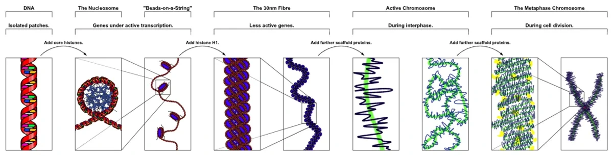
核の中身(核質)の主成分は、核酸とタンパク質の複合体ヌクレオタンパク質である。タンパク質は2種類に分かれる。非ヒストンタンパク質は酸性の小さなタンパク質で、DNAポリメラーゼやRNAポリメラーゼなどの酵素を含み、種類は1,000を超えるが量は少ない。ヒストンはリシンとアルギニンなどの塩基性アミノ酸に富み、正に帯電している。主な種類はH1・H2A・H2B・H3・H4の5つである。
DNAの大部分はヌクレオソームに巻かれている。H2A・H2B・H3・H4を2個ずつ集めたヒストン八量体に、147塩基対のDNAが約1.65回巻きつく。ヌクレオソームの間をつなぐリンカーDNA(数十塩基対)にはH1が結合し、出入り口を閉じる。ヌクレオソームにH1が加わったものをクロマトソームという。
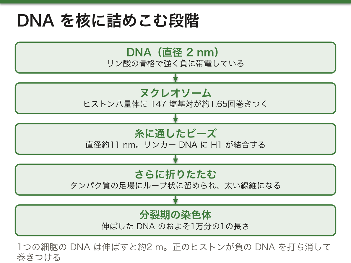
なぜヒストンは正に帯電しているのか。DNAの骨格はリン酸が並ぶので強く負に帯電している。負の電荷どうしは反発するので、DNAはそのままでは小さく折りたためない。正のヒストンがその電荷を打ち消し、イオン結合で巻きつけることで、はじめて密に詰めこめる。逆にいえば、ヒストンの正電荷を弱めれば巻きはゆるむ。リシンにアセチル基をつけると正電荷が消え、DNAが緩んで転写が起きやすくなる——これがエピジェネティクスの中心の仕組みの1つになる。
詰めこみには段階がある。直径2 nmのDNAがヌクレオソームに巻かれて約11 nmの「糸に通したビーズ」になり、それがさらに折りたたまれ、タンパク質の足場にループ状に留められて太い線維になる。分裂期の染色体では、伸ばしたDNAに比べておよそ1万分の1の長さにまで縮む。ヒトの1つの細胞のDNAを伸ばすと約2 mになるので、この段階的な詰めこみなしには核に収まらない。
English version
Core
The main component of the nuclear contents (nucleoplasm) is nucleoprotein, a complex of nucleic acid and protein. The proteins are of two kinds. Non-histone proteins are small acidic proteins, including enzymes such as DNA and RNA polymerases. There are over 1,000 kinds, but each is present in small amounts. Histones are rich in basic amino acids such as lysine and arginine, so they carry a positive charge. The main types are H1, H2A, H2B, H3 and H4.
Most DNA is wrapped into nucleosomes. Two each of H2A, H2B, H3 and H4 form a histone octamer, and 147 base pairs of DNA wrap around it about 1.65 times. The linker DNA between nucleosomes (tens of base pairs) binds H1, which closes the entry and exit points. A nucleosome plus H1 is a chromatosome.
Going further
Why are histones positively charged? The DNA backbone is a row of phosphates, so it is strongly negative. Negative charges repel each other, so DNA alone cannot fold tightly. Positive histones cancel the charge and hold the DNA by ionic bonds, which makes dense packing possible. The other side of this: weaken the histone’s positive charge and the wrapping loosens. Adding an acetyl group to lysine removes its positive charge, loosens the DNA and makes transcription easier. This is one of the central mechanisms of epigenetics.
Packing happens in steps. DNA, 2 nm wide, is wrapped into nucleosomes to make an 11 nm “beads on a string” fibre. This folds further and is held in loops on a protein scaffold, forming thicker fibres. In a mitotic chromosome, DNA is shortened to about one ten-thousandth of its stretched length. The DNA of one human cell is about 2 m long when stretched, so it could not fit in the nucleus without this stepwise packing.
ユークロマチンとヘテロクロマチン — 開いた部分と、閉じた部分Heterochromatinum
間期の核の中で、DNAとタンパク質の複合体(クロマチン)は長く伸びた状態にあり、間期染色体とも呼ばれる。詰まり方で2つに分かれる。ユークロマチンはヌクレオソームの集まりが緩く、遺伝子がよく発現し、S期の早い時期に複製される。ヘテロクロマチンは密に集まり、発現はほとんど無く、遅い時期に複製される。テロメアやセントロメアの周りに多い。
| 構成的ヘテロクロマチン | 条件的ヘテロクロマチン | |
|---|---|---|
| 中身 | 遺伝子を含まない高度な反復配列(サテライトDNA) | ふつうの遺伝子を含む領域 |
| いつ・どこで | すべての組織・すべての時期で同じ | 特定の細胞・組織でだけ閉じる |
| 例 | 1・9・16・19番とY染色体に多い | 女性の体細胞の不活性X(Barr小体) |
| 可逆性 | 安定 | 開き直して転写されうる |
ヘテロクロマチンのDNAはメチル化され、ヒストンはアセチル化が少ない。役割は、セントロメアの働きを支えること、核の中の区画づくり、そして遺伝子を黙らせることである。
間期の染色体は核の中でばらばらに混ざっているのではなく、それぞれが決まった区画(染色体テリトリー)を占める。各染色体はさらにTAD(トポロジカル関連ドメイン)という、内部どうしがよく接触するまとまりに分かれる。転写が抑えられたTADは核の周縁、ラミナのそばに、活発なTADは核の内側に集まる傾向がある。つまり「どこに置かれているか」自体が「読まれるかどうか」の情報になっている。電子顕微鏡で核の縁が黒く濃く見えるのは、この閉じたクロマチンがラミナに張りついているからである。
ヘテロクロマチンを保つメチル化が抜けると何が起きるかを示すのが、ICF症候群(免疫不全・セントロメア不安定・顔貌異常)である。DNAメチル基転移酵素の遺伝子DNMT3Bの変化で、1・9・16番のセントロメア近くのサテライトDNAのメチル化が失われ、そこの染色体が伸び、切れ、組み換わる。セントロメアの構造が乱れると、リンパ球の成熟や移動にかかわる遺伝子の発現も間接的に崩れ、免疫グロブリンが作れなくなる。
English version
Core
In the interphase nucleus, the complex of DNA and protein (chromatin) is long and stretched out; it is also called the interphase chromosome. It comes in two packing states. Euchromatin has loosely gathered nucleosomes; its genes are well expressed, and it replicates early in S phase. Heterochromatin is tightly packed; it has little or no expression and replicates late. It is common around telomeres and centromeres.
| Constitutive heterochromatin | Facultative heterochromatin | |
|---|---|---|
| Content | Highly repetitive sequences with no genes (satellite DNA) | Regions with ordinary genes |
| When / where | The same in every tissue at every stage | Closed only in certain cells or tissues |
| Example | Abundant on chromosomes 1, 9, 16, 19 and Y | The inactive X in female somatic cells (Barr body) |
| Reversible? | Stable | Can reopen and be transcribed |
Heterochromatin DNA is methylated, and its histones have little acetylation. Its roles are supporting centromere function, organising regions of the nucleus, and silencing genes.
Going further
Interphase chromosomes are not mixed up at random in the nucleus. Each one takes up its own region (chromosome territory). Each chromosome is further divided into TADs (topologically associating domains), units whose parts touch each other often. TADs with repressed transcription tend to sit at the edge of the nucleus, next to the lamina; active TADs tend to sit in the interior. So where a region sits is itself information about whether it is read. In electron microscopy, the edge of the nucleus looks dark because this closed chromatin is attached to the lamina.
In the clinic
ICF syndrome (immunodeficiency, centromeric instability, facial anomalies) shows what happens when the methylation that keeps heterochromatin closed is lost. A change in the DNA methyltransferase gene DNMT3B causes loss of methylation of satellite DNA near the centromeres of chromosomes 1, 9 and 16. These regions stretch, break and rearrange. The disturbed centromere structure also indirectly upsets genes for lymphocyte maturation and movement, and the patient cannot make enough immunoglobulin.
分裂期の染色体と核小体 — 分けて渡す形と、リボソームの工場Chromosoma
S期にDNAが複製されると、同じ染色体のコピーが2本できる。これが姉妹染色分体で、複製のときに結合するコヒーシンが分裂まで2本をつなぎとめる。分裂が始まるとコンデンシンが染色体を巻き締め、光学顕微鏡で見える分裂期(中期)染色体になる。
中期染色体のくびれがセントロメアである。独特の配列(CEN DNA)、ヒストンの変異体、専用のタンパク質からなり、ここに動原体(キネトコア)という大きなタンパク質複合体ができて紡錘体の微小管と結ぶ。セントロメアの位置で、中部着糸型(メタセントリック)・次中部着糸型(サブメタセントリック)・端部着糸型(アクロセントリック)に分ける。端部着糸型の短腕の先には小さな塊(付随体、サテライト)があり、それをつなぐ細いくびれを二次狭窄と呼ぶ。
ヒトの染色体は大きさとセントロメアの位置で7群に分けられる。
| 群 | 番号 | 特徴 |
|---|---|---|
| A | 1〜3 | 大型、中部〜次中部着糸型 |
| B | 4・5 | 大型、次中部着糸型 |
| C | 6〜12、X | 中型、次中部着糸型が中心 |
| D | 13〜15 | 中型、端部着糸型、付随体あり |
| E | 16〜18 | やや小型、中部〜次中部着糸型 |
| F | 19・20 | 小型、中部着糸型 |
| G | 21・22、Y | 小型、端部着糸型(Yには付随体が無い) |
端部着糸型の二次狭窄には、18S・5.8S・28S rRNAの遺伝子が何十も並ぶ核小体形成部位(NOR)がある。ヒトでは13・14・15・21・22番の短腕にあり、分裂が終わるとここから核小体が組み立て直される。核小体は膜をもたず、rRNA遺伝子と転写酵素のある線維中心、つくられたrRNAが加工される密線維成分、リボソームの部品が組み立てられる顆粒成分の3層からなる。なぜ核小体は膜なしでまとまれるのか——rRNA遺伝子が転写されている場所に、加工と組み立ての分子が自然に集まるからで、転写を止めると核小体はばらける。
English version
Core
When DNA is copied in S phase, each chromosome has two copies: sister chromatids. Cohesin, which binds during replication, holds the two together until division. When division starts, condensin coils the chromosome tightly and it becomes the mitotic (metaphase) chromosome seen in a light microscope.
The narrow waist of a metaphase chromosome is the centromere. It has a special sequence (CEN DNA), histone variants and its own proteins. Here a large protein complex, the kinetochore, forms and links to spindle microtubules. By centromere position, chromosomes are metacentric, submetacentric or acrocentric. The short arm of an acrocentric chromosome ends in a small knob (satellite); the thin waist that links it is the secondary constriction.
Going further
Human chromosomes are sorted into seven groups by size and centromere position.
| Group | Numbers | Features |
|---|---|---|
| A | 1–3 | Large, metacentric to submetacentric |
| B | 4, 5 | Large, submetacentric |
| C | 6–12, X | Medium, mostly submetacentric |
| D | 13–15 | Medium, acrocentric with satellites |
| E | 16–18 | Fairly small, metacentric to submetacentric |
| F | 19, 20 | Small, metacentric |
| G | 21, 22, Y | Small, acrocentric (Y has no satellites) |
The secondary constriction of acrocentric chromosomes holds a nucleolus organiser region (NOR), where dozens of 18S, 5.8S and 28S rRNA genes lie in a row. In humans these are on the short arms of chromosomes 13, 14, 15, 21 and 22, and the nucleolus is rebuilt from them after division. The nucleolus has no membrane. It has three parts: the fibrillar centre with rRNA genes and transcription enzymes, the dense fibrillar component where new rRNA is processed, and the granular component where ribosome parts are assembled. Why can the nucleolus hold together without a membrane? Molecules for processing and assembly gather by themselves where rRNA genes are being transcribed. If transcription stops, the nucleolus falls apart.
テロメアの構造と働き — 染色体の端を「切れ目」と見せない帽子Telomerum
ヒトのテロメアはTTAGGGの二本鎖の繰り返しが数〜十数kb続き、最後に一本鎖のTTAGGGが100〜200塩基ほど3′側に突き出して終わる。ここに専用のタンパク質の一群が結合する。二本鎖の部分にはTRF1とTRF2、一本鎖の突き出しにはPOT1がつく。突き出した一本鎖は、自分の二本鎖部分の中に潜りこんで輪(Tループ)をつくり、端そのものを隠す。
テロメアの働きは大きく2つある。1つはキャップ(帽子)として端を守ること、もう1つは複製のたびに少しずつ短くなることで、細胞の分裂回数を「数える」ことである。テロメラーゼの活性は幹細胞や胚の細胞では高く、ふつうの体細胞ではほとんど無い。
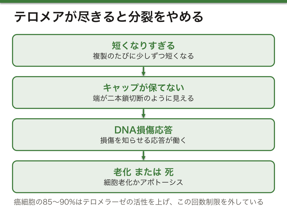
なぜ端を隠す必要があるのか。細胞にとって、DNAの「端」は本来二本鎖切断(壊れたDNA)と同じ見え方をする。むき出しの端があれば、DNA損傷への応答が働き、修復の酵素が別の染色体の端と勝手につないでしまう(末端融合)。融合した染色体は分裂のとき両極に引っぱられて切れ、さらに不安定になる。Tループと結合タンパク質は、この誤った「修復」をさせないための仕掛けである。テロメアが短くなりすぎてキャップが保てなくなると、細胞は損傷応答を起こし、分裂をやめる(細胞老化)かアポトーシスで死ぬ。これは、不安定になった細胞を増やさないための安全装置でもある。
癌細胞の85〜90%ではテロメラーゼの活性が上がっていて、短くなる仕組みを打ち消して分裂を続ける(残りは別の組み換えの仕組みでテロメアを保つ)。つまり「無限に分裂できる」ことは癌の条件の1つであり、テロメアはその鍵になっている。テロメラーゼを阻害する薬(イメテルスタット)は、骨髄異形成症候群の治療に使われ始めている。
English version
Core
Human telomeres are double-stranded TTAGGG repeats a few to over ten kb long, ending in a single-stranded TTAGGG overhang of about 100–200 bases on the 3′ side. A set of special proteins binds here: TRF1 and TRF2 on the double-stranded part and POT1 on the single-stranded overhang. The overhang tucks itself into the double-stranded part to form a loop (the T-loop), which hides the very end.
Telomeres have two main jobs. One is to protect the end as a cap. The other is to get a little shorter with each replication, so they “count” how many times a cell divides. Telomerase activity is high in stem cells and embryonic cells, and almost absent in ordinary somatic cells.
Going further
Why must the end be hidden? To a cell, an end of DNA looks just like a double-strand break (broken DNA). An exposed end triggers the DNA damage response, and repair enzymes may join it to the end of another chromosome (end fusion). A fused chromosome is pulled to both poles at division and breaks, becoming even more unstable. The T-loop and its binding proteins exist to block this wrong “repair”. When telomeres become too short to keep the cap, the cell starts a damage response and either stops dividing (cellular senescence) or dies by apoptosis. This is also a safety device to stop unstable cells from multiplying.
In the clinic
In 85–90% of cancers, telomerase activity is raised. It cancels the shortening and lets the cells keep dividing (the rest keep telomeres by a separate recombination mechanism). So unlimited division is one condition for cancer, and telomeres are a key to it. A telomerase-inhibiting drug (imetelstat) has started to be used for myelodysplastic syndrome.
核型と性染色体 — 46本の読み方と、Barr小体の数え方
中期染色体の数・大きさ・形のそろいを核型といい、生物の種ごとに決まっている。ヒトの体細胞は二倍体(2n=46)、生殖細胞は一倍体(n=23)である。性と性に関連する形質を決める性染色体はXとYで、残りの22対は男女共通の常染色体である。
| 女性 | 男性 | |
|---|---|---|
| 体細胞 | 46,XX | 46,XY |
| 配偶子 | 23,X | 23,X または 23,Y |
| Barr小体 | 1個(1本のXが不活性化) | 無し |
核型は「総数、性染色体、変化」の順に書く。たとえば21番が1本多い男性は47,XY,+21、X染色体が1本だけの女性は45,Xとなる。
XとYは大部分で相同ではないが、両端に偽常染色体領域(PAR1・PAR2)という相同な短い区間があり、ここは常染色体のようにふるまって減数分裂で組み換えを起こす。男性の減数分裂でXとYが対をつくって正しく分かれられるのは、この区間があるからである。
Barr小体の数は「X染色体の数−1」になる。理由は、体細胞は活性なXを1本だけ残し、それ以外のXをすべて不活性化するという数え方をしているからである。だから47,XXYでは1個、48,XXXYでは2個、45,Xや46,XYでは0個になる。間期の核を特殊な染色で見ると、不活性Xは核膜の内側に張りついた濃い点として見える。
出生前の染色体検査では、羊水を採取して胎児由来の細胞を培養し、分裂中期で止めて染色体を染め、核型を並べて数と形を調べる。結果が出るまで日数がかかるため、特定の染色体の数だけを早く調べる方法を組み合わせることも多い。また、PARの遺伝子は不活性化を免れるので、Xが1本しかないTurner症候群では、PARにあるSHOX遺伝子が1コピー分足りないことが低身長の一因になる。「不活性化されるはずのXが、なぜ1本欠けると症状が出るのか」への答えの1つがここにある。
English version
Core
The set of metaphase chromosomes, by number, size and shape, is the karyotype, and it is fixed for each species. Human somatic cells are diploid (2n = 46), and germ cells are haploid (n = 23). The sex chromosomes, X and Y, decide sex and sex-linked traits; the other 22 pairs are autosomes, shared by both sexes.
| Female | Male | |
|---|---|---|
| Somatic cell | 46,XX | 46,XY |
| Gamete | 23,X | 23,X or 23,Y |
| Barr body | One (one X is inactivated) | None |
A karyotype is written as “total number, sex chromosomes, change”. A male with an extra chromosome 21 is 47,XY,+21; a female with only one X is 45,X.
Going further
X and Y are mostly not homologous, but at both ends they share short matching regions, the pseudoautosomal regions (PAR1, PAR2). These behave like autosomes and recombine in meiosis. They are why X and Y can pair and separate correctly in male meiosis.
The number of Barr bodies is “the number of X chromosomes minus one”. The reason is that a somatic cell keeps only one X active and inactivates all the others. So 47,XXY has one, 48,XXXY has two, and 45,X or 46,XY has none. With special staining of an interphase nucleus, the inactive X appears as a dark dot on the inner side of the nuclear envelope.
In the clinic
In prenatal chromosome testing, amniotic fluid is taken and fetal cells are grown, stopped at metaphase, stained, and arranged as a karyotype to check number and shape. Because this takes days, faster methods that count only certain chromosomes are often added. Also, genes in the PAR escape inactivation. In Turner syndrome, with only one X, having one copy too few of the SHOX gene in the PAR is one cause of short stature. This is one answer to the question “if extra Xs are inactivated anyway, why does losing one cause symptoms?”
原核の染色体とミトコンドリアの染色体 — 核の外にある環状DNAMitochondrion
大腸菌のDNAを伸ばすと約1.4 mmになるが、細胞の長さは1〜5 µmしかない。この差を埋めるのが、DNA結合タンパク質と負の超らせん(二重らせんの巻きと逆向きのねじれ)である。超らせんの程度は2つのトポイソメラーゼで保たれる。DNAジャイレースが負の超らせんを入れ、トポイソメラーゼIがねじれすぎを防ぐ。
ミトコンドリアは細胞周期と関係なく自分で分裂して増える小器官である。外膜はポリンという通路タンパク質のおかげで小さな分子やイオンを通す。内膜はクリステというひだで面積を広げ、ATP合成の反応の場になり、通す物質を厳しく選ぶ。内側のマトリックスには、ATP合成にかかわる酵素、ミトコンドリア独自のリボソームとtRNA、そして環状で負の超らせんをもつmtDNAが数コピーずつある。ミトコンドリアで働くトポイソメラーゼは、すべて核の遺伝子にコードされている。
ミトコンドリアのDNAが細菌の染色体に似ているのは、α-プロテオバクテリアの仲間が取りこまれて住みついた名残と考えられている(細胞内共生説)。長い時間のうちに多くの遺伝子を核に移したため、mtDNAに残る遺伝子はわずかで、ミトコンドリアのタンパク質の大部分は核の遺伝子から作られて運びこまれる。
働きはATPの合成だけではない。グルコース1分子から、解糖だけでは2分子のATPしか得られないが、酸素を使えば約30分子になる。ほかにCa²⁺の貯蔵と放出、ステロイドホルモン(性ホルモンなど)の合成の初めの段階、そしてアポトーシスの引き金(シトクロムcなどを細胞質に放つ)を担う。ミトコンドリアは、細胞を生かす器官であると同時に、死なせる判断にもかかわる。
mtDNAは卵の細胞質を通して母からだけ伝わる(細胞質遺伝)。1つの細胞の中に正常なmtDNAと変異したmtDNAが混ざる状態(ヘテロプラスミー)があり、変異の割合がある閾値を超えた組織で症状が出る。そのため同じ家系でも重さがばらつく。MELAS(脳卒中様発作・乳酸アシドーシス)やLeber遺伝性視神経症が代表で、エネルギーを多く使う脳・筋・心臓・視神経が傷みやすい。なお、細菌のDNAジャイレースはキノロン系抗菌薬の標的である。
English version
Core
Stretched out, the DNA of E. coli is about 1.4 mm long, yet the cell is only 1–5 µm long. DNA-binding proteins and negative supercoiling (twisting in the direction opposite to the helix) close this gap. The level of supercoiling is kept by two topoisomerases: DNA gyrase adds negative supercoils, and topoisomerase I stops them from becoming too strong.
The mitochondrion is an organelle that divides by itself, independent of the cell cycle. Its outer membrane lets small molecules and ions through, thanks to channel proteins called porins. Its inner membrane is folded into cristae to increase surface area. It is the site of ATP synthesis and lets substances through very selectively. The inner matrix holds enzymes for ATP production, the mitochondrion’s own ribosomes and tRNAs, and several copies of circular, negatively supercoiled mtDNA. All topoisomerases working in mitochondria are encoded by nuclear genes.
Going further
Mitochondrial DNA looks like a bacterial chromosome because a relative of α-proteobacteria was taken into a cell and stayed (the endosymbiotic theory). Over a long time, many of its genes moved to the nucleus. Only a few genes remain in mtDNA, and most mitochondrial proteins are made from nuclear genes and imported.
Mitochondria do more than make ATP. From one glucose molecule, glycolysis alone gives only 2 ATP, but using oxygen gives about 30. Mitochondria also store and release Ca²⁺, carry out the first steps of steroid hormone (for example sex hormone) synthesis, and trigger apoptosis (by releasing cytochrome c and other molecules into the cytoplasm). They keep the cell alive and also take part in the decision to let it die.
In the clinic
mtDNA is passed on only from the mother, through the egg cytoplasm (cytoplasmic inheritance). One cell can hold a mix of normal and variant mtDNA (heteroplasmy), and symptoms appear in tissues where the share of variant DNA passes a threshold. So severity varies even within one family. MELAS (stroke-like episodes, lactic acidosis) and Leber hereditary optic neuropathy are key examples. The brain, muscle, heart and optic nerve, which use a lot of energy, are damaged most easily. Bacterial DNA gyrase is also the target of quinolone antibiotics.
まとめ
| 核膜と核膜孔複合体 — 2枚の膜と、見張りのついた門 | 核膜は外膜と内膜の2枚の膜でできている。 |
|---|---|
| 核ラミナとLINC複合体 — 核の形を支える網と、細胞骨格への綱 | 核ラミナは内膜の内側に張られた網で、ラミンというタンパク質の中間径フィラメントでできている。 |
| ヒストンとヌクレオソーム — 負のDNAを正のタンパク質に巻く | 核の中身(核質)の主成分は、核酸とタンパク質の複合体ヌクレオタンパク質である。 |
| ユークロマチンとヘテロクロマチン — 開いた部分と、閉じた部分 | 間期の核の中で、DNAとタンパク質の複合体(クロマチン)は長く伸びた状態にあり、間期染色体とも呼ばれる。 |
| 分裂期の染色体と核小体 — 分けて渡す形と、リボソームの工場 | S期にDNAが複製されると、同じ染色体のコピーが2本できる。 |
| テロメアの構造と働き — 染色体の端を「切れ目」と見せない帽子 | ヒトのテロメアはTTAGGGの二本鎖の繰り返しが数〜十数kb続き、最後に一本鎖のTTAGGGが100〜200塩基ほど3′側に突き出して終わる。 |
| 核型と性染色体 — 46本の読み方と、Barr小体の数え方 | 中期染色体の数・大きさ・形のそろいを核型といい、生物の種ごとに決まっている。 |
| 原核の染色体とミトコンドリアの染色体 — 核の外にある環状DNA | 大腸菌のDNAを伸ばすと約1.4 mmになるが、細胞の長さは1〜5 µmしかない。 |
このノートは医学生が授業の内容を自分の言葉でまとめたもので、診療の判断に使うものではありません。誤りに気づいたらお知らせください。