細胞が分裂するかどうかは、気まぐれでは決まらない。大きさは十分か、栄養と増殖の合図はあるか、DNAは傷ついていないか、染色体は正しく紡錘体につながったか。細胞はこうした問いに1つずつ答えを出しながら、決まった順番で周期を進む。この章では、周期そのものの形、それを前に進めるエンジン(サイクリンとCDK)、ブレーキとなるチェックポイント、傷を直すDNA修復、そして直せないときの最後の手段である細胞老化とアポトーシスを扱う。
全体を貫くのは「進めるか、止めるか、退場させるか」という判断である。癌はこの判断が壊れた状態として理解でき、多くの抗癌薬や放射線治療は、まさにこの判断の仕組みを利用している。
- 細胞周期 — 分裂から次の分裂までの一生
- サイクリンとCDK — 周期を前に進めるエンジン
- チェックポイント — 先へ進む前の点検と、p53
- 細胞周期の遺伝子と癌 — 体細胞の変異と、生まれつきの変異
- DNA損傷応答とミスマッチ修復 — 複製の写し間違いを直す
- 除去修復 — 大きな傷はまとめて切り、小さな傷は塩基ごと替える
- 二本鎖切断の修復 — 写しを見て正確に直すか、端をとにかくつなぐか
- 細胞老化とアポトーシス — 分裂をやめる道と、自分で退場する道
細胞周期 — 分裂から次の分裂までの一生Cyclus cellularis
細胞周期とは、ある分裂から次の分裂までの細胞の一生である。
細菌は二分裂で増え、周期は3つの時期に分けられる。分裂から複製開始までのB期(間隔期)、ゲノムを写すC期(複製期、大腸菌でおよそ40分)、娘染色体を分けて細胞をくびるD期(分裂期)である。新しい染色体はそれぞれ別の場所で細胞膜につながり、細胞が伸びるにつれて離れていく。分裂の中央にはFtsZというタンパク質がZリングをつくり、そこに隔壁ができる。
真核細胞の周期は、間期(G1・S・G2)とM期からなる。24時間で1周する速い細胞なら、G1が約11時間、Sが約8時間、G2が約4時間、Mが約1時間という配分になる。G1は成長と合成の時期で、長さが細胞によって最も大きく変わる。S期にDNAが倍になり、中心小体も複製される。G2でもう一度点検してからM期に入る。
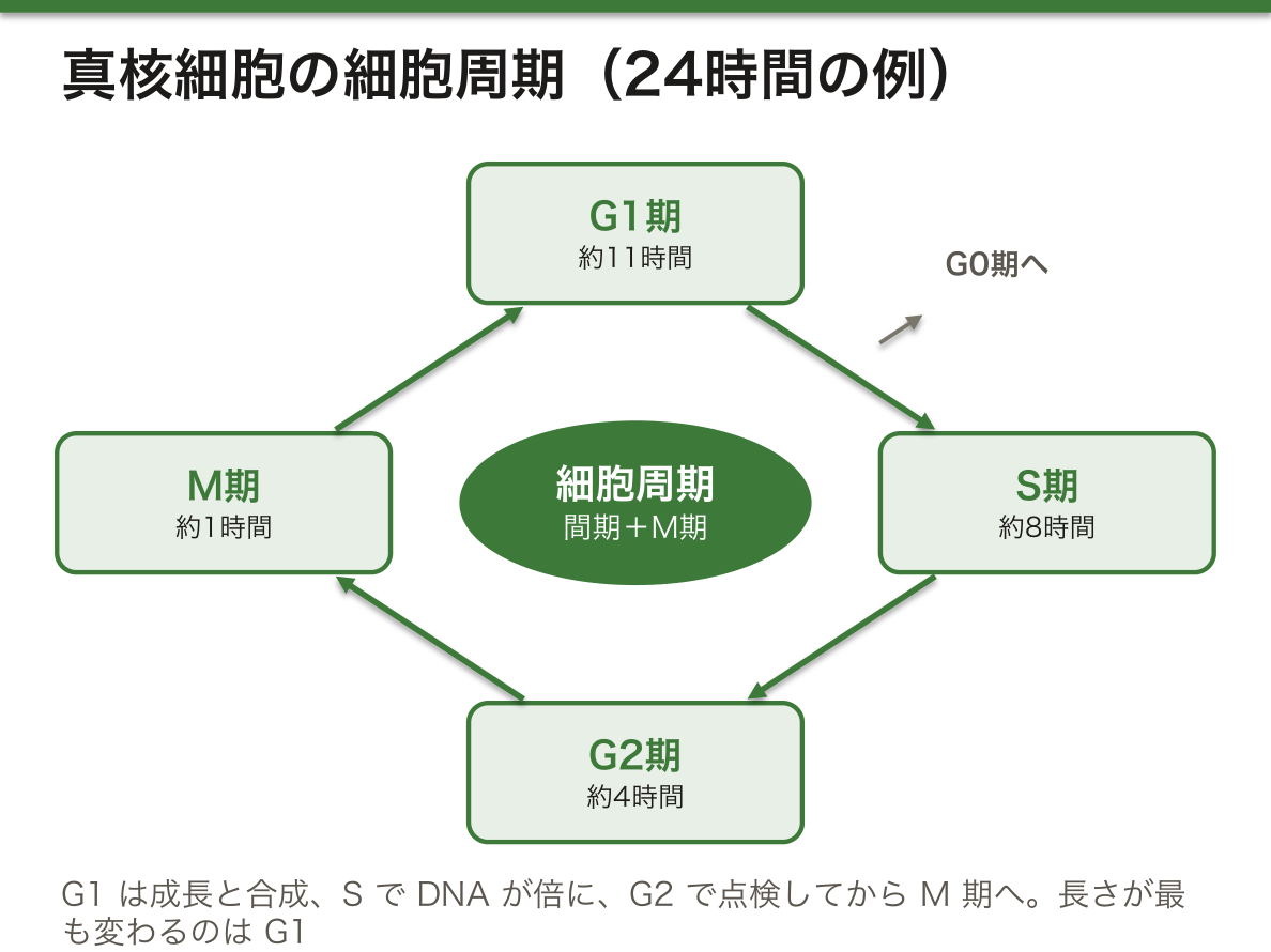
多細胞生物の細胞の多くは、周期を抜けてG0期にとどまる。G0には2種類ある。合図があれば周期に戻れる静止(quiescent)状態と、二度と戻らない老化(senescent)状態である。分化した体細胞の大半は静止したG0にいる。
分裂の能力で細胞を分けると、①ほぼ分裂しない細胞(赤血球、神経細胞など)、②刺激があれば分裂を再開する細胞(肝細胞、リンパ球など)、③常に分裂している細胞(造血幹細胞、上皮の細胞など)の3群になる。なぜ周期の長さの差は主にG1で生じるのか。S・G2・Mは「決まった仕事を決まった手順で済ませる」時期なので長さがあまり変わらないが、G1は外からの合図を待って、進むかどうかを決める時期だからである。
肝臓の一部を切除しても元の大きさに戻るのは、肝細胞がG0から周期に戻れる②の群だからである。逆に神経細胞は①の群なので、失われると数がほとんど戻らない。抗癌薬や放射線で骨髄・消化管粘膜・毛根が先に傷むのは、③の群が多い組織だからである。
English version
Core
The cell cycle is the life of a cell from one division to the next.
Bacteria multiply by binary fission, and the cycle has three periods: the B period (interval) from division to the start of replication, the C period (replication; about 40 minutes in E. coli), and the D period (division), when daughter chromosomes are separated and the cell pinches in two. Each new chromosome attaches to the membrane at its own point and moves away as the cell grows. In the middle of the cell, the protein FtsZ forms a Z ring, where the septum forms.
The eukaryotic cycle has interphase (G1, S, G2) and M phase. In a fast cell with a 24-hour cycle, G1 lasts about 11 hours, S about 8, G2 about 4 and M about 1. G1 is for growth and synthesis, and its length varies most between cells. In S phase DNA is doubled and the centrioles are duplicated. In G2 the cell checks again before entering M phase.
Going further
Many cells in a multicellular organism leave the cycle and stay in G0. There are two kinds of G0: a quiescent state, from which the cell can return when signalled, and a senescent state, from which it never returns. Most differentiated somatic cells are quiescent in G0.
By ability to divide, cells form three groups: (1) cells that hardly divide (red blood cells, neurons), (2) cells that divide again when stimulated (hepatocytes, lymphocytes), and (3) cells that divide all the time (haematopoietic stem cells, epithelial cells). Why do cycle lengths differ mainly in G1? S, G2 and M do a fixed job in a fixed order, so their length changes little. G1 is the phase in which the cell waits for outside signals and decides whether to go on.
In the clinic
The liver grows back after part of it is removed because hepatocytes belong to group (2) and can return from G0. Neurons belong to group (1), so once lost their number hardly recovers. Anticancer drugs and radiation harm bone marrow, gut lining and hair roots first because these tissues are rich in group (3) cells.
サイクリンとCDK — 周期を前に進めるエンジン
細胞周期を進める中心はサイクリン依存性キナーゼ(CDK)である。キナーゼはタンパク質のセリン・トレオニン・チロシンにリン酸をつける酵素で、CDKはサイクリンと結合したときだけ働く。CDKの量はほぼ一定だが、サイクリンの量は周期の中で大きく上下する。どのサイクリンと組むかで、どのタンパク質をリン酸化するかが決まる。
| 複合体 | サイクリン | CDK | おもな役割 |
|---|---|---|---|
| G1期 | サイクリンD | CDK4・CDK6 | 増殖因子などの外の合図に応じて働き、pRBをリン酸化してDNA合成に必要な遺伝子の発現を許す |
| サイクリンE | CDK2 | G1からS期への移行 | |
| S期 | サイクリンA | CDK2・CDK1 | DNA合成を進める(ヘリカーゼやポリメラーゼが標的) |
| M期(MPF) | サイクリンB | CDK1 | G2からM期への移行、APC/Cの活性化 |
pRBはふだん転写因子E2Fをつかまえて、S期の遺伝子を眠らせている。サイクリンD–CDK4/6、続いてサイクリンE–CDK2がpRBをリン酸化すると、pRBはE2Fを放し、S期に必要な遺伝子が一斉に読まれ始める。
なぜ周期は一方向にしか進まないのか。鍵は、サイクリンが使い終わると壊されることにある。M期の終わりにはAPC/C(後期促進複合体)がサイクリンBにユビキチンをつけ、プロテアソームが分解する。分解は後戻りできないので、一度先に進んだ周期は前の段階に戻れない。「作って、使って、壊す」のくり返しが、周期に向きを与えている。
ホルモン受容体陽性の乳癌では、サイクリンD–CDK4/6の経路に強く頼って増える。CDK4/6阻害薬(パルボシクリブなど)はここを止めてG1で細胞を足止めし、内分泌療法と組み合わせて使われる。pRBが壊れている腫瘍では、止めるべき相手がいないので効きにくい。
English version
Core
The core engine of the cell cycle is the cyclin-dependent kinase (CDK). A kinase adds phosphate to serine, threonine or tyrosine in proteins. A CDK works only when bound to a cyclin. The amount of CDK stays about the same, but cyclin levels rise and fall during the cycle. The cyclin partner decides which proteins get phosphorylated.
| Complex | Cyclin | CDK | Main role |
|---|---|---|---|
| G1 | Cyclin D | CDK4, CDK6 | Responds to outside signals such as growth factors; phosphorylates pRB so genes needed for DNA synthesis can be expressed |
| Cyclin E | CDK2 | Move from G1 to S | |
| S phase | Cyclin A | CDK2, CDK1 | Drives DNA synthesis (targets include helicases and polymerases) |
| Mitotic (MPF) | Cyclin B | CDK1 | Move from G2 to M; activates APC/C |
Going further
pRB normally holds the transcription factor E2F and keeps S-phase genes silent. When cyclin D–CDK4/6 and then cyclin E–CDK2 phosphorylate pRB, it lets go of E2F, and the genes needed for S phase are read all at once.
Why does the cycle only move forward? The key is that cyclins are destroyed after use. At the end of M phase, APC/C (the anaphase-promoting complex) tags cyclin B with ubiquitin, and the proteasome breaks it down. Destruction cannot be undone, so the cycle cannot go back to an earlier stage. The repeat of “make, use, destroy” gives the cycle its direction.
In the clinic
Hormone receptor-positive breast cancer depends strongly on the cyclin D–CDK4/6 pathway. CDK4/6 inhibitors (such as palbociclib) block it and hold cells in G1; they are used together with endocrine therapy. In tumours where pRB is broken, there is nothing left to hold back, so the drugs work poorly.
チェックポイント — 先へ進む前の点検と、p53
チェックポイントは、周期の大事な出来事が正しい順番で、欠けなく、正確に済んだかを見張る仕組みである。異常が見つかると周期をそこで止める。
| チェックポイント | 見ているもの | 異常があると |
|---|---|---|
| G1(制限点) | 細胞の大きさ、栄養、増殖因子、DNAの傷 | G1で停止、またはG0へ |
| S期 | 複製中のDNAの傷、複製フォーク | フォークを安定させ、複製を一時停止 |
| G2(DNA損傷) | 傷ついたDNAのままM期に入らないか | M期への移行を止める。傷が大きければアポトーシス |
| M期(紡錘体) | すべての染色体が紡錘体に正しくつながったか | 後期の開始(姉妹染色分体の分離)を止める |
G1の制限点は最大の決断の場で、ここを越えると、その後は増殖因子が無くても周期が最後まで進む。
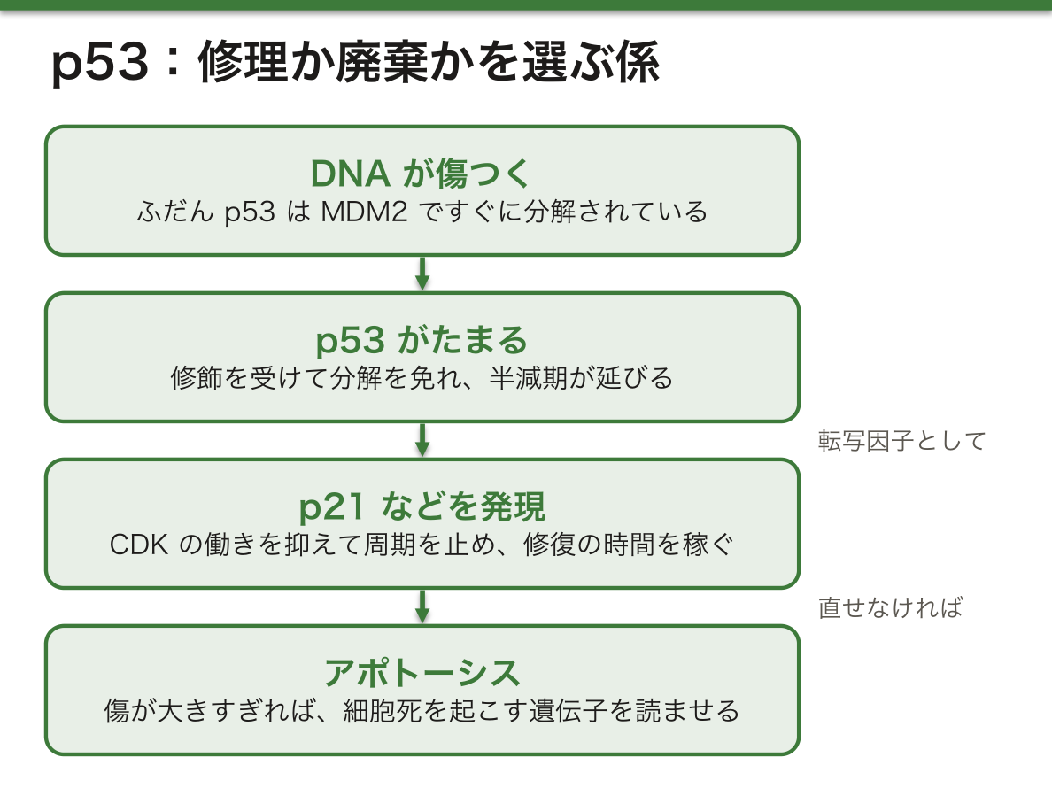
DNA損傷のチェックポイントの中心にいるのがp53である。p53は核にあるリン酸化タンパク質で、特定のDNA配列に結合する転写因子として働く。ふだんはMDM2によってすぐに分解されるので量が少ない。DNAが傷つくと、p53は修飾を受けて分解を免れ、半減期が延びてたまる。翻訳開始の速さも上がる。
たまったp53は、CDKの働きを抑えるp21などの遺伝子を発現させて周期を止め、修復の時間を稼ぐ。傷が大きすぎれば、アポトーシスを起こすタンパク質の遺伝子を読ませる。なぜ「止める」と「殺す」の両方が必要なのか。直せる傷なら細胞を残すほうが得だが、直せない傷を抱えた細胞を分裂させれば、変異が娘細胞に固定されて癌の芽になる。p53は修理か廃棄かを選ぶ係である。
M期のチェックポイントは、微小管に作用する抗癌薬(タキサン系・ビンカアルカロイド系)の効き方と関係する。紡錘体が正しくできないと、このチェックポイントが働き続けて細胞はM期で止まり、やがて死ぬ。
English version
Core
Checkpoints watch whether the key events of the cycle have happened in the right order, completely and accurately. If a problem is found, the cycle stops there.
| Checkpoint | What it checks | If something is wrong |
|---|---|---|
| G1 (restriction point) | Cell size, nutrients, growth factors, DNA damage | Arrest in G1, or entry into G0 |
| S phase | DNA damage during replication, replication forks | Stabilises forks and pauses replication |
| G2 (DNA damage) | Whether the cell would enter M with damaged DNA | Blocks entry into M; apoptosis if damage is severe |
| M phase (spindle) | Whether every chromosome is properly attached to the spindle | Blocks the start of anaphase (sister chromatid separation) |
The G1 restriction point is the biggest decision. After it, the cycle runs to the end even without growth factors.
Going further
At the centre of the DNA damage checkpoints is p53. It is a nuclear phosphoprotein that binds specific DNA sequences and acts as a transcription factor. Normally MDM2 causes it to be broken down quickly, so levels are low. When DNA is damaged, p53 is modified, escapes breakdown, its half-life becomes longer, and it builds up. Its translation also starts faster.
p53 then switches on genes such as p21, which blocks CDKs, so the cycle stops and there is time for repair. If the damage is too great, it switches on genes that cause apoptosis. Why are both “stop” and “kill” needed? If the damage can be fixed, keeping the cell is better. But if a cell with damage that cannot be fixed divides, the changes become fixed in its daughter cells and can become the seed of cancer. p53 chooses between repair and disposal.
In the clinic
The M-phase checkpoint is linked to how drugs acting on microtubules (taxanes, vinca alkaloids) work. If the spindle cannot form properly, the checkpoint stays on, the cell stops in M phase, and it eventually dies.
細胞周期の遺伝子と癌 — 体細胞の変異と、生まれつきの変異
周期のブレーキ役の遺伝子が壊れると、細胞は止まるべきところで止まれなくなる。同じ遺伝子でも、体細胞だけに変異が起きれば散発性の癌、生殖細胞系列に変異があれば家族性の癌症候群になる。
| 遺伝子 | タンパク質の働き | 体細胞の変異 | 生殖細胞系列の変異 |
|---|---|---|---|
| RB1 | 転写の調節(pRB) | 網膜芽細胞腫、骨肉腫、肺・乳・前立腺・膀胱の癌 | 家族性網膜芽細胞腫 |
| TP53 | 転写因子(p53) | 全癌のおよそ半数 | Li-Fraumeni症候群 |
| CDKN2A(p16) | CDKの阻害 | 多くの癌の約2〜3割 | 家族性黒色腫、家族性膵癌 |
| APC | β-カテニンの分解の調節など | 大腸癌 | 家族性大腸腺腫症(Gardner症候群を含む) |
pRBはG0からG1、G1からSへの移行を抑える癌抑制タンパク質である。増殖を促す外の因子(マイトジェン)は、最終的にこのブレーキを外す方向に働く。
なぜ家族性の網膜芽細胞腫は若くして、しかも両目に起きやすいのか。癌抑制遺伝子は2つのコピーの両方が壊れて初めてブレーキが失われる(Knudsonの2ヒット説)。散発性の場合は、1つの網膜の細胞に2回の偶然の変異が重ならなければならず、まれで、ふつう片目に1つである。家族性では生まれつき全身の細胞で1つ目がすでに壊れているので、あと1回の変異で腫瘍になる。網膜の細胞は数が多いので、どこかで2つ目が起きる確率が高く、早い発症・両側性・多発になる。遺伝の形式は優性に見えるが、細胞の中では劣性に働いている、という点が要である。
網膜芽細胞腫を治療した人が、のちに骨肉腫を発症することがある。全身の細胞でRB1の1つ目がすでに壊れているからで、特に放射線を当てた部位で起きやすい。家族歴のある子どもには、早期から眼底の定期検査が勧められる。
English version
Core
When genes that act as cycle brakes are broken, cells cannot stop where they should. With the same gene, a variant only in somatic cells gives sporadic cancer, while a germline variant gives a familial cancer syndrome.
| Gene | Protein function | Somatic variant | Germline variant |
|---|---|---|---|
| RB1 | Transcription regulator (pRB) | Retinoblastoma, osteosarcoma; lung, breast, prostate and bladder cancer | Familial retinoblastoma |
| TP53 | Transcription factor (p53) | About half of all cancers | Li-Fraumeni syndrome |
| CDKN2A (p16) | CDK inhibitor | About 20–30% of many cancers | Familial melanoma, familial pancreatic cancer |
| APC | Controls breakdown of β-catenin, among others | Colorectal cancer | Familial adenomatous polyposis (including Gardner syndrome) |
pRB is a tumour suppressor that holds back the moves from G0 to G1 and from G1 to S. Outside factors that promote division (mitogens) work in the end by releasing this brake.
Going further
Why does familial retinoblastoma start young and often in both eyes? A tumour suppressor loses its braking function only when both copies are broken (Knudson’s two-hit hypothesis). In sporadic cases, two chance variants must happen in the same retinal cell, which is rare, so there is usually one tumour in one eye. In familial cases, the first copy is already broken in every cell from birth, so one more variant is enough. The retina has many cells, so a second hit somewhere is likely. The result is early onset, both eyes and several tumours. The key point: the inheritance looks dominant, but inside the cell the gene acts recessively.
In the clinic
People treated for retinoblastoma may later develop osteosarcoma, because the first copy of RB1 is already broken in all their cells; it happens more often in areas that received radiation. Children with a family history are advised to have regular eye examinations from early on.
DNA損傷応答とミスマッチ修復 — 複製の写し間違いを直す
DNAは絶えず傷つく。細胞の中から生じる原因(複製の誤り、代謝でできる活性酸素など)は自然に起きる変異の、外からの原因(放射線、紫外線、化学物質)は誘発される変異のもとになる。原核細胞にも真核細胞にも修復の仕組みがあり、多くのタンパク質がかかわる。
修復は細胞周期の調節と強く結びついている。傷を感知するとDNA損傷応答(DDR)が働き、チェックポイントを通じて周期を止め、その間に修復させる。複製の前と、複製のあと分裂の前に、DNAが無傷かが確かめられる。この点検が失われると、傷が積み重なっていく。
ミスマッチ修復(MMR)は、複製の写し間違いを直す仕組みである。対にならない塩基の組(ミスマッチ)や、数塩基が余ったり欠けたりしてできる小さなループを見つけて取り除き、正しく合成し直す。
ミスマッチ修復には難しい問題がある。AとGが向かい合っていたとき、どちらが間違いなのかを知らなければ、正しいほうを壊してしまうかもしれない。そこでMMRは鎖を区別する。新しく合成された鎖には一時的に切れ目が残っていることなどを手がかりに、新しい鎖のほうだけを削って作り直す。間違いは必ず新しい鎖の側で起きるので、これで正しく直せる。
MMRが壊れると、特にマイクロサテライト(1〜4塩基の短い繰り返し)で写し間違いが残りやすい。繰り返し配列ではポリメラーゼが「滑って」数を数え間違えやすく、その誤りを直す係がいなくなるからである。この状態をマイクロサテライト不安定性(MSI)と呼ぶ。
MMR遺伝子(MLH1・MSH2・MSH6・PMS2)の生殖細胞系列の変異は、Lynch症候群(遺伝性非ポリポーシス大腸癌)を起こし、大腸癌や子宮内膜癌になりやすい。腫瘍のMSI検査やMMRタンパク質の免疫染色はこの症候群を拾い上げる手段になる。さらに、MSIが高い(dMMRの)腫瘍は変異が多く免疫に見つかりやすいため、免疫チェックポイント阻害薬が効きやすい。がんの種類を問わずこの性質で薬が選ばれることがある。
English version
Core
DNA is damaged all the time. Causes from inside the cell (replication errors, reactive oxygen from metabolism) lead to spontaneous mutations; causes from outside (radiation, UV light, chemicals) lead to induced mutations. Both prokaryotes and eukaryotes have repair systems that involve many proteins.
Repair is tightly linked to control of the cell cycle. When damage is sensed, the DNA damage response (DDR) stops the cycle through checkpoints and allows repair in the meantime. The DNA is checked before replication, and again after replication before division. If these checks are lost, damage builds up.
Mismatch repair (MMR) fixes copying errors made during replication. It finds base pairs that do not match, and small loops where a few bases are extra or missing, removes them and synthesises the stretch again.
Going further
Mismatch repair faces a hard question. If A sits opposite G, which one is wrong? Without knowing, it might destroy the correct base. So MMR tells the strands apart. Using clues such as temporary nicks left in the newly made strand, it cuts back and rebuilds only the new strand. Errors always happen in the new strand, so this fixes them correctly.
When MMR is broken, errors stay especially in microsatellites (short repeats of 1–4 bases). In repeats, the polymerase easily “slips” and miscounts, and nobody is left to fix it. This state is called microsatellite instability (MSI).
In the clinic
Germline variants in MMR genes (MLH1, MSH2, MSH6, PMS2) cause Lynch syndrome (hereditary non-polyposis colorectal cancer), with a high risk of colorectal and endometrial cancer. MSI testing of tumours and immunostaining for MMR proteins help find this syndrome. Also, MSI-high (dMMR) tumours carry many variants and are easier for the immune system to see, so immune checkpoint inhibitors tend to work well. Drugs are sometimes chosen by this feature regardless of cancer type.
除去修復 — 大きな傷はまとめて切り、小さな傷は塩基ごと替えるXeroderma pigmentosum
ヌクレオチド除去修復(NER)は、紫外線や化学物質など環境因子による傷を直す。代表は紫外線でできるピリミジン二量体で、隣り合う塩基どうしがくっついて二重らせんがゆがむ。NERはゆがんだ場所を含む数十塩基の一本鎖をまとめて切り出し、反対の鎖を型に埋め直す。転写中の遺伝子を優先して直す転写共役型(TC-NER)と、ゲノム全体を見回る全ゲノム型(GG-NER)の2つの道筋がある。
塩基除去修復(BER)は、らせんをゆがめない小さな傷を直す。酸化された塩基、アルキル化された塩基、脱アミノ化された塩基、誤って入ったウラシル、一本鎖の切れ目などである。まずDNAグリコシラーゼが傷んだ塩基を見つけて外し、そのあとを埋める。1塩基だけ替える短い道筋と、2〜10塩基を替える長い道筋がある。
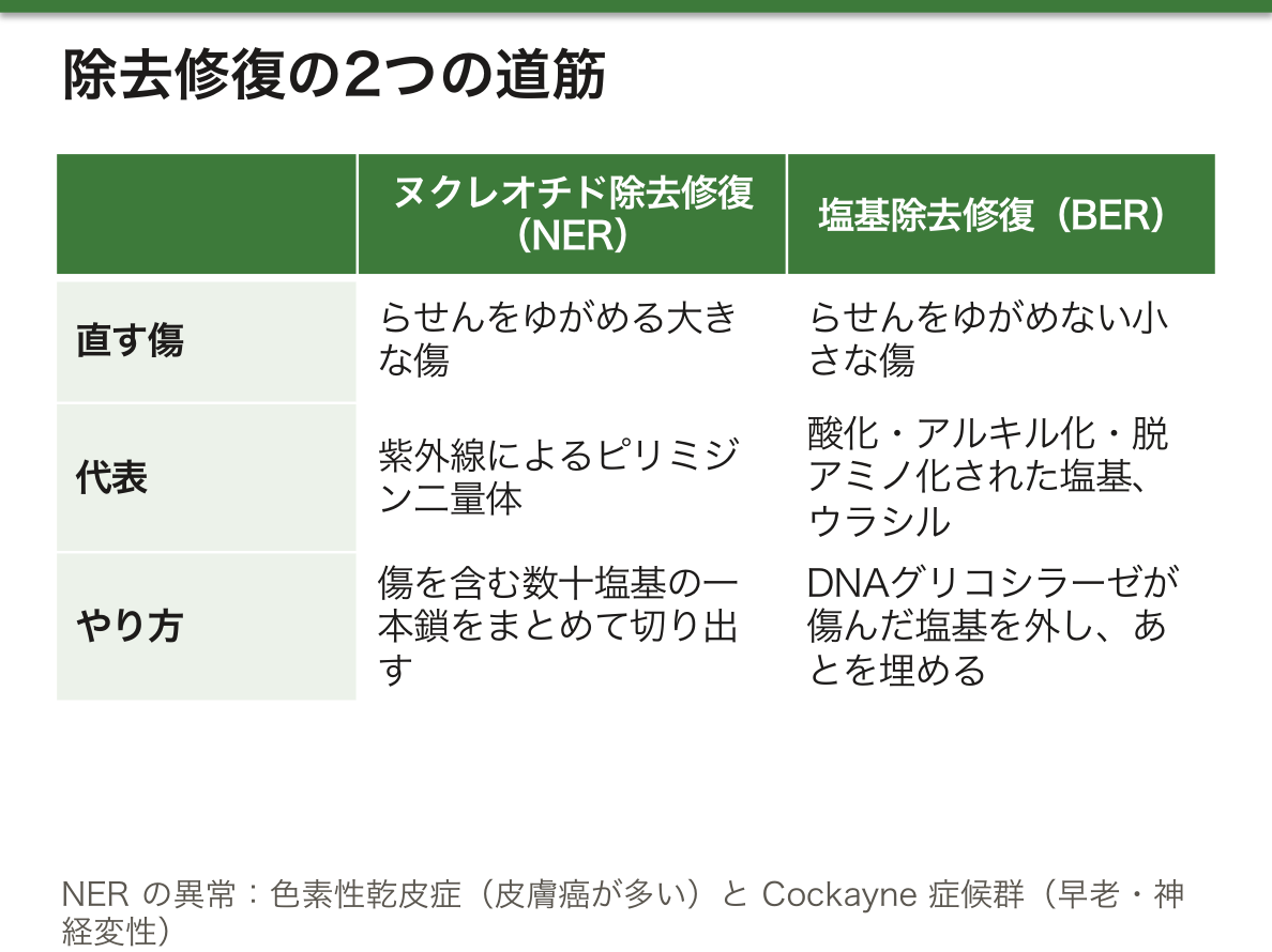
NERの異常として対照的な2つの病気がある。色素性乾皮症(XP)では光線過敏、色素の異常、皮膚の毛細血管拡張が起き、皮膚癌が非常に多い。Cockayne症候群(CS)も光線過敏を示すが、成長と発達の遅れ、感音難聴、皮下脂肪の喪失、進行する神経変性と早老が目立ち、癌はむしろ増えない。
なぜ同じNERの病気で、片方は癌に、片方は老化に向かうのか。XPではゲノム全体の傷が直らず、生き残った細胞に変異がたまるので癌になる。CSで主に失われるのは転写中の遺伝子の修復(TC-NER)で、転写が傷で止まった細胞は変異をためる前に死んでしまう。さらにCSのタンパク質は酸化ストレスの後にミトコンドリアに集まって細胞を守る働きももち、その欠如が早老の一因と考えられている。直らない細胞が「生き残る」か「死ぬ」かで、表に出る病気が分かれる。
BERの中心の酵素がまったく無いと胎生致死になるため、重い異常は病気として現れにくい。それでもBERのグリコシラーゼの1つMUTYHの両アレルの変異では、酸化した塩基の誤りが直らず、MUTYH関連ポリポーシスとして大腸に多数のポリープと癌ができる。XPの患者には、徹底した遮光と皮膚の定期的な診察が欠かせない。
English version
Core
Nucleotide excision repair (NER) fixes damage from environmental factors such as UV light and chemicals. The classic example is the UV-induced pyrimidine dimer, where neighbouring bases stick together and distort the double helix. NER cuts out a single-stranded stretch of a few dozen bases around the distortion and fills it in using the other strand as a template. There are two routes: transcription-coupled NER (TC-NER), which repairs genes being transcribed first, and global genome NER (GG-NER), which patrols the whole genome.
Base excision repair (BER) fixes small lesions that do not distort the helix: oxidised, alkylated or deaminated bases, wrongly inserted uracil, and single-strand breaks. A DNA glycosylase first finds and removes the damaged base, and the gap is then filled. A short route replaces one base; a long route replaces 2–10 bases.
Going further
Two contrasting diseases come from NER defects. In xeroderma pigmentosum (XP) there is sensitivity to light, abnormal pigmentation, small dilated skin vessels and very many skin cancers. Cockayne syndrome (CS) also shows light sensitivity, but growth and developmental delay, sensorineural hearing loss, loss of fat under the skin, progressive neurodegeneration and premature ageing dominate, and cancer is not increased.
Why does one NER disease lead to cancer and the other to ageing? In XP, damage across the genome is not fixed, surviving cells collect variants, and cancer follows. In CS, mainly repair of transcribed genes (TC-NER) is lost. Cells whose transcription is blocked by damage die before they collect variants. CS proteins also move to mitochondria after oxidative stress and protect the cell; their loss is thought to add to premature ageing. Whether unrepaired cells survive or die decides which disease appears.
In the clinic
Complete loss of core BER enzymes is lethal before birth, so severe defects rarely appear as disease. Still, biallelic variants in MUTYH, one of the BER glycosylases, leave errors from oxidised bases unrepaired and cause MUTYH-associated polyposis, with many colon polyps and cancer. Patients with XP need strict protection from light and regular skin checks.
二本鎖切断の修復 — 写しを見て正確に直すか、端をとにかくつなぐか
DNAの二本鎖切断(DSB)は、両方の鎖が同じ場所で切れる最も危険な傷である。直し方は2つある。
| 相同組換え修復(HR) | 非相同末端結合(NHEJ) | |
|---|---|---|
| 型 | 相同なDNA(ふつうは姉妹染色分体)を使う | 型を使わない |
| 正確さ | 非常に正確 | 数塩基を失うことが多い |
| 働く時期 | S期・G2期で優位 | G1期で活発 |
二本鎖切断の修復がうまくいかないと、染色体の並び替えや一部の喪失が起き、染色体不安定性になる。
なぜ修復の方法が細胞周期の時期で切り替わるのか。HRは、切れた場所と同じ配列をもつ無傷のDNAを写して直す。その最適な型は複製でできた姉妹染色分体で、これが存在するのはS期の後半からG2期だけである。G1期には型が無いので、切れた端を直接つなぐNHEJに頼るしかない。NHEJは不正確だが、切れたままにして染色体を失うよりはずっとましである。HRは減数分裂の乗り換え(交叉)とも同じ仕組みを使っている。
HRにはBRCA1やBRCA2などのタンパク質が必要である。これらが壊れると、二本鎖切断が不正確なやり方でしか直らず、変異と染色体の異常がたまる。
BRCA1/2の生殖細胞系列の変異は遺伝性乳癌卵巣癌症候群を起こす。BRCAが壊れた腫瘍にPARP阻害薬(オラパリブなど)を使うと、一本鎖の切れ目の修復も止まり、それが複製の途中で二本鎖切断に変わる。正常細胞はHRで直せるが、腫瘍は直せずに死ぬ。2つの経路の片方だけなら生きられるが両方失うと死ぬ、という合成致死の考え方である。放射線治療も、主に二本鎖切断をつくって癌細胞を殺している。
English version
Core
A double-strand break (DSB), in which both strands are cut at the same place, is the most dangerous kind of DNA damage. There are two ways to repair it.
| Homologous recombination (HR) | Non-homologous end joining (NHEJ) | |
|---|---|---|
| Template | Uses homologous DNA (usually the sister chromatid) | No template |
| Accuracy | Very accurate | Often loses a few bases |
| When | Dominant in S and G2 | Active in G1 |
If DSB repair fails, chromosomes can be rearranged or partly lost, which leads to chromosomal instability.
Going further
Why does the repair method change with the cell cycle stage? HR fixes the break by copying undamaged DNA with the same sequence. The best template is the sister chromatid made by replication, which exists only from late S phase to G2. In G1 there is no template, so the cell must use NHEJ, which joins the broken ends directly. NHEJ is not accurate, but it is far better than leaving the break and losing part of a chromosome. HR also uses the same machinery as crossing over in meiosis.
HR needs proteins such as BRCA1 and BRCA2. When they are broken, DSBs can only be fixed in inaccurate ways, and variants and chromosome abnormalities build up.
In the clinic
Germline BRCA1/2 variants cause hereditary breast and ovarian cancer syndrome. When PARP inhibitors (such as olaparib) are given to BRCA-deficient tumours, repair of single-strand nicks also stops, and these turn into DSBs during replication. Normal cells fix them by HR, but the tumour cannot and dies. This is synthetic lethality: a cell can live without one of two pathways, but not without both. Radiotherapy also kills cancer cells mainly by making DSBs.
細胞老化とアポトーシス — 分裂をやめる道と、自分で退場する道Apoptosis
細胞老化は、細胞の増殖が不可逆に止まった状態である。主に2つの経路、p53–p21とp16–pRBがこの停止を支える。テロメアの短縮やDNAの傷が引き金になる。
アポトーシスはプログラムされた細胞死で、多細胞生物では日常的に起きている正常な現象である。発生の過程(指の間の細胞が消えるなど)、ゲノムが傷ついた細胞や老化した細胞の除去、感染が終わって不要になった活性化T細胞の整理などで働く。
実行役はカスパーゼというタンパク質分解酵素の一族である。不活性な前駆体(プロカスパーゼ)として作られ、ほかのカスパーゼにアスパラギン酸の後ろで切られて活性化する。このカスケードは後戻りできない。開始の経路は2つある。
| 外因性経路 | 内因性経路 | |
|---|---|---|
| 引き金 | 細胞外の死の合図(TNF、Fasリガンドなど)が受容体に結合 | DNAの傷、ストレス、増殖因子の欠乏 |
| 調節 | 細胞死受容体 | Bcl-2ファミリーのタンパク質がミトコンドリアを制御 |
| 開始カスパーゼ | カスパーゼ8 | シトクロムcの放出 → アポトソーム → カスパーゼ9 |
アポトーシスが始まると、細胞は縮んで凝縮し、細胞骨格が崩れ、核膜が解体し、核のDNAが断片化する。細胞の表面も変わり、隣の細胞やマクロファージに食べられて片づけられる。
なぜアポトーシスでは炎症が起きないのか。細胞の中身が外に漏れる前に、膜に包まれたまま小さな断片になり、「食べてよい」という目印を表面に出して丸ごと回収されるからである。壊死のように中身がばらまかれると周りの免疫が騒ぎ出すが、アポトーシスは静かな退場になる。毎日膨大な数の細胞が入れ替わっても体に炎症が起きないのはこのためである。カスケードが一方向であることも、「死にかけて中途半端に生き残る」危険な細胞を作らないための設計と考えられる。
アポトーシスが足りないと癌が育ち、多すぎると組織が失われる。慢性リンパ性白血病の細胞はBcl-2を過剰にもって死を免れており、BCL-2阻害薬のベネトクラクスはその鎧を外して細胞を死なせる。逆に1型糖尿病では、免疫の攻撃で膵β細胞のアポトーシスが過剰に起き、インスリンを作る細胞が失われる。
English version
Core
Cellular senescence is a state in which cell proliferation has stopped for good. Two main pathways keep it in place: p53–p21 and p16–pRB. Short telomeres and DNA damage are common triggers.
Apoptosis is programmed cell death, a normal and frequent event in multicellular organisms. It works in development (for example, removing cells between the fingers), in clearing cells with damaged genomes and senescent cells, and in removing activated T cells that are no longer needed after an infection.
It is carried out by caspases, a family of protein-cutting enzymes. They are made as inactive precursors (procaspases) and are activated when other caspases cut them after aspartic acid. This cascade cannot be reversed. There are two starting pathways.
| Extrinsic pathway | Intrinsic pathway | |
|---|---|---|
| Trigger | Death signals from outside (TNF, Fas ligand) bind receptors | DNA damage, stress, lack of growth factors |
| Control | Death receptors | Bcl-2 family proteins control the mitochondria |
| Initiator caspase | Caspase-8 | Release of cytochrome c → apoptosome → caspase-9 |
Going further
Once apoptosis starts, the cell shrinks and condenses, the cytoskeleton collapses, the nuclear envelope breaks down, and nuclear DNA is cut into fragments. The cell surface changes, and neighbouring cells or macrophages eat it.
Why does apoptosis cause no inflammation? Before the contents can leak out, the cell breaks into small pieces still wrapped in membrane. It shows an “eat me” signal on its surface and is taken up whole. When contents spill out, as in necrosis, the nearby immune system reacts; apoptosis is a quiet exit. This is why the body is not inflamed even though huge numbers of cells are replaced every day. The one-way cascade can also be seen as a design that avoids dangerous cells that “half die” and survive.
In the clinic
Too little apoptosis lets cancer grow; too much destroys tissue. Chronic lymphocytic leukaemia cells carry too much Bcl-2 and escape death. The BCL-2 inhibitor venetoclax removes this armour and lets them die. In type 1 diabetes, on the other hand, immune attack causes excessive apoptosis of pancreatic β cells, and the insulin-making cells are lost.
まとめ
| 細胞周期 — 分裂から次の分裂までの一生 | 細胞周期とは、ある分裂から次の分裂までの細胞の一生である。 |
|---|---|
| サイクリンとCDK — 周期を前に進めるエンジン | 細胞周期を進める中心はサイクリン依存性キナーゼ(CDK)である。 |
| チェックポイント — 先へ進む前の点検と、p53 | チェックポイントは、周期の大事な出来事が正しい順番で、欠けなく、正確に済んだかを見張る仕組みである。 |
| 細胞周期の遺伝子と癌 — 体細胞の変異と、生まれつきの変異 | 周期のブレーキ役の遺伝子が壊れると、細胞は止まるべきところで止まれなくなる。 |
| DNA損傷応答とミスマッチ修復 — 複製の写し間違いを直す | DNAは絶えず傷つく。細胞の中から生じる原因(複製の誤り、代謝でできる活性酸素など)は自然に起きる変異の、外からの原因(放射線、紫外線、化学物質)は誘発される変異のもとになる。 |
| 除去修復 — 大きな傷はまとめて切り、小さな傷は塩基ごと替える | ヌクレオチド除去修復(NER)は、紫外線や化学物質など環境因子による傷を直す。 |
| 二本鎖切断の修復 — 写しを見て正確に直すか、端をとにかくつなぐか | DNAの二本鎖切断(DSB)は、両方の鎖が同じ場所で切れる最も危険な傷である。 |
| 細胞老化とアポトーシス — 分裂をやめる道と、自分で退場する道 | 細胞老化は、細胞の増殖が不可逆に止まった状態である。 |
このノートは医学生が授業の内容を自分の言葉でまとめたもので、診療の判断に使うものではありません。誤りに気づいたらお知らせください。