分子細胞生物学は、「細胞」という箱と、その中で情報が読まれ、写され、使われる仕組みの2つを、分子の言葉で説明する学問である。この章はその出発点として、生命と呼べるものの条件、原核細胞と真核細胞のちがい、細胞をもたない病原体(ウイルスとプリオン)、そして遺伝情報を担う核酸の構造とDNA複製を扱う。
読み進めるときは、セントラルドグマ(DNA → RNA → タンパク質)を地図として持っておくとよい。この章は地図の最初の矢印、つまりDNAがDNAを写す段階までを描く。複製の酵素の名前を並べて覚えるより、「なぜ片方の鎖は途切れ途切れにしか作れないのか」「なぜ染色体の端は少しずつ短くなるのか」という問いに答えられることを目標にする。
- 生命の性質と細胞 — 定義は無いが、条件はある
- 原核細胞と真核細胞 — 核膜1枚が分けるもの
- ウイルスとプリオン — 細胞をもたない病原体
- 遺伝情報の置き場所 — 核様体・プラスミド・核DNA・ミトコンドリアDNA
- 核酸の化学 — 塩基・糖・リン酸と、二重らせんを支える2つの力
- RNAの種類 — 情報を運ぶ・アミノ酸を運ぶ・つなぐ・調節する
- DNA複製 — 半保存的に、5′→3′の向きにしか伸ばせない
- 末端複製問題と、原核・真核の複製のちがい
生命の性質と細胞 — 定義は無いが、条件はあるCellula
生命には誰もが認める厳密な定義が無い。そこで生物学は、生きているものに共通する性質を並べて代わりにする。①細胞という決まった形をもつ、②遺伝情報を蓄え、それを使う、③代謝を行う、④内部環境を一定に保つ(恒常性)、⑤自分を増やす、の5つである。
細胞は、この5つをすべて1つの中にそろえた最小の単位である。核酸に情報を保存し、ATPを作り、タンパク質を合成し、機能に合った形をとり、分裂して数を増やす。多細胞生物では、幹細胞が分化して組織をつくり、組織が集まって器官になる。
多細胞生物の細胞は、役割で2種類に分かれる。体細胞は体そのものをつくる細胞で、遺伝情報を2組もつ二倍体(2n)である。生殖細胞は次の世代に情報を渡す細胞で、成熟した配偶子は1組だけの一倍体(n)になる。
ヒトの細胞の数は、体重70 kgの標準的な成人でおよそ30兆個(3×10¹³)と見積もられている。数のうえでは赤血球が大半を占める。赤血球は核をもたず小さいので、重さで比べると筋や脂肪の細胞のほうがずっと大きな割合になる。「数」と「重さ」で見え方がまったく変わることは、覚えておくと役に立つ。
体細胞と生殖細胞の区別は、変異が子に伝わるかどうかをそのまま決める。体細胞に起きた変異はその人の中だけで積み重なり、癌の原因になりうるが、子には伝わらない。生殖細胞の変異は遺伝性疾患として次の世代に渡る。遺伝カウンセリングで「この変異は腫瘍だけにあるのか、生まれつき全身にあるのか」を区別するのは、このためである。
English version
Core
Life has no strict definition that everyone accepts. Instead, biology lists the properties that living things share: (1) a fixed physical form, the cell; (2) genetic information that is stored and used; (3) metabolism; (4) a stable internal environment (homeostasis); (5) reproduction.
The cell is the smallest unit that has all five. It stores information in nucleic acids, makes ATP, builds proteins, takes a shape that fits its job, and divides. In multicellular organisms, stem cells differentiate into tissues, and tissues form organs.
Going further
Cells in a multicellular organism have two roles. Somatic cells build the body and are diploid (2n), with two sets of genetic information. Germ cells pass information to the next generation; mature gametes are haploid (n), with one set.
A standard 70 kg adult has about 30 trillion (3×10¹³) human cells. Red blood cells are the majority by number. They are small and have no nucleus, so by mass, muscle and fat cells make up a much larger share. Counting by number and by mass gives very different pictures.
In the clinic
The somatic/germ cell split decides whether a variant can pass to children. A variant in a somatic cell stays in that person and may lead to cancer, but it is not inherited. A germline variant is passed on as an inherited disease. This is why genetic counselling asks whether a variant is found only in the tumour or in every cell of the body from birth.
原核細胞と真核細胞 — 核膜1枚が分けるもの
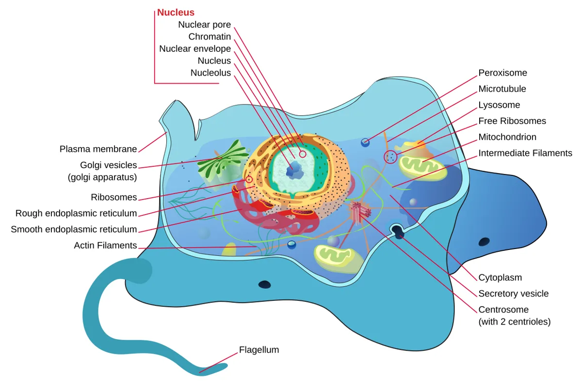
細胞は核をもつかどうかで2つに分かれる。細菌と古細菌は核をもたない原核生物、植物・動物・真菌は核をもつ真核生物である。
| 原核細胞 | 真核細胞 | |
|---|---|---|
| 生物 | 単細胞(細菌・古細菌) | 単細胞と多細胞 |
| 核 | 無い(核様体) | ある(核膜で囲まれる) |
| DNA | 環状の染色体が1本(〜数本)、一倍体、プラスミド | 線状の染色体が複数、体細胞は二倍体 |
| 膜で囲まれた小器官 | 無い | ある |
| リボソーム | 小さい(70S) | 大きい(80S) |
| 細胞壁 | ある(化学的に複雑) | ある場合は単純な成分 |
| 大きさ | およそ0.5〜10 µm | およそ10〜100 µm |
真核細胞の中でも、植物は葉緑体とセルロースの細胞壁をもち中心体を欠く。真菌はミトコンドリアはあるが葉緑体は無く、細胞壁はキチンを含む。動物の細胞は細胞壁をもたず、中心体をもち、形を柔軟に変えられる。
核膜の意味は「DNAを包む袋」だけではない。核膜があることで、mRNAを写す場所(核)と、それを読んでタンパク質を作る場所(細胞質)が空間的にも時間的にも分かれる。そのすきまに、mRNAの端に帽子をつけ、尾をつけ、イントロンを切り取る加工(RNAプロセシング)が入りこめる。原核細胞では転写の途中からリボソームが読み始めるので、この加工を挟む余地がほとんど無い。真核細胞が1つの遺伝子から複数のタンパク質を作れる(選択的スプライシング)のは、核膜がこの「待ち時間」を作っているからだと考えると理解しやすい。
原核と真核のちがいは、そのまま抗菌薬の選択毒性になる。ヒトの細胞に無いペプチドグリカンの細胞壁を狙うのがβ-ラクタム系、70Sリボソームを狙うのがアミノグリコシド系やマクロライド系である。真菌は真核生物なのでヒトとの差が小さく、抗真菌薬は細胞膜のステロールがエルゴステロールである点などを突く。真菌症の治療が難しく副作用が出やすいのは、相手が「近い親戚」だからである。
English version
Core
Cells fall into two groups by whether they have a nucleus. Bacteria and archaea are prokaryotes with no nucleus; plants, animals and fungi are eukaryotes with one.
| Prokaryotic cell | Eukaryotic cell | |
|---|---|---|
| Organisms | Unicellular (bacteria, archaea) | Unicellular and multicellular |
| Nucleus | None (nucleoid) | Yes (enclosed by the nuclear envelope) |
| DNA | One (or a few) circular chromosomes, haploid, plasmids | Several linear chromosomes, diploid in somatic cells |
| Membrane-bound organelles | None | Present |
| Ribosomes | Small (70S) | Large (80S) |
| Cell wall | Present, chemically complex | Simple, when present |
| Size | About 0.5–10 µm | About 10–100 µm |
Among eukaryotes, plants have chloroplasts and a cellulose wall but no centrosome. Fungi have mitochondria but no chloroplasts, and their wall contains chitin. Animal cells have no wall, have centrosomes, and can change shape freely.
Going further
The nuclear envelope is more than a bag for DNA. It separates, in space and time, the place where mRNA is copied (nucleus) from the place where it is read to make protein (cytoplasm). This gap allows RNA processing: adding a cap and a tail and cutting out introns. In prokaryotes, ribosomes start reading while transcription is still going on, so there is little room for processing. One useful way to see it: eukaryotes can make several proteins from one gene (alternative splicing) because the nuclear envelope creates this waiting time.
In the clinic
The prokaryote–eukaryote difference is the basis of selective toxicity of antibiotics. β-lactams target the peptidoglycan wall, which human cells lack; aminoglycosides and macrolides target the 70S ribosome. Fungi are eukaryotes, so they differ less from us. Antifungal drugs use points such as the membrane sterol being ergosterol. Fungal infections are hard to treat and the drugs have more side effects because the enemy is a close relative.
ウイルスとプリオン — 細胞をもたない病原体Virus
ウイルスは細胞の構造をもたない小さな感染性の粒子で、自分だけでは代謝も増殖もできない。核酸はDNAかRNAのどちらか一方だけをもち(両方をもつことは無い)、それをカプシドというタンパク質の殻が包む。核酸とカプシドを合わせてヌクレオカプシドと呼ぶ。細胞の外にある感染可能な粒子の形をビリオン、宿主のDNAに組み込まれたウイルスの遺伝子をプロウイルスという。
ウイルスがどの細胞に感染するか(宿主特異性)は、ウイルス表面のタンパク質と宿主細胞の受容体の相性で決まる。細菌に感染するウイルスはバクテリオファージと呼ばれる。
プリオンは核酸をまったく含まない、タンパク質だけの感染性粒子である。神経細胞にもともとある正常なタンパク質が誤った形に折りたたまれたもので、正常型に触れるとそれを次々に同じ誤った形へ変えていく。
ウイルスやプリオンが「生きているか」は、古典的な生物学では生きていないと答える。先の5つの性質のうち、遺伝情報はもっていても(プリオンはそれすら無い)、代謝と自力の増殖を欠くからである。「生命の縁にあるもの」と表現されることもある。
プリオンが核酸なしで「増える」理由は、形そのものが鋳型になるからである。誤った形のタンパク質がβシートの多い凝集体をつくり、そこに正常型が取り込まれて同じ形に変わる。情報が配列ではなく立体構造で伝わる、という点で、セントラルドグマの例外として扱われる。
SARS-CoV-2は、表面のスパイクタンパク質が宿主のACE2に結合して細胞に入る。受容体の分布が、どの臓器が傷むかを説明する手がかりになる。HIVはプロウイルスとして宿主のDNAに入りこむため、薬でウイルスを抑えても潜伏した細胞が残り、治りきらない。
プリオン病には、ウシの牛海綿状脳症(BSE)やヒトのCreutzfeldt-Jakob病(CJD)がある。プリオンは通常の加熱や消毒では失活しにくく、汚染した硬膜の移植で起きた医原性CJDは日本でも大きな問題になった。脳外科の器具の扱いが特別に定められているのはこのためである。
English version
Core
A virus is a small infectious particle with no cell structure. It cannot carry out metabolism or reproduce on its own. It contains either DNA or RNA, never both, wrapped in a protein coat called the capsid. Nucleic acid plus capsid is the nucleocapsid. The infectious particle outside the cell is the virion; viral genetic material built into the host DNA is a provirus.
Which cells a virus can infect (host specificity) depends on how well its surface proteins fit the host cell’s receptors. Viruses that infect bacteria are bacteriophages.
A prion is an infectious particle made only of protein, with no nucleic acid. It is a misfolded form of a normal protein found in nerve cells. When it meets the normal form, it converts it into the same wrong shape, again and again.
Going further
Are viruses and prions alive? Classical biology says no. Of the five properties, they may have genetic information (prions do not even have that), but they lack metabolism and self-reproduction. They are sometimes described as being “at the edge of life”.
Prions can “multiply” without nucleic acid because the shape itself acts as a template. The misfolded protein forms β-sheet-rich aggregates, and normal protein joins them and takes the same shape. The information is passed on by 3D structure, not by sequence, so prions are treated as an exception to the central dogma.
In the clinic
SARS-CoV-2 enters cells when its spike protein binds the host receptor ACE2. Where the receptor is found helps explain which organs are damaged. HIV inserts itself into host DNA as a provirus, so even when drugs control the virus, latently infected cells remain and the infection is not cured.
Prion diseases include bovine spongiform encephalopathy (BSE) in cattle and Creutzfeldt-Jakob disease (CJD) in humans. Prions resist ordinary heating and disinfection. Iatrogenic CJD from contaminated dura mater grafts was a major problem in Japan. This is why there are special rules for handling neurosurgical instruments.
遺伝情報の置き場所 — 核様体・プラスミド・核DNA・ミトコンドリアDNA
原核細胞では、DNAは膜に囲まれずに核様体として細胞質の一角にまとまる。核様体はDNAとRNA、それに結合するタンパク質の複合体で、DNAは環状、ヒストンには巻きついていない。ふつう1本で、情報は1組だけ(一倍体)である。これとは別に、小さな環状DNAのプラスミドをもつことがある。
真核細胞のDNAは3か所にある。核DNA、ミトコンドリアDNA(mtDNA)、植物ではさらに葉緑体DNAである。
| 核DNA | mtDNA | |
|---|---|---|
| 形 | 線状・二本鎖 | 環状・二本鎖 |
| コピー数 | 体細胞で2組(生殖細胞で1組) | 1つのミトコンドリアに複数、細胞の働きで数が変わる |
| もつ情報 | 個体が時間と空間の中で発生・成長するための情報 | ミトコンドリア自身の働きに必要な情報 |
プラスミドがもつのは生存に必須ではない少量の情報だが、その中身が問題になる。抗菌薬などの毒性物質への耐性を決める遺伝子が載っていることが多い。さらに一部のプラスミドは細菌から細菌へ移動でき、核様体に入りこんだり離れたりする。つまりプラスミドは、細菌が分裂を待たずに横から遺伝子を受け取るための乗り物であり、遺伝的な組み換えの道具になっている。
mtDNAが細菌の染色体に似た環状であることは偶然ではない。ミトコンドリアはもともと細菌が取りこまれたものと考えられており、その名残である。
院内感染で問題になるESBL産生菌やカルバペネマーゼ産生菌の耐性遺伝子は、多くがプラスミドに載っている。プラスミドは菌種の壁を越えて移るので、ある菌で生まれた耐性が別の菌に広がる。抗菌薬を必要な時だけ使う(適正使用)ことが強調されるのは、耐性を選び出す圧力を減らすためである。
English version
Core
In prokaryotic cells, DNA is not enclosed by a membrane. It gathers in one area of the cytoplasm as the nucleoid, a complex of DNA, RNA and binding proteins. The DNA is circular and not wrapped around histones. There is usually one molecule, so one set of information (haploid). Many bacteria also have small circular DNAs called plasmids.
In eukaryotic cells, DNA is found in three places: nuclear DNA, mitochondrial DNA (mtDNA), and in plants, chloroplast DNA.
| Nuclear DNA | mtDNA | |
|---|---|---|
| Form | Linear, double-stranded | Circular, double-stranded |
| Copies | Two sets in somatic cells (one in germ cells) | Several per mitochondrion; the number depends on cell function |
| Information | How the organism develops in time and space | What the mitochondrion needs to work |
Going further
Plasmids carry only a small amount of information that is not essential for survival, but what they carry matters. They often hold genes for resistance to toxic substances such as antibiotics. Some plasmids can also move from one bacterium to another, and can join the nucleoid or leave it. So plasmids are vehicles that let bacteria receive genes sideways, without waiting for cell division. They are tools of genetic recombination.
It is no accident that mtDNA is circular like a bacterial chromosome. Mitochondria are thought to come from bacteria that were taken into another cell long ago.
In the clinic
The resistance genes of ESBL-producing and carbapenemase-producing bacteria, which are major problems in hospital infections, are often on plasmids. Plasmids can cross species lines, so resistance that appears in one species spreads to another. Antibiotic stewardship (using antibiotics only when needed) is stressed because it reduces the pressure that selects resistant bacteria.
核酸の化学 — 塩基・糖・リン酸と、二重らせんを支える2つの力
核酸はヌクレオチドが一列につながった鎖である。塩基+五炭糖=ヌクレオシド、それにリン酸がつくとヌクレオチドになる。隣のヌクレオチドとは、糖の3′位と次の糖の5′位がリン酸を挟んでつながるホスホジエステル結合で結ばれるので、鎖には5′端と3′端という向きがある。
| DNA | RNA | |
|---|---|---|
| 塩基 | A・G・C・T | A・G・C・U |
| 糖 | デオキシリボース | リボース |
| ふつうの形 | 二本鎖 | 一本鎖 |
DNAの二本鎖ではAとT、GとCが向かい合う(相補性)。そのためDNA全体でAとTの量、GとCの量はそれぞれ等しくなる。ヒトではA 約31%、T 約29%、G・C 各約20%で、ほぼこの規則に合う。2本の鎖は逆向き(逆平行)に並び、塩基対ごとにおよそ36°ずつねじれて、約10塩基対で1回転する。
二重らせんを支える力は2つある。1つは塩基対の間の水素結合(A–Tは2本、G–Cは3本)、もう1つは、らせんの中心で平たい塩基の環が積み重なるスタッキングである。教科書の図では水素結合ばかりが目立つが、安定性への寄与はスタッキングも大きい。GCに富むDNAほど2本の鎖が離れにくい(融解温度が高い)のは、水素結合が多く、積み重なりも強いためで、PCRのプライマーを設計するときにこれが効いてくる。
なぜDNAはリボースでなくデオキシリボースを使い、UでなくTを使うのか。リボースの2′位の-OHは、隣のリン酸結合を攻撃して鎖を切りやすくする。これを持たないDNAは化学的に安定で、長期保存に向く。また、シトシンは自然に脱アミノ化してウラシルに変わることがある。DNAがもともとUを使わないので、DNAの中にUがあれば「壊れた印」と見分けられる。修復の仕組みが働けるのはこのためである。
らせんの溝には塩基の縁が露出していて、配列を読むタンパク質(転写因子など)がここから結合する。たとえばATに富む配列に結合するTATA結合タンパク質はDNAを大きく曲げ、転写が始まる足場をつくる。
English version
Core
A nucleic acid is a chain of nucleotides. Base + five-carbon sugar = nucleoside; add a phosphate and it becomes a nucleotide. Neighbouring nucleotides are joined by a phosphodiester bond: a phosphate links the 3′ carbon of one sugar to the 5′ carbon of the next. So every strand has a direction, a 5′ end and a 3′ end.
| DNA | RNA | |
|---|---|---|
| Bases | A, G, C, T | A, G, C, U |
| Sugar | Deoxyribose | Ribose |
| Usual form | Double-stranded | Single-stranded |
In double-stranded DNA, A pairs with T and G pairs with C (complementarity). So in all of a cell’s DNA, the amount of A equals T and G equals C. In humans, A is about 31%, T about 29%, and G and C about 20% each, close to this rule. The two strands run in opposite directions (antiparallel), twist about 36° per base pair, and make one turn about every 10 base pairs.
Going further
Two forces hold the double helix together. One is hydrogen bonding between base pairs (two bonds for A–T, three for G–C). The other is stacking of the flat base rings on top of each other in the centre of the helix. Textbook figures show mostly hydrogen bonds, but stacking also adds a lot of stability. GC-rich DNA is harder to separate (higher melting temperature) because it has more hydrogen bonds and stronger stacking. This matters when designing PCR primers.
Why does DNA use deoxyribose instead of ribose, and T instead of U? The 2′-OH of ribose can attack the nearby phosphate bond and break the chain. DNA lacks it, so it is chemically stable and good for long-term storage. Also, cytosine sometimes loses its amino group and turns into uracil. Because DNA does not normally use U, any U in DNA can be recognised as damage. This is what allows repair systems to work.
The edges of the bases are exposed in the grooves of the helix, and proteins that read the sequence (such as transcription factors) bind there. For example, the TATA-binding protein binds an AT-rich sequence, bends the DNA strongly, and builds a platform for starting transcription.
RNAの種類 — 情報を運ぶ・アミノ酸を運ぶ・つなぐ・調節する
役割分担は単純である。DNAは情報を保存し、RNAは情報を実行する(一部のウイルスではRNAが保存も担う)。
mRNA(伝令RNA)は転写でつくられ、写しとった情報を細胞質に運び、タンパク質合成の型になる。tRNA(転移RNA)は活性化したアミノ酸を結合してリボソームに運ぶ。少なくともアミノ酸の数(20種)だけあり、真核細胞では同じアミノ酸を運ぶ別のtRNA(イソアクセプター)もあって、タンパク質合成の速さと量の調節にかかわる。rRNA(リボソームRNA)はリボソームの骨格で、大サブユニットのrRNAがペプチド結合をつくる反応を触媒し、小サブユニットのrRNAがmRNAの正確な結合を見張る。
このほかに、タンパク質をつくらない調節性のRNA(ノンコーディングRNA)がある。snRNA、miRNA、siRNA、lncRNAなどで、遺伝子発現のエピジェネティックな調節や、分化・発生にかかわる。
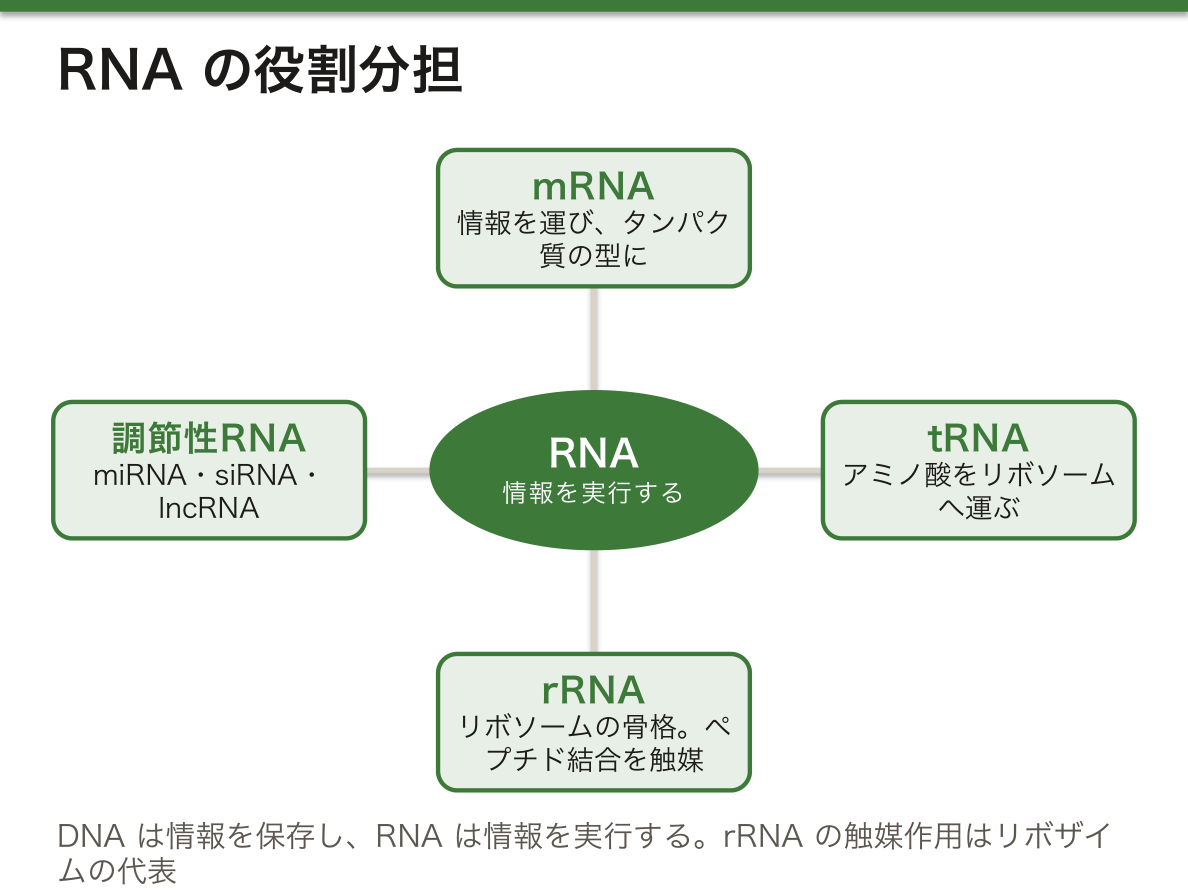
RNAはDNAより形がはるかに多彩である。一本鎖なので、鎖の中で相補的な部分同士が対をつくり、二本鎖の「茎」とループの部分が混ざった二次構造になり、さらに折りたたまれて三次構造をとる。tRNAのL字形がその代表である。形が多彩だから、RNAは情報を運ぶだけでなく、酵素として働く(リボザイム)ことができる。タンパク質合成の中心反応を、タンパク質ではなくrRNAが触媒しているという事実は、生命がRNAから始まったという仮説の有力な根拠になっている。
RNAの理解は、そのまま新しい薬につながっている。mRNAワクチンは抗原タンパク質の設計図をmRNAとして細胞に届ける。siRNA製剤は標的のmRNAを分解させて、原因タンパク質の量を減らす(遺伝性トランスサイレチンアミロイドーシスなど)。アンチセンス核酸のヌシネルセンは、脊髄性筋萎縮症でスプライシングを変えて、働くタンパク質を増やす。どれも「配列が相補的に結合する」という性質をそのまま治療に使っている。
English version
Core
The division of labour is simple: DNA stores information; RNA carries it out (in some viruses, RNA also stores it).
mRNA (messenger RNA) is made by transcription, carries the copied information to the cytoplasm, and serves as the template for protein synthesis. tRNA (transfer RNA) binds an activated amino acid and brings it to the ribosome. There are at least as many tRNAs as amino acids (20). In eukaryotes there are also isoacceptor tRNAs, different tRNAs that carry the same amino acid; they help control the speed and amount of protein synthesis. rRNA (ribosomal RNA) forms the core of the ribosome. rRNA in the large subunit catalyses peptide bond formation, and rRNA in the small subunit checks that mRNA binds correctly.
There are also regulatory RNAs (non-coding RNAs) that do not make protein: snRNA, miRNA, siRNA, lncRNA and others. They take part in epigenetic control of gene expression and in differentiation and development.
Going further
RNA is much more varied in shape than DNA. Because it is single-stranded, complementary parts of the same chain pair up. This creates a secondary structure of double-stranded “stems” and loops, which folds further into a tertiary structure. The L shape of tRNA is the classic example. Because of this variety, RNA can do more than carry information: it can act as an enzyme (ribozyme). The central reaction of protein synthesis is catalysed by rRNA, not by protein. This is strong support for the idea that life began with RNA.
In the clinic
Understanding RNA leads directly to new drugs. mRNA vaccines deliver the blueprint of an antigen protein to cells as mRNA. siRNA drugs make the target mRNA break down and lower the amount of a harmful protein (for example in hereditary transthyretin amyloidosis). The antisense drug nusinersen changes splicing in spinal muscular atrophy so that more working protein is made. All of these use the same property: sequences that bind by complementarity.
DNA複製 — 半保存的に、5′→3′の向きにしか伸ばせない
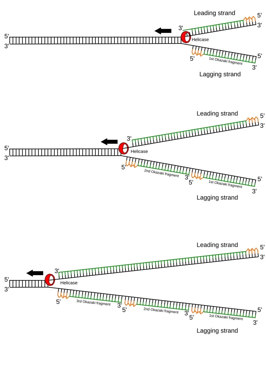
細胞が分裂する前には、すべての染色体のDNAを1回だけ正確に写す必要がある。真核細胞ではS期に、原核細胞では二分裂の始まりに複製が起きる。できた2本のDNAは、どちらも古い鎖1本と新しい鎖1本からなる(半保存的複製)。
複製は複製起点という決まった配列から始まり、開始タンパク質がそこを認識する。ヘリカーゼが塩基対の水素結合を切って二本鎖をほどき、Y字形の複製フォークができる。ほどけた先で生じるねじれはトポイソメラーゼが解消し、一本鎖結合タンパク質が鎖が再びくっつくのを防ぐ。
| 酵素・タンパク質 | 役割 |
|---|---|
| Pol α–プライマーゼ複合体 | 10〜12塩基のRNAプライマーをつくり、そこから合成を始める(リーディング鎖の最初と、各岡崎断片の最初) |
| DNAポリメラーゼε | リーディング鎖を伸ばす。誤りを直す活性ももつ |
| DNAポリメラーゼδ | ラギング鎖を伸ばす。誤りを直す活性ももつ |
| RNase H | 役目を終えたRNAプライマーを分解する |
| DNAリガーゼ | 断片の5′リン酸と3′-OHをつないで1本にする |
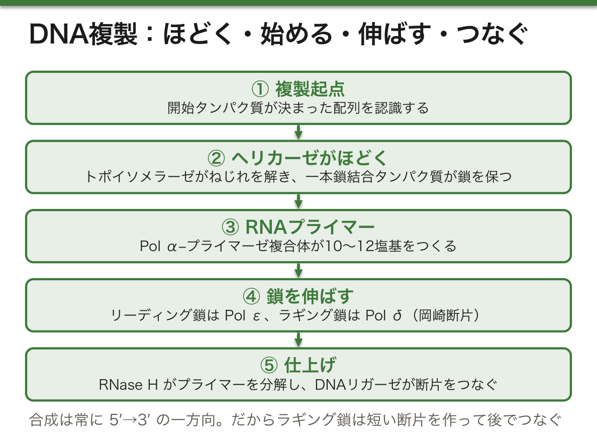
なぜ片方の鎖(ラギング鎖)は途切れ途切れにしか作れないのか。DNAポリメラーゼは、新しいdNTP(デオキシリボヌクレオシド三リン酸)を伸びている鎖の3′-OHにしかつなげない。dNTPがもつ三リン酸のうち2つがピロリン酸として外れ、その分解のエネルギーで結合ができる。だから合成は常に5′→3′の一方向になる。フォークが進む向きと同じ向きに伸ばせる鎖(リーディング鎖)は続けて作れるが、逆向きの鎖はフォークが少し開くたびに短い断片(岡崎断片)を作って後でつなぐしかない。
この「一方向しか無い」設計には利点がある。間違った塩基を入れたとき、ポリメラーゼは3′端の1塩基を削って入れ直す(校正)ことができる。もし逆向きに伸ばす設計だと、エネルギー源の三リン酸が伸びる側の端に残る形になり、誤りを削った瞬間にその端のエネルギーも失われて続きが作れなくなる。正確さと引き換えに、ラギング鎖の手間を受け入れている、と考えられている。
細胞の中ではプライマーがRNAで、プライマーゼが作る。試験管の中のPCRでは、人工的に合成したDNAのプライマーを2種類(前向きと逆向き)加える点がちがう。
複製の酵素は薬の標的として重要である。トポイソメラーゼ阻害薬のイリノテカン(I型)やエトポシド(II型)は抗癌薬として、細菌のDNAジャイレースとトポイソメラーゼIVを阻害するキノロン系は抗菌薬として使われる。抗ウイルス薬のアシクロビルは、3′-OHを欠くヌクレオシドのまがいものとして鎖に取りこまれ、そこで合成を止める。「3′-OHにしかつなげない」という性質を逆手に取った薬である。
English version
Core
Before a cell divides, it must copy the DNA of every chromosome exactly once. In eukaryotes this happens in S phase; in prokaryotes, at the start of binary fission. Each new DNA molecule has one old strand and one new strand (semiconservative replication).
Replication starts at a fixed sequence, the origin of replication, which initiation proteins recognise. Helicase breaks the hydrogen bonds between base pairs and opens the double strand, forming a Y-shaped replication fork. Topoisomerase removes the twisting tension ahead of the fork, and single-strand binding proteins stop the strands from rejoining.
| Enzyme / protein | Role |
|---|---|
| Pol α–primase complex | Makes an RNA primer of 10–12 nucleotides and starts synthesis from it (at the start of the leading strand and of each Okazaki fragment) |
| DNA polymerase ε | Extends the leading strand; also corrects errors |
| DNA polymerase δ | Extends the lagging strand; also corrects errors |
| RNase H | Breaks down RNA primers after use |
| DNA ligase | Joins the 5′ phosphate and 3′-OH of fragments into one strand |
Going further
Why can one strand (the lagging strand) only be made in pieces? DNA polymerase can add a new dNTP (deoxyribonucleoside triphosphate) only to the free 3′-OH of the growing strand. Two of the three phosphates leave as pyrophosphate, and the energy from breaking it down makes the bond. So synthesis always runs 5′→3′. The strand that grows in the same direction as the fork (leading strand) can be made continuously. The other strand must be made as short pieces (Okazaki fragments) each time the fork opens a little, and joined later.
This one-way design has a benefit. When a wrong base is added, the polymerase can cut off the last base at the 3′ end and try again (proofreading). If DNA grew the other way, the energy-carrying triphosphate would sit at the growing end. Cutting off a mistake would remove that energy too, and synthesis could not continue. The cell seems to accept the extra work on the lagging strand in exchange for accuracy.
In the cell, primers are RNA and are made by primase. In PCR in a test tube, two kinds of synthetic DNA primers (forward and reverse) are added instead.
In the clinic
Replication enzymes are important drug targets. Topoisomerase inhibitors such as irinotecan (type I) and etoposide (type II) are anticancer drugs. Quinolones, which block bacterial DNA gyrase and topoisomerase IV, are antibiotics. The antiviral drug aciclovir is built into the chain as a fake nucleoside with no 3′-OH, and synthesis stops there. It turns the rule “bonds form only at the 3′-OH” against the virus.
末端複製問題と、原核・真核の複製のちがい
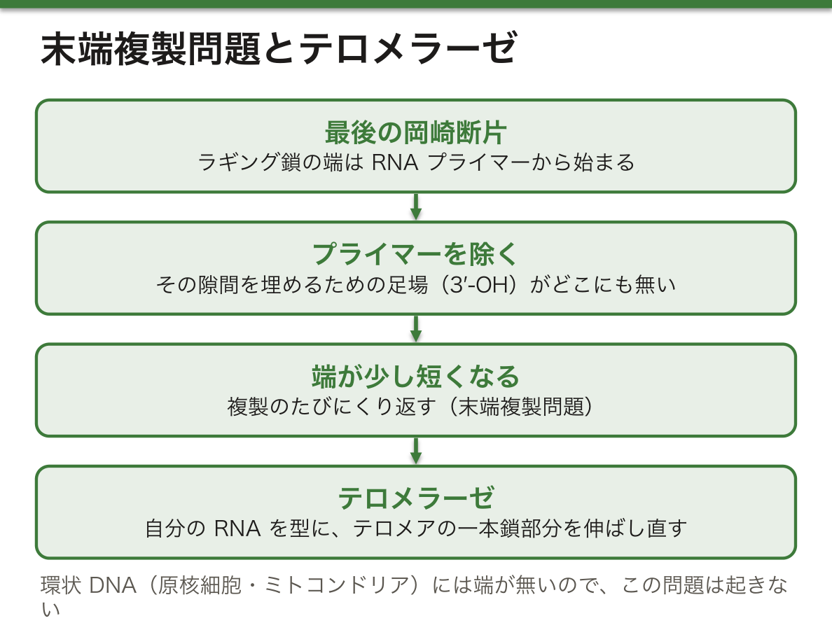
線状の染色体には端がある。ラギング鎖の最後の岡崎断片はRNAプライマーから始まるが、プライマーを取り除いたあと、その隙間を埋めるための3′-OH(足場)がどこにも無い。そのため、複製のたびに染色体の端が少しずつ短くなる。これが末端複製問題である。環状DNAには端が無いので、原核細胞やミトコンドリアではこの問題は起きない。
染色体の端には、遺伝子を含まない繰り返し配列テロメアが置かれている。ヒトではTTAGGGの繰り返しで、3′端は一本鎖のまま突き出してキャップをつくる。テロメラーゼは自分の中にRNAの型をもつ酵素で、その型を使ってテロメアの一本鎖部分を伸ばし直す。ただし働くのは発生の特定の時期や一部の細胞に限られる。
| 原核細胞 | 真核細胞 | |
|---|---|---|
| 場所 | 細胞質 | 核 |
| 複製起点 | 1か所(100〜200塩基) | 多数(ヒトでは数万か所、各約150塩基) |
| フォーク | 起点から両方向に2つ | 多数の起点で同時に進む |
| 開始タンパク質 | 少数 | 多くのサブユニットからなる複合体 |
| 岡崎断片 | 長い(1,000〜2,000塩基) | 短い(100〜200塩基) |
| 速さ | 速い(約2,000塩基/秒) | 遅い(約100塩基/秒) |
なぜ真核細胞は起点をたくさん必要とするのか。計算するとすぐにわかる。ヒトのゲノム約30億塩基対を、1か所の起点から両方向に毎秒100塩基ずつ写すと、3×10⁹÷200=1.5×10⁷秒、つまり約170日かかる。実際のS期は8時間ほどである。フォークが遅く、ゲノムが大きい真核細胞は、起点を数多く並べて同時に進めることでしか間に合わない。岡崎断片が短いのも、ヌクレオソームに巻かれたDNAが少しずつしか露出しないためと考えられている。
テロメアが極端に短くなる体質の人は、短テロメア症候群として、骨髄の働きが落ちる(再生不良性貧血、骨髄異形成症候群、急性骨髄性白血病)、肺が線維化するなどの病気を起こす。若いうちは血液の病気として、年をとってからは肺の病気として現れやすい。分裂の多い組織ほど端の消耗の影響を受けやすいからである。一方、癌細胞の多くはテロメラーゼを再び働かせて、この「回数制限」を外している。
English version
Core
Linear chromosomes have ends. The last Okazaki fragment of the lagging strand starts from an RNA primer. After the primer is removed, there is no 3′-OH (no foothold) to fill the gap. So the chromosome end gets a little shorter with each round of replication. This is the end-replication problem. Circular DNA has no ends, so prokaryotes and mitochondria do not have this problem.
Chromosome ends carry telomeres, repeated sequences that contain no genes. In humans the repeat is TTAGGG. The 3′ end sticks out as a single strand and forms a cap. Telomerase is an enzyme that carries its own RNA template and uses it to lengthen the single-stranded part of the telomere. It works only at certain stages of development and in some cells.
Going further
| Prokaryotic cell | Eukaryotic cell | |
|---|---|---|
| Location | Cytoplasm | Nucleus |
| Origins | One (100–200 nt) | Many (tens of thousands in humans, each about 150 nt) |
| Forks | Two, moving both ways from the origin | Many, working at the same time |
| Initiation proteins | Few | Large multi-subunit complexes |
| Okazaki fragments | Long (1,000–2,000 nt) | Short (100–200 nt) |
| Speed | Fast (about 2,000 nt/s) | Slow (about 100 nt/s) |
Why do eukaryotes need so many origins? A quick calculation shows it. Copying the human genome, about 3 billion base pairs, from a single origin in both directions at 100 nucleotides per second takes 3×10⁹ ÷ 200 = 1.5×10⁷ seconds, or about 170 days. The real S phase lasts about 8 hours. With slow forks and a large genome, eukaryotes can only finish in time by running many origins at once. Okazaki fragments are probably short because DNA wrapped around nucleosomes is exposed only a little at a time.
In the clinic
People born with very short telomeres have short telomere syndromes: bone marrow failure (aplastic anaemia, myelodysplastic syndrome, acute myeloid leukaemia) and lung fibrosis, among others. In young people they tend to appear as blood diseases, and in older people as lung disease, because tissues that divide often suffer most from worn-down ends. Many cancer cells, on the other hand, switch telomerase back on and remove this limit on the number of divisions.
まとめ
| 生命の性質と細胞 — 定義は無いが、条件はある | 生命には誰もが認める厳密な定義が無い。そこで生物学は、生きているものに共通する性質を並べて代わりにする。 |
|---|---|
| 原核細胞と真核細胞 — 核膜1枚が分けるもの | 細胞は核をもつかどうかで2つに分かれる。 |
| ウイルスとプリオン — 細胞をもたない病原体 | ウイルスは細胞の構造をもたない小さな感染性の粒子で、自分だけでは代謝も増殖もできない。 |
| 遺伝情報の置き場所 — 核様体・プラスミド・核DNA・ミトコンドリアDNA | 原核細胞では、DNAは膜に囲まれずに核様体として細胞質の一角にまとまる。 |
| 核酸の化学 — 塩基・糖・リン酸と、二重らせんを支える2つの力 | 核酸はヌクレオチドが一列につながった鎖である。 |
| RNAの種類 — 情報を運ぶ・アミノ酸を運ぶ・つなぐ・調節する | 役割分担は単純である。DNAは情報を保存し、RNAは情報を実行する(一部のウイルスではRNAが保存も担う)。 |
| DNA複製 — 半保存的に、5′→3′の向きにしか伸ばせない | 細胞が分裂する前には、すべての染色体のDNAを1回だけ正確に写す必要がある。 |
| 末端複製問題と、原核・真核の複製のちがい | 線状の染色体には端がある。ラギング鎖の最後の岡崎断片はRNAプライマーから始まるが、プライマーを取り除いたあと、その隙間を埋めるための3′-OH(足場)がどこにも無い。 |
このノートは医学生が授業の内容を自分の言葉でまとめたもので、診療の判断に使うものではありません。誤りに気づいたらお知らせください。