普通の光と何が違うのかを答えられることが要点。「強い光」ではなく、単色性・指向性・可干渉性という3つの性質を持つ光である。
医療での使い分けは、結局「その波長を何が吸収するか」で決まる。標的が水なのかヘモグロビンなのかメラニンなのかで、選ぶレーザーが変わる。
- レーザーの原理 — 誘導放出と共振器
- 医療用レーザーの選び方と安全
- レーザーと組織の相互作用 — なぜ波長で用途が決まるか
- 低出力レーザー療法 — 何が分かっていて、何が分かっていないか
- 回折と干渉 — 光が波であることの証拠
- 回折格子でレーザーの波長と光子1個のエネルギーを測る
レーザーの原理 — 誘導放出と共振器
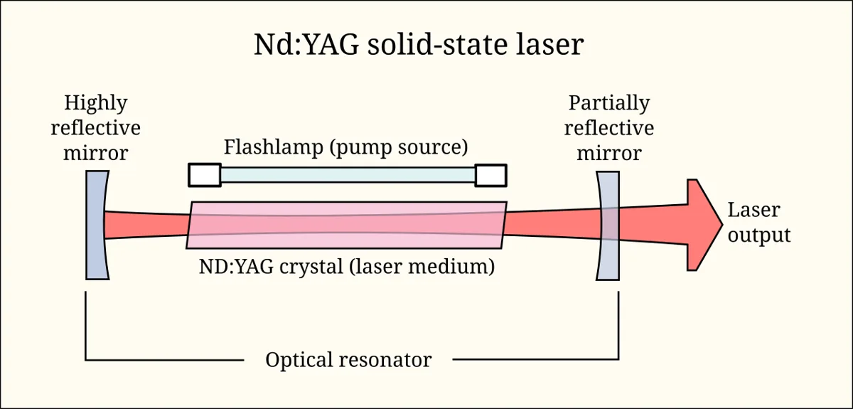
LASER = Light Amplification by Stimulated Emission of Radiation。
3つの要素:
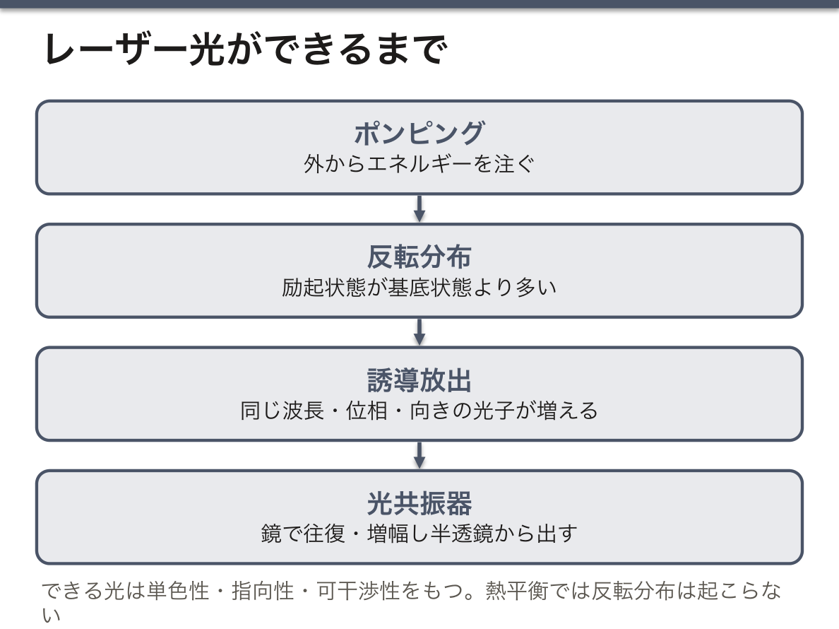
- 反転分布——励起状態の原子が基底状態より多い状態。外部からエネルギーを注ぐ(ポンピング)ことで作る。熱平衡では絶対に起こらない
- 誘導放出——励起状態の原子に光子が当たると、同じ波長・同じ位相・同じ向きの光子がもう1個放出される。1個が2個、2個が4個と増える
- 光共振器——2枚の鏡で光を往復させて増幅し、片方の鏡(半透鏡)から取り出す
3つの性質:単色性(波長がそろう)・指向性(平行に進む)・可干渉性(位相がそろう)。
「1点に集められる」ことが医療で使われる理由である。指向性が高いので、レンズで回折限界(波長程度)まで絞れる。同じ出力でも、集光できれば単位面積あたりのエネルギー(power density, W/cm²)が桁違いに大きくなる。切れるのは出力が大きいからではなく、絞れるからである。
組織への作用は照射時間と出力密度で4つに分かれる。①光熱作用(凝固・切開・蒸散)②光機械作用(衝撃波で破砕。Nd:YAG の後発白内障切開)③光化学作用(光線力学療法 PDT——光感受性物質を腫瘍に集めて照射する)④光アブレーション(分子結合を直接切る。エキシマレーザーの LASIK)。
パルス幅を短くすると、熱が周囲へ逃げる前に作用が終わる(選択的光熱融解)。だから色素レーザーで血管腫を治療しても周囲の皮膚が焼けない。「どれだけ短く当てるか」が、選択性を作っている。
English version
Core
LASER = Light Amplification by Stimulated Emission of Radiation.
Three elements:
- Population inversion — more atoms in the excited state than in the ground state. It is created by pouring in energy from outside (pumping). At thermal equilibrium it can never occur.
- Stimulated emission — a photon striking an excited atom causes another photon of the same wavelength, phase and direction to be emitted. One becomes two, two become four.
- The optical cavity — two mirrors send the light back and forth to amplify it, and it is taken out through one of them (a half-mirror).
Three properties: monochromaticity (one wavelength), directionality (it travels parallel), coherence (the phases are aligned).
Going further
That it can be gathered to a point is why it is used in medicine. Its directionality being high, a lens can focus it to the diffraction limit (about a wavelength). At the same power output, focusing makes the energy per unit area (the power density, W/cm²) greater by orders of magnitude. It cuts not because the power is great but because it can be focused.
The action on tissue divides into four by the exposure time and the power density. ① Photothermal (coagulation, incision, vaporisation) ② photomechanical (fragmentation by a shock wave; Nd:YAG capsulotomy for after-cataract) ③ photochemical (photodynamic therapy, PDT — a photosensitiser is gathered in the tumour and irradiated) ④ photoablation (molecular bonds cut directly; the excimer laser of LASIK).
Shorten the pulse and the action is over before the heat escapes into the surroundings (selective photothermolysis). So a dye laser treats a haemangioma without burning the surrounding skin. "How briefly it is applied" is what creates the selectivity.
医療用レーザーの選び方と安全
| レーザー | 波長 | 吸収するもの | 用途 |
|---|---|---|---|
| CO₂ | 10,600 nm | 水(表層で全部吸収) | 切開・蒸散(皮膚・耳鼻科) |
| Nd:YAG | 1,064 nm | 組織深部まで届く | 深部凝固・後発白内障切開 |
| Ar(アルゴン) | 488/514 nm | ヘモグロビン・メラニン | 網膜光凝固 |
| 色素(dye) | 585〜595 nm | オキシヘモグロビン | 血管腫・毛細血管拡張 |
| エキシマ(ArF) | 193 nm | 角膜(光アブレーション) | LASIK・PRK |
| Ho:YAG | 2,100 nm | 水 | 尿路結石破砕・前立腺 |
波長を選ぶのではなく、「標的が吸収する波長」を選ぶのが原則である。
眼が最も危険な臓器である。波長で傷む場所が違う:可視光と近赤外(400〜1,400 nm、「網膜危険域」)は角膜と水晶体を透過して網膜に集光する——約 10 万倍に濃縮されるので、わずかな出力で網膜が焼ける。紫外線は角膜(電気性眼炎)、遠赤外(CO₂ など)は角膜表面で吸収される。だから CO₂ レーザーの保護眼鏡と Nd:YAG の保護眼鏡は別物である。
クラス分類(IEC 60825):クラス1(安全)〜クラス4(拡散反射でも危険・火災の危険・医療用の多くがここ)。クラス4の運用では波長に合った保護眼鏡・立入制限・鏡面になる器具を避ける・可燃物(アルコール消毒・気道内の酸素)を除く。気管内チューブへの引火は実際に起こる事故で、レーザー手術では専用のチューブを使う。
プルーム(発生する煙)にも危険がある——組織の蒸散で生じる粒子には生きたウイルス(HPV)が含まれうるので、吸引装置とマスクが要る。物理の実習でありながら、感染対策に繋がる。
English version
Core
| Laser | Wavelength | What absorbs it | Use |
|---|---|---|---|
| CO₂ | 10,600 nm | Water (all absorbed at the surface) | Incision and vaporisation (skin, ENT) |
| Nd:YAG | 1,064 nm | Reaches deep into tissue | Deep coagulation, capsulotomy for after-cataract |
| Argon | 488/514 nm | Haemoglobin, melanin | Retinal photocoagulation |
| Dye | 585–595 nm | Oxyhaemoglobin | Haemangioma, telangiectasia |
| Excimer (ArF) | 193 nm | Cornea (photoablation) | LASIK, PRK |
| Ho:YAG | 2,100 nm | Water | Fragmentation of urinary stones, prostate |
The principle is not to choose a wavelength but to choose "the wavelength the target absorbs".
Going further
The eye is the organ most at risk. Where the damage falls differs with the wavelength: visible and near infrared (400–1,400 nm, the "retinal hazard region") pass through cornea and lens and are focused on the retina — concentrated about a hundred thousand-fold, so a small power burns the retina. Ultraviolet is absorbed by the cornea (photokeratitis) and far infrared (CO₂ and the like) at the corneal surface. Hence protective glasses for a CO₂ laser and for an Nd:YAG are different things.
The class system (IEC 60825): class 1 (safe) to class 4 (dangerous even by diffuse reflection, a fire risk, and where most medical lasers sit). Operating a class 4 laser requires protective glasses matched to the wavelength, restricted access, avoiding instruments with mirror surfaces, and removing combustibles (alcohol antisepsis, oxygen in the airway). Ignition of a tracheal tube is a real accident, and special tubes are used in laser surgery.
The plume (the smoke produced) is dangerous too — the particles from vaporised tissue may contain live virus (HPV), so suction and masks are required. A physics practical connecting to infection control.
レーザーと組織の相互作用 — なぜ波長で用途が決まるか
組織に何が起きるかは、①波長(何に吸収されるか)②出力密度 ③照射時間 で決まる。
波長と吸収体——水は赤外を強く吸う(CO₂ 10.6 μm、Er:YAG 2.94 μm → 表層を精密に蒸散、切開に向く)。ヘモグロビンは緑〜黄を吸う(KTP 532 nm、色素レーザー 585 nm → 血管病変)。メラニンは広く可視を吸う(Q スイッチ Nd:YAG、アレキサンドライト → 色素病変・脱毛)。Nd:YAG 1064 nm は吸収が弱いぶん深く入り、凝固に向く。
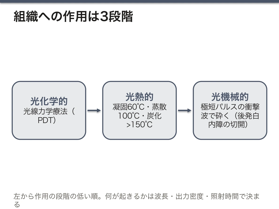
作用の段階——低い順に光化学的(光線力学療法)→光熱的(凝固 60℃・蒸散 100℃・炭化 >150℃)→光機械的/光破壊(極短パルスでプラズマを作り衝撃波で砕く。後発白内障の切開)。
選択的光熱融解——標的の熱緩和時間より短いパルスで照射すれば、熱が周りへ逃げる前に標的だけが壊れる。これが美容・血管レーザーの原理。
安全——クラス3B・4 は拡散反射でも危険。保護眼鏡は波長ごとに選ぶ(どの波長でも使える眼鏡は無い)。
English version
Core
What happens to the tissue is set by ① the wavelength (what absorbs it) ② the power density ③ the exposure time.
Wavelength and absorber — water absorbs the infrared strongly (CO₂ 10.6 μm, Er:YAG 2.94 μm → precise vaporisation of the surface, suited to incision). Haemoglobin absorbs green to yellow (KTP 532 nm, dye laser 585 nm → vascular lesions). Melanin absorbs broadly across the visible (Q-switched Nd:YAG, alexandrite → pigmented lesions, hair removal). Nd:YAG 1064 nm, being weakly absorbed, penetrates deeply and suits coagulation.
The stages of action — from the least: photochemical (photodynamic therapy) → photothermal (coagulation 60 °C, vaporisation 100 °C, carbonisation >150 °C) → photomechanical / photodisruptive (an ultrashort pulse makes a plasma and shatters by a shock wave; capsulotomy for after-cataract).
Selective photothermolysis — irradiate with a pulse shorter than the thermal relaxation time of the target and only the target is destroyed before the heat escapes. This is the principle of cosmetic and vascular lasers.
Safety — classes 3B and 4 are dangerous even by diffuse reflection. Protective glasses are chosen for the wavelength (no pair works for every wavelength).
低出力レーザー療法 — 何が分かっていて、何が分かっていないか
低出力レーザー療法(low-level laser therapy, LLLT。最近は光生物調節療法 photobiomodulation とも呼ぶ)は、赤〜近赤外(およそ 600〜1100 nm)の弱い光を当てて、組織を温めも壊しもせずに細胞の働きを変えようとする治療である。出力は mW〜数百 mW の桁で、熱で凝固させる中出力、蒸散させる高出力とはまったく別の作用を狙う。使う波長は光学的窓の中にあるので、皮膚の下の組織まで届く。
考えられている仕組みは、ミトコンドリアの呼吸鎖のシトクロム c オキシダーゼ(複合体 IV)が赤〜近赤外の光を吸収することである。光で酵素から一酸化窒素(NO)が外れて電子伝達が進み、ATP の産生が増え、活性酸素や NO を介した細胞内の信号が動く、と説明される。
効果は光の量に対して二相性を示すとされる——少なすぎれば効かず、多すぎるとかえって働きを抑える。波長・出力密度・照射時間・回数の組み合わせが研究ごとにばらばらなことが、結果がそろわない大きな理由になっている。
根拠の強さは用途によって大きく違う。教科書には「痛みや浮腫の軽減、創傷治癒の促進、微小循環の改善、脱毛の改善」などが並ぶことが多いが、どれも同じ重さで信じてよいわけではない。
比較的しっかりしているのは、がん治療に伴う口腔粘膜炎の予防である。造血幹細胞移植の前の大量化学療法や、頭頸部への放射線治療を受ける患者で、口の中に照射すると粘膜炎が減ることが複数のランダム化比較試験で示され、国際的ながん支持療法のガイドラインで勧められている。
首の痛みや一部の腱・関節の痛みでは、短い期間の痛みを減らすというメタ解析がある一方、効果を認めない試験もあり、照射条件をそろえた質の高い試験が足りない。腰痛・創傷治癒・美容目的の多くは、根拠が弱いか不十分である。米国で医療機器として認められた家庭用の育毛レーザーもあるが、それは主に安全性と既存の機器との同等性が審査されたという意味で、大きな効果が証明されたという意味ではない。
低出力でも、使う機器の多くはクラス 3B で、目に直接入れば網膜を傷めうるので保護眼鏡が要る。光感受性物質を使って腫瘍を壊す光線力学療法(PDT)とは別物である——PDT では外から入れた薬が光を吸収し、低出力レーザー療法では体にもともとある分子が光を吸収する。
English version
Core
Low-level laser therapy (LLLT, now also called photobiomodulation) shines weak red to near-infrared light (about 600–1100 nm) on tissue to change how cells work without heating or destroying the tissue. The power is in the range of milliwatts to a few hundred milliwatts, aiming at an effect quite different from coagulation (moderate power) or vaporisation (high power). The wavelengths lie inside the optical window, so the light reaches tissue under the skin.
The proposed mechanism is that cytochrome c oxidase (complex IV) of the mitochondrial respiratory chain absorbs red and near-infrared light. Light releases nitric oxide (NO) from the enzyme, electron transport speeds up, ATP production rises, and signals inside the cell are triggered through reactive oxygen species and NO.
The effect is said to be biphasic with dose: too little does nothing, and too much inhibits instead. Wavelength, power density, exposure time and number of sessions differ from study to study, and this is a major reason why results do not agree.
Going further
The strength of the evidence differs greatly between uses. Textbooks often list “less pain and swelling, faster wound healing, better microcirculation, less hair loss”, but these should not all be believed equally.
The most solid use is prevention of oral mucositis during cancer treatment. In patients receiving high-dose chemotherapy before stem cell transplantation, or radiotherapy to the head and neck, light applied inside the mouth reduced mucositis in several randomised controlled trials, and international supportive cancer care guidelines recommend it.
For neck pain and some tendon or joint pain, there are meta-analyses showing short-term pain relief, but there are also trials showing no effect, and good trials with standard settings are lacking. For low back pain, wound healing and most cosmetic uses, the evidence is weak or insufficient. Some home hair-growth lasers are accepted as medical devices in the USA, but this mainly means they were reviewed for safety and similarity to existing devices, not that a large effect was proven.
In the clinic
Even at low power, many devices are class 3B and can damage the retina if the beam enters the eye directly, so protective glasses are needed. It is not the same as photodynamic therapy (PDT), which destroys tumours with a photosensitiser: in PDT a drug given from outside absorbs the light, while in low-level laser therapy a molecule already in the body absorbs it.
回折と干渉 — 光が波であることの証拠
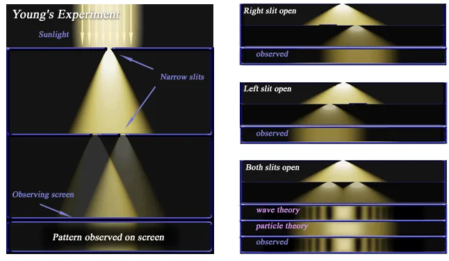
回折(diffraction)は、波が障害物の縁やすき間を通るとき、まっすぐ進むだけでなく影になるはずの所へ回り込む現象である。波面の各点が新しい小さな波の源になる(Huygens の原理)と考えると説明できる。回折がはっきり見えるのは、すき間の大きさが波長と同じくらいのときである。音(波長 約 1 m)は塀の向こうまで聞こえるが、光(波長 約 0.5 μm)は非常に細いすき間でないと回り込みが見えない。
干渉(interference)は、2つ以上の波が重なって強め合ったり弱め合ったりする現象である。2つの波の道のりの差(経路差)が
波長の整数倍(mλ)なら、山と山が重なって強め合う(constructive)——明るい
半波長の奇数倍((m + ½)λ)なら、山と谷が重なって弱め合う(destructive)——暗い
干渉の縞がはっきり見えるには、重なる波の位相の関係が一定でなければならない(可干渉性)。レーザー光は位相がそろっているので、簡単に縞が見える。Young は2本のすき間(二重スリット)で光の縞を見せ、光が波であることを示した。すき間の数を増やすほど明るい縞は細く鋭くなる——これが回折格子である。
身の回りの例——シャボン玉や水たまりの油膜の虹色(薄い膜の表と裏で反射した光の干渉で、膜の厚さによって強め合う色が変わる)、CD・DVD の虹色(細かい溝が回折格子になる)、眼鏡の反射防止コート(膜の中での波長の 1/4 の厚さにして、表と裏の反射光を弱め合わせる)、レーザーを壁に当てたときのざらざらした斑点(スペックル)、蝶やクジャクの羽の構造色。波であれば何にでも起こり、ノイズキャンセリングのヘッドホンは音の弱め合う干渉を使っている。
回折は顕微鏡の限界を決める。レンズで光を1点に集めても、回折のため点は波長程度の広がりを持つ。可視光の顕微鏡で見分けられるのは約 0.2 μm までで、多くのウイルスが光学顕微鏡で見えないのはこのためである。より細かく見るには、波長のずっと短い電子線(電子顕微鏡)や X 線を使う。DNA の二重らせんも、結晶に当てた X 線の回折像から決められた。
光干渉断層計(OCT)は、干渉を医療に使った代表である。可干渉性の短い近赤外光を、組織と参照鏡に分けて当て、戻ってきた2つの光の道のりがそろったときだけ干渉が起こることを使って、反射がどの深さから来たかを μm の精度で決める。網膜の層を断面で描き、黄斑浮腫・加齢黄斑変性・緑内障の神経線維層の菲薄化の診断に、眼科で日常的に使われている。
English version
Core
Diffraction is when a wave passing the edge of an obstacle or an opening does not only go straight but bends into the region that should be in shadow. It can be explained by thinking of every point on a wavefront as the source of a new small wave (Huygens’ principle). Diffraction is clearly seen when the opening is about the size of the wavelength. Sound (wavelength about 1 m) can be heard behind a wall, but light (wavelength about 0.5 μm) only bends visibly through very narrow slits.
Interference is when two or more waves overlap and strengthen or weaken each other. If the difference in path length of the two waves is
a whole number of wavelengths (mλ), crest meets crest and they reinforce each other (constructive): bright
an odd number of half wavelengths ((m + ½)λ), crest meets trough and they cancel (destructive): dark
To see clear fringes, the phase relation between the waves must stay fixed (coherence). Laser light is in phase, so fringes are easy to see. Young showed fringes of light with two slits (the double slit) and proved that light is a wave. The more slits, the narrower and sharper the bright fringes become; this is a diffraction grating.
Going further
Everyday examples: the rainbow colours of soap bubbles and oil films on puddles (interference of light reflected from the top and bottom of a thin film; the colour that is reinforced depends on the thickness), the rainbow on a CD or DVD (its fine grooves act as a grating), anti-reflection coatings on glasses (a layer a quarter of the wavelength thick inside the film makes the reflections from its two surfaces cancel), the grainy spots of a laser on a wall (speckle), and the structural colours of butterfly and peacock feathers. It happens with any wave; noise-cancelling headphones use destructive interference of sound.
Diffraction sets the limit of the microscope. Even when a lens focuses light to a point, diffraction spreads the point over about one wavelength. A visible-light microscope can separate details only down to about 0.2 μm, which is why most viruses cannot be seen with a light microscope. For finer detail, waves of much shorter wavelength are used: electron beams (electron microscope) or X-rays. The double helix of DNA was worked out from the X-ray diffraction pattern of a crystal.
In the clinic
Optical coherence tomography (OCT) is the leading medical use of interference. Near-infrared light of short coherence is split between the tissue and a reference mirror. Interference occurs only when the two returning paths are equal in length, and this tells from what depth each reflection comes, with micrometre precision. OCT draws the layers of the retina in cross-section and is used every day in ophthalmology to diagnose macular oedema, age-related macular degeneration and thinning of the nerve fibre layer in glaucoma.
回折格子でレーザーの波長と光子1個のエネルギーを測る
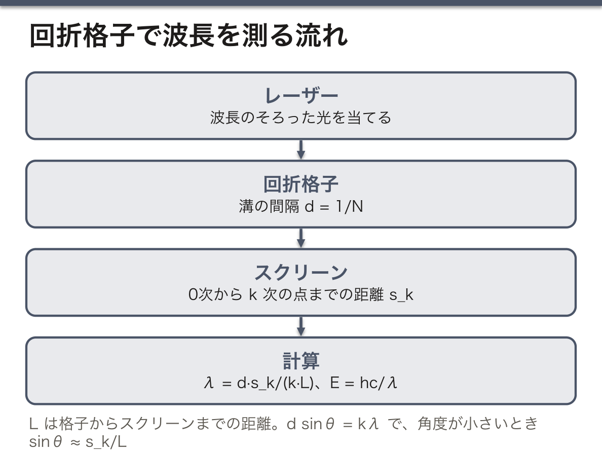
装置——光学台(長さ1 m 余り)の一端にレーザー、もう一端に mm 目盛りのスクリーンを置き、その間に回折格子を立てる。格子には 1 mm あたり N 本の細い溝が等間隔に刻まれていて、溝の間隔 d(格子定数)は d = 1/N である。たとえば 100 本/mm なら d = 0.01 mm = 1×10⁻⁵ m。
レーザー光を格子に当てると、スクリーンには中央の明るい点(0次)と、その左右に対称に並ぶ点(±1次、±2次…)が現れる。それぞれの点は、隣り合う溝から来た光の経路差が波長の整数倍になる向きにあり、
d sinθ = kλ(k = 1, 2, 3…)
が成り立つ。格子からスクリーンまでの距離を L、中央の点から k 次の点までの距離を s_k とすると、角度が小さいときは sinθ ≈ tanθ = s_k/L なので
λ = d·s_k / (k·L)
で波長が出る。波長が分かれば振動数は ν = c/λ、光子1個のエネルギーは E = hν = hc/λ(J)で、素電荷 1.6×10⁻¹⁹ C で割れば eV になる。たとえば λ = 650 nm の赤い光なら、ν ≈ 4.6×10¹⁴ Hz、E ≈ 3.1×10⁻¹⁹ J ≈ 1.9 eV である。
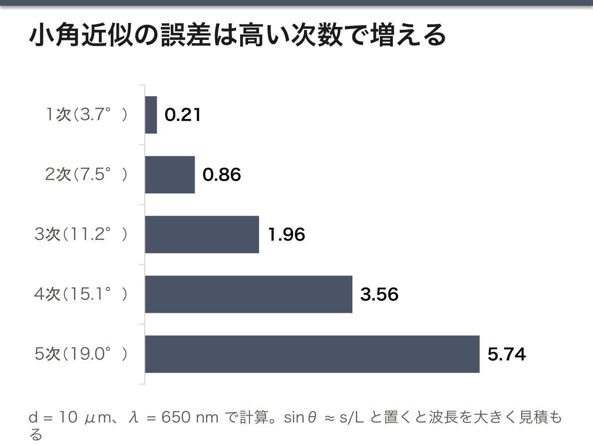
誤差を減らす工夫も物理から出てくる。①近似 sinθ ≈ s/L は角度が大きいほど狂う。d = 10 μm、λ ≈ 650 nm なら、1次(約 3.7°)では誤差 0.3% 未満だが、3次(約 11°)では波長を約 2% 大きく見積もる。高い次数を使うときは sinθ = s/√(s² + L²) で計算する。②点の中心はぼやけているので、+k 次と −k 次の点の間の距離を測って半分にすると、0次の位置の読み誤りが打ち消される。③L を長くとれば s も大きくなり、同じ目盛りの読み誤差でも相対誤差が小さくなる。④次数 k を変えて求めた λ がそろえば、測定が正しくできている確かめになる。
回折格子では波長の長い光ほど大きく曲がる(sinθ が λ に比例する)。白色光を当てると、プリズム(青のほうが大きく曲がる)とは逆の順に並んだ虹が見える。
光子1個のエネルギーを求める意味は、光が組織に何をするかが光子1個のエネルギーで決まる点にある。赤い光(約 1.9 eV)は分子の結合を切れないが、ArF エキシマレーザーの 193 nm の光子は約 6.4 eV で、結合を直接切って組織を削れる(光アブレーション)。
分光光度計や臨床検査の自動分析装置の中で、白色光から測定波長を取り出しているのも回折格子である。この測定は、その単色化の仕組みを逆向きに使って、光の波長を読んでいることになる。
English version
Core
Set-up. A laser is placed at one end of an optical bench (a little over 1 m long) and a screen with a millimetre scale at the other, with a diffraction grating standing between them. The grating has N fine grooves per millimetre at equal spacing, and the spacing d (the grating constant) is d = 1/N. For example, 100 lines/mm gives d = 0.01 mm = 1×10⁻⁵ m.
When the laser beam hits the grating, the screen shows a bright central spot (zero order) and spots placed symmetrically on both sides (±1st order, ±2nd order …). Each spot lies in the direction where the path difference between light from neighbouring grooves is a whole number of wavelengths:
d sinθ = kλ (k = 1, 2, 3 …)
If L is the distance from the grating to the screen and s_k the distance from the central spot to the k-th spot, then for small angles sinθ ≈ tanθ = s_k/L, so
λ = d·s_k / (k·L)
gives the wavelength. From it, the frequency is ν = c/λ and the energy of one photon is E = hν = hc/λ (in J); divide by the elementary charge 1.6×10⁻¹⁹ C to get eV. For red light of λ = 650 nm, ν ≈ 4.6×10¹⁴ Hz and E ≈ 3.1×10⁻¹⁹ J ≈ 1.9 eV.
Going further
Ways to reduce error also come from the physics. ① The approximation sinθ ≈ s/L gets worse at larger angles. With d = 10 μm and λ ≈ 650 nm, the error is below 0.3% for the first order (about 3.7°), but the third order (about 11°) overestimates the wavelength by about 2%. For higher orders, use sinθ = s/√(s² + L²). ② The centres of the spots are blurred, so measure the distance between the +k and −k spots and halve it; this cancels any error in reading the zero-order position. ③ A longer L makes s larger, so the same reading error on the scale gives a smaller relative error. ④ If λ found from different orders k agrees, this confirms that the measurement was done correctly.
In a grating, longer wavelengths bend more (sinθ is proportional to λ). With white light you see a rainbow in the opposite order to a prism, where blue bends more.
The point of finding the energy of one photon is that what light does to tissue depends on the energy of a single photon. Red light (about 1.9 eV) cannot break molecular bonds, but a 193 nm photon from an ArF excimer laser carries about 6.4 eV and can break bonds directly and remove tissue (photoablation).
In the clinic
Spectrophotometers and automated clinical analysers also use a diffraction grating to pick the measuring wavelength out of white light. This measurement uses the same principle the other way round, to read the wavelength of a light.
まとめ
| レーザーの原理 — 誘導放出と共振器 | LASER = Light Amplification by Stimulated Emission of Radiation。 |
|---|---|
| 医療用レーザーの選び方と安全 | レーザー波長吸収するもの用途CO₂10,600 nm水(表層で全部吸収)切開・蒸散(皮膚・耳鼻科)Nd:YAG1,064 nm組織深部まで届く深部凝固・後発白内障切開Ar(アルゴン… |
| レーザーと組織の相互作用 — なぜ波長で用途が決まるか | 組織に何が起きるかは、①波長(何に吸収されるか)②出力密度 ③照射時間 で決まる。 |
| 低出力レーザー療法 — 何が分かっていて、何が分かっていないか | 低出力レーザー療法(low-level laser therapy, LLLT。 |
| 回折と干渉 — 光が波であることの証拠 | 回折(diffraction)は、波が障害物の縁やすき間を通るとき、まっすぐ進むだけでなく影になるはずの所へ回り込む現象である。 |
| 回折格子でレーザーの波長と光子1個のエネルギーを測る | 装置——光学台(長さ1 m 余り)の一端にレーザー、もう一端に mm 目盛りのスクリーンを置き、その間に回折格子を立てる。 |
このノートは医学生が授業の内容を自分の言葉でまとめたもので、診療の判断に使うものではありません。誤りに気づいたらお知らせください。