光の進み方の変化から、溶けているものを測る2つの方法。屈折計は「どれだけ曲がるか」、旋光計は「偏光面がどれだけ回るか」を測る。
どちらも「試薬を使わず、光学的性質だけで濃度が分かる」点が共通する。臨床では尿比重と、かつての尿糖測定に使われた。
- 屈折計 — Snell の法則と全反射
- 鏡面反射と拡散反射 — 像が見えるか、面が見えるか
- 偏光の種類と作り方 — Brewster の法則
- Malus の法則と複屈折 — 偏光で測る・見分ける
- 旋光計 — 偏光面を回すのは不斉炭素
- 臨床で何を測っているか — 屈折率と旋光度の使いどころ
屈折計 — Snell の法則と全反射
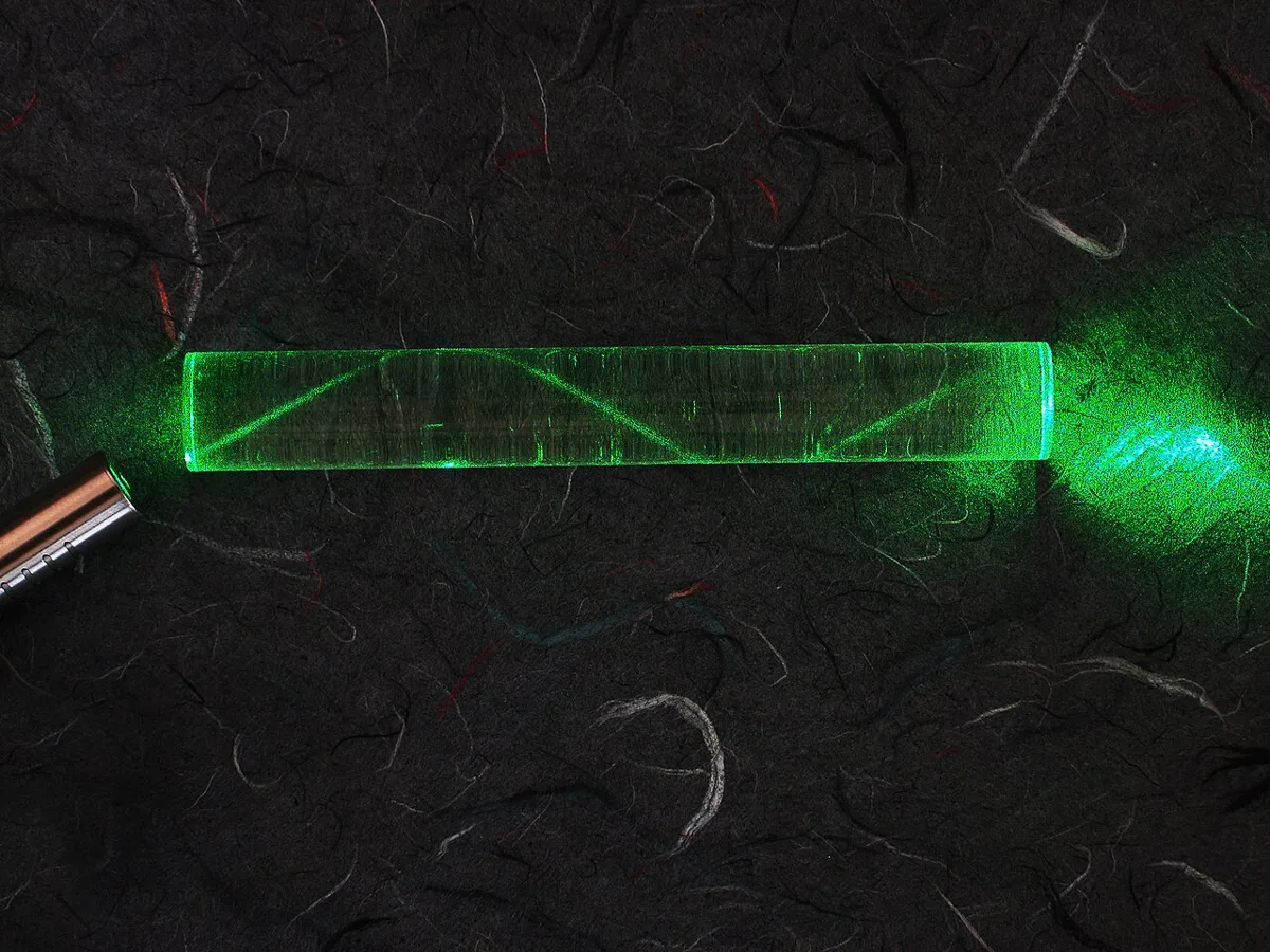
光が屈折率の違う媒質へ斜めに入ると進む向きが変わる。
n₁ sin θ₁ = n₂ sin θ₂(Snell の法則)
屈折率 n = c/v(真空中の光速 ÷ その媒質中の光速)。溶液の屈折率は溶質の濃度とともに直線的に上がるので、n を測れば濃度が出る。
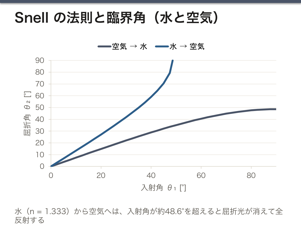
臨界角と全反射:密な媒質から疎な媒質へ向かうとき、入射角がある角度(臨界角 θc、sin θc = n₂/n₁)を超えると屈折光が消え、すべて反射する。実用の屈折計はこの臨界角を測っている——視野に明暗の境界が現れ、その位置が屈折率に対応する。
臨床応用は尿比重である。試験紙より正確で、少量(1〜2滴)で測れ、温度補正機能がある。ただし屈折率は「溶けているものの総量」を見るので、造影剤・高濃度の糖・蛋白があると偽高値になる(試験紙はイオン濃度を見るので、逆に糖と造影剤の影響を受けない)。2つの方法が測っているものが違うことを説明できるかが要点。
全反射は光ファイバーそのものである。コア(高い n)とクラッド(低い n)の境界で全反射を繰り返して光が閉じ込められる。内視鏡・ファイバースコープ・光凝固用のレーザー導光がすべてこの原理である。
眼科の屈折検査は同じ言葉を別の意味で使う。こちらは眼球全体の屈折力(D ジオプトリー = 1/焦点距離[m])で、角膜(約43 D)と水晶体(約19 D)を合わせて約60 D。用語が同じでも測っている量が違うので、混同しないこと。
English version
Core
When light enters obliquely a medium of different refractive index, its direction changes.
n₁ sin θ₁ = n₂ sin θ₂ (Snell’s law)
The refractive index n = c/v (the speed of light in vacuum ÷ its speed in the medium). The refractive index of a solution rises linearly with the concentration of solute, so measuring n gives the concentration.
The critical angle and total internal reflection: going from a denser to a less dense medium, beyond a certain angle of incidence (the critical angle θc, sin θc = n₂/n₁) the refracted ray disappears and all the light is reflected. A practical refractometer measures this critical angle — a boundary of light and dark appears in the field, and its position corresponds to the refractive index.
Going further
The clinical application is urine specific gravity. It is more accurate than a dipstick, needs only a small volume (one or two drops), and has temperature compensation. But the refractive index sees "the total amount of everything dissolved", so contrast medium, a high concentration of glucose or protein give a falsely high value (the dipstick sees ionic concentration and is conversely unaffected by glucose and contrast). The point is whether one can explain that the two methods measure different things.
Total internal reflection is the optical fibre itself. Light is confined by repeated total reflection at the boundary of core (high n) and cladding (low n). Endoscopes, fibrescopes and the delivery of laser light for photocoagulation all work on this principle.
Refraction testing in ophthalmology uses the same word in another sense. There it is the refractive power of the whole eye (D, dioptres = 1/focal length in m), the cornea (about 43 D) and the lens (about 19 D) together giving about 60 D. The same term, a different quantity measured — do not confuse them.
鏡面反射と拡散反射 — 像が見えるか、面が見えるか
反射の法則(反射角 = 入射角。入射光・反射光・法線は同じ平面の中)は、どんな面でも光線1本ごとには必ず成り立つ。2種類の反射を分けるのは面の凹凸の大きさである。
鏡面反射(specular reflection)——面の凹凸が波長よりずっと小さい(鏡・静かな水面・磨いた金属)。平行に入った光線は平行なまま1方向へ跳ね返るので、像ができる。銀めっきの鏡は光の 95% 以上を反射する。
拡散反射(diffuse reflection、乱反射)——面が波長の尺度で粗い(紙・壁・皮膚・布)。光線ごとに当たる小さな面の傾きが違うので、反射光はあらゆる方向へ散らばる。像はできないが、かわりにどこから見ても面そのものが見える。身の回りの物が見えるのは、ほとんどが拡散反射のおかげである。
実際の面は両方を混ぜて持ち、鏡面反射の成分が「つや」として見える。
面が濡れると鏡面反射が増える。水の膜が凹凸を埋めて面をなめらかにするからで、雨の路面はぎらつく。濡れた布や石の色が濃く暗く見えるのは、表面で白っぽく散らばっていた光が減り、内部に入って吸収される光が増えるからである。
理想的な拡散面は、どの方向から見ても同じ明るさに見える(Lambert 面)。照度の余弦則と対になる性質である。
非金属の面での鏡面反射光は部分的に偏光している。偏光フィルタでぎらつきを消せるのはこのためである(次の項目の Brewster の法則)。
内視鏡や腹腔鏡では、濡れた粘膜の鏡面反射(ハレーション)が白く光って病変を隠す。皮膚を拡大して見るダーモスコピーでは、皮膚の表面の反射を消すために、液体を挟んで角層表面の凹凸と空気の層を埋める(接触型)か、直交させた偏光フィルタで鏡面反射だけを除く(偏光型)。眼科の角膜反射法(Hirschberg 法)は、ペンライトの光が角膜で作る鏡面反射の点の位置から、斜視を見つける検査である。
English version
Core
The law of reflection (angle of reflection = angle of incidence; the incident ray, reflected ray and normal lie in one plane) holds for every single ray on any surface. What separates the two kinds of reflection is the size of the surface roughness.
Specular reflection: the roughness is much smaller than the wavelength (mirrors, still water, polished metal). Parallel rays stay parallel and bounce off in one direction, so an image forms. A silvered mirror reflects more than 95% of the light.
Diffuse reflection: the surface is rough on the scale of the wavelength (paper, walls, skin, cloth). Each ray meets a small facet with a different tilt, so the reflected light spreads in all directions. No image forms, but instead the surface itself can be seen from anywhere. Almost everything around us is seen thanks to diffuse reflection.
Real surfaces show both, and the specular part is seen as “gloss”.
Going further
A wet surface shows more specular reflection, because the water film fills the roughness and makes the surface smooth; that is why wet roads glare. Wet cloth or stone looks darker and deeper in colour because less light is scattered whitish from the surface and more enters and is absorbed.
An ideal diffuse surface looks equally bright from every direction (a Lambertian surface). This is the partner of the cosine law for illuminance.
Specular reflection from a non-metallic surface is partly polarised. This is why a polarising filter can remove glare (see Brewster’s law in the next item).
In the clinic
In endoscopy and laparoscopy, specular reflection from wet mucosa (halation) shines white and can hide lesions. In dermoscopy, which magnifies the skin, surface reflection is removed either by putting a liquid between lens and skin to fill the roughness and air of the surface layer (contact type), or by crossed polarising filters that remove only the specular reflection (polarised type). The corneal light reflex test (Hirschberg test) in ophthalmology detects strabismus from the position of the specular reflection that a penlight makes on the cornea.
偏光の種類と作り方 — Brewster の法則
光は横波の電磁波で、電場は進む向きに垂直に振動する。垂直な向きは無数にあるので、振動の向きに「選ぶ余地」がある——これが偏光である。音のような縦波は進む向きに振動するので偏光しない。偏光は光が横波である証拠である。
| 種類 | 電場の振動 |
|---|---|
| 無偏光(自然光) | 向きがでたらめに速く入れ替わる。太陽光・電球の光 |
| 直線偏光 | 1つの面の中だけで振動する |
| 円偏光 | 大きさの等しい直交する2つの直線偏光を 1/4 周期ずらして重ねたもの。電場の向きが回転する |
| 楕円偏光 | その中間。最も一般的な形 |
偏光を作る方法は4つある。①選択的な吸収——一方向にそろえた長い分子の膜(偏光板)が、分子の向きの振動を吸収し、それに垂直な振動を通す。②反射——下の Brewster の法則。③複屈折——結晶が偏光の向きで光を2本に分ける(次の項目)。④散乱——太陽から 90° の方向の青空の光は強く偏光している。
Brewster の法則——非金属の面で光が反射するとき、反射光と屈折光がちょうど 90° をなす入射角で、反射光は面に平行な向きに完全に偏光する。この角 θB は tan θB = n₂/n₁ で決まり、空気から水(n = 1.33)なら約 53°、ガラス(n = 1.5)なら約 56° である。
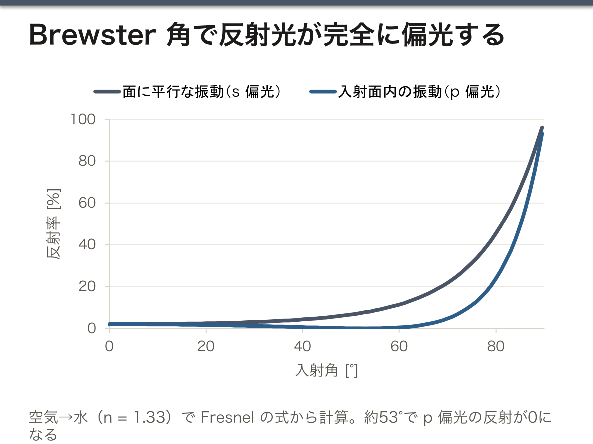
なぜその角度で完全に偏光するのか。反射光は、2つ目の媒質の中で光の電場に揺さぶられた電子が出し直した光である。揺れる電子(振動する双極子)は、自分が揺れている方向へは光を出せない。入射面の中で振動する成分は、屈折光の進む向きに垂直に揺れる。反射光と屈折光が 90° をなすとき、この揺れの向きがちょうど反射光の進む向きと一致するので、この成分は反射光に入れない。残るのは面に平行な振動だけになる。
水面や路面で反射したぎらつきは、主に水平に偏光している。偏光サングラスは透過軸を縦にしてこれを遮る。水面の反射が消えて水の中が見えるので、釣りやヨット・スキー・運転に使われる。
立体視の検査(Titmus ステレオテストなど)は、左右の目に違う向きの偏光で描いた2つの像を、偏光眼鏡で振り分けて見せる。両眼で1つに重ねて飛び出して見えるかどうかで、両眼視の働きを調べる。
English version
Core
Light is a transverse electromagnetic wave: the electric field vibrates perpendicular to the direction of travel. There are countless perpendicular directions, so there is a “choice” of vibration direction; this is polarisation. A longitudinal wave such as sound vibrates along the direction of travel and cannot be polarised. Polarisation is proof that light is a transverse wave.
| Type | Vibration of the electric field |
|---|---|
| Unpolarised (natural) | The direction changes randomly and quickly; sunlight, lamp light |
| Linear | Vibrates in a single plane only |
| Circular | Two equal linear waves at right angles, shifted by a quarter period; the field direction rotates |
| Elliptical | In between; the most general form |
There are four ways to make polarised light. ① Selective absorption: a film of long molecules lined up in one direction (a polariser) absorbs vibrations along the molecules and passes those perpendicular to them. ② Reflection: Brewster’s law, below. ③ Birefringence: a crystal splits light into two rays by polarisation (next item). ④ Scattering: skylight at 90° from the sun is strongly polarised.
Brewster’s law. When light reflects from a non-metallic surface, at the angle of incidence where the reflected and refracted rays are exactly 90° apart, the reflected light is completely polarised parallel to the surface. This angle θB is given by tan θB = n₂/n₁: about 53° from air to water (n = 1.33) and about 56° for glass (n = 1.5).
Going further
Why is it completely polarised at that angle? Reflected light is light re-emitted by electrons in the second medium that are shaken by the electric field. A vibrating electron (an oscillating dipole) cannot emit light along its own direction of vibration. The component vibrating in the plane of incidence moves perpendicular to the refracted ray. When the reflected and refracted rays are at 90°, this vibration points exactly along the reflected ray, so this component cannot enter the reflected light. Only the vibration parallel to the surface remains.
Glare reflected from water or roads is mainly horizontally polarised. Polarised sunglasses have a vertical transmission axis and block it. The reflection on the water disappears and you can see into the water, so they are used for fishing, sailing, skiing and driving.
In the clinic
Stereo vision tests (such as the Titmus stereo test) show each eye a different image drawn in light of different polarisation, separated by polarised glasses. Whether the patient fuses them into one image that seems to stand out tests binocular vision.
Malus の法則と複屈折 — 偏光で測る・見分ける
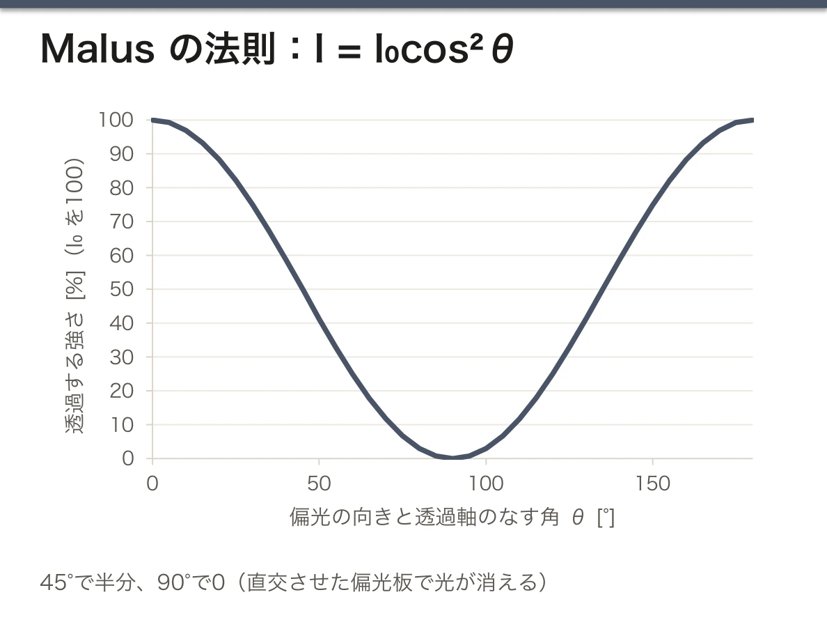
Malus の法則——直線偏光を偏光板(検光子)に通すと、透過する光の強さは
I = I₀ cos²θ
になる(θ=入ってくる光の偏光の向きと、偏光板の透過軸のなす角)。θ = 0° なら全部、45° なら半分(cos²45° = 1/2)、90° なら 0(直交させた偏光板で光が消える)。理由は単純で、偏光板は電場のうち透過軸に沿った成分 E cosθ だけを通し、光の強さは電場の2乗に比例するからである。無偏光の光を1枚目の偏光板に通すと、cos²θ をあらゆる向きで平均した 1/2 が通る。
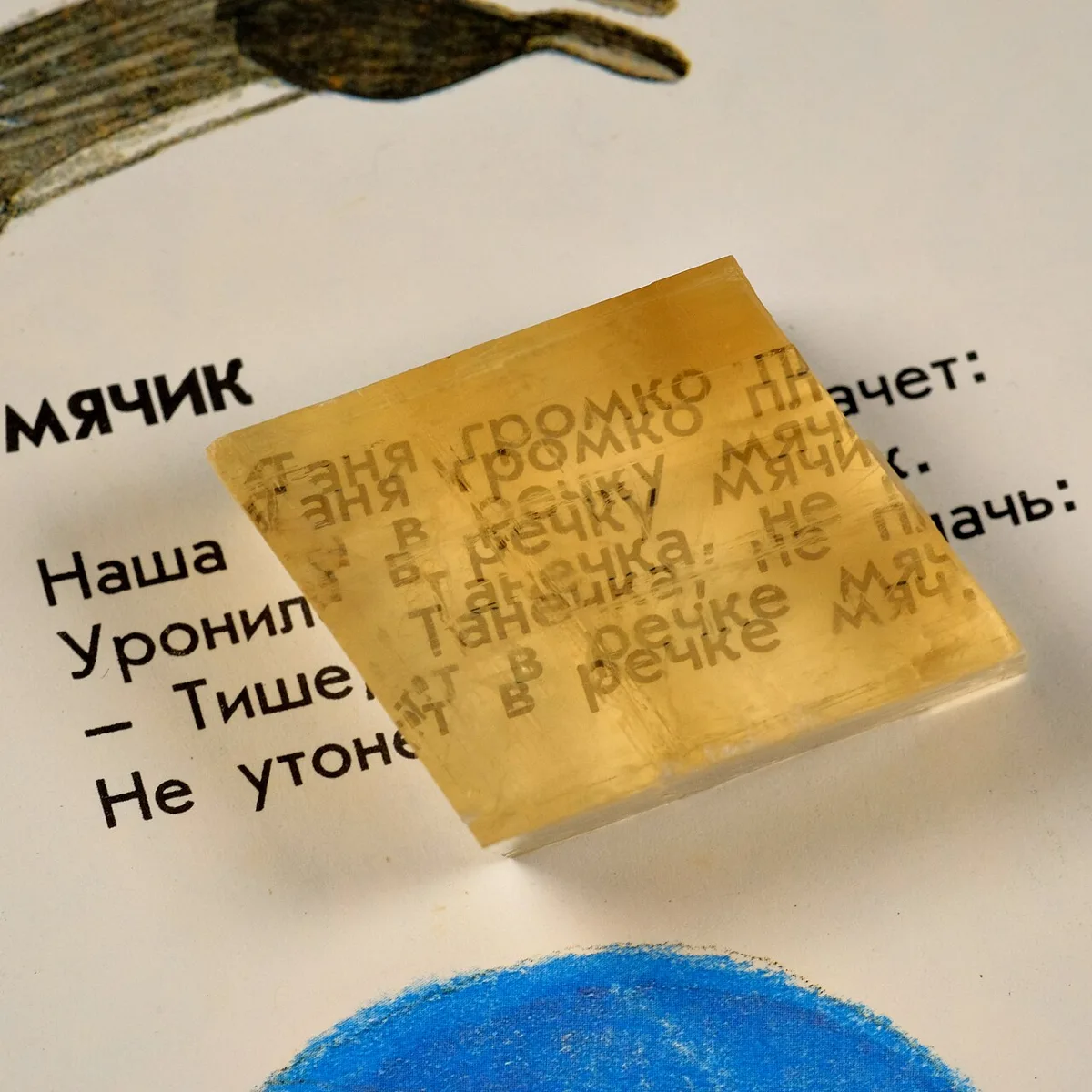
複屈折(birefringence、double refraction)——方解石のような異方性の結晶では、屈折率が偏光の向きによって違う。結晶に入った1本の光は、Snell の法則に従う常光線と従わない異常光線の2本に分かれ、2本は互いに直交する向きに偏光している。方解石を文字の上に置くと文字が二重に見える(ナトリウム D 線で常光線の屈折率は約 1.66、異常光線は約 1.49)。
旋光計に偏光が要る理由もここから分かる。旋光は「振動の面が回る」現象なので、はじめに振動の面が1つに決まっていなければ回転を測れない。自然光にはあらゆる面が混ざっていて、回っても何も変わらない。起偏子で面を1つに決め、検光子を回して Malus の法則による明るさの山(または消光の谷)の位置を読めば、試料が面を回した角度が分かる。
直交させた2枚の偏光板は光を通さないが、間に 45° の偏光板をもう1枚入れると光が通る(I₀/2 → I₀/4 → I₀/8)。間の板が光を「作った」のではなく、偏光の向きを2段階に分けて回したのである。複屈折する物質を直交させた偏光板の間に置いても、同じ理由で暗い背景の中に明るく浮かび上がる——これが偏光顕微鏡の原理である。
偏光顕微鏡は診断に使う。関節液の結晶では、痛風の尿酸ナトリウム結晶は針状で強い負の複屈折、偽痛風のピロリン酸カルシウム結晶は菱形〜棒状で弱い正の複屈折を示し、補償板を入れたときの色(黄か青か)で見分ける。アミロイドはコンゴーレッドで染めて偏光で見ると緑色(アップルグリーン)の複屈折を示す。骨格筋の横紋の A 帯(anisotropic。複屈折する)と I 帯(isotropic。しない)の名前も、偏光顕微鏡で見た性質から来ている。緑内障では、網膜神経線維層の複屈折から層の厚さを測る(走査レーザーポラリメトリー)。
English version
Core
Malus’s law. When linearly polarised light passes through a polariser (the analyser), the transmitted intensity is
I = I₀ cos²θ
where θ is the angle between the polarisation of the incoming light and the transmission axis of the polariser. At θ = 0° all of it passes, at 45° half (cos²45° = 1/2), and at 90° none (crossed polarisers block the light). The reason is simple: the polariser passes only the field component along its axis, E cosθ, and intensity is proportional to the square of the field. Unpolarised light through a first polariser gives 1/2, the average of cos²θ over all directions.
Birefringence (double refraction). In an anisotropic crystal such as calcite, the refractive index depends on the direction of polarisation. One ray entering the crystal splits into two: the ordinary ray, which obeys Snell’s law, and the extraordinary ray, which does not. The two rays are polarised at right angles to each other. A calcite crystal laid on text shows the letters double (at the sodium D line the ordinary index is about 1.66 and the extraordinary about 1.49).
Going further
This also explains why a polarimeter needs polarised light. Optical rotation is “the plane of vibration turning”, so you cannot measure a rotation unless there is one defined plane at the start. Natural light contains every plane, and turning it changes nothing. The polariser fixes one plane; turning the analyser and reading the position of the brightness peak (or the dark minimum) from Malus’s law gives the angle through which the sample turned the plane.
Two crossed polarisers pass no light, but if a third polariser at 45° is put between them, light gets through (I₀/2 → I₀/4 → I₀/8). The middle filter did not “create” light; it turned the polarisation in two steps. For the same reason, a birefringent material between crossed polarisers shines brightly on a dark background; this is the principle of the polarising microscope.
In the clinic
The polarising microscope is used in diagnosis. In joint fluid, monosodium urate crystals of gout are needle-shaped with strong negative birefringence, while calcium pyrophosphate crystals of pseudogout are rhomboid or rod-shaped with weak positive birefringence; they are told apart by their colour (yellow or blue) with a compensator. Amyloid stained with Congo red shows apple-green birefringence under polarised light. The A band (anisotropic, birefringent) and I band (isotropic, not birefringent) of skeletal muscle are named after how they look in the polarising microscope. In glaucoma, the thickness of the retinal nerve fibre layer is measured from its birefringence (scanning laser polarimetry).
旋光計 — 偏光面を回すのは不斉炭素
直線偏光を光学活性な物質の溶液に通すと、偏光面が回転する(旋光)。回転角は
α = [α] · l · c ([α]=比旋光度、l=光路長 dm、c=濃度 g/mL)
右へ回すものを右旋性(+, d)、左へ回すものを左旋性(−, l)という。旋光性を示すのは不斉炭素を持つ物質——生化学で扱う立体異性と同じ話である。
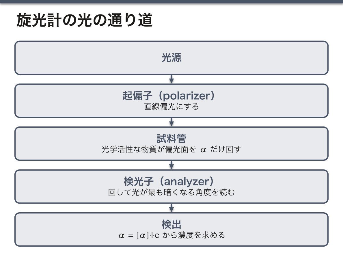
装置は光源 → 起偏子(polarizer)→ 試料管 → 検光子(analyzer)→ 検出。検光子を回して光が最も暗くなる角度を読む。
D/L と d/l(+/−)は別の分類である。D/L は構造(グリセルアルデヒドを基準にした配置)、d/l は実測した旋光の向き。両者は一致するとは限らない——たとえばD-フルクトースは左旋性(levulose と呼ばれる)。この不一致を説明できるかが理解の分かれ目で、「構造の分類と物理的性質は別物」という一般則の具体例になる。
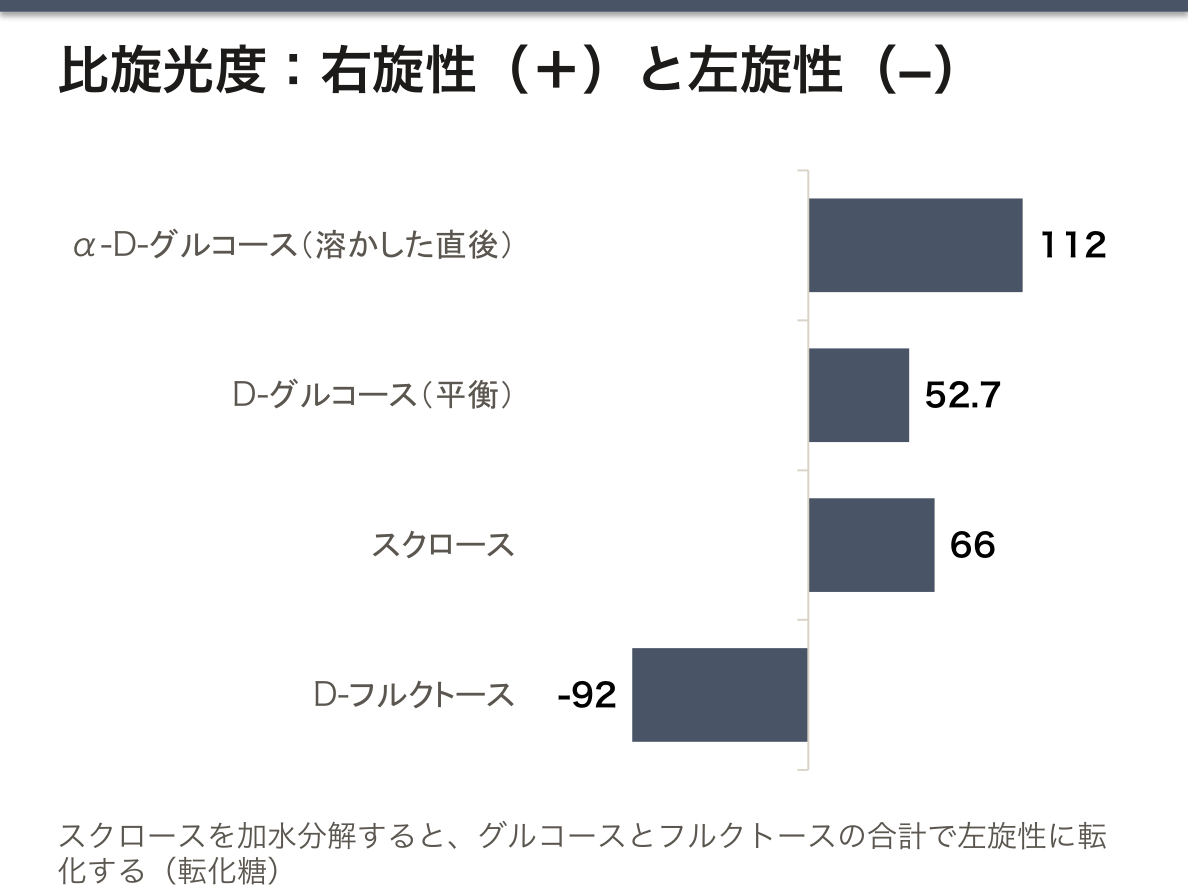
変旋光 mutarotation:結晶の α-D-グルコースを水に溶かすと比旋光度が +112° から +52.7° へ変化して落ち着く。α 型と β 型が開環形を経て平衡に達するためで、旋光度の時間変化から異性化を追える。
転化糖(invert sugar)という名前も旋光から来ている。スクロース(+66°)を加水分解するとグルコース(+52°)とフルクトース(−92°)になり、合計で左旋性に「転化」する。
かつて尿糖の定量に旋光計が使われたのは、グルコースが右旋性で試薬なしに測れたからである。現在は酵素法(グルコースオキシダーゼ)に置き換わったが、「光学的性質から濃度を出す」という発想は、後の分光光度計に受け継がれている。
English version
Core
Pass plane-polarised light through a solution of an optically active substance and the plane of polarisation rotates (optical rotation). The angle of rotation is
α = [α] · l · c ([α] = the specific rotation, l = path length in dm, c = concentration in g/mL)
What rotates to the right is dextrorotatory (+, d), to the left laevorotatory (−, l). What shows optical activity is a substance with an asymmetric carbon — the same matter of stereoisomerism taken in biochemistry.
The apparatus is source → polariser → sample tube → analyser → detection. The analyser is rotated and the angle at which the light is darkest is read.
Going further
D/L and d/l (+/−) are different classifications. D/L is structure (the configuration referred to glyceraldehyde); d/l is the measured direction of rotation. The two do not necessarily agree — D-fructose, for instance, is laevorotatory (hence "levulose"). Being able to explain this discrepancy divides real understanding, and it is a concrete instance of the general rule that a structural classification and a physical property are different things.
Mutarotation: dissolve crystalline α-D-glucose in water and the specific rotation changes from +112° and settles at +52.7°. The α and β forms reach equilibrium through the open-chain form, so isomerisation can be followed from the change of rotation with time.
The name invert sugar also comes from rotation. Hydrolyse sucrose (+66°) and it becomes glucose (+52°) and fructose (−92°), and the sum "inverts" to laevorotatory.
A polarimeter was formerly used to quantify urinary glucose, because glucose is dextrorotatory and could be measured without reagents. It has been replaced by an enzymatic method (glucose oxidase), but the idea of obtaining a concentration from an optical property was inherited by the later spectrophotometer.
臨床で何を測っているか — 屈折率と旋光度の使いどころ
屈折計——溶液の屈折率は溶質の濃度にほぼ比例する。だから屈折率を測れば濃度が分かる。
用途——①尿比重(脱水と腎の濃縮力の評価。試験紙より少量で速い)②血清総蛋白の簡易測定 ③房水・涙液の検査 ④眼科の屈折検査(屈折率ではなく眼の屈折力 [D] を測る点は別物)。
温度と波長で変わるので、ナトリウム D 線(589 nm)・20℃ を基準にする(nD20)。温度補正を言えることが要点。
旋光計——旋光度 α = [α]·l·c(比旋光度×光路長×濃度)。不斉炭素を持つ物質だけが偏光面を回す。
用途——糖の定量(かつての尿糖の測定。グルコースは右旋性で D-(+))。光学異性体の識別——薬理学的にはこれが本題で、鏡像異性体は効果も毒性も違いうる(サリドマイド)。
English version
Core
The refractometer — the refractive index of a solution is nearly proportional to the concentration of solute. So measuring the refractive index gives the concentration.
Uses — ① urine specific gravity (assessment of dehydration and of the concentrating power of the kidney; faster and on less volume than a dipstick) ② simple measurement of serum total protein ③ examination of aqueous humour and tears ④ refraction testing in ophthalmology (a separate matter, measuring the refractive power of the eye [D] rather than a refractive index).
It varies with temperature and wavelength, so the sodium D line (589 nm) at 20 °C is the reference (nD20). Being able to state the temperature correction is the point.
The polarimeter — the rotation α = [α]·l·c (specific rotation × path length × concentration). Only substances with an asymmetric carbon rotate the plane of polarisation.
Uses — quantifying sugars (the former measurement of urinary glucose; glucose is dextrorotatory, D-(+)). Identifying optical isomers — pharmacologically this is the main point, since enantiomers can differ in both effect and toxicity (thalidomide).
まとめ
| 屈折計 — Snell の法則と全反射 | 光が屈折率の違う媒質へ斜めに入ると進む向きが変わる。 |
|---|---|
| 鏡面反射と拡散反射 — 像が見えるか、面が見えるか | 反射の法則(反射角 = 入射角。入射光・反射光・法線は同じ平面の中)は、どんな面でも光線1本ごとには必ず成り立つ。 |
| 偏光の種類と作り方 — Brewster の法則 | 光は横波の電磁波で、電場は進む向きに垂直に振動する。 |
| Malus の法則と複屈折 — 偏光で測る・見分ける | Malus の法則——直線偏光を偏光板(検光子)に通すと、透過する光の強さはI = I₀ cos²θになる(θ=入ってくる光の偏光の向きと、偏光板の透過軸のなす角)。 |
| 旋光計 — 偏光面を回すのは不斉炭素 | 直線偏光を光学活性な物質の溶液に通すと、偏光面が回転する(旋光)。 |
| 臨床で何を測っているか — 屈折率と旋光度の使いどころ | 屈折計——溶液の屈折率は溶質の濃度にほぼ比例する。 |
このノートは医学生が授業の内容を自分の言葉でまとめたもので、診療の判断に使うものではありません。誤りに気づいたらお知らせください。