音は媒質の圧力の波である。空気を伝わって耳に届けば聴覚になり、周波数を人の聞こえない高さまで上げて体に送れば超音波検査になる。どちらも同じ物理——振動が媒質を伝わり、境界で反射し、進むうちに弱まる——で説明できる。
この章の前半は音と聴覚:波の基本、音の強さとデシベル、耳が強さをどう感じるか。後半は超音波:圧電素子で音をつくり、組織の境界からのエコーで像を描き、ドプラ効果で血流を測る。
鍵になる量は2つ。v = fλ(速さ=周波数×波長)と、音響インピーダンス Z = ρc。前者は「なぜ高い周波数ほど細かく見えるのに深くは見えないか」を、後者は「なぜゼリーを塗るのか」「なぜ骨とガスの奥が見えないか」を説明する。
- 波の基本 — 運ばれるのはエネルギーで、物質ではない
- 音の性質と可聴域 — 高さ・大きさ・音色
- 音の強さとデシベル — 10 dB で強さは10倍
- 耳はどう聞くか — 対数で感じ、中耳で整合する
- 超音波の発生と検出 — 同じ結晶が送って受ける
- 音響インピーダンスと反射 — なぜゼリーを塗るのか
- 減衰と周波数の選び方 — 細かく見るか、深く見るか
- ドプラ法 — 周波数のずれから血流の速さを測る
波の基本 — 運ばれるのはエネルギーで、物質ではない
波は、振動(乱れ)が空間を伝わっていく現象である。媒質の粒子はその場で振動するだけで、波と一緒に運ばれるのはエネルギーである(スタジアムのウェーブで、人は席から動かない)。
| 分け方 | 種類 |
|---|---|
| 媒質が要るか | 力学的波(媒質が必要:音・水の波)/電磁波(真空も伝わる:電波・光・X線) |
| 粒子の振動の向き | 縦波(進む向きと平行:音)/横波(進む向きと垂直:弦・水面の波) |
空気中の音は力学的な縦波で、密な部分(圧縮)と疎な部分(膨張)が交互に進む。
基本の関係は v = fλ(速さ=振動数×波長)。波長は隣り合う同じ状態の点(山と山)の間隔で、振幅とは別物である。
なぜ音は縦波なのか。気体や液体は「押す・引く」には抵抗する(圧縮されると戻ろうとする)が、「ずらす」には抵抗しない。横にずらす力(せん断)に復元力がない媒質では横波は伝わらない。固体はせん断にも抵抗するので、縦波と横波の両方が伝わる。
2つ以上の波が同じ場所を通るとき、各点の変位はそれぞれの波の変位の和になる(重ね合わせの原理)。山と山が重なれば強め合い、山と谷が重なれば弱め合う(干渉)。
速さは媒質で決まり、周波数を変えても変わらない。だから同じ媒質で周波数を上げると、波長が短くなる。
English version
Core
A wave is a vibration (disturbance) traveling through space. The particles of the medium only vibrate in place; what travels with the wave is energy (in a stadium wave, people stay in their seats).
| Division | Types |
|---|---|
| Need for a medium | Mechanical waves (need a medium: sound, water waves) / electromagnetic waves (travel through vacuum: radio, light, X-rays) |
| Direction of particle vibration | Longitudinal (parallel to travel: sound) / transverse (perpendicular to travel: strings, water surface) |
Sound in air is a mechanical longitudinal wave: dense regions (compressions) and thin regions (rarefactions) move forward in turn.
The basic relation is v = fλ (speed = frequency × wavelength). The wavelength is the distance between neighboring points in the same state (crest to crest); it is not the amplitude.
Going further
Why is sound longitudinal? Gases and liquids resist “push and pull” (they spring back when compressed) but not “sliding”. In a medium with no restoring force against sideways sliding (shear), transverse waves cannot travel. Solids also resist shear, so both longitudinal and transverse waves travel in them.
When two or more waves pass the same place, the displacement at each point is the sum of the displacements of each wave (the superposition principle). Crest on crest strengthens; crest on trough weakens (interference).
The speed is set by the medium and does not change with frequency. So in the same medium, raising the frequency shortens the wavelength.
音の性質と可聴域 — 高さ・大きさ・音色
音の3要素は物理量に対応する。高さ(pitch)は振動数、大きさは振幅(強さ)、音色は波形(含まれる倍音の割合)。振動数が高い=周期が短いほど高い音に聞こえる。
人が聞こえる範囲(可聴域)はおよそ 20 Hz〜20 kHz。それより低い音を超低周波音(infrasound)、高い音を超音波(ultrasound)という。上限は年齢とともに下がる。
音の速さは媒質で決まる:空気 約340 m/s(20 ℃)、水 約1480 m/s、軟部組織 約1540 m/s、骨 約3000〜4000 m/s。硬く(圧縮しにくく)、軽い媒質ほど速い。
例:空気中の 1 kHz の音の波長は 340/1000 ≈ 0.34 m。同じ 1 kHz でも水中では 1.5 m になる。周波数は音源が決め、速さは媒質が決め、波長はその結果として決まる。
人の声の大事な成分はおよそ 250 Hz〜4 kHz にあり、子音の聞き分けは高い側(2〜4 kHz)に頼る。高い音から聞こえなくなると、音は聞こえるのに言葉が聞き取れないという訴えになる。
聴診器は周波数を選んで聞く道具である。ベル型は低い音(Ⅲ音・Ⅳ音、僧帽弁狭窄の拡張期ランブル)、膜型は高い音(Ⅰ音・Ⅱ音、大動脈弁逆流の雑音、肺の音)に向く。打診は叩いた部位の振動の響き方を聞く:含気の多い肺は清音、実質臓器や液体は濁音、胃や腸のガスは鼓音。気胸では打診が過共鳴になり、胸水では濁音になる。
English version
Core
The three qualities of sound match physical quantities: pitch is frequency, loudness is amplitude (intensity), and timbre is waveform (the mix of overtones). The higher the frequency (the shorter the period), the higher the pitch.
The range people can hear (audible range) is about 20 Hz to 20 kHz. Lower sounds are infrasound; higher sounds are ultrasound. The upper limit falls with age.
The speed of sound depends on the medium: air about 340 m/s (20 °C), water about 1480 m/s, soft tissue about 1540 m/s, bone about 3000–4000 m/s. The stiffer (harder to compress) and lighter the medium, the faster.
Going further
Example: a 1 kHz sound in air has a wavelength of 340/1000 ≈ 0.34 m. The same 1 kHz in water has a wavelength of 1.5 m. The source sets the frequency, the medium sets the speed, and the wavelength follows from both.
The important parts of the human voice are roughly 250 Hz to 4 kHz, and telling consonants apart relies on the high side (2–4 kHz). When high sounds go first, the complaint becomes “I can hear sound but cannot understand words”.
In the clinic
A stethoscope is a tool for listening by frequency. The bell is for low sounds (S3, S4, the diastolic rumble of mitral stenosis); the diaphragm is for high sounds (S1, S2, the murmur of aortic regurgitation, lung sounds). Percussion listens to how the tapped part resonates: air-filled lung gives a resonant note, solid organs and fluid a dull note, and gas in the stomach or bowel a tympanic note. Pneumothorax gives hyperresonance; pleural effusion gives dullness.
音の強さとデシベル — 10 dB で強さは10倍
音の強さ(intensity)は、音の進む向きに垂直な単位面積を1秒間に通るエネルギーである。
I = P/A 単位 W/m2(P:音のパワー)
強さは振幅の2乗に比例し(振幅2倍→強さ4倍)、点音源からは距離の2乗に反比例して弱まる(逆2乗則)。
人が聞こえる強さは 10−12 W/m2 から痛みを感じる 1 W/m2 付近まで、1兆倍にわたる。そこで対数を使った音の強さのレベルで表す。
L = 10·log10(I/I0) [dB] (I0 = 10−12 W/m2)
デシベルは単位というより「比」である。2つの強さを比べた比の常用対数を10倍したもの。だから計算は掛け算が足し算になる。
覚えておく値:+10 dB = 強さ10倍、+20 dB = 100倍、+3 dB ≈ 2倍。「10 dB ごとに強さが2倍」は誤り(10倍である)。距離が2倍になると強さは1/4で、約 −6 dB。
目安:ささやき約30 dB、会話約60 dB、地下鉄の車内約80〜90 dB、痛みの閾値約120 dB。60 dB と 90 dB の差は30 dB、つまり強さで1000倍である。
騒音による内耳の障害は、強さ(dB)と曝露時間の積で決まる。85 dB 程度の騒音でも長時間続けば聴力を損なうので、職場の騒音管理やイヤホンの音量には上限の目安が設けられている。
English version
Core
Sound intensity is the energy passing each second through a unit area perpendicular to the direction of the sound.
I = P/A, unit W/m2 (P: sound power)
Intensity is proportional to the square of the amplitude (twice the amplitude → four times the intensity), and from a point source it falls with the square of the distance (inverse square law).
Audible intensities range from 10−12 W/m2 to about 1 W/m2, where it starts to hurt — a trillion times. So we use a logarithmic sound intensity level.
L = 10·log10(I/I0) [dB] (I0 = 10−12 W/m2)
Going further
The decibel is a ratio more than a unit: ten times the common logarithm of the ratio of two intensities. So multiplication becomes addition.
Values to remember: +10 dB = 10 times the intensity, +20 dB = 100 times, +3 dB ≈ 2 times. “Intensity doubles every 10 dB” is wrong (it is ten times). Twice the distance gives a quarter of the intensity, about −6 dB.
Rough values: whisper about 30 dB, conversation about 60 dB, inside a subway train about 80–90 dB, pain threshold about 120 dB. The difference between 60 dB and 90 dB is 30 dB — 1000 times the intensity.
In the clinic
Noise damage to the inner ear depends on intensity (dB) multiplied by exposure time. Even noise around 85 dB harms hearing if it continues for a long time, so workplace noise control and earphone volume have recommended upper limits.
耳はどう聞くか — 対数で感じ、中耳で整合する
耳の感じ方は強さに比例しない。強さが10倍(+10 dB)になると、大きさはおよそ2倍に感じる——反応は線形ではなく対数に近い。
また同じ強さでも周波数で聞こえ方が違う。人の耳は2〜5 kHz 付近で最も敏感で、低い音や非常に高い音は強くしないと同じ大きさに聞こえない。この主観的な大きさをラウドネスといい、1 kHz の音の dB 値に合わせた単位フォン(phon)で表す。等しく聞こえる点を結んだのが等ラウドネス曲線である。
聴力検査(audiometry)では、周波数ごとに聞こえる最小の音(閾値)を測り、正常な耳の閾値を基準にした値(dB HL)でグラフにする(オージオグラム)。
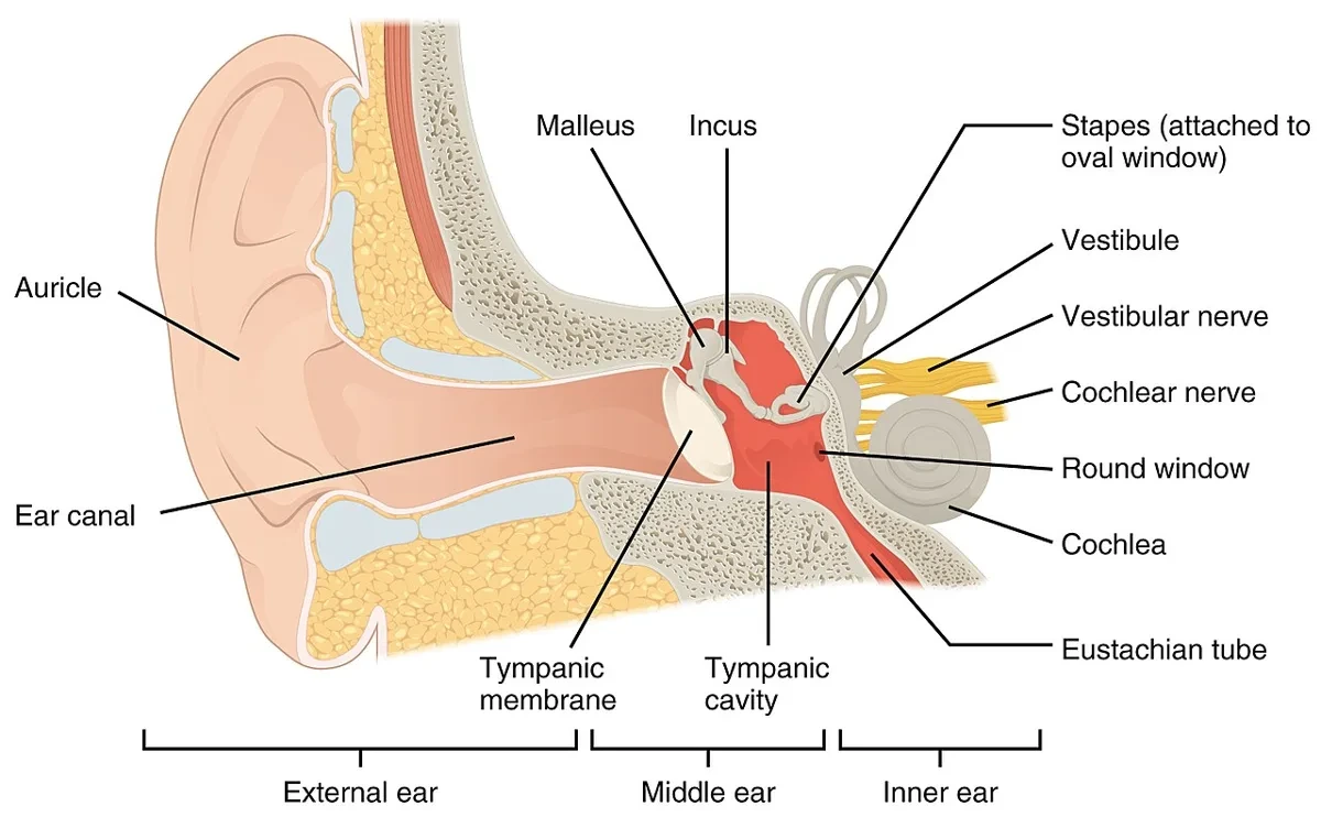
なぜ 3 kHz 付近で敏感なのか。外耳道は長さ約2.5 cm の片側の閉じた管で、波長の1/4がちょうど管の長さに合う音(約3〜4 kHz)で共鳴し、鼓膜での音圧が高まる。
中耳はインピーダンス整合器である。空気から内耳の液体へ音が直接入ると、音響インピーダンスの差でエネルギーの約99.9%が反射してしまう。鼓膜(membrana tympanica)の有効面積はあぶみ骨底の約17倍あり、耳小骨(ossicula auditus)のてこ作用(約1.3倍)と合わせて音圧を約20倍に高めて内耳に送る。
対数で感じることにも意味がある。弱い音には敏感に、強い音には鈍く反応することで、1兆倍の範囲を1つの感覚器で扱える。
オージオグラムで気導と骨導の差(気骨導差)があれば伝音難聴(外耳・中耳の障害)、両方が同じだけ悪ければ感音難聴(内耳・神経)。騒音性難聴は 4 kHz 付近の落ち込み(c5 dip)、加齢性難聴は高音域から両側対称に悪くなるのが典型である。音叉を使う Weber 法・Rinne 法も同じ区別をする。
English version
Core
The ear does not sense loudness in proportion to intensity. When intensity goes up ten times (+10 dB), sound seems about twice as loud — the response is close to logarithmic, not linear.
Also, at the same intensity, hearing depends on frequency. The human ear is most sensitive around 2–5 kHz; low sounds and very high sounds must be stronger to seem equally loud. This subjective loudness is measured in phons, matched to the dB value of a 1 kHz tone. Lines joining equally loud points are equal-loudness contours.
Audiometry measures the softest sound you can hear (the threshold) at each frequency and plots it relative to normal ears (dB HL) — the audiogram.
Going further
Why so sensitive near 3 kHz? The ear canal is a tube about 2.5 cm long, closed at one end. It resonates at the sound whose quarter wavelength matches the tube length (about 3–4 kHz), which raises the pressure at the eardrum.
The middle ear is an impedance-matching device. If sound went straight from air into the fluid of the inner ear, about 99.9% of the energy would be reflected because of the difference in acoustic impedance. The eardrum (membrana tympanica) has about 17 times the area of the stapes footplate, and together with the lever action of the ossicles (ossicula auditus, about 1.3 times) it raises the sound pressure about twenty times before passing it to the inner ear.
Logarithmic sensing also has a purpose. Being sensitive to weak sounds and less sensitive to strong ones lets a single sense organ cover a range of a trillion.
In the clinic
On an audiogram, a gap between air and bone conduction (air–bone gap) means conductive hearing loss (outer or middle ear); if both are equally poor, it is sensorineural hearing loss (inner ear or nerve). Noise-induced hearing loss shows a dip around 4 kHz (the c5 dip), and age-related hearing loss typically worsens symmetrically from the high frequencies. The Weber and Rinne tuning-fork tests make the same distinction.
超音波の発生と検出 — 同じ結晶が送って受ける
超音波は 20 kHz を超える音で、診断にはおよそ 1〜15 MHz が使われる。超音波は波そのもの、超音波検査(ultrasonography)はそれで体の中を描く方法を指す。体内では縦波として伝わる。
発生と検出には圧電効果(piezoelectric effect)を使う。ある種の結晶は、押されると電圧を生じ(正圧電効果)、電圧をかけると変形する(逆圧電効果)。プローブの結晶に交流電圧をかけると結晶が伸び縮みして超音波を出し、戻ってきたエコーが結晶をゆらすと電圧が生じて受信できる。同じ結晶が送信と受信の両方を担う。
パルス・エコー法:プローブはごく短いパルスを送ったあと、ほとんどの時間を「聞く」ことに使う。エコーが返るまでの時間 t から深さは
d = c·t/2(往復なので2で割る)
装置は軟部組織の音速を 1540 m/s と仮定して距離を計算する。1 cm の深さからの往復は約13 μs。音速が仮定と違う組織(脂肪や骨)を通ると、位置がわずかにずれて描かれる。
なぜ MHz なのか。細かいものを見分ける力(分解能)は波長で決まる。1540 m/s で 5 MHz なら λ ≈ 0.3 mm。可聴音(数 cm〜数 m)ではとても臓器の構造は見えない。
超音波は電離放射線を使わないので、妊婦・小児に安心して繰り返し使え、ベッドサイドで即座に動きを見られる。そのため胎児の観察、心臓・腹部・甲状腺・乳腺・血管の検査、穿刺の誘導など、使われる範囲はきわめて広い。ただしエネルギーは熱として吸収されるので、出力と時間には安全の目安がある。
English version
Core
Ultrasound is sound above 20 kHz; diagnosis uses roughly 1–15 MHz. Ultrasound means the waves themselves, and ultrasonography means the method of imaging the body with them. In the body it travels as a longitudinal wave.
It is produced and detected by the piezoelectric effect. Some crystals produce a voltage when pressed (direct effect) and deform when a voltage is applied (reverse effect). An alternating voltage on the crystal in the probe makes it expand and contract and send out ultrasound; a returning echo shakes the crystal, which produces a voltage that is received. The same crystal both sends and receives.
Going further
The pulse–echo method: the probe sends a very short pulse and then spends most of its time “listening”. From the time t until the echo returns, the depth is
d = c·t/2 (divide by 2 because it is a round trip)
The machine assumes a soft-tissue speed of 1540 m/s. The round trip from 1 cm deep takes about 13 μs. Passing through tissue with a different speed (fat or bone) shifts the position slightly in the image.
Why MHz? The ability to tell fine things apart (resolution) is set by the wavelength. At 1540 m/s and 5 MHz, λ ≈ 0.3 mm. Audible sound (centimeters to meters) could never show the structure of organs.
In the clinic
Ultrasound uses no ionizing radiation, so it can be repeated safely in pregnancy and in children, and it shows movement at once at the bedside. Its uses are very wide: watching the fetus, examining the heart, abdomen, thyroid, breast and vessels, and guiding needles. But its energy is absorbed as heat, so there are safety guides for output and time.
音響インピーダンスと反射 — なぜゼリーを塗るのか
音響インピーダンス Z は、媒質が音の伝わりに示す「抵抗」のような量で、密度と音速の積である。
Z = ρ·c 単位 kg/(m2·s)
厚さにはよらない。2つの媒質の境界では、Z の差が大きいほど多く反射する。強さの反射率と透過率は
R = [(Z2 − Z1)/(Z2 + Z1)]2、T = 1 − R
Z が近い境界ではほとんど反射せず、そのまま奥へ進む。
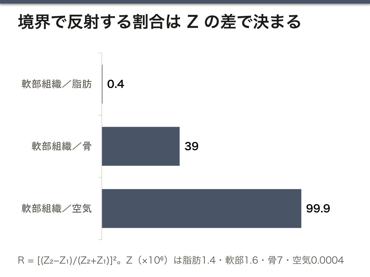
おおよその Z(×106 kg/(m2·s)):空気 0.0004、脂肪 1.4、軟部組織 1.6、骨 約7。
| 境界 | 反射 | 画像では |
|---|---|---|
| 軟部組織どうし | ごくわずか | 灰色の濃淡。組織の内部構造が見える |
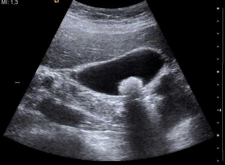
| 軟部組織/骨 | 約4割 | 表面が白く光り、奥は暗い影(音響陰影) |
| 軟部組織/空気 | ほぼ100% | 奥は何も見えない |
大事なのは差の大きさで、増えるか減るかは問わない。画像の白さ(エコーの強さ)は境界での Z の変化の大きさを表している。
プローブと皮膚の間にわずかでも空気が残ると、超音波はそこでほぼ全部はね返る。ゼリーは空気を追い出して Z の差を小さくするために塗る(水とプロピレングリコールが主成分)。同じ理由で、腸管ガスの奥や肺は見えにくく、結石や骨の奥には音響陰影ができる。逆に、胆嚢結石の後ろの陰影はそれ自体が診断の手がかりになる。
English version
Core
Acoustic impedance Z is a kind of “resistance” of a medium to the passage of sound. It is the product of density and the speed of sound.
Z = ρ·c, unit kg/(m2·s)
It does not depend on thickness. At a boundary between two media, the larger the difference in Z, the more is reflected. The intensity reflection and transmission coefficients are
R = [(Z2 − Z1)/(Z2 + Z1)]2, T = 1 − R
At a boundary with similar Z, almost nothing is reflected and the wave goes on deeper.
Going further
Approximate Z (×106 kg/(m2·s)): air 0.0004, fat 1.4, soft tissue 1.6, bone about 7.
| Boundary | Reflection | In the image |
|---|---|---|
| Soft tissue / soft tissue | Very small | Shades of gray; the inner structure of tissues is visible |
| Soft tissue / bone | About 40% | A bright surface with a dark shadow behind (acoustic shadow) |
| Soft tissue / air | Almost 100% | Nothing visible behind |
What matters is the size of the change, not whether Z goes up or down. The brightness of an echo shows how large the change in Z is at that boundary.
In the clinic
If even a little air stays between the probe and the skin, almost all the ultrasound bounces back there. Gel is used to push out the air and make the difference in Z small (its main parts are water and propylene glycol). For the same reason, it is hard to see behind bowel gas and into the lungs, and stones and bone cast acoustic shadows. In turn, the shadow behind a gallstone is itself a clue to the diagnosis.
減衰と周波数の選び方 — 細かく見るか、深く見るか
超音波は組織を進むうちに弱まる(減衰 attenuation)。原因は4つ:吸収(エネルギーが熱に変わる)、散乱(小さな不均一で四方に散る)、反射(境界ではね返る)、屈折(音速の違う媒質で曲がる)。
軟部組織での減衰はおよそ 0.5 dB/(cm·MHz)、つまり周波数が高いほど、深いほど大きい。
画像の種類:Bモード(2次元の白黒の断面。毎秒約30枚でリアルタイム)、Mモード(1本の線上の動きを時間で追う。心臓の弁や壁)、3D(2次元像の組み合わせ)、4D(3D+時間)。
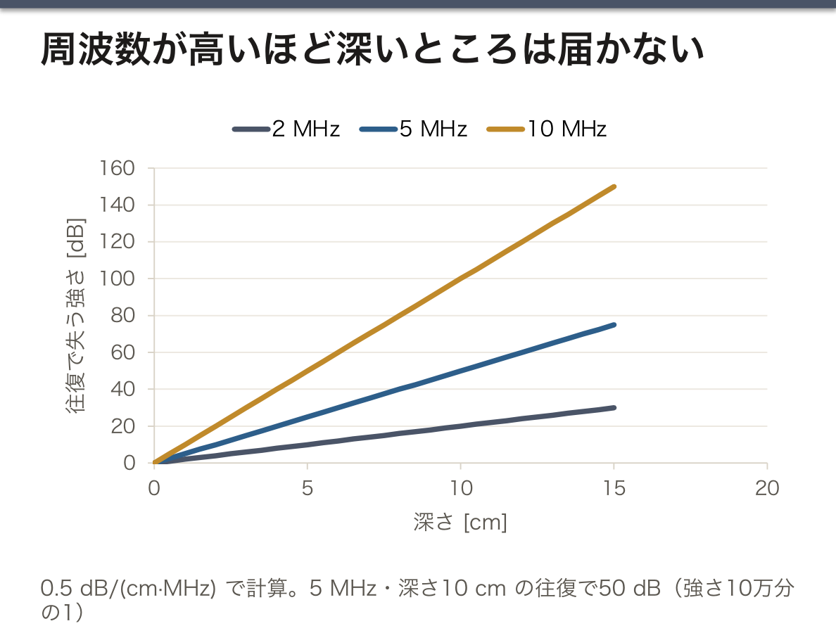
周波数の選択はトレードオフである。周波数を上げると波長が短くなり分解能は上がるが、減衰が大きくなって深くまで届かない。
| 周波数 | 得意なもの |
|---|---|
| 低め(2〜5 MHz) | 深い臓器:腹部、心臓、妊娠子宮 |
| 高め(7〜15 MHz) | 浅い臓器:甲状腺、乳腺、頸動脈、筋骨格、皮膚 |
例:5 MHz で深さ 10 cm を往復すると、20 cm × 0.5 × 5 = 50 dB、強さは10万分の1になる。装置は深いエコーほど増幅を強めて(深さ補正)、明るさをそろえている。
超音波のエネルギーを治療に使うこともある。理学療法の超音波治療は深部組織を温めて血流を増やし、痛みの軽減や組織の回復を助ける。強く集束させた超音波(HIFU)は焦点だけを高温にして組織を壊す治療に用いられる。
English version
Core
Ultrasound weakens as it travels through tissue (attenuation). There are four causes: absorption (energy turns into heat), scattering (spreading in all directions from small irregularities), reflection (bouncing back at boundaries) and refraction (bending in a medium with a different speed).
Attenuation in soft tissue is about 0.5 dB/(cm·MHz): the higher the frequency and the deeper, the greater the loss.
Image types: B-mode (a 2D gray-scale cross-section, about 30 frames per second in real time), M-mode (motion along one line over time — heart valves and walls), 3D (combined 2D images) and 4D (3D plus time).
Going further
Choosing the frequency is a trade-off. A higher frequency gives a shorter wavelength and better resolution, but stronger attenuation, so it does not reach deep.
| Frequency | Best for |
|---|---|
| Lower (2–5 MHz) | Deep organs: abdomen, heart, pregnant uterus |
| Higher (7–15 MHz) | Superficial organs: thyroid, breast, carotid artery, muscles and joints, skin |
Example: a round trip to 10 cm at 5 MHz loses 20 cm × 0.5 × 5 = 50 dB, so the intensity becomes one hundred-thousandth. The machine amplifies deeper echoes more (depth compensation) to even out the brightness.
In the clinic
Ultrasound energy is also used for treatment. Therapeutic ultrasound in physiotherapy warms deep tissue, increases blood flow and helps reduce pain and heal tissue. Strongly focused ultrasound (HIFU) heats only the focus and is used to destroy tissue.
ドプラ法 — 周波数のずれから血流の速さを測る
音源や反射体が近づくと周波数は高く、遠ざかると低くなる。これがドプラ効果である(振幅ではなく周波数が変わる)。
超音波では、送った周波数 f0 と、動く赤血球から返ってきた周波数の差(ドプラ偏移)から血流の速さ v を求める。
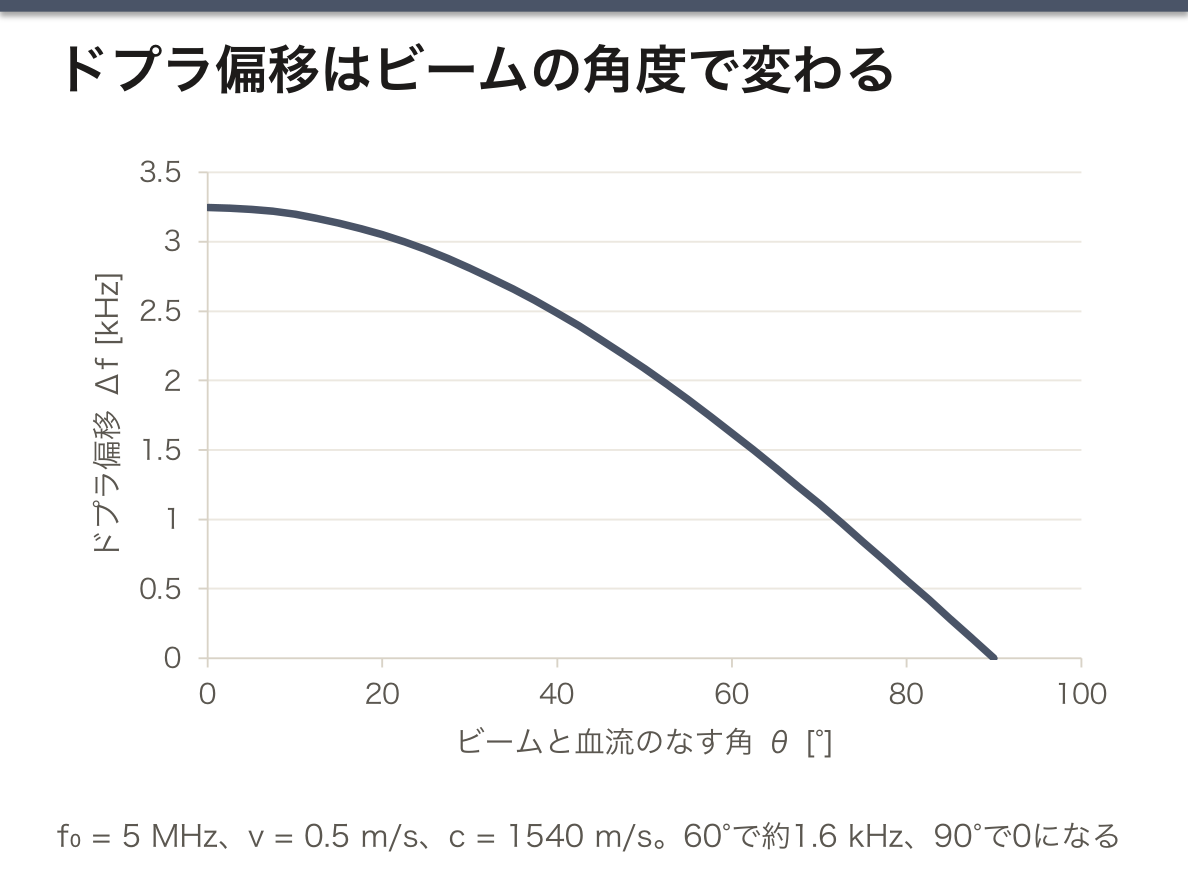
Δf = 2f0·v·cosθ/c
θ はビームと血流のなす角、c は組織の音速。速さがわかれば、血管の断面積 A から流量 Q = A·v(v は断面の平均流速)が求まる。
2がつくのはなぜか。赤血球は「動く受信者」として音を受け、さらに「動く音源」として送り返すので、ずれが2回かかる。
角度が決定的に効く。cos 90° = 0 なので、ビームが血管に直角だとずれは出ない。角度の誤差の影響は大きな角ほど増えるので、計測は60°以下で行うのが原則である。
例:f0 = 5 MHz、v = 0.5 m/s、θ = 60° なら Δf = 2 × 5×106 × 0.5 × 0.5/1540 ≈ 1.6 kHz。偏移は可聴域に入るので、装置は血流を音として聞かせることができる。
流量の例:内径 6 mm、平均流速 20 cm/s なら A = π(3×10⁻³)2 ≈ 2.8×10⁻⁵ m2、Q ≈ 5.7×10⁻⁶ m3/s ≈ 0.34 L/min。
カラードプラはプローブに近づく流れを赤、遠ざかる流れを青で重ねて表示する。深部静脈血栓症(DVT)では、プローブで押しても静脈がつぶれず、血流信号が途絶える。狭窄部では流速が上がるので、頸動脈や弁の狭窄の程度をピーク流速から推定できる。
English version
Core
When a source or reflector comes closer, the frequency gets higher; when it moves away, lower. This is the Doppler effect (the frequency changes, not the amplitude).
In ultrasound, the blood flow speed v is found from the difference (Doppler shift) between the sent frequency f0 and the frequency returned by moving red blood cells.
Δf = 2f0·v·cosθ/c
θ is the angle between the beam and the flow, and c is the speed of sound in tissue. With the speed known, the cross-sectional area A of the vessel gives the flow rate Q = A·v (v is the mean velocity over the cross-section).
Going further
Why the 2? A red blood cell receives the sound as “a moving receiver” and sends it back as “a moving source”, so the shift happens twice.
The angle matters decisively. cos 90° = 0, so if the beam is perpendicular to the vessel there is no shift. Errors in the angle matter more at large angles, so the rule is to measure at 60° or less.
Example: with f0 = 5 MHz, v = 0.5 m/s and θ = 60°, Δf = 2 × 5×106 × 0.5 × 0.5/1540 ≈ 1.6 kHz. The shift is in the audible range, so the machine can let you hear the flow as sound.
Flow example: with an inner diameter of 6 mm and a mean velocity of 20 cm/s, A = π(3×10⁻³)2 ≈ 2.8×10⁻⁵ m2 and Q ≈ 5.7×10⁻⁶ m3/s ≈ 0.34 L/min.
In the clinic
Color Doppler overlays flow toward the probe in red and flow away in blue. In deep vein thrombosis (DVT), the vein does not collapse when pressed with the probe, and the flow signal is absent. Flow speeds up at a narrowing, so the degree of carotid or valve stenosis can be estimated from the peak velocity.
まとめ
| 波の基本 — 運ばれるのはエネルギーで、物質ではない | 波は、振動(乱れ)が空間を伝わっていく現象である。 |
|---|---|
| 音の性質と可聴域 — 高さ・大きさ・音色 | 音の3要素は物理量に対応する。高さ(pitch)は振動数、大きさは振幅(強さ)、音色は波形(含まれる倍音の割合)。 |
| 音の強さとデシベル — 10 dB で強さは10倍 | 音の強さ(intensity)は、音の進む向きに垂直な単位面積を1秒間に通るエネルギーである。 |
| 耳はどう聞くか — 対数で感じ、中耳で整合する | 耳の感じ方は強さに比例しない。強さが10倍(+10 dB)になると、大きさはおよそ2倍に感じる——反応は線形ではなく対数に近い。 |
| 超音波の発生と検出 — 同じ結晶が送って受ける | 超音波は 20 kHz を超える音で、診断にはおよそ 1〜15 MHz が使われる。 |
| 音響インピーダンスと反射 — なぜゼリーを塗るのか | 音響インピーダンス Z は、媒質が音の伝わりに示す「抵抗」のような量で、密度と音速の積である。 |
| 減衰と周波数の選び方 — 細かく見るか、深く見るか | 超音波は組織を進むうちに弱まる(減衰 attenuation)。 |
| ドプラ法 — 周波数のずれから血流の速さを測る | 音源や反射体が近づくと周波数は高く、遠ざかると低くなる。 |
このノートは医学生が授業の内容を自分の言葉でまとめたもので、診療の判断に使うものではありません。誤りに気づいたらお知らせください。