伝導路を覚えるときに要るのは3つだけ。①どこで始まるか ②どこで乗り換えるか ③どこで交叉するか。この3つを言えれば、症状が同側か対側か、どの高さで切れたかが自分で導ける。路の名前を暗記しても、交叉の場所を言えなければ使えない。
感覚路は「3ニューロン・視床を経る」、小脳へ行く路は「2ニューロン・視床を経ない」。意識にのぼるかどうかが、そのまま経路の段数を決めている。
- 錐体路 — 随意運動の道
- 錐体外路 — 姿勢と筋緊張の道
- 意識にのぼる感覚路 — 3ニューロンと交叉の場所
- 意識にのぼらない深部感覚路 — 小脳への道
- 運動協調の経路 — 小脳への入りと出
- 辺縁系 — 情動と記憶の輪
錐体路 — 随意運動の道Tractus pyramidalis
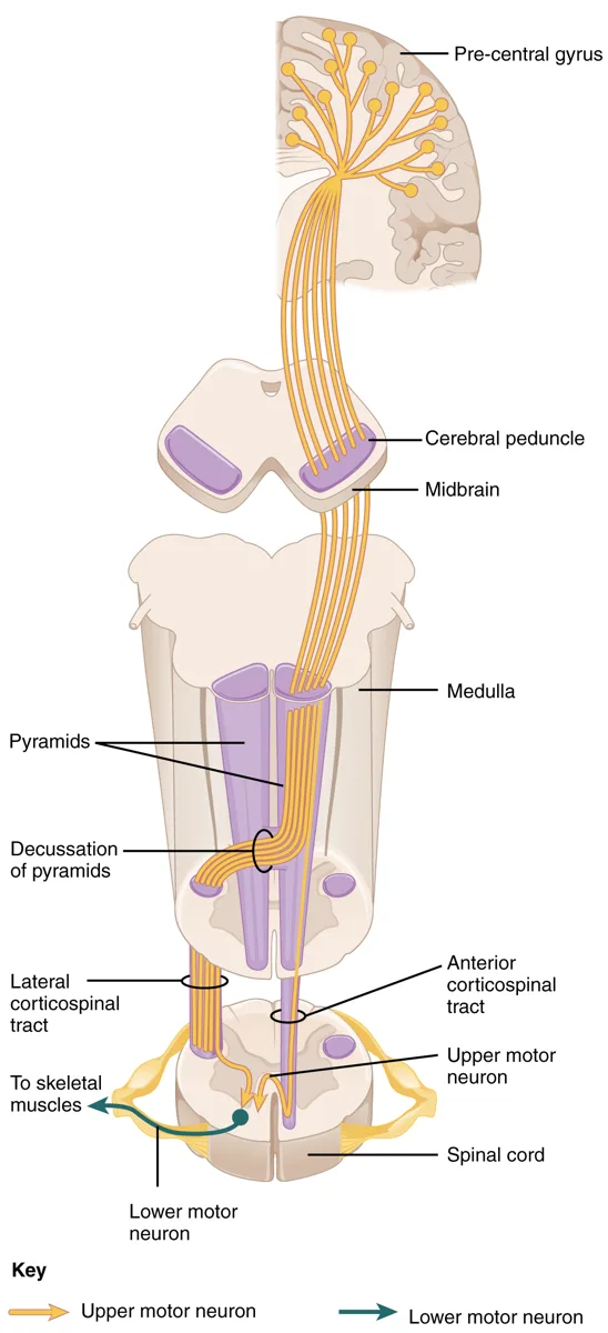
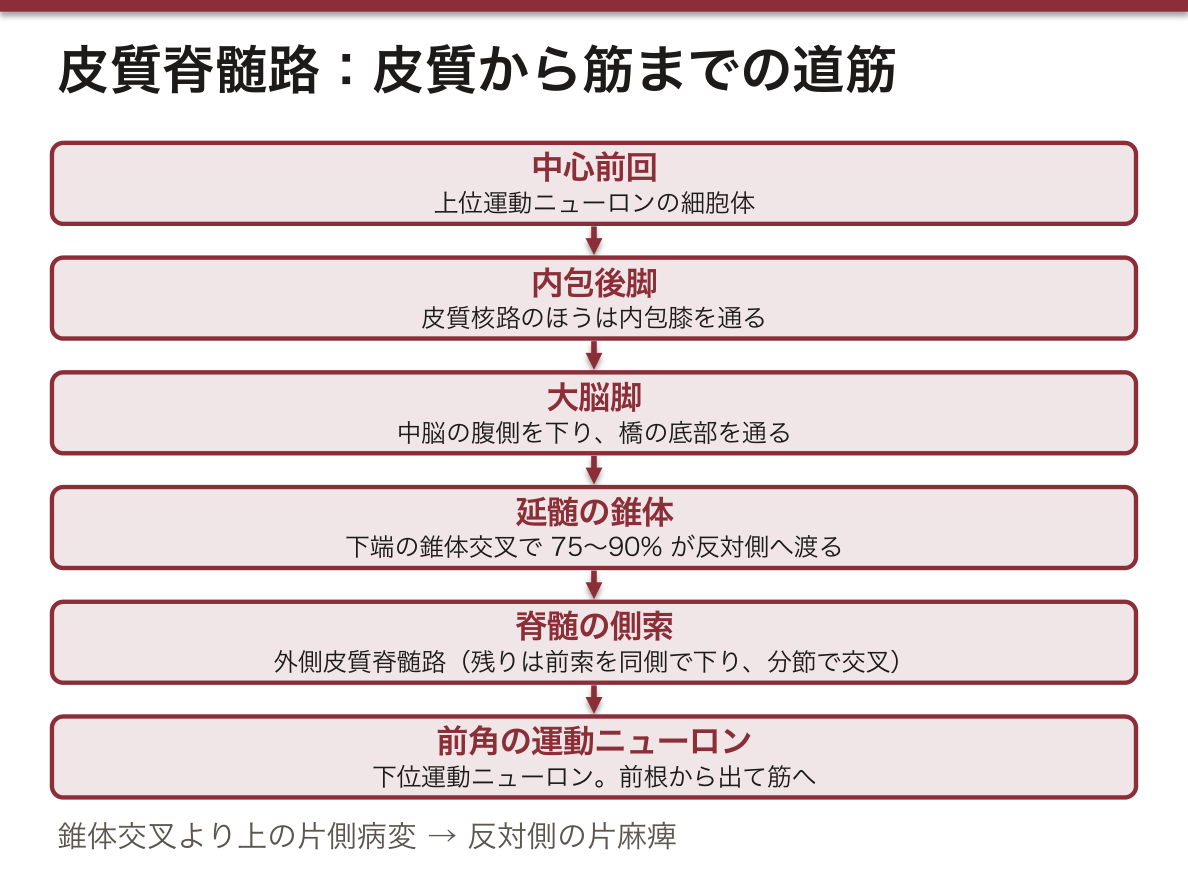
大脳皮質から運動ニューロンへ直接下る2本。いずれも中心前回(と中心後回・補足運動野)から出て、内包後脚を通り、大脳脚を下る。
- Tractus corticospinalis(皮質脊髄路)— 延髄下端の錐体交叉で 75〜90% が交叉して側索(lateralis)へ、残りが前索(anterior)を同側で下り分節で交叉。前角の運動ニューロンで終わる
- Tractus corticonuclearis(皮質核路)— 内包膝を通り、脳幹の運動性脳神経核で終わる
上位運動ニューロン(皮質→脊髄前角)と下位運動ニューロン(前角→筋)の2段構え。どちらが壊れたかで所見が正反対になる。
| 上位ニューロン障害 | 下位ニューロン障害 | |
|---|---|---|
| 麻痺の型 | 痙性 | 弛緩性 |
| 腱反射 | 亢進 | 消失 |
| 病的反射 | Babinski 陽性 | なし |
| 萎縮 | 軽い(廃用性) | 強い |
| 線維束攣縮 | なし | あり |
皮質核路はほとんどの核へ両側性に投射する。だから片側の皮質が傷んでも、多くの脳神経症状は出ない。例外が2つ——①顔面神経核の下半分(対側支配のみ)→ 中枢性顔面神経麻痺では額のしわ寄せが残る。②舌下神経核(対側支配優位)→ 舌が麻痺と反対側から押されて病側へ偏る。
この2つの例外が、末梢性と中枢性を分ける唯一の所見である。Bell 麻痺(末梢性)では額も動かない。
交叉する場所が分かれば、病変の高さが決まる。皮質脊髄路は延髄下端の錐体交叉で反対側へ渡る。したがって錐体交叉より上の片側病変では、反対側の片麻痺になる。
交叉性片麻痺——脳幹の片側病変では、反対側の上下肢の麻痺に病変側の脳神経麻痺が加わる。脳神経核はその高さで出てしまうため交叉しないからである。「脳神経は病変側、手足は反対側」と読めば、麻痺の組み合わせから病変の高さが逆算できる。
中脳=Weber 症候群(同側の動眼神経+対側片麻痺)。橋下部=Millard–Gubler 症候群(同側の外転神経・顔面神経+対側片麻痺)。
皮質核路は両側性に投射するので、片側の皮質が傷んでも多くの脳神経症状は出ない。例外が顔面神経の下半分で、ここだけ対側支配。だから中枢性顔面神経麻痺では額のしわが残り、末梢性では残らない。
出典:イラスト解剖学 誌面739頁「交叉性片麻痺」
English version
Core
Two tracts descending directly from the cerebral cortex to motor neurones. Both arise from the precentral gyrus (and the postcentral gyrus and supplementary motor area), pass through the posterior limb of the internal capsule and descend in the cerebral peduncle.
- Tractus corticospinalis — at the pyramidal decussation at the lower end of the medulla, 75–90 per cent cross into the lateral funiculus, the rest descending ipsilaterally in the anterior funiculus and crossing at their segment. They end on the motor neurones of the anterior horn.
- Tractus corticonuclearis — passes through the genu of the internal capsule and ends on the motor cranial nerve nuclei of the brainstem.
Two storeys: the upper motor neurone (cortex → anterior horn) and the lower motor neurone (anterior horn → muscle). Which of them is destroyed makes the findings exactly opposite.
Going further
| Upper motor neurone lesion | Lower motor neurone lesion | |
|---|---|---|
| Type of paralysis | Spastic | Flaccid |
| Tendon reflexes | Brisk | Absent |
| Pathological reflexes | Babinski positive | None |
| Wasting | Slight (disuse) | Marked |
| Fasciculation | None | Present |
The corticonuclear tract projects bilaterally to almost every nucleus. So damage to one cortex produces few cranial nerve signs. There are two exceptions — ① the lower half of the facial nucleus (contralateral supply only) → in a central facial palsy the ability to wrinkle the forehead is preserved; ② the hypoglossal nucleus (predominantly contralateral) → the tongue is pushed from the side opposite the paralysis and deviates towards the lesion. These two exceptions are the only findings that separate peripheral from central. In Bell’s palsy (peripheral) the forehead does not move either.
In the clinic
Know where it crosses and the level of the lesion is settled. The corticospinal tract crosses at the pyramidal decussation at the lower end of the medulla, so a unilateral lesion above the decussation gives a contralateral hemiplegia.
Crossed hemiplegia — a unilateral brainstem lesion adds a cranial nerve palsy on the side of the lesion to the hemiplegia on the opposite side, because the cranial nerve nuclei leave at that level and do not cross. Read it as "cranial nerve on the side of the lesion, limbs on the opposite side" and the level can be worked backwards from the combination.
- Midbrain = Weber’s syndrome (ipsilateral oculomotor + contralateral hemiplegia).
- Lower pons = Millard–Gubler syndrome (ipsilateral abducens and facial + contralateral hemiplegia).
The corticonuclear tract projects bilaterally, so damage to one cortex gives few cranial nerve signs. The exception is the lower half of the facial nerve, supplied contralaterally only. Hence in a central facial palsy the wrinkling of the forehead is preserved, and in a peripheral one it is not.
錐体外路 — 姿勢と筋緊張の道Tractus extrapyramidales
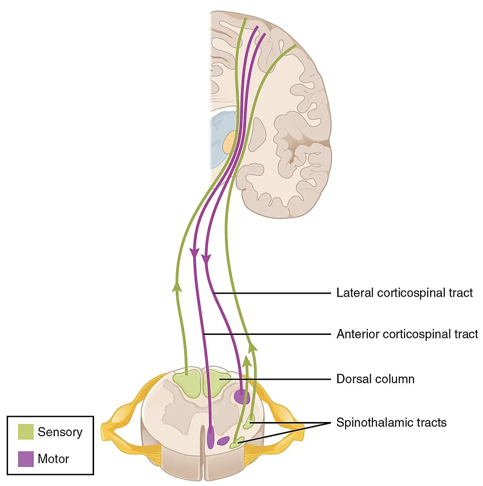
錐体路以外の下行路。「錐体を通らない」から錐体外路という。姿勢・筋緊張・自動的な運動を調節する。
| 路 | 起こる場所 | 働き |
|---|---|---|
| Tractus rubrospinalis | 赤核(中脳) | 上肢の屈筋を促進 |
| Tractus tectospinalis | 上丘 | 視覚刺激への頭の向き変え |
| Tractus vestibulospinalis | 前庭核 | 抗重力筋(伸筋)を促進・平衡 |
| Tractus reticulospinalis | 網様体(橋・延髄) | 筋緊張と歩行の自動性 |
| Tractus olivospinalis | 下オリーブ核 | 運動の調整 |
大脳基底核と小脳は、脊髄へ直接下りない。どちらも視床を介して大脳皮質へ戻り、皮質から改めて下る(ループ構造)。だから「錐体外路系の疾患」と呼ばれるパーキンソン病やハンチントン病でも、下行しているのは結局錐体路である。
除脳硬直と除皮質硬直の違いは、赤核より上か下かで決まる。赤核より上で切れると、赤核脊髄路(上肢屈筋を促進)が残るので上肢は屈曲する(除皮質硬直)。赤核より下で切れると赤核脊髄路も失われ、前庭脊髄路(伸筋促進)だけが残るので上肢も伸展する(除脳硬直)。肢位から病変の高さが言える——解剖が予後判定にそのまま使われる例である。
異常姿勢は、どの下行路が生き残ったかを示す。
除皮質硬直——病変は大脳(内包・大脳半球)で、赤核脊髄路が生きている。赤核脊髄路は上肢の屈筋を促進するので、上肢は屈曲し下肢は伸展する。
除脳硬直——病変が中脳(赤核より下)まで及び、赤核脊髄路が落ちる。残るのは前庭脊髄路・網様体脊髄路(伸筋を促進)だけなので、四肢がすべて伸展し、前腕は回内する。
「腕が曲がっていればまだ上、伸びきったら中脳まで来た」——除皮質から除脳へ変わればヘルニアの進行を意味し、緊急である。
なお Parkinson 病やハンチントン病は「錐体外路疾患」と呼ばれるが、基底核も小脳も脊髄へ直接下りていない。どちらも視床を介して皮質へ戻り、最終的に下るのは錐体路である。
English version
Core
The descending tracts other than the pyramidal — "extrapyramidal" because they do not pass through the pyramid. They regulate posture, muscle tone and automatic movement.
| Tract | Arises from | Function |
|---|---|---|
| Tractus rubrospinalis | Red nucleus (midbrain) | Facilitates the flexors of the upper limb |
| Tractus tectospinalis | Superior colliculus | Turning the head towards a visual stimulus |
| Tractus vestibulospinalis | Vestibular nuclei | Facilitates the antigravity (extensor) muscles; balance |
| Tractus reticulospinalis | Reticular formation (pons, medulla) | Muscle tone and the automatism of walking |
| Tractus olivospinalis | Inferior olivary nucleus | Adjustment of movement |
Going further
Neither the basal ganglia nor the cerebellum descends directly to the cord. Both return through the thalamus to the cerebral cortex, and it is from the cortex that the descent begins again (a loop). So even in the diseases called "extrapyramidal" — Parkinson’s, Huntington’s — what is descending is, in the end, the pyramidal tract.
The difference between decerebrate and decorticate rigidity is decided by whether the lesion is above or below the red nucleus. Cut above it and the rubrospinal tract (facilitating the flexors of the upper limb) survives, so the arms flex (decorticate). Cut below it and the rubrospinal tract is lost too, leaving only the vestibulospinal tract (facilitating extensors), so the arms extend as well (decerebrate).
The posture states the level of the lesion — an example of anatomy used directly in judging prognosis.
In the clinic
An abnormal posture shows which descending tract has survived.
Decorticate rigidity — the lesion is cerebral (internal capsule, hemisphere) and the rubrospinal tract is alive. It facilitates the flexors of the upper limb, so the arms flex and the legs extend.
Decerebrate rigidity — the lesion has reached the midbrain (below the red nucleus) and the rubrospinal tract is lost. Only the vestibulospinal and reticulospinal tracts remain (facilitating extensors), so all four limbs extend and the forearms pronate.
"Arms flexed, still high; arms fully extended, it has reached the midbrain" — a change from decorticate to decerebrate means the herniation is progressing, and is an emergency.
Note that although Parkinson’s and Huntington’s diseases are called extrapyramidal, neither the basal ganglia nor the cerebellum descends directly to the cord: both return through the thalamus to the cortex, and what finally descends is the pyramidal tract.
意識にのぼる感覚路 — 3ニューロンと交叉の場所Tractus sensorii conscii
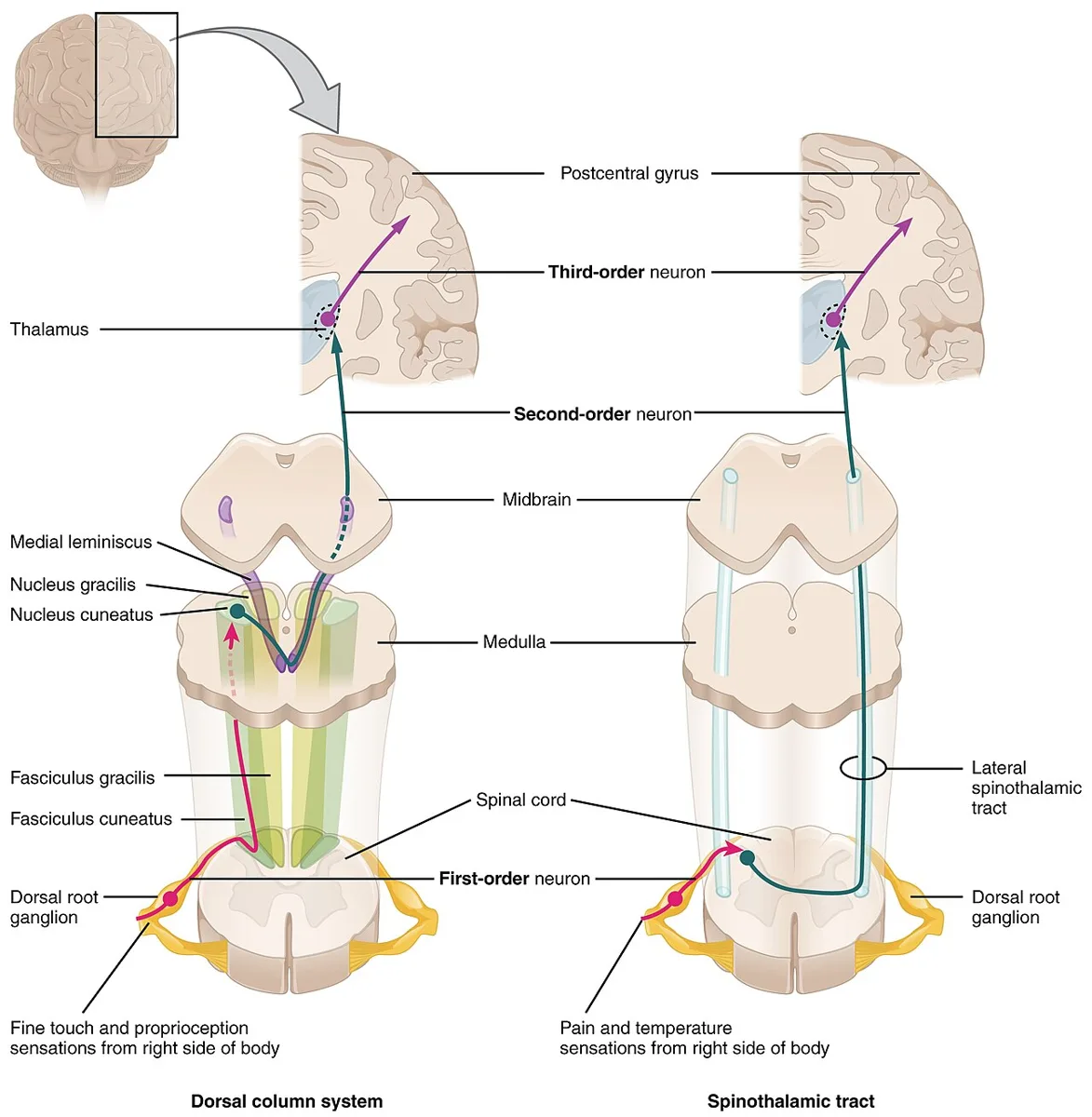
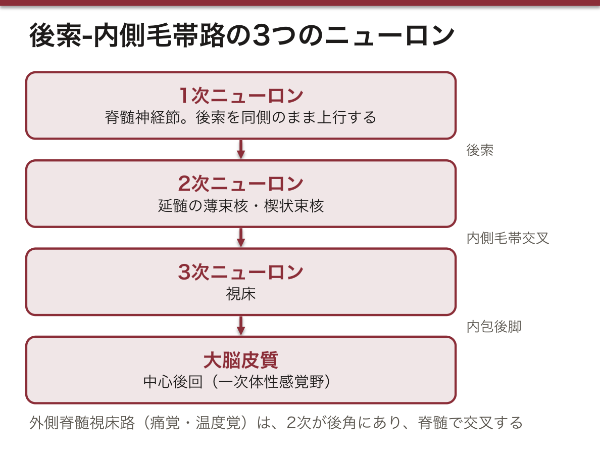
意識に達する感覚はすべて3つのニューロンを継ぐ。1次=脊髄神経節(または三叉神経節)、3次=視床 → 皮質は共通で、違うのは2次ニューロンがどこにあり、どこで交叉するかだけである。
| 路 | 運ぶもの | 2次 | 交叉する場所 |
|---|---|---|---|
| 後索‐内側毛帯路 | 識別性触覚・振動覚・位置覚 | 薄束核・楔状束核(延髄) | 延髄(内側毛帯交叉) |
| Tractus spinothalamicus lateralis | 痛覚・温度覚 | 後角(脊髄) | 脊髄(入った1〜2分節上・白交連) |
| Tractus spinothalamicus anterior | 粗大触覚・圧覚 | 後角 | 脊髄 |
後索は脊髄では同側、脊髄視床路は脊髄ですでに対側。この1点が Brown-Séquard の所見を全部説明する。
後索の中の配列(体部位局在)。内側が fasciculus gracilis(下半身、T6 以下)、外側が fasciculus cuneatus(上半身、T6 以上)。下から入った線維が内側へ押しやられるため、下肢が内側になる。だから延髄の tuberculum gracile が内側、tuberculum cuneatum が外側にある——延髄の外形で見た2つの結節の位置が、ここで意味を持つ。
脊髄視床路は入ってすぐ交叉しない。後角に入った線維は1〜2分節上行してから白交連を渡る(Lissauer 路)。だから半側切断では切断のレベルから1〜2分節下の対側から感覚が落ちる。
脊髄空洞症は中心管の周りが壊れるので、白交連を渡ろうとしている痛覚・温度覚だけが両側で落ちる。後索は無事なので触覚は残る——宙吊り型の解離性感覚障害である。「交叉する場所が中心にある」という解剖から、症状の形が直接出てくる。
Brown–Séquard 症候群(脊髄半側切断)——交叉する場所が路によって違うことが、そのまま症状の左右差になる。感覚路を覚える最大の理由がこれである。
同側——深部感覚(触覚・振動覚・位置覚)の消失(後索は延髄まで交叉しないため)と、痙性麻痺(外側皮質脊髄路)。
反対側——損傷高位の1〜2分節下から温痛覚の消失(外側脊髄視床路は脊髄に入ってすぐ交叉するため)。
「動かないほうと、熱さが分からないほうが逆」という奇妙な組み合わせは、これで全部説明がつく。損傷高位そのものでは同側の帯状の全感覚脱失が出る。
脊髄空洞症では中心管の周りが広がるため、そこで交叉する温痛覚だけが両側で落ち、後索は保たれる——宙吊り型(腕から胸だけ)の解離性感覚障害になる。
出典:イラスト解剖学 誌面750頁「ブラウン・セカール症候群」
English version
Core
Every sensation reaching consciousness passes through three neurones. The first (in a spinal or trigeminal ganglion) and the third (thalamus → cortex) are common to all; what differs is only where the second neurone lies and where it crosses.
| Pathway | Carries | Second neurone | Where it crosses |
|---|---|---|---|
| Posterior column–medial lemniscus | Discriminative touch, vibration, position | Gracile and cuneate nuclei (medulla) | Medulla (decussation of the lemnisci) |
| Tractus spinothalamicus lateralis | Pain and temperature | Posterior horn (cord) | Cord (one or two segments above entry, in the white commissure) |
| Tractus spinothalamicus anterior | Crude touch and pressure | Posterior horn | Cord |
The posterior columns are ipsilateral within the cord; the spinothalamic tracts are already contralateral there. This single point explains all the findings of Brown-Séquard.
Going further
The somatotopy within the posterior columns. Medial is the fasciculus gracilis (lower body, below T6), lateral the fasciculus cuneatus (upper body, above T6). Fibres entering from below are pushed medially, so the leg lies medially. Hence in the medulla the tuberculum gracile is medial and the tuberculum cuneatum lateral — the positions of the two tubercles seen on the external form of the medulla acquire their meaning here.
The spinothalamic tract does not cross immediately on entry. Fibres entering the posterior horn ascend one or two segments before crossing in the white commissure (Lissauer’s tract). So in a hemisection, sensation is lost on the opposite side beginning one or two segments below the level of the cut.
Syringomyelia destroys the region around the central canal, so only the pain and temperature fibres in the act of crossing the white commissure are lost, on both sides. The posterior columns are intact, so touch remains — a suspended dissociated sensory loss. The anatomy that the crossing happens in the centre produces the shape of the symptom directly.
In the clinic
Brown-Séquard syndrome (hemisection of the cord) — that the pathways cross at different places is directly the asymmetry of the symptoms. This is the single greatest reason for learning the sensory pathways.
Ipsilateral — loss of deep sensation (touch, vibration, position), the posterior columns not crossing until the medulla, and spastic paralysis (lateral corticospinal tract).
Contralateral — loss of pain and temperature from one or two segments below the level, the lateral spinothalamic tract crossing soon after entering the cord.
The strange combination — the side that cannot move and the side that cannot feel heat are opposite — is entirely explained by this. At the level of the injury itself there is a band of complete ipsilateral sensory loss.
In syringomyelia the region around the central canal widens, so only the pain and temperature fibres crossing there are lost bilaterally while the posterior columns are preserved — a suspended (arms and chest only) dissociated sensory loss.
意識にのぼらない深部感覚路 — 小脳への道Tractus spinocerebellares
筋紡錘・腱器官・関節からの情報を小脳へ運ぶ。意識にはのぼらないので、2ニューロンで済む(視床を経ない)。
- Tractus spinocerebellaris posterior(Flechsig)— 2次は Clarke 柱(C8〜L2)。交叉しない。下小脳脚から入る。下肢と体幹の情報
- Tractus spinocerebellaris anterior(Gowers)— 2次は後角基部。脊髄で交叉し、上小脳脚の手前でもう一度交叉して戻る(二重交叉)。上小脳脚から入る
どちらも最終的には同側の小脳に届く——後脊髄小脳路は交叉しないから、前脊髄小脳路は2回交叉するから。
「小脳の障害が同側に出る」ことの根拠がここにある。入力(脊髄小脳路)は同側から届き、出力(歯状核 → 上小脳脚 → 交叉 → 対側赤核・視床 → 対側皮質 → 錐体交叉 → 元の側)は2回交叉して戻る。入りも出も同側に閉じる。
Clarke 柱は C8〜L2 にしかない。それより下(下肢の下部)から来た線維は、いったん後索を上ってから L2 の Clarke 柱に入る。だから後索の下部には、意識にのぼる線維と小脳へ行く線維が混在している。
運動の協調は「命令の写しと結果の報告」を突き合わせて成り立つ。前脊髄小脳路は脊髄の運動ニューロンへ届いた命令の写しを、後脊髄小脳路は筋紡錘からの実際の結果を運ぶ。この2つを小脳が比べることで誤差が修正される。比較器としての小脳という見方をすると、失調の症状(測定障害・企図振戦=誤差修正の失敗)が理解できる。
後索が落ちると、目を閉じた瞬間に倒れる。深部感覚を失っても、見えているあいだは視覚が代償するためである。これを確かめるのが Romberg 試験で、閉眼で動揺が増せば陽性=後索性(感覚性)失調。小脳性失調では開眼でもふらつくので、ここで区別できる。
脊髄癆(神経梅毒)——後索と後根が両側性・進行性に変性する。電撃痛・深部感覚消失・失調性歩行(踵を打ちつけて歩く)・Argyll Robertson 瞳孔。
亜急性連合性脊髄変性症——ビタミン B12 欠乏。後索と側索(錐体路)が同時にやられるので、深部感覚障害+痙性麻痺という組み合わせになる。胃全摘後・悪性貧血・厳格な菜食で起こる。
Friedreich 失調症——後索・脊髄小脳路・錐体路が変性する常染色体潜性遺伝病。失調+深部感覚障害+腱反射消失+Babinski 陽性という、一見矛盾する組み合わせが手がかりになる。
English version
Core
They carry information from muscle spindles, tendon organs and joints to the cerebellum. Since it does not reach consciousness, two neurones suffice (it does not pass through the thalamus).
- Tractus spinocerebellaris posterior (Flechsig) — the second neurone is Clarke’s column (C8–L2). It does not cross. Enters by the inferior cerebellar peduncle. Information from the leg and trunk.
- Tractus spinocerebellaris anterior (Gowers) — the second neurone is at the base of the posterior horn. It crosses in the cord and crosses back again just before the superior cerebellar peduncle (a double decussation). Enters by the superior peduncle.
Both end in the cerebellum of the same side — the posterior because it never crosses, the anterior because it crosses twice.
Going further
Here is the basis for cerebellar signs appearing ipsilaterally. The input (spinocerebellar) arrives from the same side, and the output (dentate nucleus → superior peduncle → decussation → contralateral red nucleus and thalamus → contralateral cortex → pyramidal decussation → back to the original side) crosses twice and returns. Both in and out close on the same side.
Clarke’s column exists only from C8 to L2. Fibres from below it (the lower part of the leg) ascend first in the posterior columns and enter Clarke’s column at L2. So the lower part of the posterior columns contains a mixture of fibres destined for consciousness and fibres destined for the cerebellum.
Coordination of movement works by matching a copy of the command against a report of the result. The anterior spinocerebellar tract carries a copy of the command that reached the motor neurones, the posterior tract the actual result from the muscle spindles. The cerebellum compares the two and corrects the error. Seen as a comparator, the symptoms of ataxia (dysmetria, intention tremor = failure of error correction) become intelligible.
In the clinic
When the posterior columns fail, the patient falls the moment the eyes close, because vision compensates for the lost deep sensation while it is available. Romberg’s test checks this: increased sway on eye closure is positive = posterior column (sensory) ataxia. In cerebellar ataxia the patient sways with the eyes open too, and the two can be separated here.
Tabes dorsalis (neurosyphilis) — bilateral progressive degeneration of the posterior columns and posterior roots. Lightning pains, loss of deep sensation, ataxic gait (stamping the heels), Argyll Robertson pupils.
Subacute combined degeneration — vitamin B12 deficiency. The posterior columns and the lateral columns (pyramidal tract) are affected together, giving the combination of loss of deep sensation with spastic paralysis. It occurs after total gastrectomy, in pernicious anaemia, and in a strict vegan diet.
Friedreich’s ataxia — an autosomal recessive degeneration of the posterior columns, spinocerebellar tracts and pyramidal tracts. The apparently contradictory combination of ataxia, loss of deep sensation, absent tendon reflexes and a positive Babinski is the clue.
運動協調の経路 — 小脳への入りと出Tractus cerebellares
「小脳が何を受け取り、どこへ返すか」を一本の回路として言えるようにする。
入る道(下小脳脚・中小脳脚)——①脊髄小脳路(意識にのぼらない深部感覚。いま身体がどうなっているか)②皮質橋小脳路(大脳が何をしようとしているか。橋核で乗り換え、交叉して中小脳脚へ)③前庭小脳路(平衡)④オリーブ小脳線維(誤差の信号)。
出る道(上小脳脚)——小脳皮質 → 歯状核 → 上小脳脚 → 上小脳脚交叉 → 赤核と視床(VL 核) → 大脳の運動野。
要点は「意図と実際を突き合わせて誤差を修正する」——小脳は運動を起こさず、調整だけをする。
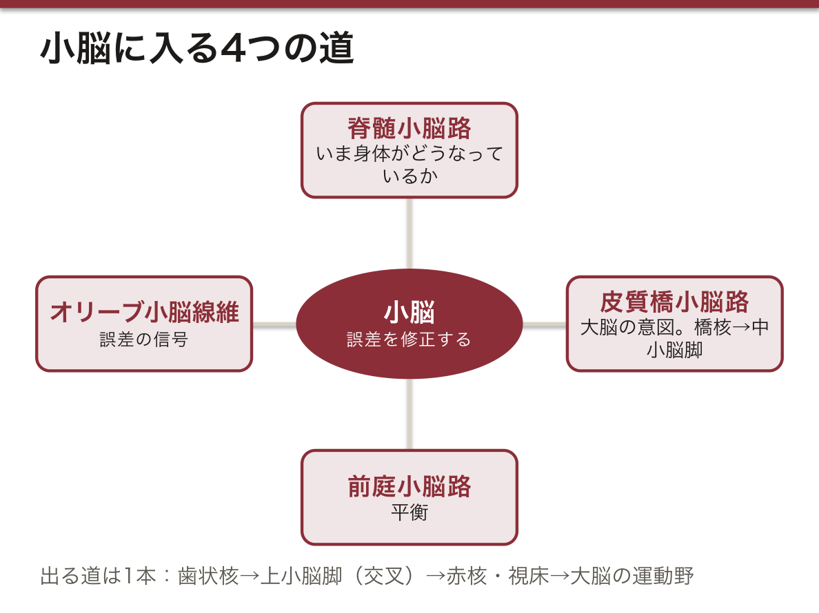
なぜ症状が同側に出るか——出力路が上小脳脚交叉で1回、皮質脊髄路が錐体交叉でもう1回、計2回交叉するため、結局もとの側に戻る。この「二重交叉」を言えることが決め手になる。
障害では測定障害・企図振戦・反復拮抗運動不能・失調性歩行・断綴性言語。
English version
Core
Be able to state, as one circuit, what the cerebellum receives and where it sends it back.
The ways in (inferior and middle peduncles) — ① the spinocerebellar tracts (unconscious proprioception: what the body is actually doing) ② the cortico-ponto-cerebellar tract (what the cerebrum intends to do; relaying in the pontine nuclei and crossing into the middle peduncle) ③ the vestibulocerebellar fibres (balance) ④ the olivocerebellar fibres (the error signal).
The way out (superior peduncle) — cerebellar cortex → dentate nucleus → superior peduncle → its decussation → red nucleus and thalamus (VL) → motor cortex.
The point is that it matches intention against actuality and corrects the error — the cerebellum does not produce movement, it only adjusts it.
Going further
Why the symptoms are ipsilateral — the output crosses once in the decussation of the superior peduncles and the corticospinal tract crosses again at the pyramidal decussation: twice in all, so it returns to the original side. Being able to state this double decussation is the decisive point.
Lesions give dysmetria, intention tremor, dysdiadochokinesia, ataxic gait and scanning speech.
辺縁系 — 情動と記憶の輪Systema limbicum
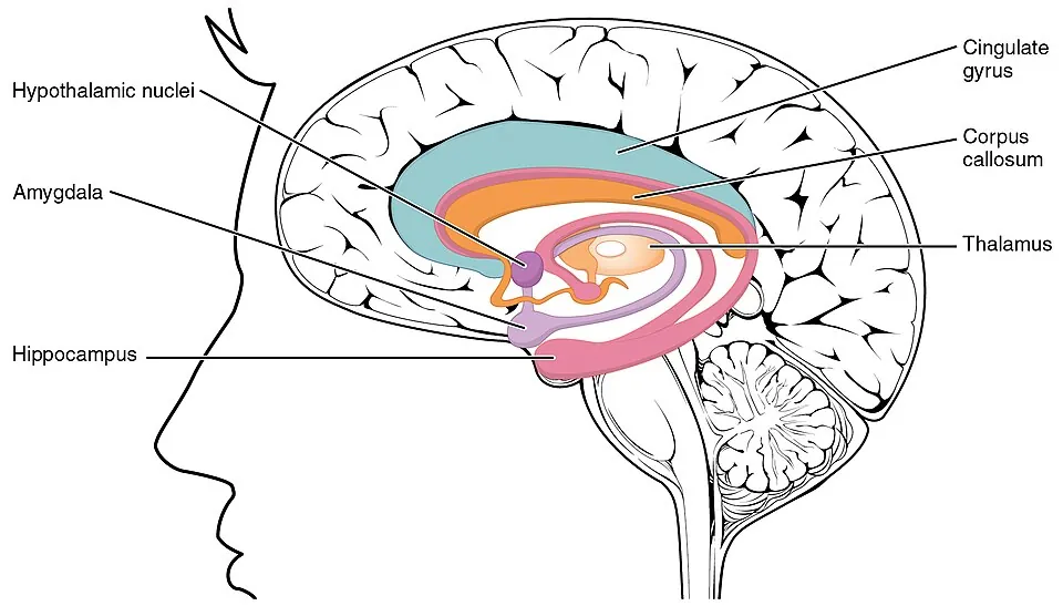
脳梁を取り巻く輪としてまとまる古い皮質と皮質下核。
構成——海馬、扁桃体、帯状回、海馬傍回、脳弓、乳頭体、視床前核、中隔核、嗅覚野。
Papez 回路——海馬 → 脳弓 → 乳頭体 → 乳頭視床束 → 視床前核 → 帯状回 → 海馬傍回 → 海馬。この順を言えることが要点。
役割——海馬は新しい記憶の固定、扁桃体は恐怖と情動の色づけ、帯状回は情動と注意の統合。
臨床——両側海馬の障害で前向性健忘(新しいことを覚えられない)。乳頭体の障害=Wernicke-Korsakoff 症候群。扁桃体の障害=Klüver-Bucy 症候群(恐怖の消失)。側頭葉てんかんの起始部でもある。
English version
Core
The old cortex and subcortical nuclei that group together as a ring encircling the corpus callosum.
Components — hippocampus, amygdala, cingulate gyrus, parahippocampal gyrus, fornix, mamillary body, anterior thalamic nucleus, septal nuclei, olfactory areas.
The Papez circuit — hippocampus → fornix → mamillary body → mamillothalamic tract → anterior thalamic nucleus → cingulate gyrus → parahippocampal gyrus → hippocampus. Being able to give that order is the point.
Functions — the hippocampus consolidates new memory, the amygdala colours things with fear and emotion, the cingulate gyrus integrates emotion and attention.
Going further
Clinically — bilateral hippocampal damage gives anterograde amnesia (an inability to learn anything new). Damage to the mamillary bodies = Wernicke–Korsakoff syndrome. Damage to the amygdala = Klüver–Bucy syndrome (loss of fear). It is also the site of onset of temporal lobe epilepsy.
まとめ
| 錐体路 — 随意運動の道 | 大脳皮質から運動ニューロンへ直接下る2本。 |
|---|---|
| 錐体外路 — 姿勢と筋緊張の道 | 錐体路以外の下行路。「錐体を通らない」から錐体外路という。 |
| 意識にのぼる感覚路 — 3ニューロンと交叉の場所 | 意識に達する感覚はすべて3つのニューロンを継ぐ。 |
| 意識にのぼらない深部感覚路 — 小脳への道 | 筋紡錘・腱器官・関節からの情報を小脳へ運ぶ。 |
| 運動協調の経路 — 小脳への入りと出 | 「小脳が何を受け取り、どこへ返すか」を一本の回路として言えるようにする。 |
| 辺縁系 — 情動と記憶の輪 | 脳梁を取り巻く輪としてまとまる古い皮質と皮質下核。 |
このノートは医学生が授業の内容を自分の言葉でまとめたもので、診療の判断に使うものではありません。誤りに気づいたらお知らせください。