内臓学はここから始まる。消化管を「入口から順に1本の管として読む」のが基本方針で、この章はその最初の区間である。
口腔は歯列を境に2つの部屋に分かれる(前庭と固有口腔)。咽頭は3階建て(鼻部・口部・喉頭部)で、そのそれぞれに前方から別の腔が開く。「いくつに分かれるか」を先に言ってから中身に入ると、説明が崩れない。
この範囲の芯は「空気の道と食物の道が咽頭で交差する」ことである。鼻から来た空気は咽頭を下って前の喉頭へ、口から来た食物は咽頭を下って後ろの食道へ。交差点だからこそ、嚥下のたびに喉頭蓋が閉じ、耳管が開き、扁桃輪が守っている。
- 口腔 — 2つの部屋と壁
- 舌 — 粘膜と筋
- 歯 — 構造と歯式
- 大唾液腺 — 3対と導管の開口
- 小唾液腺 — 口の粘膜に散らばる数百の腺
- 口蓋 — 硬口蓋と軟口蓋
- 咽頭 — 3階と、それぞれに開く口
- 耳下腺
- 顎下腺
- 舌下腺
- 舌尖・舌体・舌根
- 茸状乳頭
- 有郭乳頭
- 葉状乳頭
- 舌小帯
- 舌扁桃
- 硬口蓋
- 軟口蓋
- 口蓋垂
- 口蓋扁桃
- 咽頭鼻部
- 咽頭扁桃(アデノイド)
- 耳管咽頭口
- 咽頭陥凹(Rosenmüller 窩)
- 咽頭口部
- 咽頭喉頭部
- 梨状陥凹
- 正中舌喉頭蓋ひだ
- 外側舌喉頭蓋ひだ
- 歯冠
- 歯頸
- 歯根
- 頬と唇 — 構造
- 舌筋 — 内舌筋と外舌筋
- 咽頭の壁と筋
- 発生 — 咽頭弓・咽頭嚢と、舌・甲状腺のでき方
- 口腔 — 2つの部屋と壁Cavitas oris
- 舌 — 粘膜と筋Lingua
- 歯 — 構造と歯式Dentes
- 大唾液腺 — 3対と導管の開口Glandula salivaria major
- 小唾液腺 — 口の粘膜に散らばる数百の腺Glandulae salivariae minores
- 口蓋 — 硬口蓋と軟口蓋Palatum
- 咽頭 — 3階と、それぞれに開く口Pharynx
- 耳下腺Glandula parotidea
- 顎下腺Glandula submandibularis
- 舌下腺Glandula sublingualis
- 舌尖・舌体・舌根Apex, corpus, radix linguae
- 茸状乳頭Papillae fungiformes
- 有郭乳頭Papillae vallatae
- 葉状乳頭Papillae foliatae
- 舌小帯Frenulum linguae
- 舌扁桃Tonsilla lingualis
- 硬口蓋Palatum durum
- 軟口蓋Palatum molle, velum palatinum
- 口蓋垂Uvula
- 口蓋扁桃Tonsilla palatina
- 咽頭鼻部Pars nasalis pharyngis
- 咽頭扁桃(アデノイド)Tonsilla pharyngealis
- 耳管咽頭口Ostium pharyngeum tubae auditivae
- 咽頭陥凹(Rosenmüller 窩)Recessus pharyngeus
- 咽頭口部Pars oralis pharyngis
- 咽頭喉頭部Pars laryngea pharyngis
- 梨状陥凹Recessus piriformis
- 正中舌喉頭蓋ひだPlica glossoepiglottica mediana
- 外側舌喉頭蓋ひだPlica glossoepiglottica lateralis
- 歯冠Corona dentis
- 歯頸Cervix dentis
- 歯根Radix dentis
- 頬と唇 — 構造Buccae et labia
- 舌筋 — 内舌筋と外舌筋Musculi linguae
- 咽頭の壁と筋Musculi pharyngis
- 発生 — 咽頭弓・咽頭嚢と、舌・甲状腺のでき方Arcus pharyngei
- まとめ
口腔 — 2つの部屋と壁Cavitas oris
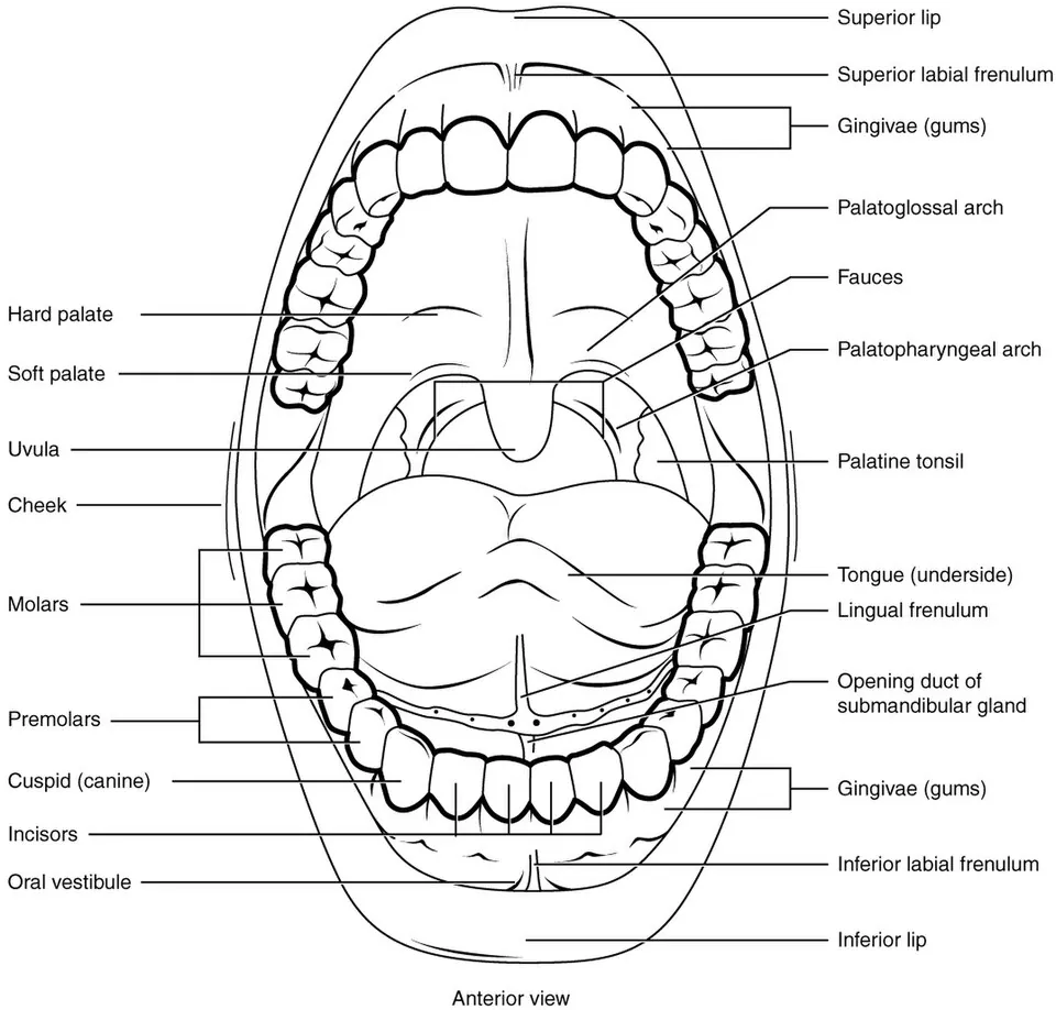
歯列弓と歯槽突起を境に2つに分かれる。
- Vestibulum oris(口腔前庭)— 唇・頬と歯列のあいだの馬蹄形の隙間。上下の frenulum labii superioris / inferioris が正中で粘膜を張る。Papilla parotidea(耳下腺乳頭)が上顎第2大臼歯の高さの頬粘膜に開く
- Cavitas oris propria(固有口腔)— 歯列より内側。後方は isthmus faucium(口峡)で咽頭へ続く
壁:上壁=口蓋、下壁=口腔底(顎舌骨筋)、前・側壁=唇と頬、後方=口峡。
顔面の目印として sulcus nasolabialis(鼻唇溝)・philtrum(人中)・sulcus mentolabialis(オトガイ唇溝)も押さえておく。
口峡の枠は上が口蓋帆・口蓋垂、両側が arcus palatoglossus(前)と arcus palatopharyngeus(後)、下が舌根。2本の弓のあいだの窪み sinus tonsillaris に tonsilla palatina が収まる——標本では「弓を2本示してから、そのあいだの扁桃を指す」。
Papilla parotidea の位置が臨床で使われる。耳下腺の導管(Stensen 管)の開口部で、流行性耳下腺炎では発赤し、唾石症では圧迫すると膿や唾液が出ない。口の中から耳下腺の状態が分かる唯一の窓である。
口腔前庭と固有口腔は、歯を食いしばると口裂でしか繋がらない。だから顎間固定をした患者でも、前庭側から流動食を入れられる。
耳下腺管は上顎第2大臼歯の高さの頬粘膜に開く。流行性耳下腺炎(ムンプス)ではこの耳下腺乳頭が発赤する。
口腔底の腫脹は気道緊急である。下顎の歯性感染が口腔底へ広がったLudwig アンギナでは、舌が押し上げられて短時間で窒息しうる。「口の中の腫れ」を軽く見ないこと。
English version
Core
It divides in two at the dental arches and alveolar processes.
- Vestibulum oris — the horseshoe-shaped space between lips and cheeks and the teeth. The frenulum labii superioris / inferioris stretch the mucosa in the midline. The papilla parotidea opens on the buccal mucosa at the level of the upper second molar.
- Cavitas oris propria — inside the dental arches. Behind, it continues into the pharynx through the isthmus faucium.
Walls: roof = palate, floor = the floor of the mouth (mylohyoid), anterior and lateral = lips and cheeks, posterior = the isthmus of the fauces.
The sulcus nasolabialis, philtrum and sulcus mentolabialis are also to be known as facial landmarks.
Going further
The frame of the isthmus of the fauces is the soft palate and uvula above, the arcus palatoglossus (in front) and arcus palatopharyngeus (behind) at the sides, and the root of the tongue below. In the hollow between the two arches, the sinus tonsillaris, lies the tonsilla palatina — on the specimen, show the two arches first and then point to the tonsil between them.
The position of the papilla parotidea is used clinically. It is the opening of the parotid duct (Stensen’s), reddened in mumps, and yielding neither pus nor saliva on pressure in sialolithiasis. It is the one window through which the state of the parotid can be seen from inside the mouth.
In the clinic
With the teeth clenched, the vestibule and the cavity proper communicate only at the oral fissure. So a patient in intermaxillary fixation can still be fed liquids from the vestibular side.
The parotid duct opens on the buccal mucosa at the level of the upper second molar. In mumps this papilla is reddened.
Swelling of the floor of the mouth is an airway emergency. In Ludwig’s angina, in which a dental infection of the mandible spreads to the floor of the mouth, the tongue is pushed up and asphyxia can follow within a short time. Never take "a swelling in the mouth" lightly.
舌 — 粘膜と筋Lingua
3部:apex(尖)・corpus(体)・radix(根)。背面 dorsum linguae の後方に V 字の sulcus terminalis があり、その頂点の小孔が foramen caecum(甲状舌管の遺残)。この V 字の前が体、後ろが根——発生の由来も神経支配もここで変わる。
乳頭 papillae:糸状(機械的・味蕾なし)、茸状 fungiformes(尖と縁・味蕾あり)、有郭 vallatae(分界溝の前に7〜12個・味蕾多数)、葉状 foliatae(後外側縁)。舌根には tonsilla lingualis。
下面正中に frenulum linguae。舌根の後方に plica glossoepiglottica mediana / lateralis があり、そのあいだの窪みが vallecula epiglottica。
筋は内舌筋と外舌筋の2群。内舌筋(上下縦・横・垂直)は形を変える、外舌筋は位置を変える。外舌筋4つは起始で覚える:m. genioglossus(オトガイ棘 → 前へ突き出す)・m. hyoglossus(舌骨 → 下げる)・m. styloglossus(茎状突起 → 後上へ引く)・m. palatoglossus(口蓋腱膜 → 舌根を上げる)。
神経支配は「1つだけ例外」で覚える。運動は全て XII 舌下神経——ただし m. palatoglossus だけ X 迷走神経(口蓋の筋だから)。感覚は分界溝の前が V3(一般)+VII 鼓索神経(味覚)、後ろが IX 舌咽神経(一般も味覚も)、喉頭蓋近くが X。「前は2本、後ろは1本」。
Genioglossus が舌を前へ出すので、片側麻痺では健側が押して舌尖が麻痺側へ偏る。仰臥位で意識が落ちると両側が緩んで舌根が沈む(気道閉塞)——下顎挙上法はこの筋の起始を前へ動かす手技である。
舌の神経支配は分界溝で切り替わる。ここが試験でも臨床でも要になる。
前2/3 — 一般感覚は舌神経(V3)、味覚は鼓索神経(VII)。後1/3 — 一般感覚も味覚も舌咽神経(IX)。運動はすべて舌下神経(XII)(口蓋舌筋だけが迷走神経)。
舌下神経麻痺では、舌を出すと患側へ偏る。健側のオトガイ舌筋だけが前へ押し出すためである。「舌は悪いほうを向く」と覚える。
盲孔は甲状舌管の遺残であり、下降の途中に組織が残ると正中頸嚢胞(甲状舌管嚢胞)になる。嚥下や舌の突出で上下に動くのが側頸嚢胞との鑑別点である。
English version
Core
Three parts: apex, corpus and radix. On the dorsum, behind, is the V-shaped sulcus terminalis, with the foramen caecum at its apex (the remnant of the thyroglossal duct). In front of that V is the body, behind it the root — and both the developmental origin and the innervation change there.
Papillae: filiform (mechanical, no taste buds), fungiformes (tip and margin, with taste buds), vallatae (7–12 in front of the sulcus terminalis, many taste buds), foliatae (posterolateral margin).
The root carries the tonsilla lingualis; the midline of the under surface has the frenulum linguae. Behind the root are the median and lateral plicae glossoepiglotticae, and the hollow between them is the vallecula epiglottica.
Going further
Two groups of muscle: intrinsic and extrinsic. The intrinsic (superior and inferior longitudinal, transverse, vertical) change the shape; the extrinsic change the position. Learn the four extrinsic by their origins: genioglossus (mental spine → protrudes), hyoglossus (hyoid → depresses), styloglossus (styloid process → retracts and elevates), palatoglossus (palatine aponeurosis → elevates the root).
Learn the innervation by its single exception. Motor supply is entirely by XII, the hypoglossal — except the palatoglossus, which is X, the vagus (it is a muscle of the palate). Sensation in front of the sulcus terminalis is V3 (general) plus the chorda tympani of VII (taste); behind it, IX (both general and taste); near the epiglottis, X. "Two nerves in front, one behind".
Genioglossus protrudes the tongue, so in a unilateral palsy the sound side pushes and the tip deviates towards the paralysed side. When consciousness is lost supine, both relax and the root falls back (airway obstruction) — the jaw thrust is a manoeuvre that moves the origin of this muscle forwards.
In the clinic
The innervation of the tongue changes over at the sulcus terminalis, and this is the crux both in the examination and in the clinic.
Anterior two thirds — general sensation by the lingual nerve (V3), taste by the chorda tympani (VII). Posterior third — both general sensation and taste by the glossopharyngeal (IX). Motor supply entirely by the hypoglossal (XII) (the palatoglossus alone by the vagus).
In hypoglossal palsy the protruded tongue deviates to the affected side, because only the sound genioglossus pushes forward. Remember: the tongue points to the bad side.
The foramen caecum is the remnant of the thyroglossal duct, and tissue left along its descent gives a midline neck (thyroglossal) cyst. That it moves up and down on swallowing and on protruding the tongue distinguishes it from a lateral cervical cyst.
歯 — 構造と歯式Dentes
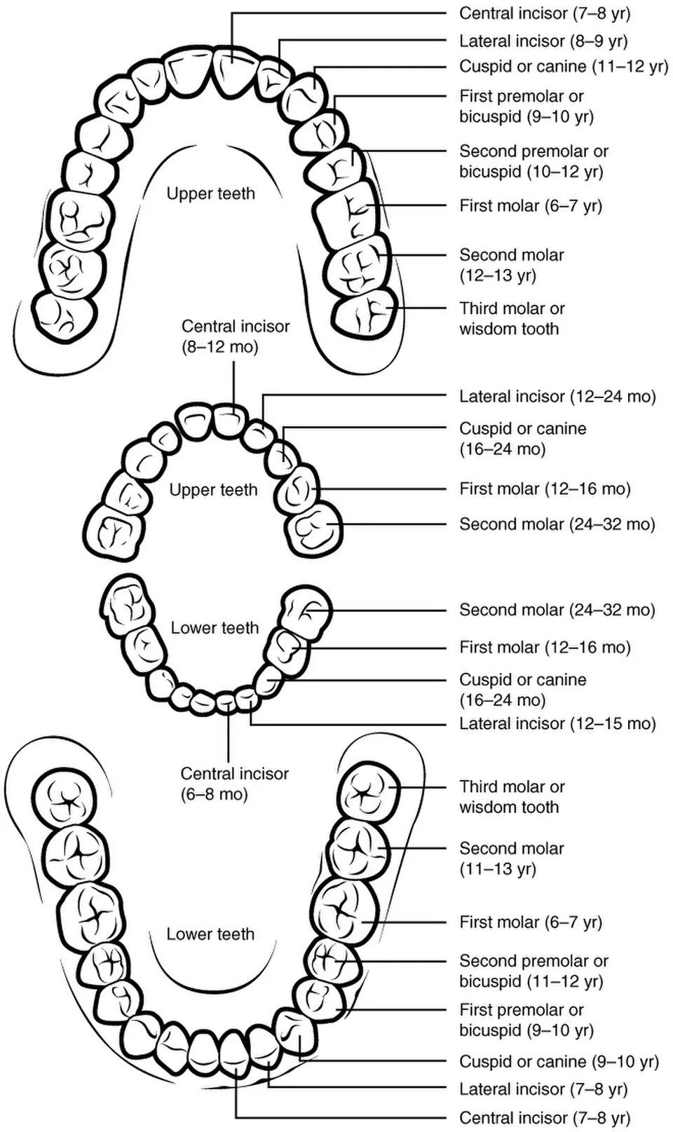
3部:corona dentis(歯冠・エナメル質に覆われ口腔に露出)、cervix (collum) dentis(歯頸・歯肉に囲まれる境)、radix dentis(歯根・歯槽に埋まる)。
内部は cavitas dentis(歯髄腔)→ canalis radicis dentis(根管)→ foramen apicis(根尖孔)と続き、ここから血管・神経が入る。硬組織は外から enamelum(エナメル質)→ dentinum(象牙質)→ pulpa(歯髄)、根の表面は cementum。
種類:dentes incisivi(切歯)・canini(犬歯)・premolares(小臼歯)・molares(大臼歯)。
歯式は「片側の上下」で書く。乳歯 2・1・0・2 = 片側5本 ×4 = 20本(小臼歯が無い)。永久歯 2・1・2・3 = 片側8本 ×4 = 32本。「乳歯に小臼歯は無い」が唯一の違いである。
萌出の時期:乳歯は生後6か月ごろ下顎中切歯から始まり2〜3歳で揃う。永久歯は6歳ごろ第1大臼歯(6歳臼歯)から——乳歯が抜けずに生える最初の永久歯なので見落とされやすい。第3大臼歯(智歯)は17〜25歳、生えないこともある。
歯髄には侵害受容器しかない。温度でも圧でも「痛み」としてしか感じられないため、齲蝕の初期から冷水痛が出る。根尖孔が唯一の出入口なので、感染が根尖を越えると顎骨・軟部組織へ広がる(歯性感染症)。
歯髄腔は硬い壁に囲まれた閉じた空間である。だから炎症で腫れても逃げ場がなく、歯髄炎の痛みは激烈になる。
上顎大臼歯の根尖は上顎洞底に接する。根尖病巣がそのまま歯性上顎洞炎になり、片側だけの膿性鼻漏・悪臭で気づかれる。「片側性の副鼻腔炎を見たら歯を疑う」。
下顎大臼歯の根尖は下顎管に近い。智歯(第三大臼歯)の抜歯で下歯槽神経が傷つくと、同側の下唇とオトガイのしびれが残る。
歯式は法医学でも使う。歯は焼損・腐敗に最も強い組織で、個人識別の決め手になる。
English version
Core
Three parts: corona dentis (covered with enamel, exposed in the mouth), cervix (collum) dentis (the junction, surrounded by gingiva) and radix dentis (buried in the alveolus). Internally the cavitas dentis (pulp cavity) → canalis radicis dentis → foramen apicis, through which vessels and nerves enter.
The hard tissues from outside in are enamelum → dentinum → pulpa, with cementum on the surface of the root.
Types: dentes incisivi, canini, premolares, molares.
Going further
The dental formula is written for one side, upper and lower. Deciduous 2·1·0·2 = 5 per side × 4 = 20 teeth (no premolars). Permanent 2·1·2·3 = 8 per side × 4 = 32 teeth. The only difference is that there are no deciduous premolars.
Times of eruption: the deciduous teeth begin at about six months with the lower central incisors and are complete at 2–3 years. The permanent teeth begin at about six years with the first molar (the six-year molar) — the first permanent tooth to erupt without a deciduous tooth being shed, and so readily missed. The third molar erupts at 17–25 years, or not at all.
The pulp contains only nociceptors. Neither temperature nor pressure can be felt as anything but pain, so cold sensitivity appears from the earliest caries. The apical foramen is the only way in or out, so infection passing beyond the apex spreads to the jaw and the soft tissues (odontogenic infection).
In the clinic
The pulp cavity is a closed space surrounded by hard walls. So inflammatory swelling has nowhere to escape and the pain of pulpitis is agonising.
The apices of the upper molars touch the floor of the maxillary sinus. A periapical lesion becomes an odontogenic maxillary sinusitis, noticed as unilateral purulent, foul-smelling nasal discharge. "Seeing a unilateral sinusitis, suspect a tooth."
The apices of the lower molars are close to the mandibular canal. Injury to the inferior alveolar nerve during extraction of a third molar leaves numbness of the ipsilateral lower lip and chin.
The dental formula is used in forensic medicine too — teeth are the tissue most resistant to burning and putrefaction, and are decisive in identification.
大唾液腺 — 3対と導管の開口Glandula salivaria major
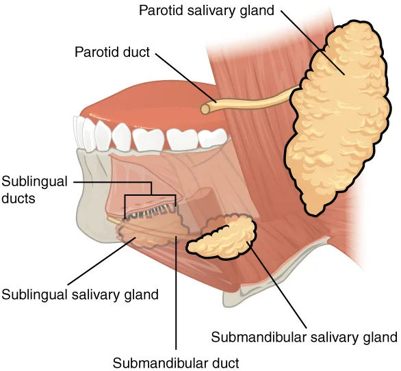
| 腺 | 位置 | 導管と開口 | 分泌 |
|---|---|---|---|
| Glandula parotidea | 耳の前下・咬筋の上 | Ductus parotideus(Stensen)→ 上顎第2大臼歯の高さの頬粘膜(papilla parotidea) | 純漿液性 |
| Glandula submandibularis | 顎下三角 | Ductus submandibularis(Wharton)→ 舌下小丘 caruncula sublingualis | 混合(漿液優位) |
| Glandula sublingualis | 口腔底・舌下ヒダの下 | 大導管(Bartholin)→ 舌下小丘、小導管多数 → plica sublingualis | 混合(粘液優位) |
耳下腺の中を3つの構造が貫く(浅い順に):顔面神経(VII)→ 下顎後静脈 → 外頸動脈。だから耳下腺腫瘍・手術では顔面神経麻痺が最大の合併症になる。腺の中で神経が枝分かれする(鵞足 pes anserinus)ため、腺を「浅葉・深葉」に分けるのは神経の面を基準にした外科的な区分である。
唾石は顎下腺に多い(約80%)。導管が長く上向きに走ること、分泌が粘稠でカルシウムに富むことによる。食事のたびに腫れて痛む(唾疝痛)のが典型で、口腔底で導管を触れると石が触れることがある。
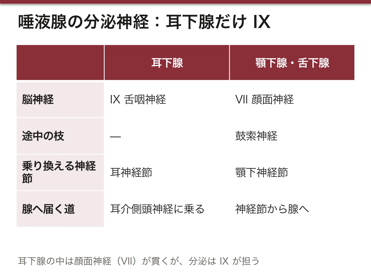
分泌の神経は腺ごとに違う。耳下腺は IX 舌咽神経 → 耳神経節 → 耳介側頭神経、顎下腺・舌下腺は VII 顔面神経(鼓索神経)→ 顎下神経節。耳下腺だけ IX と覚える。
耳下腺の中を顔面神経が貫く。これが耳下腺手術の最大の要点で、耳下腺腫瘍の摘出では顔面神経麻痺が最大の合併症である。神経より浅いか深いかで腫瘍を浅葉・深葉に分ける。
流行性耳下腺炎(ムンプス) — 両側の耳下腺が腫れる。合併症として思春期以降の精巣炎(不妊の原因)・無菌性髄膜炎・難聴。難聴は片側性・不可逆で、ワクチンで防ぐべき理由になる。
唾石は顎下腺に最も多い。顎下腺管(Wharton 管)が長く、上向きに走り、唾液が粘稠だからである。食事のたびに腫れて痛む(唾疝痛)のが特徴。
Sjögren 症候群では涙腺・唾液腺が破壊され、乾燥性角結膜炎と口腔乾燥。齲蝕が急増する。
English version
Core
| Gland | Position | Duct and opening | Secretion |
|---|---|---|---|
| Glandula parotidea | In front of and below the ear, on the masseter | Ductus parotideus (Stensen) → buccal mucosa at the level of the upper second molar (papilla parotidea) | Purely serous |
| Glandula submandibularis | The submandibular triangle | Ductus submandibularis (Wharton) → caruncula sublingualis | Mixed (serous predominant) |
| Glandula sublingualis | Floor of the mouth, beneath the sublingual fold | A major duct (Bartholin) → caruncula sublingualis; many minor ducts → plica sublingualis | Mixed (mucous predominant) |
Going further
Three structures pierce the parotid (from superficial to deep): the facial nerve (VII) → the retromandibular vein → the external carotid artery. So facial palsy is the greatest complication of parotid tumours and of parotid surgery. The nerve branches within the gland (the pes anserinus), so dividing the gland into superficial and deep lobes is a surgical division based on the plane of the nerve.
Calculi are commoner in the submandibular gland (about 80 per cent), because its duct is long and runs upwards and its secretion is viscid and rich in calcium. Swelling and pain at every meal (salivary colic) is typical, and the stone may be palpable along the duct in the floor of the mouth.
The secretomotor supply differs by gland. The parotid by IX → otic ganglion → auriculotemporal nerve; the submandibular and sublingual by VII (chorda tympani) → submandibular ganglion. Remember the parotid alone as IX.
In the clinic
The facial nerve pierces the parotid. This is the chief point of parotid surgery, and facial palsy is the greatest complication of removing a parotid tumour. Tumours are assigned to the superficial or deep lobe according to whether they lie superficial or deep to the nerve.
Mumps — both parotids swell. Complications are orchitis after puberty (a cause of infertility), aseptic meningitis and deafness. The deafness is unilateral and irreversible, and is the reason it should be prevented by vaccination.
Calculi are commonest in the submandibular gland, because Wharton’s duct is long, runs upwards, and the saliva is viscid. Swelling and pain at every meal (salivary colic) is characteristic.
In Sjögren’s syndrome the lacrimal and salivary glands are destroyed, giving keratoconjunctivitis sicca and a dry mouth. Caries increases sharply.
小唾液腺 — 口の粘膜に散らばる数百の腺Glandulae salivariae minores
唾液腺は大唾液腺3対(耳下腺・顎下腺・舌下腺)と、口腔の粘膜の下に散らばる小唾液腺に分かれる。小唾液腺は1つ1つが小さく、短い導管で粘膜にじかに開く。場所で名前が付く。
| 腺 | 場所 | 分泌 |
|---|---|---|
| glandulae labiales(口唇腺) | 上下の口唇の粘膜の下(口唇の中間層) | 主に粘液 |
| glandulae buccales(頬腺) | 頬の粘膜の下。臼歯の近くのものを glandulae molares(臼歯腺)とも呼ぶ | 混合 |
| glandulae palatinae(口蓋腺) | 硬口蓋の後部と軟口蓋の粘膜の下 | 粘液 |
| glandulae linguales(舌腺) | 舌尖の下面の前舌腺、有郭乳頭・葉状乳頭の溝に開く漿液腺(Ebner 腺)、舌根の粘液腺 | 部位で異なる |
大唾液腺は「食べるときに大量に」、小唾液腺は「いつも少しずつ」働く。小唾液腺の唾液は全体の1割に満たないが、粘液が多く、眠っている間も粘膜をぬらして守る。口の中が渇かないのは、主にこちらの仕事である。
Ebner 腺が有郭乳頭の溝に漿液を流し込むのは、溝の底にある味蕾を洗い流し、次の味を感じられるようにするためである。
小唾液腺は粘膜のすぐ下にあるので、唇をかんだりして導管が切れると、唾液が組織にもれて粘液嚢胞ができる(下唇に多い青みがかった小さなふくらみ)。口底にできる大きなものはがま腫で、こちらは大唾液腺の舌下腺から出る。
下唇の内側から小唾液腺を数個取る口唇腺生検は、Sjögren 症候群の診断に使う——唾液腺にリンパ球が集まる病気なので、取りやすい小唾液腺で確かめる。
小唾液腺の腫瘍は口蓋に最も多い。腺が小さいほど、できた腫瘍が悪性である割合が高い(耳下腺では2割ほど、小唾液腺では半分近く)。
English version
Core
Salivary glands are divided into three pairs of major glands (parotid, submandibular, sublingual) and the minor salivary glands scattered under the oral mucosa. Each minor gland is small and opens directly onto the mucosa by a short duct. They are named by location.
| Glands | Location | Secretion |
|---|---|---|
| glandulae labiales | Under the mucosa of the upper and lower lips | Mainly mucous |
| glandulae buccales | Under the cheek mucosa; those near the molars are also called glandulae molares | Mixed |
| glandulae palatinae | Under the mucosa of the back of the hard palate and the soft palate | Mucous |
| glandulae linguales | Anterior lingual gland under the tip; serous glands of von Ebner opening into the trenches of vallate and foliate papillae; mucous glands of the root | Varies by site |
Going further
Major glands work “a lot at meal times”; minor glands work “a little all the time”. Minor glands give less than a tenth of total saliva, but it is rich in mucus and keeps the mucosa moist and protected even during sleep. They are the main reason the mouth does not dry out.
Von Ebner’s glands pour serous fluid into the trenches of the vallate papillae to wash the taste buds at the bottom, so that the next taste can be detected.
In the clinic
Because the minor glands lie just under the mucosa, a cut duct (for example after biting the lip) lets saliva leak into the tissue and forms a mucocele — a small bluish swelling, most often on the lower lip. A large swelling in the floor of the mouth is a ranula, which comes from the sublingual (major) gland.
A labial gland biopsy, taking a few minor glands from inside the lower lip, is used to diagnose Sjögren’s syndrome: the disease gathers lymphocytes in salivary glands, so the easily reached minor glands are checked.
Tumours of minor salivary glands are most common in the palate. The smaller the gland, the higher the share of malignant tumours (about one fifth in the parotid, close to half in minor glands).
口蓋 — 硬口蓋と軟口蓋Palatum
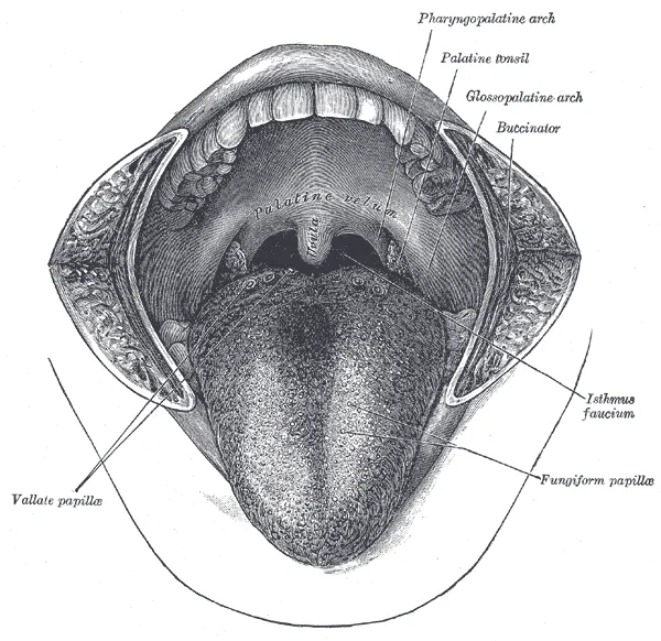
Palatum durum(硬口蓋)— 前2/3。骨(上顎骨の口蓋突起+口蓋骨の水平板)に粘膜が密着し、前部に plicae palatinae transversae(横口蓋ヒダ)と正中の raphe palati。
Palatum molle(軟口蓋・velum palatinum)— 後1/3。骨は無く、口蓋腱膜と筋。後縁の正中から uvula(口蓋垂)が垂れ、両側へ arcus palatoglossus と arcus palatopharyngeus が下る。
筋は5つ:m. tensor veli palatini(緊張・V3)、m. levator veli palatini(挙上)、m. uvulae、m. palatoglossus、m. palatopharyngeus。
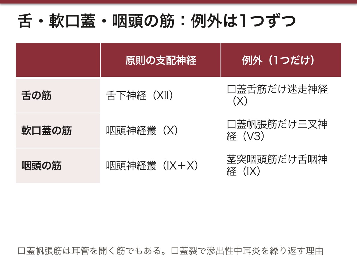
神経支配は「1つだけ例外」でまた覚える。軟口蓋の筋のうち m. tensor veli palatini だけが V3(三叉神経下顎枝)で、残り4つは咽頭神経叢(X 迷走神経)。咽頭・喉頭の筋で例外的に V3 と XI を持つものを押さえる——咽頭では m. stylopharyngeus だけが IX である。
Tensor veli palatini は耳管を開く筋でもある。嚥下やあくびのときに耳が「抜ける」のはこの筋の働きで、耳管咽頭口が引き開けられて中耳の圧が平衡する。口蓋裂ではこの筋の走行が乱れるため、滲出性中耳炎を繰り返す——口蓋の筋の話が耳の疾患に直結する。
片側の迷走神経麻痺では、発声時に口蓋垂が健側へ偏る(健側の挙上筋だけが働くため)。「アー」と言わせて口蓋垂の動きを見るのは、この解剖を使った診察である。
軟口蓋は嚥下と発声の弁である。迷走神経(咽頭神経叢)が支配し、口蓋帆張筋だけが V3——ここだけ例外なので問われやすい。
片側の迷走神経麻痺では、「アー」と言わせると軟口蓋が健側へ引かれ、口蓋垂が健側を向く。咽頭後壁が健側へ寄るカーテン徴候もこれである。延髄外側症候群の所見の1つ。
口蓋裂 — 一次口蓋と二次口蓋の癒合不全。哺乳障害・鼻咽腔閉鎖不全による開鼻声に加え、口蓋帆張筋が働かず耳管が開かないため滲出性中耳炎を繰り返す。
硬口蓋の後外側には大口蓋孔があり、歯科の大口蓋神経ブロックの目印になる。
English version
Core
- Palatum durum — the anterior two thirds. Mucosa adherent to bone (the palatine processes of the maxillae and the horizontal plates of the palatine bones), with the plicae palatinae transversae in front and the median raphe palati.
- Palatum molle (velum palatinum) — the posterior third. No bone; the palatine aponeurosis and muscle. From the midline of its posterior margin hangs the uvula, and to each side descend the arcus palatoglossus and arcus palatopharyngeus.
Five muscles: m. tensor veli palatini (tenses; V3), m. levator veli palatini (elevates), m. uvulae, m. palatoglossus, m. palatopharyngeus.
Going further
Again the innervation is learnt by its single exception. Of the muscles of the soft palate only the tensor veli palatini is V3 (mandibular division of the trigeminal); the other four are supplied by the pharyngeal plexus (X). Note the exceptions among the muscles of pharynx and larynx — in the pharynx only the stylopharyngeus is IX.
Tensor veli palatini is also the muscle that opens the auditory tube. The "popping" of the ears on swallowing or yawning is its action, drawing the pharyngeal opening of the tube apart and equalising the pressure in the middle ear. In cleft palate its course is disordered, so serous otitis media recurs — a discussion of a palatal muscle leading straight to a disease of the ear.
In a unilateral vagus palsy the uvula deviates to the sound side on phonation (only the sound levator acting). Asking the patient to say "ah" and watching the uvula is an examination that uses this anatomy.
In the clinic
The soft palate is the valve of swallowing and of speech. It is supplied by the vagus (pharyngeal plexus), with the tensor veli palatini alone by V3 — the one exception, and therefore readily asked.
In a unilateral vagus palsy, on saying "ah" the soft palate is drawn towards the sound side and the uvula points that way. The curtain sign, in which the posterior pharyngeal wall shifts to the sound side, is the same thing. One of the findings of the lateral medullary syndrome.
Cleft palate — failure of fusion of the primary and secondary palates. Besides feeding difficulty and hypernasal speech from velopharyngeal incompetence, the tensor veli palatini does not work, the auditory tube does not open, and serous otitis media recurs.
The greater palatine foramen lies posterolaterally in the hard palate and is the landmark for the dental greater palatine nerve block.
咽頭 — 3階と、それぞれに開く口Pharynx
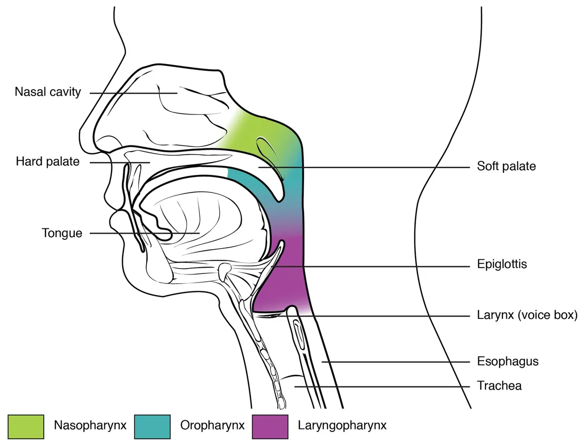
頭蓋底から C6(輪状軟骨の下縁)までの約12cm の筋膜性の管。前方が3つの腔に開くので3部に分かれる。
| 部 | 前に開くもの | そこにあるもの |
|---|---|---|
| Pars nasalis(上咽頭) | Choanae(後鼻孔) | Fornix pharyngis・tonsilla pharyngealis・ostium pharyngeum tubae auditivae・torus tubarius・recessus pharyngeus(Rosenmüller 窩) |
| Pars oralis(中咽頭) | Isthmus faucium(口峡) | Tonsilla palatina・舌根 |
| Pars laryngea(下咽頭) | Aditus laryngis(喉頭口) | Recessus piriformis・下端の ostium oesophageum |
壁の筋は2群。輪走する収縮筋 3つ(m. constrictor pharyngis superior / medius / inferior)が瓦を重ねたように並び、縦走する挙上筋 3つ(m. stylopharyngeus・palatopharyngeus・salpingopharyngeus)が内側を下る。収縮筋は食物を送り、挙上筋は嚥下時に咽頭を持ち上げて喉頭口を塞ぐ。
支配は咽頭神経叢(IX・X)。例外は m. stylopharyngeus だけで IX 単独——「舌咽神経が運動を出す唯一の筋」として問われる。
Anulus lymphoideus pharyngis(Waldeyer 扁桃輪)は、空気と食物の入口を輪で囲む免疫の関所。上から咽頭扁桃(アデノイド)→ 耳管扁桃 → 口蓋扁桃 → 舌扁桃。小児で咽頭扁桃が肥大すると鼻閉・いびき・滲出性中耳炎(耳管咽頭口を塞ぐため)を起こす。
Recessus piriformis は魚骨が刺さる場所、recessus pharyngeus は上咽頭癌の好発部位——どちらも「袋になっている」という形がそのまま臨床の意味を持つ。
Waldeyer 咽頭輪——咽頭扁桃・耳管扁桃・口蓋扁桃・舌扁桃が入口をぐるりと囲む。空気と食物の両方が通る場所を免疫で守る仕組みである。
アデノイド(咽頭扁桃)増殖症は小児で耳管咽頭口を塞ぎ、滲出性中耳炎・口呼吸・いびき・閉塞性睡眠時無呼吸を起こす。
扁桃周囲膿瘍 — 片側の強い咽頭痛・開口障害・含み声・口蓋垂の健側偏位。被膜を越えて広がると咽後膿瘍から縦隔炎に至るため、頸部の深部感染は縦隔まで落ちうることを常に意識する。
上咽頭癌は Rosenmüller 窩に好発し、EBV と関連。耳管を塞ぐので、成人の片側性滲出性中耳炎は上咽頭癌を除外するまで安心しない。
出典:イラスト解剖学 誌面391頁「扁桃の臨床関連事項」
English version
Core
A fibromuscular tube about 12 cm long, from the base of the skull to C6 (the lower border of the cricoid cartilage). Three cavities open into it in front, so it divides into three parts.
| Part | Opens in front into | What is found there |
|---|---|---|
| Pars nasalis | Choanae | Fornix pharyngis, tonsilla pharyngealis, ostium pharyngeum tubae auditivae, torus tubarius, recessus pharyngeus (fossa of Rosenmüller) |
| Pars oralis | Isthmus faucium | Tonsilla palatina, root of the tongue |
| Pars laryngea | Aditus laryngis | Recessus piriformis; the oesophageal opening below |
Going further
Two groups of muscle in the wall. Three circular constrictors (superior, middle, inferior) overlapping like roof tiles, and three longitudinal elevators (stylopharyngeus, palatopharyngeus, salpingopharyngeus) descending inside them. The constrictors propel the food; the elevators lift the pharynx during swallowing and close the laryngeal inlet.
The supply is from the pharyngeal plexus (IX and X). The exception is the stylopharyngeus alone, supplied by IX — asked as "the only muscle to which the glossopharyngeal gives motor fibres".
The anulus lymphoideus pharyngis (Waldeyer’s ring) is an immunological guard-post encircling the entrance of air and of food: from above, the pharyngeal tonsil (adenoids) → tubal tonsil → palatine tonsil → lingual tonsil. Hypertrophy of the pharyngeal tonsil in a child causes nasal obstruction, snoring and serous otitis media (by blocking the pharyngeal opening of the tube).
The piriform recess is where a fish bone lodges, and the pharyngeal recess is the site of predilection of nasopharyngeal carcinoma — in both, the fact of being a pocket is itself the clinical meaning.
In the clinic
Waldeyer’s ring — pharyngeal, tubal, palatine and lingual tonsils encircling the entrance. An arrangement guarding immunologically the place through which both air and food must pass.
Adenoid hypertrophy in children blocks the pharyngeal opening of the auditory tube, causing serous otitis media, mouth breathing, snoring and obstructive sleep apnoea.
Peritonsillar abscess — severe unilateral sore throat, trismus, a muffled voice and deviation of the uvula to the sound side. Spreading beyond the capsule it passes from a retropharyngeal abscess to mediastinitis, so always keep in mind that a deep infection of the neck can descend to the mediastinum.
Nasopharyngeal carcinoma has a predilection for the fossa of Rosenmüller and is associated with EBV. It blocks the auditory tube, so a unilateral serous otitis media in an adult is not safe until nasopharyngeal carcinoma has been excluded.
耳下腺Glandula parotidea
3大唾液腺で最大。耳の前下、下顎枝と胸鎖乳突筋の間の窩を埋める。純漿液性。
導管(Stensen 管)は咬筋の上を前へ走り、頬粘膜を貫いて上顎第2大臼歯の高さに開く。腺の実質を顔面神経が貫くので、手術で麻痺が問題になる。流行性耳下腺炎(おたふく)で腫れるのもここ。
English version
Core
The largest of the three major salivary glands, filling the fossa between the ramus of the mandible and the sternocleidomastoid, below and in front of the ear. Purely serous.
The duct (Stensen’s) runs forwards over the masseter, pierces the buccal mucosa and opens at the level of the upper second molar.
The facial nerve traverses the substance of the gland, so palsy is a concern in surgery. It is also the gland that swells in mumps.
顎下腺Glandula submandibularis
下顎体の内側、顎下三角にある。混合性(漿液優位)。
導管(Wharton 管)は口腔底を前へ走り、舌小帯の両側の舌下小丘に開く。唾石ができやすいのはこの腺——導管が上向きに長いため。
English version
Core
Medial to the body of the mandible, in the submandibular triangle. Mixed (serous predominant).
The duct (Wharton’s) runs forwards in the floor of the mouth and opens at the sublingual caruncle beside the frenulum.
This is the gland in which calculi most readily form — because the duct is long and runs upwards.
舌下腺Glandula sublingualis
口腔底の粘膜の直下、舌下ひだの中にある最小の大唾液腺。混合性(粘液優位)。
多数の小導管が舌下ひだに沿って直接開く。3腺は「大きさ・性状・開口部」の3点セットで問われる。
English version
Core
The smallest of the major glands, lying immediately beneath the mucosa of the floor of the mouth within the sublingual fold. Mixed (mucous predominant). Numerous small ducts open directly along the sublingual fold.
The three glands are asked as a set of three points: size, character of secretion, and site of opening.
舌尖・舌体・舌根Apex, corpus, radix linguae
舌は分界溝 sulcus terminalis を境に、前2/3の舌体(先端が舌尖)と後1/3の舌根に分かれる。
この境は発生と神経支配の境でもある。体は一般知覚が舌神経(V3)・味覚が鼓索(VII)、根は舌咽神経(IX)が両方を担う。「なぜ2/3と1/3で切るのか」は必ず聞かれる。
English version
Core
The tongue divides at the sulcus terminalis into the anterior two thirds, the body (with the apex at its tip), and the posterior third, the root. This boundary is also the boundary of development and of innervation.
In the body, general sensation is by the lingual nerve (V3) and taste by the chorda tympani (VII); in the root, the glossopharyngeal (IX) carries both. "Why is it divided at two thirds and one third?" is always asked.
茸状乳頭Papillae fungiformes
舌背に散らばるきのこ形の赤い点。特に舌尖と縁に多い。味蕾を持つ。
English version
Core
The red mushroom-shaped points scattered over the dorsum, most numerous at the tip and margins. They carry taste buds.
有郭乳頭Papillae vallatae
分界溝に沿ってV字に並ぶ、7〜12個の大きな乳頭。堀に囲まれ、その壁に味蕾が密集する。
最も大きく数が少ないので、標本で最初に指せる。堀の底に Ebner 腺が開く。
English version
Core
The 7–12 large papillae arranged in a V along the sulcus terminalis. Each is surrounded by a trench, in whose wall the taste buds are densely packed.
Being the largest and the fewest, they are the first thing to point to on the specimen. Von Ebner’s glands open into the floor of the trench.
葉状乳頭Papillae foliatae
舌の後外側縁にある、数本の縦のひだ。ヒトでは退化的だが味蕾を持つ。
3種はいずれも味蕾を持つ。味蕾を持たないのは糸状乳頭だけ——ここが引っかけになる。
English version
Core
The few vertical folds on the posterolateral margin of the tongue. Vestigial in humans but bearing taste buds.
All three of these have taste buds. Only the filiform papillae lack them — this is where the trick question lies.
舌小帯Frenulum linguae
舌下面と口腔底を結ぶ正中のひだ。両側に舌下小丘(顎下腺管の開口)がある。
短すぎると舌の動きが制限される(舌小帯短縮症)。
English version
Core
The median fold joining the under surface of the tongue to the floor of the mouth, with the sublingual caruncles (the openings of the submandibular ducts) on either side.
If it is too short, the movement of the tongue is restricted (ankyloglossia).
舌扁桃Tonsilla lingualis
舌根の粘膜下に散在するリンパ小節の集まり。表面がでこぼこに見える。
Waldeyer 咽頭輪の一員。
English version
Core
The collection of lymphoid nodules scattered beneath the mucosa of the root of the tongue, giving the surface a lumpy appearance. A member of Waldeyer’s ring.
硬口蓋Palatum durum
口蓋の前2/3。上顎骨口蓋突起と口蓋骨水平板が骨性の土台をつくる。粘膜に横口蓋ひだがある。
English version
Core
The anterior two thirds of the palate. The palatine processes of the maxillae and the horizontal plates of the palatine bones form the bony foundation. The mucosa carries the transverse palatine folds.
軟口蓋Palatum molle, velum palatinum
口蓋の後1/3。骨を持たず、筋と腱膜でできた可動部。
嚥下のとき持ち上がって咽頭鼻部を閉じ、食塊が鼻へ逆流するのを防ぐ。挙げるのは口蓋帆挙筋(X 支配)で、迷走神経麻痺では健側へ引かれる。
English version
Core
The posterior third of the palate. Having no bone, it is a mobile part made of muscle and aponeurosis. On swallowing it is raised to close the nasopharynx and prevent the bolus refluxing into the nose.
It is raised by the levator veli palatini (X), so in a vagus palsy it is drawn towards the sound side.
口蓋垂Uvula
軟口蓋の後縁から下がる正中の突起。
「アー」と言わせて左右差を見るのが迷走神経の診察。麻痺側と反対へ偏る。
English version
Core
The median process hanging from the posterior margin of the soft palate.
Asking the patient to say "ah" and looking for asymmetry is the examination of the vagus: it deviates away from the paralysed side.
口蓋扁桃Tonsilla palatina
口蓋舌弓と口蓋咽頭弓の間の扁桃窩に収まる。口を開ければ直接見える唯一の扁桃。
表面に陰窩が深く入り込む。急性扁桃炎・扁桃周囲膿瘍の場。標本では2つの弓を先に指してから、その間として示す。
English version
Core
Housed in the tonsillar fossa between the palatoglossal and palatopharyngeal arches. The only tonsil visible directly on opening the mouth. Deep crypts run into its surface.
The site of acute tonsillitis and of peritonsillar abscess. On the specimen, point to the two arches first and then show it as lying between them.
咽頭鼻部Pars nasalis pharyngis
軟口蓋より上の部分。前は後鼻孔で鼻腔に続く。ここだけは呼吸専用で、常に開いている。
English version
Core
The part above the soft palate, continuous in front through the choanae with the nasal cavity. This part alone is purely respiratory and is always open.
咽頭扁桃(アデノイド)Tonsilla pharyngealis
咽頭鼻部の天井から後壁にあるリンパ組織。小児で大きく、思春期に退縮する。
肥大すると鼻閉・いびき・睡眠時無呼吸、耳管を塞げば滲出性中耳炎になる。
English version
Core
The lymphoid tissue on the roof and posterior wall of the nasopharynx. Large in children and regressing at puberty.
Hypertrophy causes nasal obstruction, snoring and sleep apnoea, and if it blocks the auditory tube, serous otitis media.
耳管咽頭口Ostium pharyngeum tubae auditivae
咽頭鼻部の側壁、下鼻甲介の後端の高さに開く。後上縁の膨らみが耳管隆起。
ここから中耳へ空気が入る。小児では耳管が短く水平なので、感染が中耳へ上がりやすい。
English version
Core
Opens on the lateral wall of the nasopharynx at the level of the posterior end of the inferior concha, with the swelling of the torus tubarius at its posterosuperior margin. Air enters the middle ear from here.
In children the tube is short and horizontal, so infection ascends readily to the middle ear.
咽頭陥凹(Rosenmüller 窩)Recessus pharyngeus
耳管隆起のすぐ後ろのくぼみ。
上咽頭癌の好発部位として臨床で名前が出る。標本では耳管咽頭口を先に見つけ、その後方として示す。
English version
Core
The hollow immediately behind the torus tubarius. Named clinically as the site of predilection of nasopharyngeal carcinoma.
On the specimen, find the pharyngeal opening of the tube first and show it as lying behind that.
咽頭口部Pars oralis pharyngis
軟口蓋から喉頭蓋の上縁まで。前は口峡で口腔に開く。
空気と食物の両方が通る唯一の区間で、交差点にあたる。
English version
Core
From the soft palate to the upper border of the epiglottis, opening in front through the isthmus of the fauces into the mouth. The only stretch through which both air and food pass — the crossing.
咽頭喉頭部Pars laryngea pharyngis
喉頭蓋上縁から輪状軟骨下縁(C6)まで。ここで食道に移行する。前壁は喉頭口が占める。
English version
Core
From the upper border of the epiglottis to the lower border of the cricoid cartilage (C6), where it becomes the oesophagus. The anterior wall is occupied by the laryngeal inlet.
梨状陥凹Recessus piriformis
咽頭喉頭部で、喉頭口の両側にある梨形のくぼみ。食塊はここを通って食道へ落ちる。
魚骨などの異物が引っかかる場所。粘膜下を上喉頭神経内枝が走るので、ここへの麻酔で喉頭の知覚が取れる。
English version
Core
The pear-shaped hollow on either side of the laryngeal inlet in the laryngopharynx. The bolus passes through it into the oesophagus, and it is where foreign bodies such as fish bones lodge.
The internal branch of the superior laryngeal nerve runs beneath its mucosa, so anaesthetising it removes sensation from the larynx.
正中舌喉頭蓋ひだPlica glossoepiglottica mediana
舌根と喉頭蓋を結ぶ正中のひだ。両側のくぼみが喉頭蓋谷 vallecula。
喉頭蓋谷は気管挿管でマッキントッシュ喉頭鏡の先端を当てる場所。
English version
Core
The median fold joining the root of the tongue to the epiglottis, with the hollows on either side forming the vallecula.
The vallecula is where the tip of a Macintosh laryngoscope is placed for tracheal intubation.
外側舌喉頭蓋ひだPlica glossoepiglottica lateralis
正中ひだの両外側にあるひだ。正中ひだとの間に喉頭蓋谷をつくる。
English version
Core
The folds lateral to the median one, forming the vallecula with it.
歯冠Corona dentis
歯の歯肉より上に出ている部分。表面をエナメル質——体で最も硬い組織——が覆う。
English version
Core
The part of the tooth projecting above the gingiva, covered by enamel, the hardest tissue in the body.
歯頸Cervix dentis
歯冠と歯根の境のくびれ。ここで歯肉が歯に付着する。
English version
Core
The constriction between crown and root, where the gingiva attaches to the tooth.
歯根Radix dentis
歯槽に埋まる部分。表面はセメント質で覆われ、歯根膜を介して歯槽骨と結ばれる(釘植 gomphosis)。
先端の根尖孔から血管と神経が入る。3部は「歯冠・歯頸・歯根」=「エナメル質・境・セメント質」で組にして覚える。
English version
Core
The part buried in the alveolus, covered by cementum and bound to the alveolar bone through the periodontal ligament (gomphosis). Vessels and nerves enter through the apical foramen.
Learn the three parts as a set: crown, neck, root = enamel, junction, cementum.
頬と唇 — 構造Buccae et labia
どちらも外から「皮膚 → 筋 → 粘膜」の3層でできている。
唇 labia——筋は口輪筋。赤唇縁 vermilion は角化の薄い重層扁平上皮で、真皮の毛細血管が透けて赤く見える。汗腺も唾液腺も無いので乾きやすい。皮膚との境が赤唇皮膚境界で、口唇裂の修復ではここを合わせる。粘膜側の正中に上唇小帯・下唇小帯。
頬 buccae——筋は頬筋 m. buccinator。頬筋は口を閉じたまま食物を歯の間に押し戻す働きで、顔面神経(VII)支配。麻痺すると食物が頬に溜まる。粘膜下に頬腺があり、耳下腺管が上顎第2大臼歯の高さで開く。
口腔前庭(歯列の外側)と固有口腔を隔てるのがこの2つである。
頬筋は咀嚼筋ではない(表情筋で VII 支配)点が問われる。咀嚼筋は V3 支配。頬脂肪体(Bichat 脂肪体)は乳児で吸啜を助ける。
English version
Core
Both are built in three layers from outside in — skin → muscle → mucosa.
Lips — the muscle is orbicularis oris. The vermilion is thinly keratinised stratified squamous epithelium through which the dermal capillaries show red. Having neither sweat glands nor salivary glands it dries readily. Its junction with the skin is the vermilion border, which is what is aligned in repairing a cleft lip. On the mucosal side, the upper and lower labial frenula lie in the midline.
Cheeks — the muscle is the buccinator, which with the mouth closed pushes food back between the teeth; supplied by the facial nerve (VII). Paralysed, food collects in the cheek. Buccal glands lie beneath the mucosa, and the parotid duct opens at the level of the upper second molar.
These two separate the oral vestibule (outside the dental arches) from the cavity proper.
Going further
That the buccinator is not a muscle of mastication (it is a muscle of facial expression, supplied by VII) is what is asked. The muscles of mastication are supplied by V3.
The buccal fat pad (of Bichat) assists suckling in the infant.
舌筋 — 内舌筋と外舌筋Musculi linguae
内舌筋(形を変える)——上縦舌筋・下縦舌筋・横舌筋・垂直舌筋。骨に付かず舌の中で完結し、舌を短く・細く・平たくする。
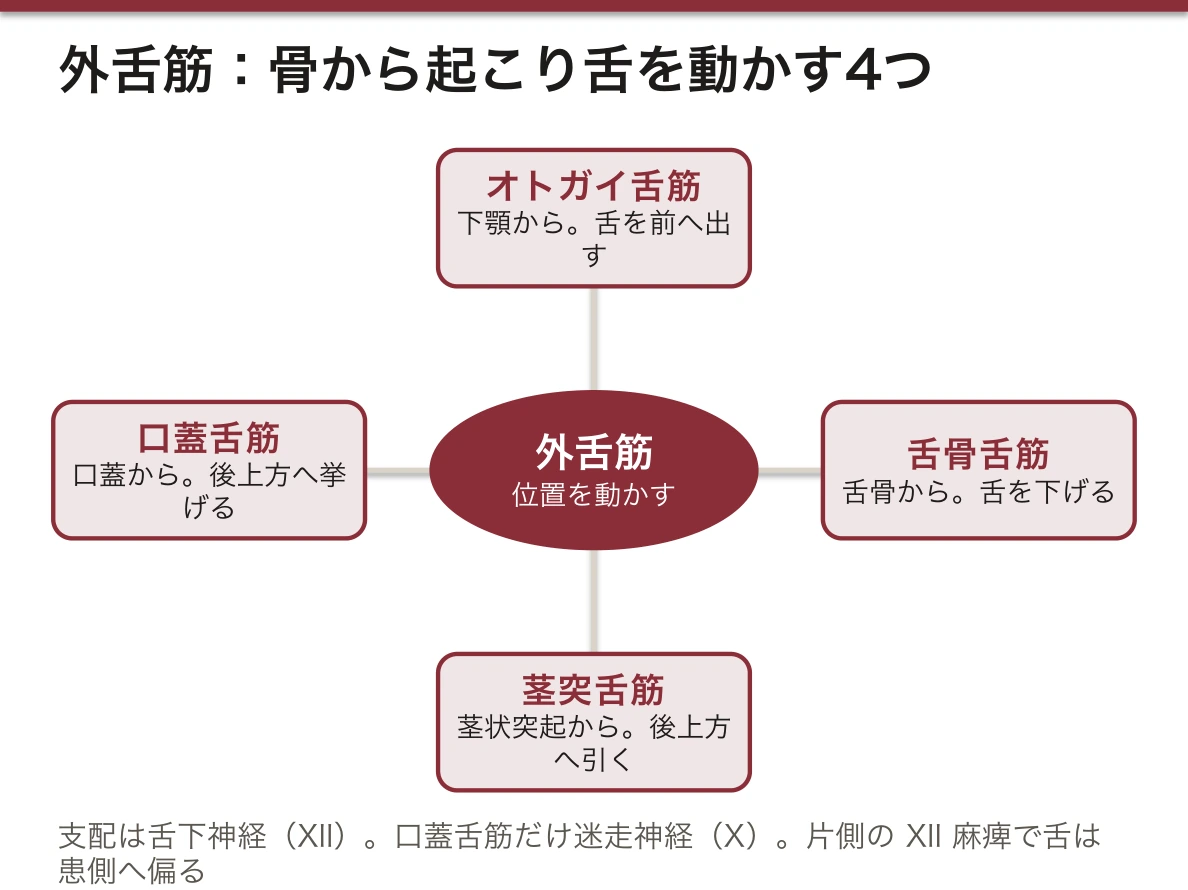
外舌筋(位置を動かす)——4つ、いずれも骨から起こる。オトガイ舌筋(下顎から。舌を前へ出す)、舌骨舌筋(舌骨から。下げる)、茎突舌筋(茎状突起から。後上方へ引く)、口蓋舌筋(口蓋から。後上方へ挙げる)。
神経支配——舌下神経(XII)がすべて。ただし口蓋舌筋だけが迷走神経(X)支配。この1つだけの例外が必ず問われる(口蓋舌筋は名前も働きも口蓋の筋だから)。
片側の XII 麻痺で舌は患側へ偏る——健側のオトガイ舌筋が押し出すため。
舌は正中の線維性の舌中隔で左右に分かれ、筋も血管も神経も正中を越えない。だから片側麻痺で偏りが出る。
English version
Core
Intrinsic muscles (change the shape) — superior and inferior longitudinal, transverse and vertical. Attached to no bone, they are contained within the tongue and make it short, narrow or flat.
Extrinsic muscles (change the position) — four, each arising from bone. Genioglossus (from the mandible; protrudes), hyoglossus (from the hyoid; depresses), styloglossus (from the styloid process; draws back and up), palatoglossus (from the palate; elevates the root).
Innervation — all by the hypoglossal (XII), except the palatoglossus alone, by the vagus (X). This single exception is invariably asked (the palatoglossus is a muscle of the palate in name and in action).
In a unilateral XII palsy the tongue deviates to the affected side, the sound genioglossus pushing it out.
Going further
The tongue is divided into halves by the median fibrous lingual septum, and neither muscle, vessel nor nerve crosses the midline. This is why a unilateral palsy produces deviation.
咽頭の壁と筋Musculi pharyngis
壁は4層——内から粘膜・咽頭粘膜下層(咽頭頭底板)・筋層・外膜(頬咽頭筋膜)。粘膜下層が無く、代わりに線維性の板があるのが特徴で、上端で頭蓋底に固定される。
上皮——鼻部は多列線毛円柱(呼吸専用だから)、口部と喉頭部は重層扁平(食物が擦れるから)。この使い分けが問われる。
筋は2群。収縮筋(輪走)——上・中・下咽頭収縮筋が瓦を重ねるように並び、順に収縮して食塊を下へ送る。下咽頭収縮筋の下部が輪状咽頭筋=上食道括約筋。挙上筋(縦走)——茎突咽頭筋・耳管咽頭筋・口蓋咽頭筋が嚥下時に咽頭と喉頭を引き上げる。
神経支配は咽頭神経叢(IX + X)。例外は2つ——茎突咽頭筋だけが舌咽神経(IX)の運動線維、口蓋帆張筋だけが三叉神経(V3)。
下咽頭収縮筋の斜走部と輪状咽頭筋の間の弱い部分が Killian 三角で、ここから粘膜が脱出するのが Zenker 憩室。
English version
Core
The wall is four layers — from within: mucosa, pharyngeal submucosa (the pharyngobasilar fascia), muscle, and adventitia (the buccopharyngeal fascia). Characteristically there is no submucosa proper but a fibrous plate instead, fixed above to the base of the skull.
Epithelium — pseudostratified ciliated columnar in the nasal part (purely respiratory), stratified squamous in the oral and laryngeal parts (food rubs over it). This division of labour is asked.
Two groups of muscle. Constrictors (circular) — superior, middle and inferior overlap like roof tiles and contract in turn to drive the bolus down. The lower part of the inferior constrictor is the cricopharyngeus = the upper oesophageal sphincter. Elevators (longitudinal) — stylopharyngeus, salpingopharyngeus and palatopharyngeus raise pharynx and larynx during swallowing.
Supply is from the pharyngeal plexus (IX + X). Two exceptions — the stylopharyngeus alone by motor fibres of the glossopharyngeal (IX), and the tensor veli palatini alone by the trigeminal (V3).
Going further
The weak area between the oblique part of the inferior constrictor and the cricopharyngeus is Killian’s triangle, and herniation of mucosa through it is a Zenker’s diverticulum.
発生 — 咽頭弓・咽頭嚢と、舌・甲状腺のでき方Arcus pharyngei
頭頸部は、胎生4〜5週に咽頭の両側に並ぶ咽頭弓(鰓弓)から作られる。ヒトでは第1・2・3・4・6の5対で、外からは4つが見える。各弓は筋・骨格・動脈・神経を1組ずつ持ち、弓の内側(咽頭の内面)のくぼみが咽頭嚢、外側の溝が咽頭溝である。弓ごとの筋と神経は呼吸器の章の発生の項目に表にした。
| 咽頭嚢 | できるもの |
|---|---|
| 第1 | 鼓室と耳管 |
| 第2 | 口蓋扁桃の陰窩の上皮 |
| 第3 | 下上皮小体と胸腺 |
| 第4 | 上上皮小体と、甲状腺の傍濾胞細胞(C 細胞) |
舌は前2/3と後1/3で出身が違う。前2/3は第1弓(外側舌隆起と無対結節)から、後1/3は第3・第4弓の隆起から生じ、その境が分界溝である。甲状腺は舌の分界溝の頂点(のちの舌盲孔)から内胚葉の芽として下り、甲状舌管を引きながら頸の前へ移る。
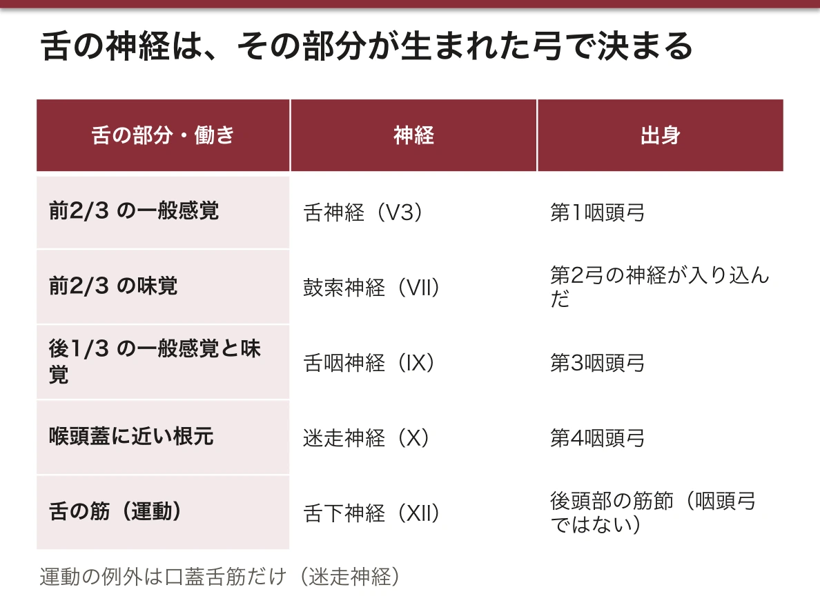
舌の神経が4本もある理由がここで分かる。前2/3の一般感覚は第1弓の三叉神経(舌神経)、後1/3は第3弓の舌咽神経、喉頭蓋に近い根元は第4弓の迷走神経。ところが舌の筋は咽頭弓ではなく後頭部の筋節から移動してくるので、運動は舌下神経がまとめて担う。前2/3の味覚が顔面神経(鼓索神経)なのは、第2弓の神経が第1弓の領域に入り込んだためである。
上皮小体の上下が「入れ替わる」のも同じ理屈である。第3嚢の上皮小体は胸腺と一緒に下へ引かれるので、下の上皮小体になる。
甲状舌管嚢胞は甲状腺が下りた道の途中に管が残ったもので、頸の正中にでき、舌を出したり飲み込んだりすると上下に動く——舌盲孔とつながっているからである。甲状腺が下りきらないと舌根に残る(舌甲状腺)。
口唇裂・口蓋裂は、顔の突起(内側鼻突起と上顎突起)や左右の口蓋突起が癒合しないことで起こる。側頸嚢胞は第2咽頭溝の名残で、胸鎖乳突筋の前縁にできる。
English version
Core
The head and neck are built from the pharyngeal (branchial) arches that line up on both sides of the pharynx in weeks 4–5. In humans there are five pairs (1, 2, 3, 4 and 6), of which four are visible from outside. Each arch carries its own set of muscle, skeleton, artery and nerve. The pockets on the inner (pharyngeal) side are the pharyngeal pouches; the grooves outside are the pharyngeal clefts. The muscles and nerves of each arch are tabled in the development item of the respiratory chapter.
| Pouch | Derivatives |
|---|---|
| 1st | Tympanic cavity and auditory tube |
| 2nd | Epithelium of the crypts of the palatine tonsil |
| 3rd | Inferior parathyroid gland and thymus |
| 4th | Superior parathyroid gland and the parafollicular (C) cells of the thyroid |
The tongue has different origins in front and behind: the anterior two thirds from the 1st arch (lateral lingual swellings and tuberculum impar), the posterior third from swellings of the 3rd and 4th arches, with the sulcus terminalis as the border. The thyroid starts as an endodermal bud at the apex of the sulcus terminalis (later the foramen caecum) and descends to the front of the neck, trailing the thyroglossal duct.
Going further
This explains why the tongue has four nerves. General sensation of the anterior two thirds comes from the 1st-arch trigeminal (lingual nerve), the posterior third from the 3rd-arch glossopharyngeal, and the root near the epiglottis from the 4th-arch vagus. Tongue muscles, however, migrate from occipital myotomes rather than an arch, so the hypoglossal nerve controls all movement. Taste on the anterior two thirds uses the facial nerve (chorda tympani) because the 2nd-arch nerve invaded the 1st-arch territory.
The parathyroids “swap places” for the same kind of reason: the 3rd-pouch parathyroid is pulled down with the thymus and ends up as the inferior gland.
In the clinic
A thyroglossal duct cyst is a leftover of the duct along the path of thyroid descent. It sits in the midline of the neck and moves up and down when the tongue is pushed out or the patient swallows, because it is still tied to the foramen caecum. If the thyroid fails to descend it stays at the tongue base (lingual thyroid).
Cleft lip and palate arise when facial processes (medial nasal and maxillary) or the two palatine shelves fail to fuse. A branchial cleft cyst is a remnant of the 2nd pharyngeal cleft and appears along the front border of the sternocleidomastoid.
まとめ
| 口腔 — 2つの部屋と壁 | 歯列弓と歯槽突起を境に2つに分かれる。Vestibulum oris(口腔前庭)— 唇・頬と歯列のあいだの馬蹄形の隙間。 |
|---|---|
| 舌 — 粘膜と筋 | 3部:apex(尖)・corpus(体)・radix(根)。 |
| 歯 — 構造と歯式 | 3部:corona dentis(歯冠・エナメル質に覆われ口腔に露出)、cervix (collum) dentis(歯頸・歯肉に囲まれる境)、radix dentis(歯根・歯槽… |
| 大唾液腺 — 3対と導管の開口 | 腺位置導管と開口分泌Glandula parotidea耳の前下・咬筋の上Ductus parotideus(Stensen)→ 上顎第2大臼歯の高さの頬粘膜(papilla pa… |
| 小唾液腺 — 口の粘膜に散らばる数百の腺 | 唾液腺は大唾液腺3対(耳下腺・顎下腺・舌下腺)と、口腔の粘膜の下に散らばる小唾液腺に分かれる。 |
| 口蓋 — 硬口蓋と軟口蓋 | Palatum durum(硬口蓋)— 前2/3。 |
| 咽頭 — 3階と、それぞれに開く口 | 頭蓋底から C6(輪状軟骨の下縁)までの約12cm の筋膜性の管。 |
| 耳下腺 | 3大唾液腺で最大。耳の前下、下顎枝と胸鎖乳突筋の間の窩を埋める。 |
| 顎下腺 | 下顎体の内側、顎下三角にある。混合性(漿液優位)。 |
| 舌下腺 | 口腔底の粘膜の直下、舌下ひだの中にある最小の大唾液腺。 |
| 舌尖・舌体・舌根 | 舌は分界溝 sulcus terminalis を境に、前2/3の舌体(先端が舌尖)と後1/3の舌根に分かれる。 |
| 茸状乳頭 | 舌背に散らばるきのこ形の赤い点。特に舌尖と縁に多い。 |
| 有郭乳頭 | 分界溝に沿ってV字に並ぶ、7〜12個の大きな乳頭。 |
| 葉状乳頭 | 舌の後外側縁にある、数本の縦のひだ。ヒトでは退化的だが味蕾を持つ。 |
| 舌小帯 | 舌下面と口腔底を結ぶ正中のひだ。両側に舌下小丘(顎下腺管の開口)がある。 |
| 舌扁桃 | 舌根の粘膜下に散在するリンパ小節の集まり。 |
| 硬口蓋 | 口蓋の前2/3。上顎骨口蓋突起と口蓋骨水平板が骨性の土台をつくる。 |
| 軟口蓋 | 口蓋の後1/3。骨を持たず、筋と腱膜でできた可動部。 |
| 口蓋垂 | 軟口蓋の後縁から下がる正中の突起。「アー」と言わせて左右差を見るのが迷走神経の診察。 |
| 口蓋扁桃 | 口蓋舌弓と口蓋咽頭弓の間の扁桃窩に収まる。 |
| 咽頭鼻部 | 軟口蓋より上の部分。前は後鼻孔で鼻腔に続く。 |
| 咽頭扁桃(アデノイド) | 咽頭鼻部の天井から後壁にあるリンパ組織。 |
| 耳管咽頭口 | 咽頭鼻部の側壁、下鼻甲介の後端の高さに開く。 |
| 咽頭陥凹(Rosenmüller 窩) | 耳管隆起のすぐ後ろのくぼみ。上咽頭癌の好発部位として臨床で名前が出る。 |
| 咽頭口部 | 軟口蓋から喉頭蓋の上縁まで。前は口峡で口腔に開く。 |
| 咽頭喉頭部 | 喉頭蓋上縁から輪状軟骨下縁(C6)まで。 |
| 梨状陥凹 | 咽頭喉頭部で、喉頭口の両側にある梨形のくぼみ。 |
| 正中舌喉頭蓋ひだ | 舌根と喉頭蓋を結ぶ正中のひだ。両側のくぼみが喉頭蓋谷 vallecula。 |
| 外側舌喉頭蓋ひだ | 正中ひだの両外側にあるひだ。正中ひだとの間に喉頭蓋谷をつくる。 |
| 歯冠 | 歯の歯肉より上に出ている部分。表面をエナメル質——体で最も硬い組織——が覆う。 |
| 歯頸 | 歯冠と歯根の境のくびれ。ここで歯肉が歯に付着する。 |
| 歯根 | 歯槽に埋まる部分。表面はセメント質で覆われ、歯根膜を介して歯槽骨と結ばれる(釘植 gomphosis)。 |
| 頬と唇 — 構造 | どちらも外から「皮膚 → 筋 → 粘膜」の3層でできている。 |
| 舌筋 — 内舌筋と外舌筋 | 内舌筋(形を変える)——上縦舌筋・下縦舌筋・横舌筋・垂直舌筋。 |
| 咽頭の壁と筋 | 壁は4層——内から粘膜・咽頭粘膜下層(咽頭頭底板)・筋層・外膜(頬咽頭筋膜)。 |
| 発生 — 咽頭弓・咽頭嚢と、舌・甲状腺のでき方 | 頭頸部は、胎生4〜5週に咽頭の両側に並ぶ咽頭弓(鰓弓)から作られる。 |
このノートは医学生が授業の内容を自分の言葉でまとめたもので、診療の判断に使うものではありません。誤りに気づいたらお知らせください。