男性生殖器は「精子の通り道を1本たどる」と全部が繋がる——精巣 → 精巣上体 → 精管 → 射精管 → 尿道。標本の上でも同じ順で示せばよい。
被膜の層は腹壁の層がそのまま降りてきたものである。精巣が腹腔内から陰嚢へ下降するとき、腹壁の各層を押し出して持って行った——だから層の数も順番も腹壁と一対一で対応する。
この範囲の芯は「下降」である。精巣の位置・被膜の層・血管が腹部から来ること・リンパが腰リンパ節へ行くこと・停留精巣・鼠径ヘルニア——すべて下降という1つの出来事の帰結である。
- 精巣と精巣上体
- 精索と精管 — 腹壁の層がそのまま被膜になる
- 付属生殖腺 — 精嚢・前立腺・尿道球腺
- 陰茎と陰嚢
- 精巣
- 精巣上体
- 精管・精管膨大部
- 精嚢
- 射精管
- 精索
- 陰嚢
- 前立腺(底・尖)
- 尿道前立腺部・膜様部
- 尿道海綿体部・舟状窩
- 陰茎根・陰茎体・亀頭
- 陰茎海綿体
- 尿道海綿体
- 坐骨海綿体筋
- 球海綿体筋
- 肛門挙筋
- 外肛門括約筋
- 発生 — 未分化の生殖腺から男性の生殖器へ
- 精巣と精巣上体Testis et epididymis
- 精索と精管 — 腹壁の層がそのまま被膜になるFuniculus spermaticus et ductus deferens
- 付属生殖腺 — 精嚢・前立腺・尿道球腺Glandula genitalis accessoria
- 陰茎と陰嚢Penis et scrotum
- 精巣Testis
- 精巣上体Epididymis
- 精管・精管膨大部Ductus deferens, ampulla ductus deferentis
- 精嚢Vesicula seminalis
- 射精管Ductus ejaculatorius
- 精索Funiculus spermaticus
- 陰嚢Scrotum
- 前立腺(底・尖)Prostata
- 尿道前立腺部・膜様部Pars prostatica, pars membranacea urethrae
- 尿道海綿体部・舟状窩Pars spongiosa urethrae, fossa navicularis
- 陰茎根・陰茎体・亀頭Radix, corpus, glans penis
- 陰茎海綿体Corpora cavernosa penis
- 尿道海綿体Corpus spongiosum penis
- 坐骨海綿体筋Musculus ischiocavernosus
- 球海綿体筋Musculus bulbospongiosus
- 肛門挙筋Musculus levator ani
- 外肛門括約筋Musculus sphincter ani externus
- 発生 — 未分化の生殖腺から男性の生殖器へDuctus mesonephricus et paramesonephricus
- まとめ
精巣と精巣上体Testis et epididymis
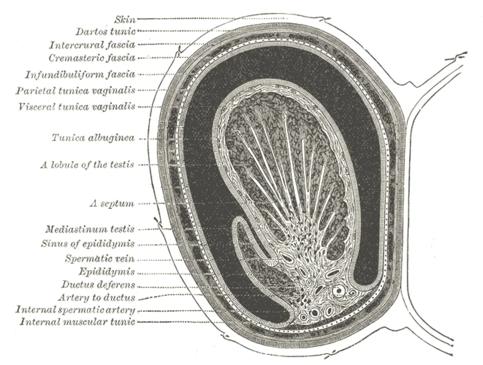
Testis:陰嚢内、卵形、約4×3×2.5cm。左が右より低いことが多い。表面を厚い tunica albuginea(白膜)が覆い、後縁で内へ入って mediastinum testis を作り、そこから中隔が放射状に伸びて 約250の lobuli testis に分ける。各小葉に tubuli seminiferi contorti(曲精細管)。
Epididymis:精巣の後上面に乗る。caput(頭・輸出小管が入る)・corpus(体)・cauda(尾・精管へ続く)。精巣と精巣上体のあいだの窪みが sinus epididymidis。
Tunica vaginalis testis:腹膜の続き(鞘状突起の遺残)で、壁側板と臓側板が精巣の前面を包む。後面には無い。
精路は「曲がる → まっすぐ → 網 → 出る → 貯める」:曲精細管 → tubuli seminiferi recti(直精細管)→ rete testis(精巣網・縦隔内)→ ductuli efferentes(輸出小管・10〜15本)→ ductus epididymidis(約6m を折り畳んだ1本)。精子が運動能と受精能を得るのは精巣上体を通るあいだである。
鞘膜が後面に無いことが精巣捻転を説明する。後面で固定されているから通常は捻れない。Bell clapper 変形(鞘膜が精巣を全周包み、後面の固定が無い)があると、精巣が鐘の舌のようにぶら下がって捻れる。解剖学的変異が疾患の必要条件になっている。
血管とリンパが「下降」の履歴を残している。精巣動脈は腹大動脈から直接(L2 の高さ)起こり、静脈は右が下大静脈、左が左腎静脈へ。リンパも陰嚢ではなく腰(傍大動脈)リンパ節へ行く。だから精巣腫瘍は鼠径リンパ節ではなく傍大動脈リンパ節へ転移する——陰嚢の皮膚の癌なら浅鼠径リンパ節へ行くのと対照的である。
精巣が腹腔外にある理由は温度で、体温より2〜3℃低い必要がある。停留精巣を放置すると造精機能が失われ、悪性化の危険が上がるのはこのためである。
精巣は腹腔内で発生し、下降して陰嚢へ入る。だから血管とリンパは腹部に残ったままである。
精巣のリンパは傍大動脈リンパ節へ、陰嚢の皮膚のリンパは浅鼠径リンパ節へ流れる。精巣腫瘍で鼠径リンパ節は腫れない——この区別が転移の検索先を決める。
精巣捻転 — 突然の激痛・挙睾筋反射の消失・挙上しても痛みが軽くならない(Prehn 徴候陰性)。6時間以内に整復しないと壊死する。思春期に好発。
停留精巣は精巣腫瘍と不妊のリスクを上げる。固定術をしても腫瘍のリスクは下がらないが、触知できる位置に置くこと自体に意味がある。
陰嚢水腫は腟状突起の遺残で、透光性があることが充実性腫瘤との鑑別点になる。
出典:イラスト解剖学 誌面525頁「精巣の臨床関連事項」
English version
Core
Testis: within the scrotum, ovoid, about 4 × 3 × 2.5 cm; the left usually lower than the right. Covered by the thick tunica albuginea, which passes inwards at the posterior border as the mediastinum testis, whence septa radiate to divide it into about 250 lobuli testis, each containing tubuli seminiferi contorti.
Epididymis: riding on the posterosuperior aspect — caput (entered by the efferent ductules), corpus, cauda (continuing into the ductus deferens). The hollow between testis and epididymis is the sinus epididymidis.
Tunica vaginalis testis: a continuation of peritoneum (the remnant of the processus vaginalis), its parietal and visceral layers covering the front of the testis. There is none behind.
Going further
The seminal path is "coiled → straight → net → out → stored": convoluted seminiferous tubules → tubuli seminiferi recti → rete testis (in the mediastinum) → ductuli efferentes (10–15) → ductus epididymidis (about 6 m folded into one). Sperm acquire motility and the capacity to fertilise while passing through the epididymis.
That there is no tunica vaginalis behind explains testicular torsion. Being fixed behind, it does not normally twist. With a bell-clapper deformity (the tunica enclosing the testis all round with no posterior fixation) the testis hangs like the clapper of a bell and twists. An anatomical variant as the necessary condition of a disease.
The vessels and lymphatics preserve the record of the descent. The testicular artery arises directly from the aorta (at L2), the vein drains to the inferior vena cava on the right and the left renal vein on the left, and the lymph goes not to the scrotum but to the lumbar (para-aortic) nodes. So a testicular tumour metastasises to the para-aortic and not the inguinal nodes — in contrast to a carcinoma of the scrotal skin, which goes to the superficial inguinal nodes.
The testis lies outside the abdomen for temperature, needing to be 2–3 °C below core. An undescended testis left alone loses spermatogenesis and carries an increased risk of malignancy.
In the clinic
The testis develops in the abdomen and descends into the scrotum, so its vessels and lymphatics remain abdominal. Testicular lymph goes to the para-aortic nodes, scrotal skin to the superficial inguinal nodes. The inguinal nodes do not enlarge in testicular tumour — and this distinction decides where the metastases are sought.
Testicular torsion — sudden severe pain, loss of the cremasteric reflex, and no relief on elevation (Prehn’s sign negative). Unless reduced within six hours it necroses. Commonest at puberty.
Undescended testis raises the risk of tumour and of infertility. Orchidopexy does not reduce the risk of tumour, but placing it where it can be palpated has value in itself.
A hydrocele is a remnant of the processus vaginalis; that it transilluminates distinguishes it from a solid mass.
精索と精管 — 腹壁の層がそのまま被膜になるFuniculus spermaticus et ductus deferens
Ductus deferens:精巣上体尾から始まり、精索の中を上って鼠径管を通り、深鼠径輪から骨盤へ入る。尿管を前から越えて(water under the bridge の男性版)膀胱底の後ろへ回り、ampulla ductus deferentis に膨らんだあと、精嚢の導管と合して ductus ejaculatorius となり前立腺を貫いて尿道前立腺部へ開く。壁が厚い平滑筋で、索状に硬く触れるのが特徴。
Funiculus spermaticus(精索)の中身は「3つの動脈・3つの神経・3つのその他」:精巣動脈・精管動脈・精巣挙筋動脈/腸骨鼠径神経(表面)・陰部大腿神経陰部枝・自律神経/精管・蔓状静脈叢・リンパ管。
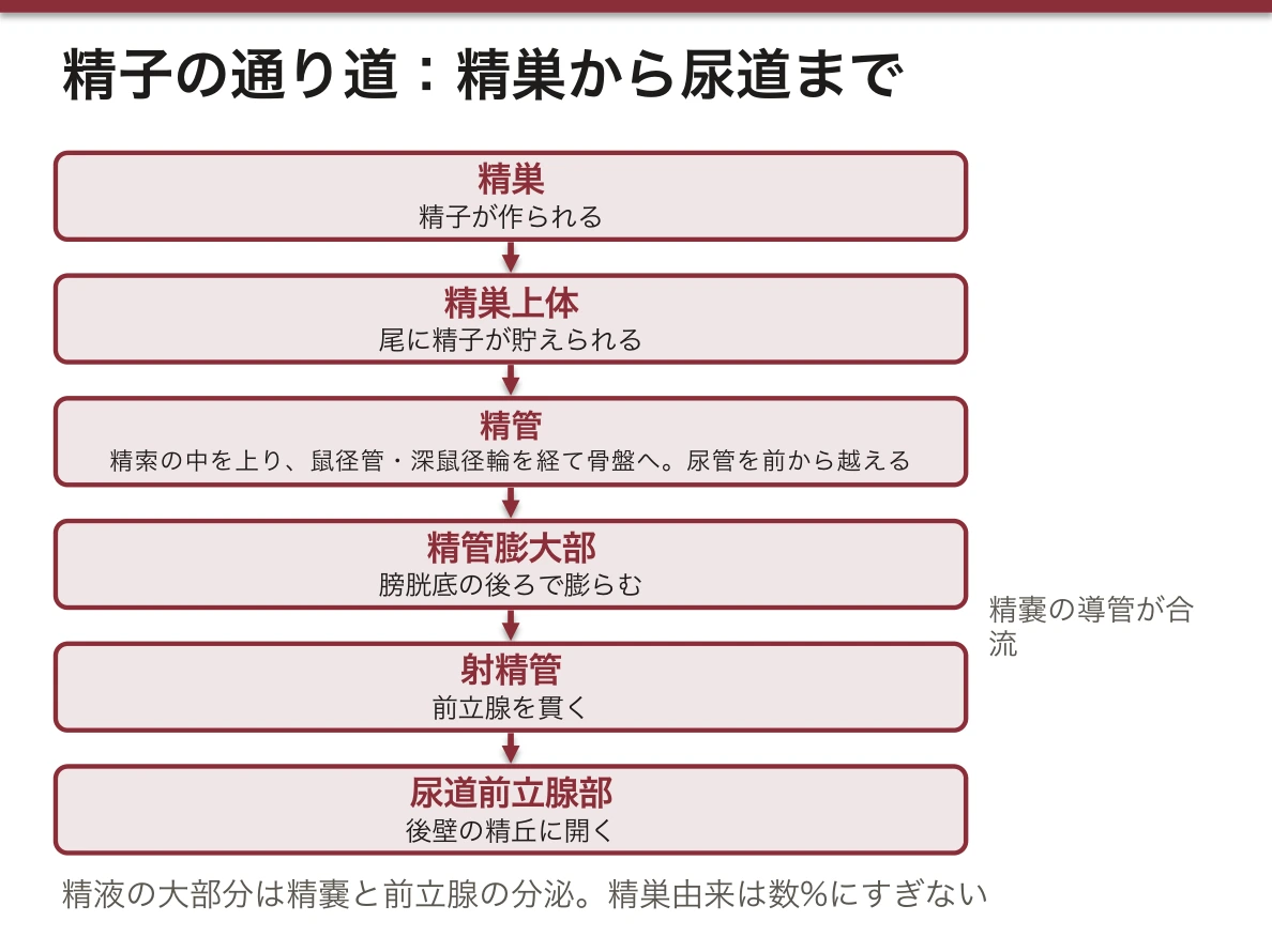
被膜は腹壁の層と一対一で対応する。
| 精索・陰嚢の層 | 由来する腹壁の層 |
|---|---|
| Fascia spermatica externa | 外腹斜筋腱膜 |
| M. cremaster と fascia cremasterica | 内腹斜筋 |
| Fascia spermatica interna | 横筋筋膜 |
| Tunica vaginalis | 腹膜(鞘状突起) |
腹横筋に由来する層は無い——精巣が下降する経路に腹横筋が及んでいないためで、これも問われる。
蔓状静脈叢は対向流熱交換器である。温かい動脈血が冷たい静脈血と熱を交換し、精巣に達する頃には温度が下がっている。精索静脈瘤で造精機能が落ちるのは、この冷却装置が壊れるからである。
精管結紮(vasectomy)は陰嚢上部で行う。精索の中で最も硬く索状に触れる構造が精管なので、皮膚の上から確実に同定できる。結紮後も精液量はほとんど減らない——精液の大部分は精嚢と前立腺の分泌で、精巣由来は数%にすぎないからである。
精索の被膜は腹壁の層がそのまま伸びたものである。外精筋膜=外腹斜筋腱膜、挙睾筋=内腹斜筋、内精筋膜=横筋筋膜。下降の途中で腹壁を引き連れていった履歴がそのまま層になっている。
間接(外)鼠径ヘルニアは深鼠径輪から鼠径管を通り、下腹壁動脈の外側を通る。腟状突起の遺残によるもので若年に多く、陰嚢まで下りる。
直接(内)鼠径ヘルニアはHesselbach 三角から下腹壁動脈の内側に直接押し出る。高齢者の腹壁の脆弱化による。下腹壁動脈との位置関係が両者を分ける唯一の確実な指標である。
精管結紮術(パイプカット)は精索の中の精管だけを切る。精子は精巣で作られ続けるので、ホルモンにも性機能にも影響しない。
English version
Core
Ductus deferens: begins at the tail of the epididymis, ascends in the spermatic cord through the inguinal canal and enters the pelvis at the deep ring. It crosses in front of the ureter (the male version of "water under the bridge"), passes behind the fundus of the bladder, dilates as the ampulla ductus deferentis, and joins the duct of the seminal vesicle to form the ductus ejaculatorius, which pierces the prostate and opens into the prostatic urethra. Its wall is thick smooth muscle, so it is felt as a firm cord.
The contents of the funiculus spermaticus are three arteries, three nerves and three others: testicular, deferential and cremasteric arteries / ilioinguinal (on the surface), genital branch of the genitofemoral, autonomic fibres / ductus deferens, pampiniform plexus, lymphatics.
Going further
The coverings correspond one to one with the layers of the abdominal wall.
| Layer of cord and scrotum | Derived from |
|---|---|
| Fascia spermatica externa | Aponeurosis of external oblique |
| M. cremaster and fascia cremasterica | Internal oblique |
| Fascia spermatica interna | Transversalis fascia |
| Tunica vaginalis | Peritoneum (processus vaginalis) |
There is no layer from transversus abdominis — the muscle does not extend over the path of descent — and this too is asked.
The pampiniform plexus is a countercurrent heat exchanger. Warm arterial blood exchanges heat with cool venous blood and is cooled before reaching the testis. Spermatogenesis falls in varicocele because this cooling device is broken.
Vasectomy is performed high in the scrotum. The ductus is the firmest cord-like structure in the spermatic cord and so can be identified reliably through the skin. The volume of the ejaculate scarcely falls afterwards — most of the semen is the secretion of the seminal vesicles and prostate, and only a few per cent comes from the testis.
In the clinic
The coverings of the cord are the layers of the abdominal wall drawn out: external spermatic fascia = external oblique aponeurosis; cremaster = internal oblique; internal spermatic fascia = transversalis fascia. The record of having dragged the wall along during descent, left as layers.
An indirect inguinal hernia passes from the deep ring through the canal, lateral to the inferior epigastric artery. It arises from a persistent processus vaginalis, is commoner in the young, and descends into the scrotum. A direct hernia pushes straight out through Hesselbach’s triangle, medial to the inferior epigastric artery, from weakening of the wall in the elderly. The relation to the inferior epigastric artery is the only reliable way of distinguishing them.
Vasectomy divides only the ductus within the cord. Sperm continue to be made in the testis, so neither the hormones nor sexual function is affected.
付属生殖腺 — 精嚢・前立腺・尿道球腺Glandula genitalis accessoria
| 腺 | 位置 | 分泌 | 導管 |
|---|---|---|---|
| Vesicula seminalis (glandula seminalis) | 膀胱底の後ろ・精管膨大部の外側 | 精液の約60〜70%。果糖(精子のエネルギー源)・アルカリ性 | 精管と合して射精管へ |
| Prostata | 膀胱頸の下・尿道を取り巻く | 約20〜30%。PSA・酸性ホスファターゼ・クエン酸 | 多数の小孔が尿道前立腺部へ |
| Glandula bulbourethralis(Cowper) | 尿生殖隔膜の中 | ごく少量の粘液(潤滑・尿道の中和) | 尿道海綿体部へ |
前立腺は栗の実大(約20g)。Basis(上・膀胱に接する)と apex(下・尿生殖隔膜に接する)。後面は直腸に接し、直腸診で触れる。
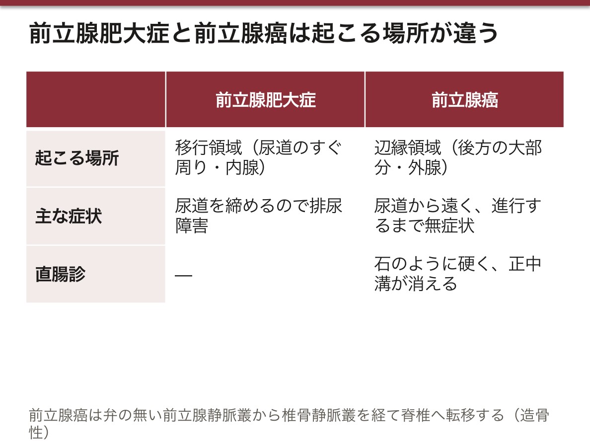
前立腺は区域(zone)で分けると疾患が説明できる。移行領域(transitional zone)は尿道のすぐ周りで前立腺肥大症(BPH)の起こる場所——だから肥大すると尿道が圧迫されて排尿障害が出る。辺縁領域(peripheral zone)は後方の大部分で癌の約70%がここ——直腸に面しているので直腸診で硬結として触れるし、生検も経直腸で行う。「肥大は内側で尿が出ない、癌は外側で触れる」。
精嚢は精子を貯めない。名前に反して分泌腺であり、精子は精巣上体尾に貯えられる。精液の果糖を測るのは精嚢と射精管の開通性を見るためで、閉塞性無精子症では果糖が低下する。
前立腺静脈叢は椎骨静脈叢(Batson)と交通する。弁が無く、腹圧で逆流しうる。前立腺癌が肺を経ずに腰椎・骨盤へ骨転移するのはこの経路による。転移は造骨性で、ALP が上がる。
前立腺は直腸のすぐ前にある。だから直腸診で触れる——正常は栗大・弾性硬・正中溝あり。癌では石のように硬く、正中溝が消える。
前立腺肥大症は内腺(移行域)から起こる。尿道を内側から締めるので排尿障害が主症状になる。
前立腺癌は外腺(辺縁域)から起こる。尿道から遠いので排尿障害が出にくく、進行するまで無症状——PSA と直腸診で拾う理由がここにある。
前立腺静脈叢は椎骨静脈叢と交通する(Batson 静脈叢)。弁が無く、腹圧で逆流するため、前立腺癌は肺を経由せずに脊椎へ転移する。造骨性の骨転移が特徴で、ALP が上がる。
精嚢の分泌液は果糖を含む——精液中の果糖の有無が精管・精嚢の閉塞の指標になる。
English version
Core
| Gland | Position | Secretion | Duct |
|---|---|---|---|
| Vesicula seminalis | Behind the fundus of the bladder, lateral to the ampulla of the ductus | 60–70 per cent of the semen. Fructose (energy for the sperm), alkaline | Joins the ductus deferens as the ejaculatory duct |
| Prostata | Below the neck of the bladder, encircling the urethra | 20–30 per cent. PSA, acid phosphatase, citrate | Many small openings into the prostatic urethra |
| Glandula bulbourethralis (Cowper) | Within the urogenital diaphragm | A little mucus (lubrication, neutralising the urethra) | Into the spongy urethra |
The prostate is the size of a chestnut (about 20 g), with a basis above (against the bladder) and an apex below (against the urogenital diaphragm). Its posterior surface lies against the rectum and is palpable per rectum.
Going further
Divide the prostate into zones and the diseases explain themselves. The transition zone, immediately around the urethra, is where benign prostatic hyperplasia arises — so enlargement compresses the urethra and causes voiding difficulty. The peripheral zone, the bulk of it behind, is where about 70 per cent of carcinomas arise — facing the rectum, so it is felt as induration per rectum and biopsied transrectally. "Hyperplasia inside, and the urine will not come; cancer outside, and it can be felt."
The seminal vesicle does not store sperm. Despite the name it is a secretory gland; the sperm are stored in the tail of the epididymis. Fructose in the semen is measured to test the patency of the vesicles and ejaculatory ducts, and is reduced in obstructive azoospermia.
The prostatic venous plexus communicates with the vertebral venous plexus (of Batson). It has no valves and can reflux under abdominal pressure. This is the route by which prostate cancer metastasises to the lumbar spine and pelvis without passing through the lung. The metastases are osteoblastic and the alkaline phosphatase rises.
In the clinic
The prostate lies immediately in front of the rectum, so it can be felt per rectum — normally the size of a chestnut, firm and elastic, with a median sulcus. In carcinoma it is stony hard and the sulcus is lost.
Benign hyperplasia arises from the inner (transition) zone. It compresses the urethra from within, so voiding symptoms predominate.
Carcinoma arises from the outer (peripheral) zone. Being far from the urethra it causes few voiding symptoms and is silent until advanced — which is why it is picked up by PSA and rectal examination.
The prostatic venous plexus communicates with the vertebral plexus (of Batson). Having no valves and refluxing under abdominal pressure, prostate cancer metastasises to the spine without passing through the lung. The osteoblastic metastases are characteristic and the alkaline phosphatase rises.
The secretion of the seminal vesicles contains fructose — its presence or absence in the semen is an index of obstruction of the ductus or the vesicles.
陰茎と陰嚢Penis et scrotum
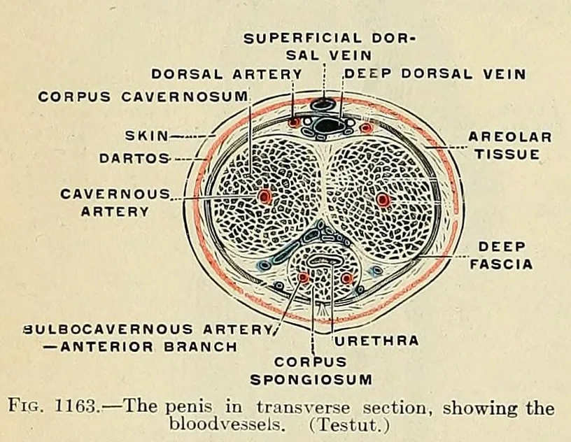
Penis:radix(根・会陰に固定)・corpus(体)・glans penis(亀頭)。亀頭を覆う皮膚のひだが preputium、下面で繋ぐのが frenulum preputii。
3本の海綿体:背側に corpora cavernosa(2本・恥骨下枝に付く crura として始まる)、腹側に corpus spongiosum(1本・尿道を包み、先が亀頭、根元が球)。
尿道は3部を通る:pars prostatica → pars membranacea → pars spongiosa(末端が fossa navicularis)。
Scrotum:皮膚+tunica dartos(平滑筋を含む浅会陰筋膜)。中隔で左右に分かれ、外面に raphe scroti。
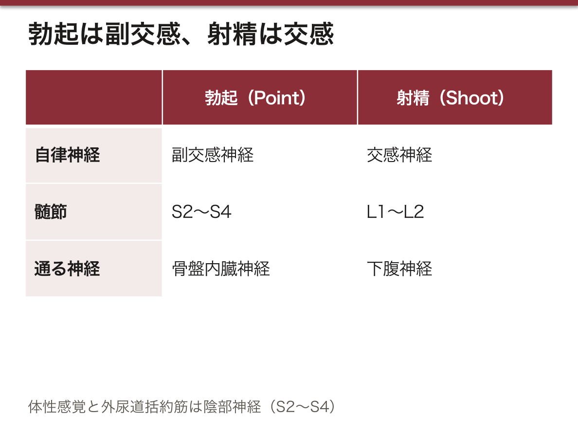
勃起は海綿体洞への動脈流入と静脈流出の遮断で起こる。海綿体は白膜に包まれており、洞が充満すると白膜の下を走る導出静脈が押しつぶされる——だから血が閉じ込められて硬くなる。尿道を包む海綿体(corpus spongiosum)だけは白膜が薄く、完全には硬くならない:硬くなると射精時に尿道が閉じてしまうためである。構造の差に機能的な理由がある。
神経支配は「勃起は副交感、射精は交感」。Point and Shoot——Parasympathetic(S2〜S4・骨盤内臓神経)で勃起、Sympathetic(L1〜L2・下腹神経)で射精。体性感覚と外尿道括約筋は陰部神経(S2〜S4)。
陰嚢と精巣ではリンパの行き先が違う。陰嚢の皮膚は浅鼠径リンパ節(体表由来)、精巣は傍大動脈リンパ節(腹腔内で発生し下降した)。同じ袋の中にあるのに転移先が違うのは、発生の由来が違うためである。
Tunica dartos が温度で収縮する。寒いと縮んで精巣を体幹へ近づけ、暑いと弛緩して離す。精巣挙筋(横紋筋・陰部大腿神経)と併せて、精巣の温度を能動的に調節している。
尿道海綿体は勃起しても内腔をつぶさない。白膜が薄く、内圧が上がりすぎないためで、そうでなければ射精ができない。
持続勃起症(プリアピズム)は陰茎海綿体だけが硬く、亀頭と尿道海綿体は柔らかいのが典型。鎌状赤血球症・薬剤(PDE5 阻害薬・抗精神病薬)が原因になる。4時間以上続けば緊急で、放置すると線維化して不可逆になる。
嵌頓包茎(パラフィモーシス) — 翻転した包皮が戻らなくなり、静脈還流が絶たれて亀頭が腫脹する。用手整復を急ぐ。
陰嚢は精巣を体温より2〜3℃ 低く保つ。肉様膜と挙睾筋が温度で伸縮し、蔓状静脈叢が対向流熱交換をする。精索静脈瘤で温度が上がると造精機能が落ちるのはこの仕組みが壊れるからで、左に多い(左精巣静脈が左腎静脈へ直角に注ぐため)。
English version
Core
Penis: radix (fixed in the perineum), corpus, glans penis. The fold of skin covering the glans is the preputium, tethered beneath by the frenulum preputii.
Three erectile bodies: dorsally the paired corpora cavernosa (beginning as crura attached to the inferior pubic rami), ventrally the single corpus spongiosum enclosing the urethra, expanded in front as the glans and behind as the bulb.
The urethra passes through three parts: pars prostatica → pars membranacea → pars spongiosa (ending in the fossa navicularis).
Scrotum: skin with the tunica dartos (superficial perineal fascia containing smooth muscle), divided by a septum, with the raphe scroti externally.
Going further
Erection occurs by arterial inflow into the cavernous sinuses and blockage of venous outflow. The corpora cavernosa are enclosed in the tunica albuginea, and when the sinuses fill, the emissary veins beneath it are crushed — so the blood is shut in and the organ becomes rigid.
Only the corpus spongiosum, enclosing the urethra, has a thin tunica and does not become fully rigid: were it to do so, the urethra would be closed at ejaculation. A structural difference with a functional reason.
The innervation is "erection parasympathetic, ejaculation sympathetic" — point and shoot. Parasympathetic (S2–S4, pelvic splanchnic) for erection, Sympathetic (L1–L2, hypogastric) for ejaculation. Somatic sensation and the external urethral sphincter are pudendal (S2–S4).
The lymph of scrotum and testis goes to different places. Scrotal skin to the superficial inguinal nodes (a body surface), the testis to the para-aortic nodes (having developed in the abdomen and descended). They lie within the same bag yet drain differently, because their origins differ.
The tunica dartos contracts with temperature, drawing the testis towards the trunk in cold and relaxing in heat. Together with the cremaster (striated, genitofemoral nerve) it actively regulates the temperature of the testis.
In the clinic
The corpus spongiosum does not crush its lumen even in erection. Its tunica is thin and the internal pressure does not rise too far — otherwise ejaculation would be impossible.
Priapism — typically the corpora cavernosa alone are rigid while glans and corpus spongiosum stay soft. Sickle cell disease and drugs (PDE5 inhibitors, antipsychotics) are causes. Beyond four hours it is an emergency; left alone it fibroses irreversibly.
Paraphimosis — a retracted foreskin that will not return, cutting off venous return and swelling the glans. Manual reduction is urgent.
The scrotum keeps the testis 2–3 °C below core temperature. The dartos and cremaster contract and relax with temperature and the pampiniform plexus exchanges heat countercurrently. Spermatogenesis falls when the temperature rises in varicocele because this mechanism is broken — commoner on the left, the left testicular vein entering the left renal vein at a right angle.
精巣Testis
陰嚢内の卵形の腺。白膜に包まれ、中隔で250ほどの小葉に分かれ、精細管が詰まる。
腹腔内で発生して下降するため、血管と神経は腹大動脈と L1 の高さから来る。痛みが腰へ放散するのはこのため。リンパも傍大動脈リンパ節へ行く(鼠径ではない)。
English version
Core
The ovoid gland within the scrotum, enclosed in the tunica albuginea and divided by septa into some 250 lobules packed with seminiferous tubules.
Developing in the abdomen and descending, its vessels and nerves come from the aorta at the level of L1 — which is why pain radiates to the loin. Its lymph likewise goes to the para-aortic nodes, not the inguinal.
精巣上体Epididymis
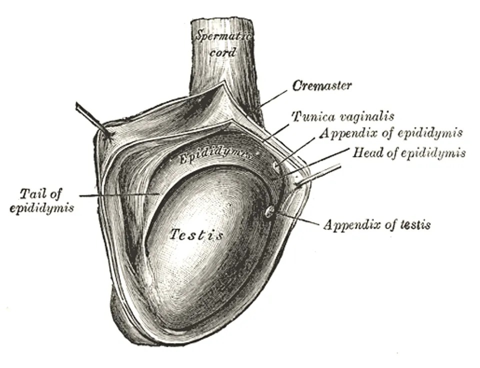
精巣の後縁に乗る。頭・体・尾の3部で、輸出管と長い管が畳まれている。
精子はここで運動能と受精能を獲得し、尾に蓄えられる。尾が精管に移行する。
English version
Core
Rides on the posterior border of the testis, in three parts — head, body and tail — containing the efferent ductules and a long folded duct.
Sperm acquire motility and the capacity to fertilise here and are stored in the tail, which continues into the ductus deferens.
精管・精管膨大部Ductus deferens, ampulla ductus deferentis
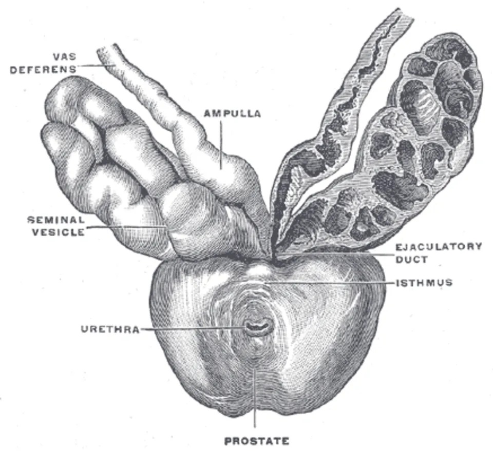
精巣上体尾から始まり、精索の中を上って鼠径管を通り、骨盤へ入って尿管を上から越え、膀胱底の後ろで膨大部になる。
壁が厚く硬いので、精索の中で紐のように触れる——精管結紮の目印。
English version
Core
Begins at the tail of the epididymis, ascends in the cord through the inguinal canal, enters the pelvis, crosses above the ureter and dilates as the ampulla behind the fundus of the bladder.
Its wall is thick and firm, so it is felt like a cord within the spermatic cord — the landmark for vasectomy.
精嚢Vesicula seminalis
膀胱底の後ろ、精管膨大部の外側にある蛇行した袋。精子は入っていない。
精液の約7割を作り、果糖を出して精子のエネルギー源にする。
English version
Core
The tortuous sac behind the fundus of the bladder, lateral to the ampulla of the ductus. It contains no sperm.
It makes about 70 per cent of the semen and secretes fructose as the energy source for the sperm.
射精管Ductus ejaculatorius
精管膨大部+精嚢の管が合してできる短い管。前立腺を貫き、尿道前立腺部の精丘に開く。
English version
Core
The short duct formed by the union of the ampulla of the ductus deferens with the duct of the seminal vesicle, piercing the prostate to open on the seminal colliculus of the prostatic urethra.
精索Funiculus spermaticus
鼠径管を通って精巣へ向かう束。精管・精巣動脈・蔓状静脈叢・神経・リンパ管を、3層の被膜が包む。
蔓状静脈叢が左で拡張しやすい(左精巣静脈が左腎静脈へ直角に入るため)=精索静脈瘤。
English version
Core
The bundle passing through the inguinal canal to the testis, containing the ductus deferens, testicular artery, pampiniform plexus, nerves and lymphatics within three coverings.
The pampiniform plexus dilates more readily on the left (the left testicular vein entering the left renal vein at a right angle) = varicocele.
陰嚢Scrotum
精巣を入れる皮膚の袋。肉様膜という平滑筋を含み、温度で縮む。
体温より1〜2度低く保つことが精子形成に要る。皮膚のリンパは浅鼠径リンパ節へ——中の精巣とは行き先が違う点が問われる。
English version
Core
The bag of skin containing the testes, with the dartos (containing smooth muscle) which contracts with temperature. Keeping the testes 1–2 degrees below core temperature is necessary for spermatogenesis.
The lymph of the skin goes to the superficial inguinal nodes — a different destination from the testis within, which is what is asked.
前立腺(底・尖)Prostata
膀胱の下、栗の実大。底が上(膀胱に接する)、尖が下——上下が名前と逆に感じるので注意。尿道を貫かせる。
直腸のすぐ前にあるので直腸診で触れる。肥大するのは内側の移行域(尿が出にくくなる)、癌ができるのは外側の辺縁域(触れる)——この住み分けが要点。
English version
Core
Below the bladder, the size of a chestnut. The base is above (against the bladder) and the apex below — the reverse of what the names suggest, so take care. The urethra passes through it.
Lying immediately in front of the rectum it is palpable per rectum. Hyperplasia arises in the inner transition zone (and obstructs the urine); carcinoma arises in the outer peripheral zone (and can be felt) — this division is the point.
尿道前立腺部・膜様部Pars prostatica, pars membranacea urethrae
前立腺部は最も広く、後壁の精丘に射精管と前立腺小室が開く。
膜様部は尿生殖隔膜を貫く約1cm。最も短く、最も狭く、最も動きにくいので、骨盤骨折で断裂しやすい。ここに外尿道括約筋(随意)がある。
English version
Core
The prostatic part is the widest, with the ejaculatory ducts and the prostatic utricle opening on the seminal colliculus of its posterior wall.
The membranous part, about 1 cm, pierces the urogenital diaphragm. It is the shortest, the narrowest and the least mobile, and so ruptures readily in pelvic fracture. The external urethral sphincter (voluntary) lies here.
尿道海綿体部・舟状窩Pars spongiosa urethrae, fossa navicularis
尿道海綿体の中を走る最長部(約15cm)。亀頭内で広がる部分が舟状窩。
男性尿道は S 字に2回曲がるので、カテーテルは陰茎を引き上げて曲がりを1つ減らしてから入れる。
English version
Core
The longest part (about 15 cm), running within the corpus spongiosum; the dilatation within the glans is the fossa navicularis.
The male urethra curves twice in an S, so the penis is drawn upwards to remove one of the curves before a catheter is passed.
陰茎根・陰茎体・亀頭Radix, corpus, glans penis
根は会陰に固定される部分(2つの脚と球)、体は遊離部、亀頭は尿道海綿体の先端の膨らみ。
English version
Core
The root is the part fixed in the perineum (the two crura and the bulb), the body the free part, and the glans the expansion at the end of the corpus spongiosum.
陰茎海綿体Corpora cavernosa penis
背側の一対。後端の脚が坐骨恥骨枝に付く。厚い白膜に包まれる。
勃起の主役はここ——白膜が硬いため、血が満ちると圧が上がり静脈が閉じる。
English version
Core
The dorsal pair, their crura attached posteriorly to the ischiopubic rami, enclosed in a thick tunica albuginea.
They are the protagonists of erection — the tunica being rigid, the pressure rises as they fill and the veins are closed.
尿道海綿体Corpus spongiosum penis
腹側の1本で、尿道を包む。後端が球、前端が亀頭。
白膜が薄く勃起しても硬くなりすぎない——硬くなると尿道が潰れて射精できないため。
English version
Core
The single ventral body enclosing the urethra, expanded behind as the bulb and in front as the glans.
Its tunica is thin so it does not become too rigid in erection — were it to do so, the urethra would be crushed and ejaculation impossible.
坐骨海綿体筋Musculus ischiocavernosus
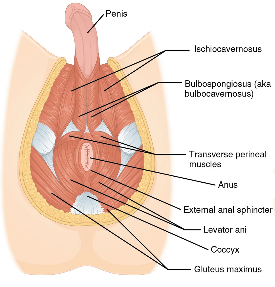
坐骨結節から陰茎脚(女性では陰核脚)を覆う。脚を圧して静脈の還流を妨げ、勃起を保つ。会陰神経支配。
English version
Core
Passes from the ischial tuberosity over the crus of the penis (or of the clitoris in the woman). It compresses the crus, impedes venous return and maintains the erection. Supplied by the perineal nerve.
球海綿体筋Musculus bulbospongiosus
会陰腱中心から陰茎球を覆う。射精と、排尿の最後に尿道に残った尿を押し出す。女性では腟口を狭める。
English version
Core
Passes from the perineal body over the bulb of the penis. It serves ejaculation and expels the last of the urine remaining in the urethra. In the woman it narrows the vaginal orifice.
肛門挙筋Musculus levator ani
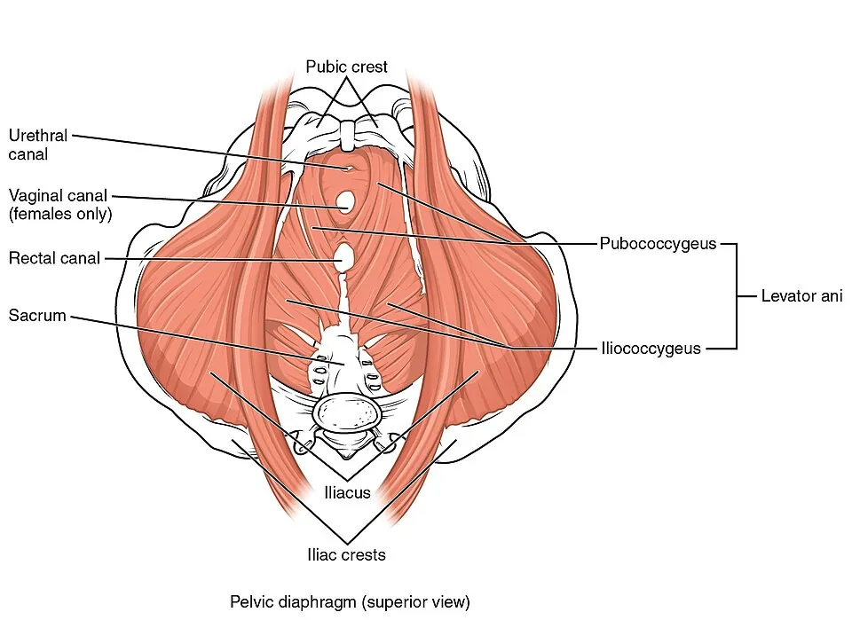
骨盤隔膜の主役。恥骨尾骨筋・腸骨尾骨筋・恥骨直腸筋からなる漏斗。
骨盤内臓を下から支える。恥骨直腸筋が直腸を前へ引いて肛門直腸角を作るのが便禁制の要。分娩で傷むと骨盤臓器脱になる。
English version
Core
The chief muscle of the pelvic diaphragm, a funnel formed by pubococcygeus, iliococcygeus and puborectalis, supporting the pelvic viscera from below.
Puborectalis pulls the rectum forward and creates the anorectal angle — the crux of faecal continence. Damage in childbirth leads to pelvic organ prolapse.
外肛門括約筋Musculus sphincter ani externus
肛門管を取り巻く横紋筋。皮下部・浅部・深部の3層。陰部神経(S2〜S4)支配で随意。
平滑筋・不随意の内肛門括約筋と必ず対にして答える。
English version
Core
The striated muscle encircling the anal canal, in three parts — subcutaneous, superficial and deep. Supplied by the pudendal nerve (S2–S4) and voluntary.
Always answer it as a pair with the internal sphincter (smooth muscle, involuntary).
発生 — 未分化の生殖腺から男性の生殖器へDuctus mesonephricus et paramesonephricus
第7週までは男女の区別がつかない(未分化期)。原始生殖細胞は第3〜4週に卵黄嚢の壁に現れ、後腸の背側間膜を伝って、中腎の内側の生殖堤へ第5〜6週に移ってくる。生殖堤の上皮が原始性索を作って生殖細胞を囲み、未分化の生殖腺になる。男女どちらにも2対の管がある——中腎管(Wolff 管)と中腎傍管(Müller 管)。
男性では Y 染色体の SRY が働いて生殖腺が精巣になる。性索は髄質へ伸びて精巣索(のちの精細管・精巣網)となり、その外を白膜が包む。そこから3つの信号が出る。
| 細胞 | 出すもの | 結果 |
|---|---|---|
| Sertoli 細胞 | 抗 Müller 管ホルモン(AMH) | Müller 管が消える(名残:精巣垂・前立腺小室) |
| Leydig 細胞 | テストステロン | 中腎管が残る → 精巣上体管・精管・精嚢・射精管。中腎の細管は精巣輸出管に |
| (標的組織で) | ジヒドロテストステロン(5α還元酵素で作る) | 外生殖器と前立腺が男性型になる |
外生殖器は、生殖結節が陰茎(亀頭)に、尿道ひだが閉じて尿道海綿体部と陰茎の腹側に、陰唇陰嚢隆起が陰嚢になる。
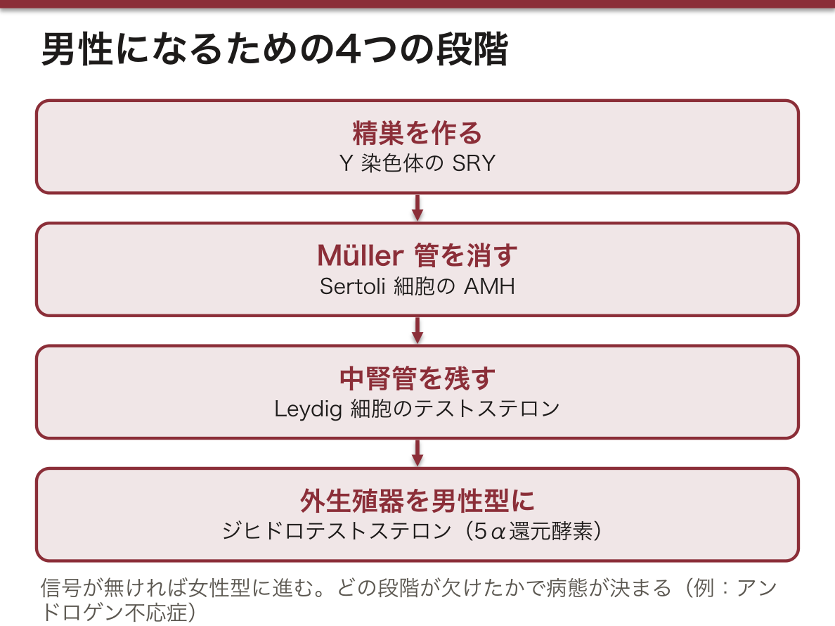
何も信号が無ければ女性型に進む。男性になるには、精巣を作る(SRY)、Müller 管を消す(AMH)、中腎管を残す(テストステロン)、外生殖器を男性型にする(ジヒドロテストステロン)——という別々の段階が全部必要で、どの段階が欠けたかで病態が決まる。
たとえばアンドロゲン不応症(46,XY でテストステロンの受容体が働かない)では、精巣はあり AMH も効くので子宮は無いが、外生殖器は女性型になる。5α還元酵素欠損症では生まれたとき外生殖器があいまいで、思春期にテストステロンが増えると男性化が進む。
尿道下裂は尿道ひだが閉じきらず、外尿道口が陰茎の腹側(裏側)に開くもので、男児で比較的多い先天異常である。治療(尿道形成)に包皮を使うので、手術の前に割礼をしてはいけない。
精管は中腎管、尿管は中腎管から出る尿管芽が元なので、片側の精管欠損には同じ側の腎の無形成が合併しやすい——出身が同じ管だからである。精巣の下降と被膜の層は本章の「精索と精管」を参照。
English version
Core
Until week 7 the sexes cannot be told apart (indifferent stage). Primordial germ cells appear in the wall of the yolk sac in weeks 3–4 and migrate along the dorsal mesentery of the hindgut to the genital ridges medial to the mesonephros in weeks 5–6. The ridge epithelium forms primitive sex cords around the germ cells, making the indifferent gonad. Both sexes have two pairs of ducts: the mesonephric (Wolffian) and the paramesonephric (Müllerian) ducts.
In males, SRY on the Y chromosome turns the gonad into a testis. The sex cords grow into the medulla as testis cords (later seminiferous tubules and rete testis), and the tunica albuginea forms around them. Three signals follow.
| Cell | Product | Result |
|---|---|---|
| Sertoli cell | Anti-Müllerian hormone (AMH) | Müllerian duct regresses (remnants: appendix testis, prostatic utricle) |
| Leydig cell | Testosterone | Mesonephric duct is kept → duct of epididymis, ductus deferens, seminal vesicle, ejaculatory duct. Mesonephric tubules become efferent ductules |
| (in target tissue) | Dihydrotestosterone (made by 5α-reductase) | External genitalia and prostate become male |
Externally, the genital tubercle becomes the penis (glans), the urethral folds close to form the spongy urethra and the underside of the penis, and the labioscrotal swellings become the scrotum.
Going further
Without any signal, development goes female. Becoming male needs separate steps — make a testis (SRY), remove the Müllerian duct (AMH), keep the mesonephric duct (testosterone), masculinise the external genitalia (dihydrotestosterone) — and the missing step decides the condition.
In androgen insensitivity (46,XY with a non-working testosterone receptor), testes are present and AMH works, so there is no uterus, but the external genitalia are female. In 5α-reductase deficiency the genitalia are ambiguous at birth, and masculinisation progresses at puberty when testosterone rises.
In the clinic
In hypospadias the urethral folds do not close fully and the urethra opens on the underside of the penis; it is a relatively common birth defect in boys. The foreskin is used for the repair, so the boy must not be circumcised before surgery.
The ductus deferens comes from the mesonephric duct and the ureter from the ureteric bud of the same duct, so absence of the vas on one side often goes with absence of the kidney on that side — they share an origin. For descent of the testis and the layers of its coverings, see “Spermatic cord and ductus deferens” in this chapter.
まとめ
| 精巣と精巣上体 | Testis:陰嚢内、卵形、約4×3×2.5cm。 |
|---|---|
| 精索と精管 — 腹壁の層がそのまま被膜になる | Ductus deferens:精巣上体尾から始まり、精索の中を上って鼠径管を通り、深鼠径輪から骨盤へ入る。 |
| 付属生殖腺 — 精嚢・前立腺・尿道球腺 | 腺位置分泌導管Vesicula seminalis (glandula seminalis)膀胱底の後ろ・精管膨大部の外側精液の約60〜70%。 |
| 陰茎と陰嚢 | Penis:radix(根・会陰に固定)・corpus(体)・glans penis(亀頭)。 |
| 精巣 | 陰嚢内の卵形の腺。白膜に包まれ、中隔で250ほどの小葉に分かれ、精細管が詰まる。 |
| 精巣上体 | 精巣の後縁に乗る。頭・体・尾の3部で、輸出管と長い管が畳まれている。 |
| 精管・精管膨大部 | 精巣上体尾から始まり、精索の中を上って鼠径管を通り、骨盤へ入って尿管を上から越え、膀胱底の後ろで膨大部になる。 |
| 精嚢 | 膀胱底の後ろ、精管膨大部の外側にある蛇行した袋。 |
| 射精管 | 精管膨大部+精嚢の管が合してできる短い管。 |
| 精索 | 鼠径管を通って精巣へ向かう束。精管・精巣動脈・蔓状静脈叢・神経・リンパ管を、3層の被膜が包む。 |
| 陰嚢 | 精巣を入れる皮膚の袋。肉様膜という平滑筋を含み、温度で縮む。 |
| 前立腺(底・尖) | 膀胱の下、栗の実大。底が上(膀胱に接する)、尖が下——上下が名前と逆に感じるので注意。 |
| 尿道前立腺部・膜様部 | 前立腺部は最も広く、後壁の精丘に射精管と前立腺小室が開く。 |
| 尿道海綿体部・舟状窩 | 尿道海綿体の中を走る最長部(約15cm)。 |
| 陰茎根・陰茎体・亀頭 | 根は会陰に固定される部分(2つの脚と球)、体は遊離部、亀頭は尿道海綿体の先端の膨らみ。 |
| 陰茎海綿体 | 背側の一対。後端の脚が坐骨恥骨枝に付く。 |
| 尿道海綿体 | 腹側の1本で、尿道を包む。後端が球、前端が亀頭。 |
| 坐骨海綿体筋 | 坐骨結節から陰茎脚(女性では陰核脚)を覆う。 |
| 球海綿体筋 | 会陰腱中心から陰茎球を覆う。射精と、排尿の最後に尿道に残った尿を押し出す。 |
| 肛門挙筋 | 骨盤隔膜の主役。恥骨尾骨筋・腸骨尾骨筋・恥骨直腸筋からなる漏斗。 |
| 外肛門括約筋 | 肛門管を取り巻く横紋筋。皮下部・浅部・深部の3層。 |
| 発生 — 未分化の生殖腺から男性の生殖器へ | 第7週までは男女の区別がつかない(未分化期)。 |
このノートは医学生が授業の内容を自分の言葉でまとめたもので、診療の判断に使うものではありません。誤りに気づいたらお知らせください。