体のすべての細胞は、ほぼ同じゲノムをもっている。それでも肝細胞は肝細胞のまま、神経細胞は神経細胞のままでいる。どの遺伝子を使い、どれを眠らせるかという選択が、配列とは別の層に書き込まれ、分裂しても受け継がれるからである。この層を扱うのがエピジェネティクスである。
この章では、印をつける仕組み(DNAメチル化・ヒストン修飾)と、印をどこにつけるかを決める仕組み(DNA結合タンパク質・ノンコーディングRNA)を、エピジェネーター → イニシエーター → メインテナーという3段階の流れで整理する。そのうえで、片方のアレルだけが働く現象(ゲノム刷り込み・X染色体の不活性化)と、環境・老化・癌がエピゲノムをどう変えるかを見る。「遺伝子の変異」と「エピジェネティックな変化」を混同しないことが、この章を通しての注意点である。
- エピジェネティクスとは — エピジェネーター・イニシエーター・メインテナー
- イニシエーター — 場所を決めるのは、タンパク質かRNAか
- DNAメチル化 — CpGのシトシンに付く、もっとも安定な印
- ヒストン修飾とクロマチンリモデリング — 尾に付く印で、巻き方が変わる
- アレルの発現の偏り — 2つあるのに、片方しか使わない
- ゲノム刷り込み — 父から来たか、母から来たかで働きが変わる
- X染色体の不活性化 — 女性の体は、2種類の細胞のモザイク
- 環境・老化・癌とエピゲノム — ゲノムと環境をつなぐ接点
エピジェネティクスとは — エピジェネーター・イニシエーター・メインテナー
エピジェネティックな形質とは、DNAの配列を変えずに、クロマチンの状態の変化によって生じ、細胞分裂を越えて安定に受け継がれる性質である。ただし元に戻すこともできる(可逆)。哺乳類では、生殖細胞ができる時期と初期胚で、印の大規模な消去と書き直しが起きる。ゲノム全体の水準で見た印の全体像をエピゲノム、それを調べる分野をエピゲノミクスという。
役割は遺伝子発現の調節だけではない。セントロメア・テロメア・転移因子を眠らせておくことで、微小管が正しくセントロメアに付く、反復配列どうしの余計な組み換えが減る、転移因子が跳び回らない、という形でゲノムの安定を守っている。
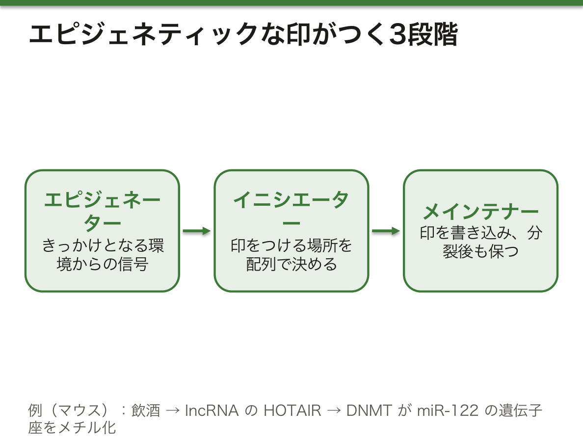
印がつく流れは3段階で考える。①エピジェネーター=きっかけとなる環境からの信号、②イニシエーター=信号を受けて染色体のどこに印をつけるかを配列で決めるもの(DNA結合タンパク質やノンコーディングRNA)、③メインテナー=印を書き込み、分裂後も保つもの(DNAメチル化・ヒストン修飾など)である。
なぜ3段階に分けるのか。信号は一時的で、場所の情報をもたない。場所は配列で指定するしかない。そして印は信号が消えたあとも残らなければ「記憶」にならない。一時的・場所の指定・持続という3つの要求を、それぞれ別の分子が受け持っていると考えると整理しやすい。
マウスでの研究から組み立てられた例で流れを追う。飲酒(エピジェネーター)が続くと、肝で長いノンコーディングRNAのHOTAIR(イニシエーター)が増える。HOTAIRはDNAメチル基転移酵素(メインテナー)をmiR-122の遺伝子座に呼び寄せ、そこをメチル化してmiR-122を沈黙させる。するとmiR-122が抑えていたサイクリンG1が増え、長い目で見ると肝細胞癌の増殖や治療抵抗性につながる。どの分子がどの段階にあたるかを言えることが、この分野の理解の目安になる。
English version
Core
An epigenetic trait is a property that arises from changes in chromatin without changing the DNA sequence, and is passed on stably through cell division. It is also reversible. In mammals, marks are widely erased and rewritten when germ cells form and in the early embryo. The full set of marks across the genome is the epigenome; the field that studies it is epigenomics.
Its role is not only to control gene expression. By keeping centromeres, telomeres and transposable elements silent, it protects genome stability: microtubules attach correctly to centromeres, extra recombination between repeats is reduced, and transposable elements do not jump.
Think of marking in three steps. (1) Epigenator: a signal from the environment that starts the process. (2) Initiator: something that receives the signal and uses sequence to decide where on the chromosome to mark (DNA-binding proteins, non-coding RNAs). (3) Maintainer: something that writes the mark and keeps it after division (DNA methylation, histone modification and so on).
Going further
Why split it into three steps? A signal is brief and carries no information about location. Location can only be given by sequence. And a mark must stay after the signal is gone, or it is not a “memory”. It helps to see three needs, brief, addressed and lasting, each handled by different molecules.
Here is an example built from studies in mice. Ongoing alcohol intake (epigenator) raises the long non-coding RNA HOTAIR (initiator) in the liver. HOTAIR brings DNA methyltransferases (maintainer) to the miR-122 locus, methylates it and silences miR-122. Cyclin G1, which miR-122 normally holds down, then increases. In the long run this links to growth and treatment resistance of hepatocellular carcinoma. Being able to say which molecule belongs to which step is a good test of understanding.
イニシエーター — 場所を決めるのは、タンパク質かRNAか
DNA結合タンパク質は、自分のα-ヘリックスをDNAの主溝に差しこみ、そこに露出した塩基の縁と水素結合などの非共有結合をつくって配列を読み分ける。リン酸の骨格や副溝との接触も結合を助ける。
ノンコーディングRNA(ncRNA)は、DNAから転写されるがタンパク質には翻訳されないRNAで、転写の段階と転写後の段階の両方で遺伝子発現を調節する。
| 種類 | 長さ | 働き |
|---|---|---|
| miRNA | 約22塩基 | RISCという複合体に入り、相補的な標的mRNAを切る・分解する・翻訳を止める |
| siRNA | 約21塩基 | 完全に相補的なmRNAを分解させる(転写後の遺伝子サイレンシング) |
| piRNA | 約26〜31塩基 | PIWIタンパク質と組み、主に生殖細胞で転移因子を抑え、クロマチンを調節する |
| lncRNA | 200塩基以上 | クロマチンを修飾するタンパク質と複合体をつくり、その酵素活性を特定の場所に運ぶ |
なぜRNAは「場所を決める役」に向いているのか。RNAは塩基対合で相手の配列を見つけられるので、配列の住所を安く、柔軟に指定できる。タンパク質が新しい配列を読むには新しい立体構造が必要だが、RNAなら配列を変えるだけでよい。
miRNAとsiRNAのちがいは相補性の度合いにある。siRNAは標的と完全に対合して切断を起こすので、ほぼ1つの標的にだけ効く。miRNAは主に標的mRNAの3′UTRと部分的に対合するため、1つのmiRNAが数百のmRNAを少しずつ抑えられる。網の目のように発現を微調整する仕組みである。
piRNAが特に生殖細胞で働くのも理にかなっている。生殖細胞で転移因子が跳ぶと、その傷は子孫のすべての細胞に受け継がれてしまうからである。
C型肝炎ウイルスは、肝に多いmiR-122を自分の増殖に利用する。miR-122に結合して働かなくするアンチセンス核酸が臨床試験で試された。ncRNAは、病気の目印(バイオマーカー)や治療の標的としても研究が進んでいる。
English version
Core
DNA-binding proteins insert an α-helix into the DNA major groove. They form non-covalent bonds, such as hydrogen bonds, with the exposed edges of the bases and so read the sequence. Contacts with the phosphate backbone and the minor groove also help binding.
Non-coding RNAs (ncRNAs) are transcribed from DNA but not translated into protein. They control gene expression both at transcription and after it.
| Type | Length | Function |
|---|---|---|
| miRNA | About 22 nt | Joins the RISC complex; cuts or degrades a complementary target mRNA, or blocks its translation |
| siRNA | About 21 nt | Causes breakdown of a fully complementary mRNA (post-transcriptional gene silencing) |
| piRNA | About 26–31 nt | Works with PIWI proteins, mainly in germ cells; holds down transposable elements and controls chromatin |
| lncRNA | Over 200 nt | Forms complexes with chromatin-modifying proteins and brings their enzyme activity to specific sites |
Going further
Why is RNA good at deciding location? RNA finds its partner sequence by base pairing, so it can give a sequence address cheaply and flexibly. For a protein to read a new sequence it needs a new 3D shape; for RNA, only the sequence needs to change.
The difference between miRNA and siRNA is how complementary they are. siRNA pairs fully with its target and causes cutting, so it acts on almost one target only. miRNA pairs partly, mainly with the 3′UTR, so one miRNA can lower hundreds of mRNAs a little each. It is a way to fine-tune expression like a net.
It also makes sense that piRNA works mainly in germ cells. If a transposable element jumps in a germ cell, the damage passes to every cell of the offspring.
In the clinic
Hepatitis C virus uses miR-122, which is abundant in the liver, for its own growth. An antisense drug that binds and blocks miR-122 has been tested in clinical trials. ncRNAs are also studied as disease markers (biomarkers) and as drug targets.
DNAメチル化 — CpGのシトシンに付く、もっとも安定な印
DNAメチル化は、ピリミジン塩基であるシトシンの5位の炭素にメチル基がつく反応で、主にCpG(シトシンの次にグアニンが続く並び)で起きる。担うのはDNAメチル基転移酵素(DNMT)で、複製後に古い鎖の印を新しい鎖に写すDNMT1(維持型)と、新しい場所に印をつけるDNMT3A・DNMT3B(新規型)がある。
遺伝子のプロモーターにあるCpGアイランドがメチル化されると、その遺伝子の発現は抑えられる。細胞の種類ごとにメチル化の模様がちがい、それが細胞ごとの発現のちがいの一部をつくっている。CpG以外のシトシンのメチル化は幹細胞で多く見られ、分化に伴ってそれが失われることが重要だと考えられている。
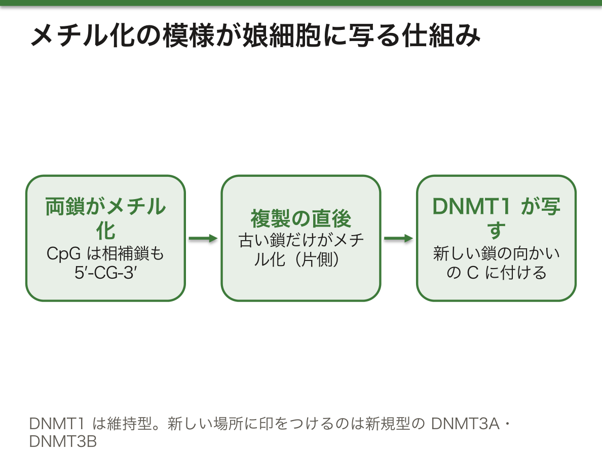
なぜメチル化は分裂を越えて「記憶」されるのか。CpGは相補鎖も5′-CG-3′になる回文の並びである。複製の直後は、古い鎖だけがメチル化された片側メチル化の状態になる。DNMT1はこの片側だけの印を見つけて、新しい鎖の向かい側のCにメチル基をつける。だから模様がそのまま娘細胞に写る。
メチル化が発現を抑える仕組みは2つある。1つは、転写因子の結合部位をメチル基がふさぐこと。もう1つは、メチル化CpGに結合するタンパク質(MeCP2など)がヒストン脱アセチル化酵素などを呼びこみ、クロマチンを固く閉じることである。
副作用もある。メチル化シトシンは自然に脱アミノ化するとチミンになり、DNAの中で異常として見分けにくい。そのためCpGは点変異の起きやすい場所になり、ゲノム全体ではCpGの並びが予想より少ない。
DNMTを阻害するアザシチジンやデシタビンは、骨髄異形成症候群などで、メチル化で眠らされた遺伝子を目覚めさせる目的で使われる。Rett症候群は、X染色体上のMECP2の変異で起きる神経発達症で、主に女児にみられる。メチル化CpGを読んで発現を抑えるタンパク質が働かず、神経細胞の発現調節が乱れる。印そのものではなく印を読む装置の遺伝子の病気である。
English version
Core
DNA methylation adds a methyl group to carbon 5 of the pyrimidine base cytosine, mainly at CpG sites (a cytosine followed by a guanine). It is done by DNA methyltransferases (DNMTs): DNMT1 (maintenance) copies the mark from the old strand to the new one after replication, and DNMT3A and DNMT3B (de novo) place marks at new sites.
When a CpG island in a gene’s promoter is methylated, that gene’s expression is repressed. Each cell type has its own methylation pattern, and this is part of what makes gene expression differ between cells. Methylation of cytosines outside CpG is common in stem cells, and its loss during differentiation is thought to be important.
Going further
Why is methylation “remembered” through division? CpG is a palindrome: the other strand also reads 5′-CG-3′. Right after replication, only the old strand is methylated (hemimethylated). DNMT1 finds this one-sided mark and methylates the C on the new strand opposite it. So the pattern is copied into the daughter cells.
Methylation represses genes in two ways. First, methyl groups can block the binding sites of transcription factors. Second, proteins that bind methylated CpG (such as MeCP2) bring in histone deacetylases and other enzymes and close the chromatin tightly.
There is a side effect. When methylcytosine loses its amino group, it becomes thymine, which is hard to spot as damage. So CpG sites are hot spots for point variants, and CpG is rarer in the genome than expected.
In the clinic
The DNMT inhibitors azacitidine and decitabine are used in myelodysplastic syndrome and similar diseases to wake up genes silenced by methylation. Rett syndrome is a neurodevelopmental disorder caused by variants in MECP2 on the X chromosome, seen mainly in girls. The protein that reads methylated CpG and represses genes does not work, so gene control in neurons is disturbed. It is a disease of the gene for the reading machine, not of the mark itself.
ヒストン修飾とクロマチンリモデリング — 尾に付く印で、巻き方が変わる
ヒストンは、球状の芯の部分と、ヌクレオソームの外に突き出したN末端の尾からなる。修飾の大部分はこの尾で起きる(翻訳後修飾)。修飾はクロマチンの詰まり具合を変え、転写装置がDNAに近づけるかどうかを左右する。
| 修飾 | 対象の残基 | 付ける/外す酵素 | 主な意味 |
|---|---|---|---|
| アセチル化 | リシン(K) | HAT/HDAC | 正電荷を消し、DNAとの結合をゆるめる → 活性化 |
| メチル化 | リシン(1〜3個)・アルギニン(1〜2個) | HMT/HDM | 位置で逆になる。H3K4me3は活性、H3K9me・H3K27me3は抑制 |
| リン酸化 | セリン・トレオニン・チロシン | キナーゼ/ホスファターゼ | 負電荷がDNAと反発 → 多くは活性化 |
| ユビキチン化 | H2A・H2Bのリシン | リガーゼなど | H2Aは抑制、H2Bは活性・抑制の両方 |
| SUMO化 | リシン | リガーゼ/プロテアーゼ | 抑制 |
このほか、別の遺伝子にコードされたヒストンバリアント(数アミノ酸だけちがう)への入れ替えや、ATPのエネルギーでヌクレオソームをほどく・組み直す・入れ替える・滑らせるリモデリングも、クロマチンの状態を変える。
なぜメチル化だけは、場所によって意味が逆になるのか。アセチル化やリン酸化は電荷を変えるので、それ自体がDNAとの結合を弱める。ところがメチル基は電荷を変えない。メチル化の意味は、その位置のその数のメチル基を認識して結合する「読み手」のタンパク質が何を呼びこむかで決まる。H3の4番目のリシンの印には活性化の装置が、27番目には抑制の装置が結合する。印が文字で、読み手が意味を与える、と考えるとわかりやすい。
修飾は互いに連動する。H2Bのユビキチン化がH3K4のメチル化の前提になるように、印どうしが掛け合わされ、DNAメチル化とも協調して働く。
歌舞伎症候群は、H3K4にメチル基を付けるKMT2D(MLL2)や、H3K27から抑制の印を外すKDM6Aの変異で起きる。どちらも遺伝子を「開く」側の酵素である。ここで大事なのは、これらはDNA配列の変化=遺伝的な変化であり、それ自体はエピジェネティックな変化ではないことだ。壊れているのはメインテナーの段階の装置である。治療では、HDACを阻害する薬(ボリノスタットなど)が一部のT細胞リンパ腫に使われている。
English version
Core
A histone has a globular core and an N-terminal tail that sticks out of the nucleosome. Most modifications (post-translational modifications) happen on this tail. They change how tightly chromatin is packed and so decide whether the transcription machinery can reach the DNA.
| Modification | Residue | Added / removed by | Main meaning |
|---|---|---|---|
| Acetylation | Lysine (K) | HAT / HDAC | Removes positive charge, loosens DNA binding → activation |
| Methylation | Lysine (1–3 groups), arginine (1–2) | HMT / HDM | Depends on position: H3K4me3 active; H3K9me and H3K27me3 repressive |
| Phosphorylation | Serine, threonine, tyrosine | Kinase / phosphatase | Negative charge repels DNA → mostly activation |
| Ubiquitination | Lysines of H2A and H2B | Ligases and others | H2A repressive; H2B both active and repressive |
| Sumoylation | Lysine | Ligase / protease | Repressive |
Chromatin also changes by swapping in histone variants (coded by other genes, differing by a few amino acids), and by remodelling, which uses ATP to take apart, rebuild, exchange or slide nucleosomes.
Going further
Why does methylation alone have opposite meanings at different positions? Acetylation and phosphorylation change charge, so they weaken DNA binding by themselves. A methyl group does not change charge. Its meaning depends on which “reader” protein recognises that number of methyl groups at that position, and what the reader brings in. A mark on lysine 4 of H3 attracts activating machinery; a mark on lysine 27 attracts repressing machinery. Think of the mark as a letter and the reader as what gives it meaning.
Modifications work together. H2B ubiquitination is needed before H3K4 methylation, for example. Marks combine with each other and also act together with DNA methylation.
In the clinic
Kabuki syndrome is caused by variants in KMT2D (MLL2), which adds methyl groups to H3K4, or KDM6A, which removes the repressive mark from H3K27. Both are enzymes on the side that “opens” genes. The key point: these are changes in DNA sequence, that is, genetic changes, not epigenetic changes in themselves. What is broken is the machinery of the maintainer step. In treatment, HDAC inhibitors (such as vorinostat) are used for some T-cell lymphomas.
アレルの発現の偏り — 2つあるのに、片方しか使わない
二倍体の細胞では、ふつう1つの遺伝子の2つのコピー(アレル)が同じくらい発現する。この比が1対1でない状態をアレルの発現の偏り(アレル不均衡)といい、常染色体の遺伝子の5〜20%にみられる。原因は、調節配列の個人差や、片方のコピーだけに付いたエピジェネティックな印で、多くは初期胚の時期に決まる。
偏りの極端な形が、片方のアレルだけが働き、もう片方は黙っている状態、つまり単一アレル発現である。
| 種類 | どちらを使うか | 例 |
|---|---|---|
| 体細胞での遺伝子再構成 | ランダム(DNAそのものを組み替える) | B細胞・T細胞の抗原受容体遺伝子 |
| ランダムなアレルの不活性化・活性化 | ランダム、細胞の種類ごと | 嗅覚受容体遺伝子、一部の免疫系の遺伝子 |
| ゲノム刷り込み | 親の由来で決まる | 100以上の遺伝子(発生・成長にかかわる) |
| X染色体の不活性化 | ランダム、初期胚で決まる | 女性のX染色体上のほとんどの遺伝子 |
4つのうち、体細胞での遺伝子再構成だけはDNAの配列そのものを変える。B細胞やT細胞は、抗原受容体の遺伝子の断片を切ってつなぎ直し、膨大な種類の受容体(抗体)を作り出す。片方のアレルで再構成に成功すると、もう片方の再構成は止められる(アレル排除)。なぜ片方だけにするのか。1つの細胞が2種類の受容体をもつと、どの抗原に反応した細胞を増やせばよいかがあいまいになるからである。1細胞=1種類の受容体という原則が、免疫のクローン選択を成り立たせている。
嗅覚の神経細胞も、数百ある受容体遺伝子のうち1つの、しかも片方のアレルだけを選ぶ。こちらも「1つの細胞に1つの役」を与える仕組みと考えられる。
再構成はクローンごとに固有なので、リンパ球の集団を調べると、反応性の増殖か、腫瘍(リンパ腫)かを見分ける手がかりになる。さまざまな再構成が混ざっていれば多クローン性の反応、同じ再構成ばかりなら単クローン性の増殖が疑われる。
English version
Core
In a diploid cell, the two copies (alleles) of a gene are usually expressed about equally. When the ratio is not 1:1, this is called allelic imbalance. It is seen in 5–20% of autosomal genes. Causes include individual differences in regulatory sequences and epigenetic marks on only one copy. It is mostly set in the early embryo.
The extreme form is when only one allele works and the other is silent: monoallelic expression.
| Type | Which copy is used | Examples |
|---|---|---|
| Somatic rearrangement | Random (the DNA itself is rearranged) | Antigen receptor genes of B and T cells |
| Random allelic silencing or activation | Random, cell type specific | Olfactory receptor genes, some immune genes |
| Genomic imprinting | Decided by the parent of origin | Over 100 genes (development and growth) |
| X inactivation | Random, set in the early embryo | Most genes on the X chromosome in females |
Going further
Of the four, only somatic rearrangement changes the DNA sequence itself. B and T cells cut and rejoin pieces of their antigen receptor genes to make a huge variety of receptors (antibodies). Once one allele is rearranged successfully, rearrangement of the other is stopped (allelic exclusion). Why use only one? If one cell had two kinds of receptor, it would be unclear which antigen should make it multiply. The rule one cell = one receptor makes clonal selection in immunity possible.
Olfactory neurons also choose only one of several hundred receptor genes, and only one allele of it. This too seems to give each cell a single role.
In the clinic
Because each rearrangement is unique to a clone, looking at a lymphocyte population helps to tell a reactive expansion from a tumour (lymphoma). Many different rearrangements suggest a polyclonal reaction; one rearrangement everywhere suggests monoclonal growth.
ゲノム刷り込み — 父から来たか、母から来たかで働きが変わる
ゲノム刷り込みとは、ある遺伝子が父由来のコピーだけ、あるいは母由来のコピーだけから発現し、もう一方がエピジェネティックに黙らされている現象である。印は主に、刷り込み領域のDNAメチル化のちがい(メチル化可変領域)として書かれる。100以上の遺伝子が知られ、多くが発生と成長にかかわる。
印は、親が配偶子をつくる時に、親の性に従ってつけられる。たとえば成長因子IGF2は父由来のコピーから発現し、母由来のコピーは黙っている。
刷り込みはどうやって次の世代に渡るのか。受精卵には、父から来た染色体に父型の印、母から来た染色体に母型の印がついている。胚の体細胞はこの印を一生保ち続ける。ところが、その胚の中で将来の生殖細胞になる始原生殖細胞では、印がいったんすべて消され、その胚自身の性に合わせて書き直される。息子なら、母からもらったコピーにも父型の印を書く。だから祖母から来た遺伝子のコピーは、息子を通ると父型になって孫に渡る。「消して、書き直す」がなければ、印は世代ごとに混ざってしまう。
なぜ刷り込みという仕組みがあるのかについては、父由来の遺伝子は胎児の成長を促し、母由来の遺伝子はそれを抑える方向に働く、という見方がある。IGF2(父由来で発現・成長促進)はその典型例として挙げられる。
15番染色体のq11–q13は刷り込みの代表的な領域である。同じ欠失でも、父由来の染色体で欠ければPrader-Willi症候群、母由来で欠ければAngelman症候群になる。同じ病気は、2本とも片親から受け継ぐ片親性ダイソミー(UPD)でも起きる(母由来2本ならPrader-Willi)。配列は正常でも、どちらの親から来たかで病気が決まる——刷り込みの存在を最もはっきり示す例である。メチル化を調べる検査は、欠失とUPDの両方を拾える。
English version
Core
Genomic imprinting means a gene is expressed only from the father’s copy or only from the mother’s copy, while the other copy is silenced epigenetically. The marks are written mainly as differences in DNA methylation in imprinted regions (differentially methylated regions). Over 100 genes are known; many are involved in development and growth.
The marks are placed when a parent makes gametes, according to that parent’s sex. For example, the growth factor IGF2 is expressed from the father’s copy, and the mother’s copy is silent.
Going further
How is imprinting passed to the next generation? In the fertilised egg, chromosomes from the father carry paternal marks and those from the mother carry maternal marks. The embryo’s body cells keep these marks for life. But in the primordial germ cells of that embryo, which will become its own gametes, the marks are all erased and then rewritten to match the embryo’s own sex. A son writes paternal marks even on the copy he got from his mother. So a copy that came from a grandmother becomes paternal when it passes through a son to a grandchild. Without “erase and rewrite”, the marks would get mixed up each generation.
As for why imprinting exists, one view is that the father’s genes push foetal growth and the mother’s genes hold it back. IGF2 (expressed from the father, promotes growth) is a typical example.
In the clinic
Chromosome 15q11–q13 is a classic imprinted region. The same deletion causes Prader-Willi syndrome if it is on the father’s chromosome and Angelman syndrome if it is on the mother’s. The same diseases also arise from uniparental disomy (UPD), where both copies come from one parent (two maternal copies give Prader-Willi). The sequence may be normal, yet which parent it came from decides the disease. This is the clearest proof that imprinting exists. A methylation test can detect both the deletion and UPD.
X染色体の不活性化 — 女性の体は、2種類の細胞のモザイク
ヒトの女性はX染色体を2本、男性は1本もつ。X上の遺伝子の量を男女でそろえるために(遺伝子量補償)、女性の細胞ではX染色体の1本が丸ごと不活性化される。どちらのX(父由来か母由来か)を眠らせるかは細胞ごとにランダムに決まり、選ばれた後は、その細胞から分裂してできるすべての細胞に受け継がれる。
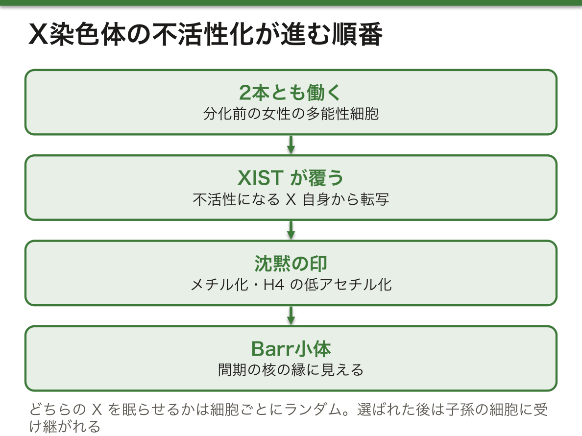
分化前の女性の多能性細胞では、2本のXがどちらも働いている。分化が始まると、将来不活性になるXからXISTという長いノンコーディングRNA(イニシエーター)が転写され、そのX自身を覆う。続いてシトシンのメチル化、ヒストンH4の低アセチル化などの印がつき、Xはほぼ全体が沈黙する。不活性化したXは、間期の核の縁にBarr小体として見える。
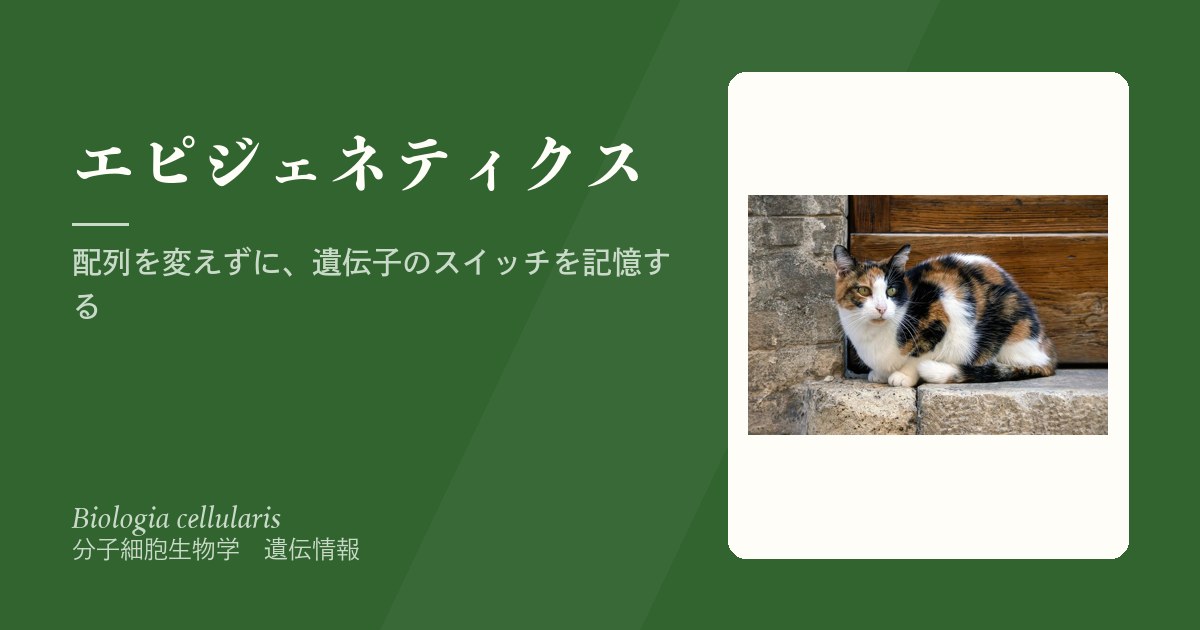
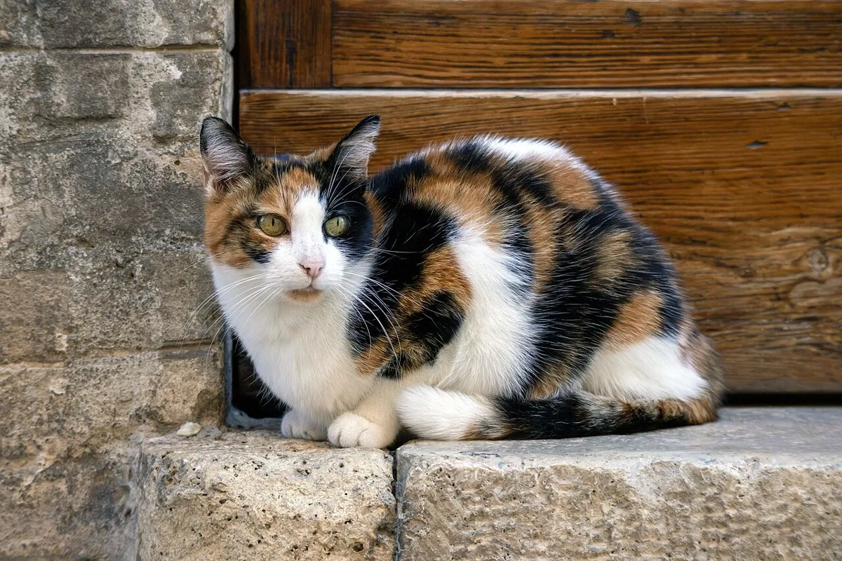
なぜ女性の体はモザイクになるのか。選択がランダムで、しかも選択の後はクローンとして受け継がれるからである。発生の早い時期にある細胞が父由来のXを眠らせると、その子孫の細胞はすべて母由来のXだけを使う。こうして体は、父由来Xを使う細胞の集団と、母由来Xを使う細胞の集団のつぎはぎになる。三毛猫の毛色の模様は、この現象が目に見える例である。
男性でXISTが働かない理由は、Xが1本だと、不活性化を逃れる遺伝子からの産物の量が少なく、XISTを誘導するのに足りないためと説明されている。なお、不活性化は生殖細胞ができる時期に解除される。
Fabry病は、X染色体上のGLA遺伝子の変異でα-ガラクトシダーゼAが不足し、リソソームに糖脂質が溜まる病気である。男性は子どもの頃から重く発症しやすい。ヘテロ接合の女性では、正常なXを使う細胞の割合が人によって大きくちがうため、酵素活性は正常から0近くまで幅があり、症状も軽いものから重いものまでさまざまで、出始めも遅いことが多い。女性では酵素活性が正常でも保因者を否定できないので、遺伝子の検査が必要になる。Duchenne型筋ジストロフィーの女性保因者に症状が出ることがあるのも同じ理由である。
English version
Core
Human females have two X chromosomes; males have one. To make the dose of X-linked genes equal between the sexes (dosage compensation), one whole X is inactivated in each female cell. Which X (from the father or the mother) is silenced is chosen at random in each cell. Once chosen, the choice is passed to every cell that comes from it.
In female pluripotent cells before differentiation, both X chromosomes are active. When differentiation starts, the future inactive X makes a long non-coding RNA called XIST (the initiator), which coats that same X. Then marks such as cytosine methylation and histone H4 hypoacetylation follow, and almost the whole X becomes silent. The inactive X can be seen at the edge of the interphase nucleus as the Barr body.
Going further
Why is the female body a mosaic? The choice is random, and after it is made it is passed on clonally. If an early embryonic cell silences the father’s X, all its descendants use only the mother’s X. So the body becomes a patchwork of cell groups using the father’s X and groups using the mother’s X. The coat pattern of a calico cat is a visible example.
XIST does not work in males because, with only one X, there is too little product from the genes that escape inactivation to induce XIST. Inactivation is also reversed when germ cells form.
In the clinic
Fabry disease is caused by variants in the GLA gene on the X chromosome. α-galactosidase A is lacking, and glycolipids build up in lysosomes. Males are often severely affected from childhood. In heterozygous females, the share of cells using the normal X differs greatly between people, so enzyme activity ranges from normal to almost zero. Symptoms range from mild to severe and often start later. In women, normal enzyme activity does not rule out being a carrier, so a gene test is needed. For the same reason, female carriers of Duchenne muscular dystrophy can show symptoms.
環境・老化・癌とエピゲノム — ゲノムと環境をつなぐ接点
エピゲノムは、環境とゲノムの間の接点である。食事・ストレス・薬物・年齢などが印を変え、その変化が発現を変える。
よく引かれるのが、1944〜45年のオランダの飢餓の冬である。妊娠の終わりの数か月に飢餓にさらされた子は小さく生まれ、その後も小柄なままだった。一方、妊娠初期にさらされた子は、成人後に肥満が多かった。約60年後に調べると、妊娠のごく初期にさらされた人では、刷り込み遺伝子IGF2のメチル化可変領域のメチル化が少なかった。ただしこれは関連を示したもので、メチル化の差が肥満を起こしたと証明されたわけではない。
一卵性双生児も手がかりになる。配列は同じでも、子どもの頃はよく似ていた印が、年齢とともにちがってくる。別々の環境で過ごした時間が長いほど差が大きい。
ラットでは、生後間もない時期に母親からの世話が少ないと、海馬のグルココルチコイド受容体遺伝子のプロモーターがメチル化され、転写因子が結合しにくくなる。受容体が減るとストレスホルモンへの負のフィードバックが弱まり、大人になってもストレス反応が強いままになる。早い時期の環境が、印を通じて長く残る仕組みの例である。動物では、慢性のコカイン投与や慢性の社会的ストレスで、脳のヒストン修飾やDNAメチル化が変わることも報告されている。
癌のエピゲノムは「全体は薄く、要所は濃く」と覚えるとよい。ゲノム全体ではメチル化が減り、反復配列が目覚めて染色体が不安定になる。同時に、癌抑制遺伝子やアポトーシスにかかわる遺伝子(FASなど)のプロモーターは過剰にメチル化されて黙らされる。老化でも全体のメチル化はゆっくり減っていき、細胞を幹細胞に近い状態に寄せて、癌の下地をつくると考えられている。
大腸癌の一部では、ミスマッチ修復遺伝子MLH1のプロモーターが過剰メチル化で黙らされている。同じようにミスマッチ修復ができない腫瘍でも、生まれつきの変異によるLynch症候群とは家族への意味がまったくちがう。腫瘍でMLH1のメチル化を調べることは、遺伝性かどうかを見分ける手順の一部になっている。
English version
Core
The epigenome is the interface between environment and genome. Diet, stress, drugs and age change the marks, and these changes alter gene expression.
A well-known example is the Dutch Hunger Winter of 1944–45. Children exposed to famine in the last months of pregnancy were born small and stayed small. Children exposed in early pregnancy had more obesity as adults. About 60 years later, people exposed very early in pregnancy had less methylation in the differentially methylated region of the imprinted gene IGF2. But this shows an association; it does not prove that the methylation difference caused obesity.
Identical twins are another clue. Their sequence is the same, but marks that were very similar in childhood become different with age. The longer they live in different environments, the bigger the difference.
Going further
In rats, if a pup gets little maternal care soon after birth, the promoter of the glucocorticoid receptor gene in the hippocampus becomes methylated, and a transcription factor binds less. With fewer receptors, negative feedback on stress hormones is weaker, and the stress response stays strong into adulthood. This shows how an early environment can last through marks. In animals, chronic cocaine and chronic social stress have also been reported to change histone modification and DNA methylation in the brain.
Remember the cancer epigenome as “thin overall, thick at key points“. Methylation falls across the genome, repeats wake up, and chromosomes become unstable. At the same time, promoters of tumour suppressor genes and apoptosis genes (such as FAS) become over-methylated and silenced. With ageing, overall methylation also falls slowly. This is thought to push cells toward a stem-cell-like state and prepare the ground for cancer.
In the clinic
In some colorectal cancers, the promoter of the mismatch repair gene MLH1 is silenced by over-methylation. These tumours also cannot repair mismatches, but they mean something very different for the family than Lynch syndrome, caused by an inherited variant. Testing the tumour for MLH1 methylation is part of the steps used to tell whether a cancer is hereditary.
まとめ
| エピジェネティクスとは — エピジェネーター・イニシエーター・メインテナー | エピジェネティックな形質とは、DNAの配列を変えずに、クロマチンの状態の変化によって生じ、細胞分裂を越えて安定に受け継がれる性質である。 |
|---|---|
| イニシエーター — 場所を決めるのは、タンパク質かRNAか | DNA結合タンパク質は、自分のα-ヘリックスをDNAの主溝に差しこみ、そこに露出した塩基の縁と水素結合などの非共有結合をつくって配列を読み分ける。 |
| DNAメチル化 — CpGのシトシンに付く、もっとも安定な印 | DNAメチル化は、ピリミジン塩基であるシトシンの5位の炭素にメチル基がつく反応で、主にCpG(シトシンの次にグアニンが続く並び)で起きる。 |
| ヒストン修飾とクロマチンリモデリング — 尾に付く印で、巻き方が変わる | ヒストンは、球状の芯の部分と、ヌクレオソームの外に突き出したN末端の尾からなる。 |
| アレルの発現の偏り — 2つあるのに、片方しか使わない | 二倍体の細胞では、ふつう1つの遺伝子の2つのコピー(アレル)が同じくらい発現する。 |
| ゲノム刷り込み — 父から来たか、母から来たかで働きが変わる | ゲノム刷り込みとは、ある遺伝子が父由来のコピーだけ、あるいは母由来のコピーだけから発現し、もう一方がエピジェネティックに黙らされている現象である。 |
| X染色体の不活性化 — 女性の体は、2種類の細胞のモザイク | ヒトの女性はX染色体を2本、男性は1本もつ。 |
| 環境・老化・癌とエピゲノム — ゲノムと環境をつなぐ接点 | エピゲノムは、環境とゲノムの間の接点である。 |
このノートは医学生が授業の内容を自分の言葉でまとめたもので、診療の判断に使うものではありません。誤りに気づいたらお知らせください。