単位を正しく使い分けられるかが最大の要点である。Bq・Gy・Sv は測っているものが全部違う。
この章では、放射線のもとになる原子核と放射能(崩壊の法則と半減期)、いつも浴びている自然放射線、放射線を数えるGM 計数管から始め、防護・相互作用・生物学的影響、そして電子の発見から X線、CT と PET までをつなげる。
防護の三原則(距離・遮蔽・時間)は暗記事項ではなく、逆二乗の法則と指数減衰から導ける。
- 放射線の種類と単位 — Bq・Gy・Sv は別物
- 原子核と核種 — 強い力・弱い力と、安定な核の条件
- 放射能と崩壊の法則 — 1個はいつ壊れるか分からないが、全体は指数関数で減る
- 自然放射線とバックグラウンド — 私たちは毎年 2〜3 mSv 浴びている
- 放射線を測る — 電離箱と GM 計数管
- 放射線防護 — 3原則は物理から導ける
- 放射線は物質とどう相互作用するか
- 電子の発見と X線スペクトル — 陰極線から X線管へ
- CT と PET — 外から透かすか、中から光らせるか
- 生物学的影響 — 確定的影響と確率的影響
放射線の種類と単位 — Bq・Gy・Sv は別物
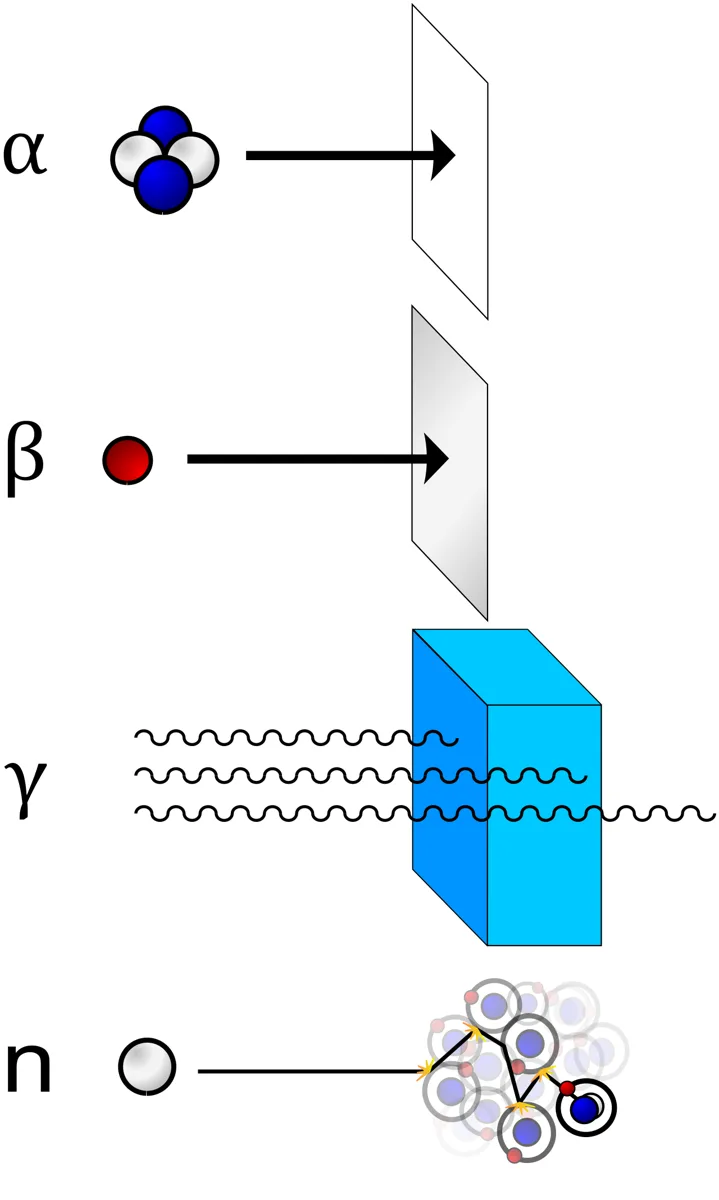
電離放射線=物質から電子をはじき出せるだけのエネルギーを持つ放射線。
| 種類 | 実体 | 透過力 | 止めるもの |
|---|---|---|---|
| α | He 原子核 | 最小(電離作用は最大) | 紙・皮膚 |
| β | 電子 | 中 | アルミ数 mm |
| γ・X | 電磁波 | 大 | 鉛・コンクリート |
| 中性子 | 中性子 | 大 | 水・パラフィン(軽い核) |
| 単位 | 測るもの | 定義 |
|---|---|---|
| Bq(ベクレル) | 線源の強さ | 1秒あたりの崩壊数 |
| Gy(グレイ) | 吸収線量 | 1 kg の物質が吸収したエネルギー(J/kg) |
| Sv(シーベルト) | 人体への影響 | Gy × 放射線加重係数(× 組織加重係数) |
Gy と Sv の違いを1文で言えること。「同じ 1 Gy でも、α 線は γ 線の約20倍の生物学的影響を持つ」——だから 1 Gy の γ 線 = 1 Sv だが、1 Gy の α 線 = 20 Sv になる。放射線加重係数は γ・X・β が 1、中性子が 5〜20、α が 20。
半減期には2種類ある。物理学的半減期(崩壊で半分になる時間)と生物学的半減期(代謝で体外へ排出されて半分になる時間)。両方が同時に働くので実効半減期は
1/T効 = 1/T物 + 1/T生
短いほうに引きずられる。核医学で使う 99mTc(物理学的半減期6時間)が選ばれるのは、診断に十分な時間があり、かつ被曝が長引かないためである。
確定的影響と確率的影響:確定的(皮膚紅斑・脱毛・白内障・不妊)はしきい値があり、線量が増えると重症度が上がる。確率的(発癌・遺伝的影響)はしきい値が無いとされ、線量が増えると発生確率が上がる(重症度は変わらない)。この2分法が防護基準の考え方を決めている——確定的影響は「しきい値を超えさせない」、確率的影響は「合理的に達成できる限り低く(ALARA)」。
English version
Core
Ionising radiation = radiation with enough energy to knock an electron out of matter.
| Kind | What it is | Penetration | What stops it |
|---|---|---|---|
| α | Helium nucleus | Least (greatest ionising power) | Paper, the skin |
| β | Electron | Intermediate | A few mm of aluminium |
| γ, X | Electromagnetic waves | Great | Lead, concrete |
| Neutron | Neutron | Great | Water, paraffin (light nuclei) |
| Unit | What it measures | Definition |
|---|---|---|
| Bq (becquerel) | The strength of the source | Disintegrations per second |
| Gy (gray) | Absorbed dose | Energy absorbed by 1 kg of matter (J/kg) |
| Sv (sievert) | The effect on the human body | Gy × the radiation weighting factor (× the tissue weighting factor) |
Going further
Be able to state the difference between Gy and Sv in one sentence. "The same 1 Gy of α radiation has about twenty times the biological effect of γ" — so 1 Gy of γ = 1 Sv while 1 Gy of α = 20 Sv. The radiation weighting factor is 1 for γ, X and β, 5–20 for neutrons and 20 for α.
There are two kinds of half-life. The physical half-life (the time to halve by decay) and the biological half-life (the time to halve by excretion through metabolism). Both act at once, so the effective half-life is
1/Teff = 1/Tphys + 1/Tbiol
It is dragged towards the shorter. ⁹⁹ᵐTc (physical half-life 6 hours) is chosen in nuclear medicine because there is time enough for the diagnosis and the exposure does not drag on.
Deterministic and stochastic effects: deterministic (erythema, epilation, cataract, infertility) have a threshold, and the severity increases with dose. Stochastic (carcinogenesis, hereditary effects) are taken to have no threshold, and the probability of occurrence increases with dose (the severity does not). This dichotomy decides how protection standards are framed — for deterministic effects, "do not let the threshold be exceeded"; for stochastic effects, "as low as reasonably achievable (ALARA)".
原子核と核種 — 強い力・弱い力と、安定な核の条件
原子は、中心の原子核(陽子と中性子)と、そのまわりの電子からできている。原子核の大きさは原子全体の約1万分の1〜10万分の1しかないが、質量のほとんどを担う。今の理解では、陽子と中性子はさらにクォークからできており(陽子は u・u・d、中性子は u・d・d)、電子は決まった軌道を回る粒ではなく、存在する確率の雲(軌道, orbital)として表される。
原子核は記号 AZX で表す。原子番号 Z =陽子の数で、これが元素を決める。質量数 A =陽子+中性子の数。Z が同じで A が違うものが同位体(isotope。例:12C・13C・14C)で、陽子数と中性子数の組で区別した原子核の種類を核種(nuclide)という。不安定で放射線を出して変わっていく核種が放射性核種(radionuclide)である。
| 化学反応 | 核反応 | |
|---|---|---|
| 変わるもの | 外側の電子の配置(結合の組みかえ) | 原子核そのもの |
| 元素 | 変わらない | 別の元素に変わることが多い |
| エネルギーの大きさ | 1原子あたり eV のけた | MeV のけた(約百万倍) |
| 同位体の違い | ほぼ同じようにふるまう | まったく違うふるまいをしうる |
なぜ原子核はばらばらにならないのか。陽子どうしは正電荷でクーロン力により強く反発する。それを上回って核子(陽子と中性子)をつなぎとめているのが強い力(strong nuclear force)である。非常に強いが、届く距離は 10−15 m(原子核の大きさ)程度しかない。一方の弱い力(weak nuclear force)は、中性子と陽子を互いに変える力で、β 崩壊を起こす。
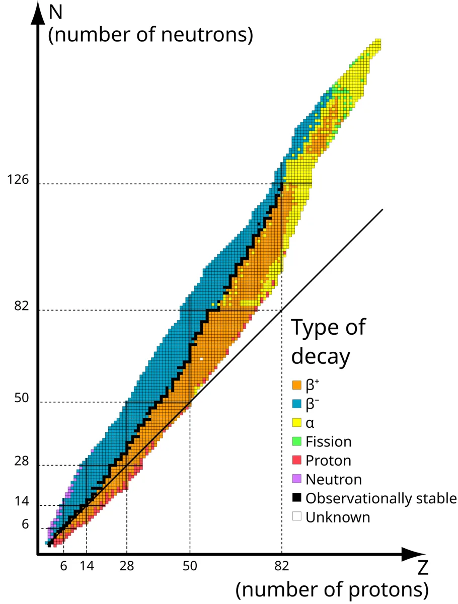
安定な核の条件:軽い元素(Z が20くらいまで)では、陽子と中性子がほぼ同数のときが安定である。重くなると陽子どうしの反発が増えるので、それを薄めるために中性子が陽子より多く必要になる。それでも 鉛(Z = 82)を超えると、どんな数の中性子でも安定にできない。安定な同位体を持つ元素は約80種類で、Z が 82 以下でもテクネチウム(43)とプロメチウム(61)には安定な同位体がない。中性子が多すぎる核は β− 崩壊、少なすぎる核は β+ 崩壊で、安定な組み合わせに近づこうとする。
同位体が「化学的にはほぼ同じ」ことが、核医学の土台である。放射性ヨウ素 131I は、ふつうのヨウ素 127I と同じように甲状腺に取りこまれるので、甲状腺の病気の治療に使える。18F をつけたブドウ糖の類似体(FDG)は、ブドウ糖と同じように細胞に取りこまれる。化学は同じ、核だけが違って放射線を出すから、体の中での行き先を外から追跡できる(トレーサー)。
English version
Core
An atom is made of a central nucleus (protons and neutrons) and electrons around it. The nucleus is only about 1/10,000 to 1/100,000 the size of the atom but carries nearly all its mass. In today’s picture, protons and neutrons are themselves made of quarks (proton u-u-d, neutron u-d-d), and electrons are not balls on fixed tracks but clouds of probability (orbitals).
A nucleus is written AZX. The atomic number Z = number of protons, which sets the element. The mass number A = protons + neutrons. Atoms with the same Z but different A are isotopes (e.g. 12C, 13C, 14C). A kind of nucleus defined by its numbers of protons and neutrons is a nuclide; an unstable one that changes by giving off radiation is a radionuclide.
| Chemical reaction | Nuclear reaction | |
|---|---|---|
| What changes | Arrangement of outer electrons (bonds rearranged) | The nucleus itself |
| Element | Unchanged | Often becomes another element |
| Energy | eV per atom | MeV (about a million times more) |
| Isotopes | Behave almost the same | Can behave completely differently |
Going further
Why doesn’t the nucleus fly apart? Protons are positive and repel each other strongly by the Coulomb force. What overcomes this and holds the nucleons (protons and neutrons) together is the strong nuclear force. It is very strong but reaches only about 10−15 m (the size of a nucleus). The weak nuclear force turns neutrons and protons into each other and causes beta decay.
What makes a nucleus stable: in light elements (up to about Z = 20), nuclei with about equal numbers of protons and neutrons are stable. In heavier ones the repulsion between protons grows, so more neutrons than protons are needed to dilute it. Even so, beyond lead (Z = 82) no number of neutrons can make a stable nucleus. About 80 elements have stable isotopes; even at Z ≤ 82, technetium (43) and promethium (61) have none. A nucleus with too many neutrons undergoes β− decay, and one with too few undergoes β+ decay, moving toward a stable combination.
In the clinic
That isotopes are "chemically almost the same" is the base of nuclear medicine. Radioactive iodine 131I is taken up by the thyroid just like ordinary 127I, so it can treat thyroid disease. A glucose analogue labelled with 18F (FDG) enters cells like glucose. Same chemistry, different nucleus that gives off radiation — so its path in the body can be followed from outside (a tracer).
放射能と崩壊の法則 — 1個はいつ壊れるか分からないが、全体は指数関数で減る
放射能(radioactivity)とは、不安定な原子核が外からの刺激なしに自発的に放射線を出して、別の核種に変わっていく性質である。1896年に Becquerel が、日光に当てていないウラン鉱石が黒い紙で包んだ写真乾板を感光させることから発見し、Curie 夫妻がポロニウムとラジウムを見つけて「放射能」という言葉をつくった。
崩壊の種類:
| 崩壊 | 核の中で起きること | Z と A の変化 | 例 |
|---|---|---|---|
| α 崩壊 | He の原子核(陽子2・中性子2)を出す | Z −2、A −4 | 238U → 234Th |
| β− 崩壊 | n → p + e− + 反ニュートリノ | Z +1、A 不変 | 14C → 14N |
| β+ 崩壊 | p → n + e+(陽電子)+ ニュートリノ | Z −1、A 不変 | 18F → 18O |
| γ 放射 | 励起状態の核が余分なエネルギーを光子で出す | 変わらない | 99mTc → 99Tc |
崩壊の法則:残っている核の数 N は時間とともに指数関数的に減る。
N = N0e−λt、半減期 T1/2 = ln2/λ ≈ 0.693/λ(λ:壊変定数)
放射能の強さ(1秒あたりの崩壊数, Bq)は A = λN で、これも同じ半減期で減る。
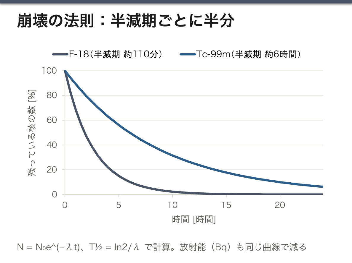
なぜ指数関数になるのか。個々の核がいつ壊れるかは予測できず、どの核も単位時間あたり同じ確率 λ で壊れる。核には「年齢」がなく、古い核ほど壊れやすいということはない。すると、単位時間に壊れる数は残っている数に比例し(dN/dt = −λN)、解は指数関数になる。だから半減期は試料の量・温度・圧力・化学状態によらず一定で、1半減期で 1/2、2半減期で 1/4、10半減期で約 1/1000 になる。核が「消える」のではなく、別の核種(最後は安定な核種)に変わる。
| 核種 | 半減期 | どこにあるか・何に使うか |
|---|---|---|
| 238U | 約 45億年 | 土壌・岩石 |
| 40K | 約 13億年 | 土壌・食品・人体 |
| 14C | 約 5,730 年 | 生物(年代測定) |
| 226Ra | 約 1,600 年 | 土壌(ラドンの親) |
| 131I | 約 8 日 | 甲状腺の治療 |
| 222Rn | 約 3.8 日 | 土壌・空気 |
| 99mTc | 約 6 時間 | 核医学の画像検査 |
| 18F | 約 110 分 | PET |
放射性炭素年代測定:生きている生物は食べ物や呼吸で炭素を入れかえ続けるので、14C の割合は大気と同じに保たれる。死ぬと補充が止まり、14C は半減期約 5,730 年で減っていく。残っている割合が 1/4 なら約 2 半減期、つまり約 11,500 年前と分かる。
放射性核種は、出す放射線の種類で使い分ける。画像診断には体の外まで届く γ 線(99mTc など)や、β+ がつくる消滅放射線(18F の PET)を使う。治療には、近くの細胞だけを壊す飛程の短い β− 線や α 線を使う——131I によるバセドウ病や甲状腺癌の治療、カルシウムに似て骨に集まる 223Ra(α 線)による前立腺癌の骨転移の治療がその例である。治療用は体内に入れたときに周囲へ届きにくく、診断用は外で検出できる、という物理の違いが、そのまま使い分けになっている。
English version
Core
Radioactivity is the property of unstable nuclei to give off radiation spontaneously, with no outside trigger, and change into other nuclides. In 1896 Becquerel discovered it when uranium ore that had not been in sunlight fogged a photographic plate wrapped in black paper. Marie and Pierre Curie found polonium and radium and coined the word "radioactivity".
Types of decay:
| Decay | What happens in the nucleus | Change in Z and A | Example |
|---|---|---|---|
| α decay | Emits a helium nucleus (2 protons, 2 neutrons) | Z −2, A −4 | 238U → 234Th |
| β− decay | n → p + e− + antineutrino | Z +1, A same | 14C → 14N |
| β+ decay | p → n + e+ (positron) + neutrino | Z −1, A same | 18F → 18O |
| γ emission | An excited nucleus gives off extra energy as a photon | No change | 99mTc → 99Tc |
Decay law: the number of nuclei left, N, falls exponentially with time.
N = N0e−λt, half-life T1/2 = ln2/λ ≈ 0.693/λ (λ: decay constant)
Activity (decays per second, Bq) is A = λN, and it falls with the same half-life.
Going further
Why exponential? When a single nucleus will decay cannot be predicted, and every nucleus has the same chance λ of decaying per unit time. Nuclei have no "age"; older ones are not more likely to decay. So the number decaying per unit time is proportional to the number left (dN/dt = −λN), and the solution is exponential. That is why the half-life is fixed, whatever the amount, temperature, pressure or chemical state: after one half-life 1/2 is left, after two 1/4, after ten about 1/1000. The nuclei do not "disappear"; they become other nuclides (in the end, stable ones).
| Nuclide | Half-life | Where found / use |
|---|---|---|
| 238U | about 4.5 billion years | Soil, rock |
| 40K | about 1.3 billion years | Soil, food, the body |
| 14C | about 5,730 years | Living things (dating) |
| 226Ra | about 1,600 years | Soil (parent of radon) |
| 131I | about 8 days | Thyroid treatment |
| 222Rn | about 3.8 days | Soil, air |
| 99mTc | about 6 hours | Nuclear medicine imaging |
| 18F | about 110 minutes | PET |
Radiocarbon dating: living things keep exchanging carbon through food and breathing, so their share of 14C stays the same as the air’s. At death the supply stops, and 14C falls with a half-life of about 5,730 years. If 1/4 is left, about two half-lives — about 11,500 years — have passed.
In the clinic
Radionuclides are chosen by the radiation they give off. Imaging uses gamma rays that reach outside the body (e.g. 99mTc) or the annihilation photons from β+ (18F in PET). Treatment uses short-range β− or α radiation that destroys only nearby cells — for example 131I for Graves’ disease and thyroid cancer, and 223Ra (α), which behaves like calcium and gathers in bone, for bone metastases of prostate cancer. Treatment nuclides should not reach far once inside the body; imaging nuclides must be detectable outside — the difference in physics becomes the choice.
自然放射線とバックグラウンド — 私たちは毎年 2〜3 mSv 浴びている
人は、宇宙・大地・食べ物・空気から、いつも低い線量の放射線を受けている。これが自然放射線(natural background radiation)で、世界平均で年間約 2.4 mSv(実効線量)とされる(国連科学委員会 UNSCEAR の見積もり)。
| 線源 | 世界平均(mSv/年) | 中身 |
|---|---|---|
| 吸入(主にラドン) | 約 1.26 | 土壌・建材のウラン系列から出る気体 |
| 大地からの γ 線 | 約 0.48 | 土壌・岩石のウラン・トリウム・カリウム 40 |
| 宇宙線 | 約 0.39 | 高度・緯度が高いほど多い |
| 食品から(体内) | 約 0.29 | 主にカリウム 40 |
大地や体内の放射性核種の多くは、地球ができたときから残っている長い半減期の核種(238U・232Th・40K など)と、その崩壊でできる核種である。40K はふつうのカリウムに一定の割合で混じっているので、体重 60〜70 kg の成人の体の中には常に約 4,000 Bq の 40K がある。
人工の線源では医療被曝が最大で、世界平均で年 0.6 mSv 程度とされる。日本は CT などの検査が多く、医療による被曝が平均で自然放射線と同じくらいかそれ以上と見積もられている。
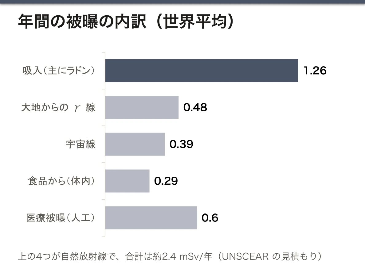
ラドンが最大の線源になる理由:ラドン 222Rn は土壌や岩石の中のラジウム 226 から生まれる貴ガスで、化学的に結合しないので地面から空気中へしみ出す。換気の悪い地下室や家の中にたまり、吸いこまれる。ラドン自身は息とともに出ていくが、崩壊してできるポロニウムなどの子孫核種は固体の粒子で、気管支の壁に付着して、そこで α 線を出す。α 線は飛程が短く電離密度が高いので、気管支上皮の細胞に線量が集中する。地域の地質と家の構造で濃度が大きく変わる。
なぜバックグラウンドを知る必要があるか:放射線を測る測定器は、試料が無くても自然放射線を数えている。試料の測定値から、試料なしで測った値(バックグラウンド)を引いた差(正味の計数)が、その試料の寄与になる。低い放射能を測るときほど、バックグラウンドの測定が結果を左右する。
線量の物差しとして使える:胸部単純写真1枚(約 0.02 mSv)は自然放射線の約3日分、頭部 CT(約 2 mSv)は約10か月分にあたる。
ラドンは、喫煙に次ぐ肺癌の原因とされている(WHO)。しかも喫煙者では危険が掛け算で大きくなる。住居のラドン濃度を測り、高ければ換気や床下の対策で下げることが、各国で公衆衛生の対策になっている。
放射線の利益とリスクを患者に説明するとき、「自然放射線の何日分・何か月分」という比較は分かりやすい。ただし、比べられるのは線量の大きさであって、必要な検査を避ける理由にはならない。
English version
Core
People constantly receive low doses of radiation from space, the ground, food and air. This is natural background radiation, about 2.4 mSv per year (effective dose) as a world average (estimate of UNSCEAR, the UN Scientific Committee).
| Source | World average (mSv/year) | What it is |
|---|---|---|
| Inhalation (mainly radon) | about 1.26 | A gas from the uranium series in soil and building materials |
| Gamma rays from the ground | about 0.48 | Uranium, thorium and potassium-40 in soil and rock |
| Cosmic rays | about 0.39 | More at high altitude and latitude |
| Food (inside the body) | about 0.29 | Mainly potassium-40 |
Many radionuclides in the ground and the body are long-lived nuclides left over from Earth’s formation (238U, 232Th, 40K and others) and the nuclides formed when they decay. 40K is mixed into ordinary potassium in a fixed proportion, so an adult of 60–70 kg always carries about 4,000 Bq of 40K.
Among man-made sources, medical exposure is the largest, about 0.6 mSv a year as a world average. In Japan, with many CT and other examinations, medical exposure is estimated to be about the same as, or more than, natural background on average.
Going further
Why radon is the largest source: radon-222 is a noble gas born from radium-226 in soil and rock. It does not bond chemically, so it seeps from the ground into the air, builds up in poorly ventilated basements and houses, and is breathed in. Radon itself is breathed out again, but its decay products, such as polonium, are solid particles that stick to the bronchial walls and give off alpha radiation there. Alpha radiation has a short range and dense ionisation, so the dose is concentrated in the bronchial lining cells. Levels vary widely with local geology and house construction.
Why background must be known: a radiation counter counts natural radiation even with no sample. The sample’s contribution is the reading with the sample minus the reading without it (the background) — the net count. The lower the activity being measured, the more the background measurement decides the result.
It works as a yardstick for dose: one chest film (about 0.02 mSv) is about 3 days of natural background; a head CT (about 2 mSv) is about 10 months.
In the clinic
Radon is regarded as the second cause of lung cancer after smoking (WHO), and in smokers the risks multiply. Measuring radon in homes and lowering high levels by ventilation or under-floor measures is a public health action in many countries.
When explaining the benefits and risks of radiation to patients, a comparison such as "so many days or months of natural background" is easy to understand. But it compares dose size only; it is not a reason to avoid a needed test.
放射線を測る — 電離箱と GM 計数管
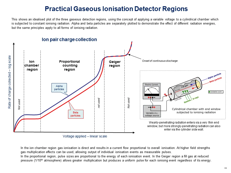
放射線は見えず、聞こえず、感じない。だから測定器で測る。多くの測定器は、放射線が気体を電離してできるイオン対を電気信号として集める。
気体を満たした容器の中に電極を置いて電圧をかけると、放射線が通った跡にできた電子と陽イオンが電極へ引かれて電流(パルス)になる。かける電圧の大きさで、働き方が変わる。
| 検出器 | 電圧 | 何が起きるか | 得られる情報 |
|---|---|---|---|
| 電離箱 | 低い | 放射線がつくったイオン対をそのまま全部集める(増幅なし) | 電流が電離の量に比例 → 線量(率)を正確に |
| 比例計数管 | 中くらい | 電子が加速されて気体を追加で電離(一定倍に増幅) | パルスの大きさがエネルギーに比例 |
| GM 計数管 | 高い(数百 V) | 1個の電離がきっかけで、陽極線全体に電子なだれが広がる | どの放射線でも同じ大きさのパルス → 数だけ(高感度) |
GM 計数管(Geiger–Müller counter)は、陰極になる金属の筒の中心に陽極の細い線を張り、低い圧力の気体を満たした管である。放射線が1個入るたびに大きなパルスが出るので、スピーカーなら「カチッ」と1回鳴る。結果は1分あたりの計数(cpm)や1秒あたりの計数(cps)で表す。
GM 計数管の強みと弱み:電子なだれで信号が大きく増幅されるので、放射線1個でも確実に数えられる(汚染の検出に向く)。しかしパルスの大きさが入ってきた放射線のエネルギーに関係しないので、種類やエネルギーを区別できない。μSv/h で表示する機種は、特定の γ 線(例えば 137Cs)で校正した換算値を出しているので、エネルギーの違う放射線では値がずれる。また、1回放電した後しばらく(約 100 μs)は次を数えられない不感時間があり、線量率が高いと数え落とす。α 線や弱い β 線は管の壁を通れないので、薄い膜の窓のついた管を使う。
数えた値にはゆらぎがある:崩壊は確率的に起きるので、同じ条件で測っても計数はばらつく。N 個数えたときのばらつき(標準偏差)は約 √N で、相対誤差は 1/√N。100 カウントなら約 10%、10,000 カウントなら約 1%。精度を上げたければ長く測って計数を増やす。試料の値からはバックグラウンドを引く。
そのほかの測定器:シンチレーション検出器は、放射線が当たると光る結晶(ヨウ化ナトリウムなど)の光を光電子増倍管で電気に変えるもので、感度が高く、光の量からエネルギーも分かる。γ カメラや PET の検出器はこの仲間である。
病院では目的で測定器を使い分ける。放射線治療装置の出力の校正には、正確さを優先して電離箱を使う。核医学で放射性物質を扱った後の手や床の汚染検査には、わずかな量でも見つけられる GM サーベイメータを使う。放射線を扱う職員は、一定期間の被曝線量を記録する個人線量計(ガラス線量計・OSL 線量計など)を体幹部(防護衣を着る場合はその内側など)につけ、線量限度を超えていないかを管理する。
English version
Core
Radiation cannot be seen, heard or felt, so it is measured with instruments. Many of them collect, as an electrical signal, the ion pairs that radiation creates when it ionises a gas.
Put electrodes in a gas-filled container and apply a voltage. The electrons and positive ions formed along the path of the radiation are pulled to the electrodes and give a current (a pulse). How the detector behaves depends on the voltage.
| Detector | Voltage | What happens | Information |
|---|---|---|---|
| Ionisation chamber | Low | Collects all ion pairs made by the radiation as they are (no amplification) | Current proportional to ionisation → accurate dose (rate) |
| Proportional counter | Medium | Electrons are accelerated and ionise more gas (amplified by a fixed factor) | Pulse size proportional to energy |
| GM counter | High (hundreds of V) | A single ionisation triggers an electron avalanche along the whole anode wire | Same-size pulse for any radiation → count only (very sensitive) |
A Geiger–Müller counter is a metal tube (the cathode) with a thin anode wire along its centre, filled with gas at low pressure. Each particle that enters gives a large pulse — one "click" from a speaker. Results are given as counts per minute (cpm) or per second (cps).
Going further
Strengths and weaknesses of the GM counter: the avalanche amplifies the signal greatly, so even a single particle is counted reliably (good for finding contamination). But the pulse size has nothing to do with the energy of the incoming radiation, so it cannot tell types or energies apart. Models that show μSv/h give a converted value calibrated with a particular gamma source (for example 137Cs), so the value is off for radiation of other energies. After each discharge there is a dead time (about 100 μs) when it cannot count, so at high dose rates it misses counts. Alpha and weak beta particles cannot pass the tube wall, so tubes with a thin window are used.
Counts fluctuate: decay is random, so repeated counts under the same conditions vary. For N counts the spread (standard deviation) is about √N, and the relative error is 1/√N. 100 counts gives about 10%; 10,000 counts about 1%. To improve precision, count longer to get more counts. Subtract the background from the sample reading.
Other detectors: a scintillation detector uses a crystal (such as sodium iodide) that flashes when hit, and a photomultiplier tube turns the light into an electrical signal. It is sensitive and the amount of light also gives the energy. Gamma cameras and PET detectors belong to this family.
In the clinic
Hospitals choose the instrument by purpose. To calibrate the output of radiotherapy machines, accuracy comes first, so an ionisation chamber is used. To check hands and floors for contamination after handling radioactive material in nuclear medicine, a GM survey meter, which finds even tiny amounts, is used. Staff who work with radiation wear a personal dosimeter (glass or OSL dosimeter, etc.) on the trunk (under the lead apron when one is worn, among other places) to record dose over a period and make sure limits are not exceeded.
放射線防護 — 3原則は物理から導ける
外部被曝の3原則:距離・遮蔽・時間。
- 距離——点線源からの線量率は距離の2乗に反比例(逆二乗の法則)。2倍離れれば1/4、3倍で1/9。最も安価で確実な手段
- 遮蔽——透過する線量は厚さに対して指数関数的に減る(I = I₀e^(−μx))。半分にする厚さを半価層 HVL という
- 時間——被曝線量は照射時間に比例する
内部被曝は防護の考え方が違い、取り込ませないこと(吸入・経口・創傷からの侵入を防ぐ)が中心になる。
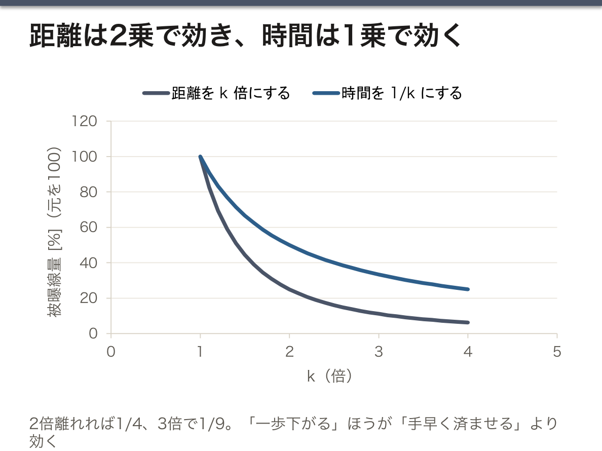
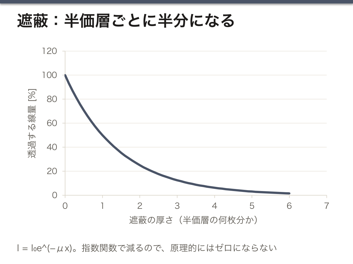
3原則が並列でないことに注意する。距離は2乗で効き、時間は1乗でしか効かない。だから「一歩下がる」ほうが「手早く済ませる」より効果が大きい。実務で「散乱線は患者から出る」と教えられるのは、線源が管球ではなく患者が二次線源になるためで、患者から離れることが最も有効になる。
遮蔽材は放射線の種類で変える。γ・X には原子番号の大きい鉛(光電効果は Z³ に比例)、β にはまずアクリルなど軽い物質(重い物質だと制動放射で X 線が出る=二次的に危険が増える)、中性子には水・パラフィン(軽い核との弾性散乱でエネルギーを奪う)。「重ければ良い」わけではないのが要点である。
ALARA(As Low As Reasonably Achievable)と正当化・最適化・線量限度の3原則(ICRP)。医療被曝には線量限度が設定されていない——患者本人に利益があるためで、代わりに正当化(その検査が必要か)と最適化(必要最小限の線量か)で管理する。職業被曝には限度がある(実効線量 5年で 100 mSv かつ年 50 mSv、眼の水晶体は年 20 mSv)。
妊婦への配慮:器官形成期(受精後2〜8週)の被曝が最も催奇形性の危険が高い。ただし 100 mGy 未満では奇形の危険は有意に増えないとされ、必要な検査を避けるべきではない——放射線を恐れて診断が遅れるほうが害になる、という判断が要る。
English version
Core
The three principles for external exposure: distance, shielding, time.
- Distance — the dose rate from a point source is inversely proportional to the square of the distance (the inverse square law). Twice as far is a quarter, three times is a ninth. The cheapest and surest measure.
- Shielding — the transmitted dose falls exponentially with thickness (I = I₀e^(−μx)). The thickness that halves it is the half-value layer, HVL.
- Time — the dose is proportional to the time of exposure.
Internal exposure is thought of differently: the centre of it is not letting it be taken in (preventing entry by inhalation, ingestion or through a wound).
Going further
Note that the three principles are not on a par. Distance acts as a square and time only linearly. So "take a step back" is more effective than "get it over with quickly". That one is taught in practice that "the scattered radiation comes from the patient" is because the source is not the tube but the patient acting as a secondary source, so moving away from the patient is the most effective thing.
The shielding material is chosen for the kind of radiation. For γ and X, lead, of high atomic number (the photoelectric effect is proportional to Z³); for β, a light material such as acrylic first (with a heavy material, bremsstrahlung produces X-rays — the danger increases secondarily); for neutrons, water or paraffin (energy is taken by elastic scattering from light nuclei). It is not a matter of "the heavier the better", and this is the point.
ALARA (As Low As Reasonably Achievable) and the three principles of justification, optimisation and dose limits (ICRP). No dose limit is set for medical exposure — because the patient benefits — and it is managed instead by justification (is this examination necessary) and optimisation (is the dose the minimum necessary). Occupational exposure does have limits (effective dose 100 mSv in five years and 50 mSv in a year; the lens of the eye 20 mSv a year).
Care in pregnancy: exposure during organogenesis (2–8 weeks after conception) carries the greatest teratogenic risk. But below 100 mGy the risk of malformation is not held to rise significantly, and a necessary examination should not be avoided — the judgement is needed that delaying a diagnosis out of fear of radiation does more harm.
放射線は物質とどう相互作用するか
荷電粒子(α・β)は直接電離する。α は重く電荷が2なので電離密度が高く、飛程が極めて短い(空気数cm、紙で止まる)——体外では無害、体内に入ると最も危険。β は数 mm〜cm 進み、アルミ板で止まる。制動放射を出す。
光子(X 線・γ 線)は間接電離——3つの過程で電子を叩き出し、その電子が電離する。①光電効果(低エネルギー、原子番号 Z の3乗に比例——だから骨とヨード造影剤が白く写る)②Compton 散乱(中エネルギー。診断領域の主役で、散乱線が画質を落とす)③電子対生成(1.02 MeV 以上。治療領域)。
光子は指数関数的に減弱する(I = I₀e^(−μx))ので、原理的にはゼロにならない——だから遮蔽は「半価層で何枚分」で考える。荷電粒子に飛程があるのと対照的で、ここが要点である。
中性子は電荷を持たず、水素との衝突で反跳陽子を作る。遮蔽には水・パラフィンを使う(鉛では止まらない)。
English version
Core
Charged particles (α, β) ionise directly. α is heavy and doubly charged, so its ionisation density is high and its range extremely short (a few cm of air, stopped by paper) — harmless outside the body, most dangerous once inside. β travels millimetres to centimetres and is stopped by aluminium. It produces bremsstrahlung.
Photons (X and γ rays) ionise indirectly — by three processes they knock out an electron, and that electron ionises. ① The photoelectric effect (low energy, proportional to the cube of the atomic number Z — which is why bone and iodinated contrast appear white) ② Compton scattering (intermediate energy; the principal one in the diagnostic range, and scattered radiation degrades the image) ③ pair production (above 1.02 MeV; the therapeutic range).
Photons are attenuated exponentially (I = I₀e^(−μx)), so in principle they never reach zero — hence shielding is thought of as "so many half-value layers". In contrast to charged particles, which have a range; this is the point.
Neutrons carry no charge and produce recoil protons by collision with hydrogen. Water and paraffin are used for shielding (lead does not stop them).
電子の発見と X線スペクトル — 陰極線から X線管へ
19世紀後半、真空にしたガラス管の両端に高い電圧をかけると、陰極から何かが出て反対側のガラスを光らせることが知られていた(陰極線, cathode rays)。その正体が粒子なのか波なのかが議論されていた。
1897年、J. J. Thomson は、陰極線が電場でも磁場でも曲がることを示し、曲がる向きから負の電荷を持つ粒子の流れだと結論した。さらに、電場と磁場を直角にかけて、2つの力が釣り合ってまっすぐ進む条件(qE = qvB、つまり v = E/B)から粒子の速さを求め、磁場だけで曲がる半径(r = mv/qB)と組み合わせて電荷と質量の比 e/m を測った。その値(約 1.76×10¹¹ C/kg)は水素イオンの約1,800倍で、原子よりはるかに軽い、原子の中の粒子が存在することを示した。これが電子の発見である。電荷 e そのものは、のちに Millikan の油滴実験(1909年)で測られた。
X線の発見(Röntgen, 1895年)は、電子の正体が分かる2年前に、同じ陰極線管の実験から生まれた。今の X線管は、加速した電子(陰極線)を金属に当てる装置そのものである。
X線管から出る X線のスペクトルには2つの成分がある。①制動放射:電子が原子核のそばで曲げられて減速し、失ったエネルギーを光子として出す。0 から最大値(管電圧 × 電子の電荷 = eV)までの連続スペクトルになる。②特性X線:電子が内殻の電子をはじき出し、外側の電子が落ちてくるときに殻のエネルギー差を光子として出す。ターゲットの元素で決まる鋭いピークになる。
スペクトルから分かること:連続スペクトルの上端から管電圧が、ピークの位置からターゲットの元素が分かる。特性X線の位置が原子番号で規則的に変わること(Moseley の法則)は、元素を原子番号で並べる根拠になり、今も試料の成分元素を調べる分析法に使われている。詳しくは X線の物理の章で扱った。
電子の発見のあと、Thomson は「正電荷の中に電子が埋まっている」原子の模型を考えたが、1911年に Rutherford は、金箔に α 線を当てるとごく一部が大きく跳ね返されることから、正電荷と質量が中心の小さな核に集まっていることを示した(この実験を行った一人が、GM 計数管に名を残す Geiger である)。
English version
Core
In the late 19th century it was known that when a high voltage is applied across an evacuated glass tube, something leaves the cathode and makes the glass at the other end glow (cathode rays). Whether they were particles or waves was debated.
In 1897 J. J. Thomson showed that cathode rays are bent by both electric and magnetic fields, and from the direction concluded they are a stream of negatively charged particles. Then, with electric and magnetic fields at right angles, he found the speed from the condition where the two forces balance and the beam goes straight (qE = qvB, so v = E/B). Combining this with the radius of bending in the magnetic field alone (r = mv/qB), he measured the charge-to-mass ratio e/m. The value (about 1.76×10¹¹ C/kg) was about 1,800 times that of the hydrogen ion, showing that there is a particle inside atoms, far lighter than an atom. This was the discovery of the electron. The charge e itself was measured later in Millikan’s oil-drop experiment (1909).
The discovery of X-rays (Röntgen, 1895) came from the same cathode-ray tube experiments, two years before the electron was identified. Today’s X-ray tube is exactly a device that fires accelerated electrons (cathode rays) at metal.
Going further
The spectrum of an X-ray tube has two parts. (1) Bremsstrahlung: electrons bent and slowed near nuclei give off the lost energy as photons, forming a continuous spectrum from 0 to a maximum (tube voltage × electron charge = eV). (2) Characteristic X-rays: an electron knocks out an inner-shell electron, and when an outer electron drops in, the energy difference between shells leaves as a photon — sharp peaks set by the target element.
What the spectrum tells us: the top end of the continuous spectrum gives the tube voltage, and the peak positions give the target element. That the characteristic X-ray positions change regularly with atomic number (Moseley’s law) was the basis for ordering elements by atomic number, and is still used to analyse which elements a sample contains. Details are in the X-ray physics chapter.
After the electron was found, Thomson pictured the atom as "electrons embedded in positive charge". In 1911 Rutherford showed, from the fact that a few alpha particles fired at gold foil bounce back sharply, that positive charge and mass are gathered in a small central nucleus (one of those who did the experiment was Geiger, whose name lives on in the GM counter).
CT と PET — 外から透かすか、中から光らせるか
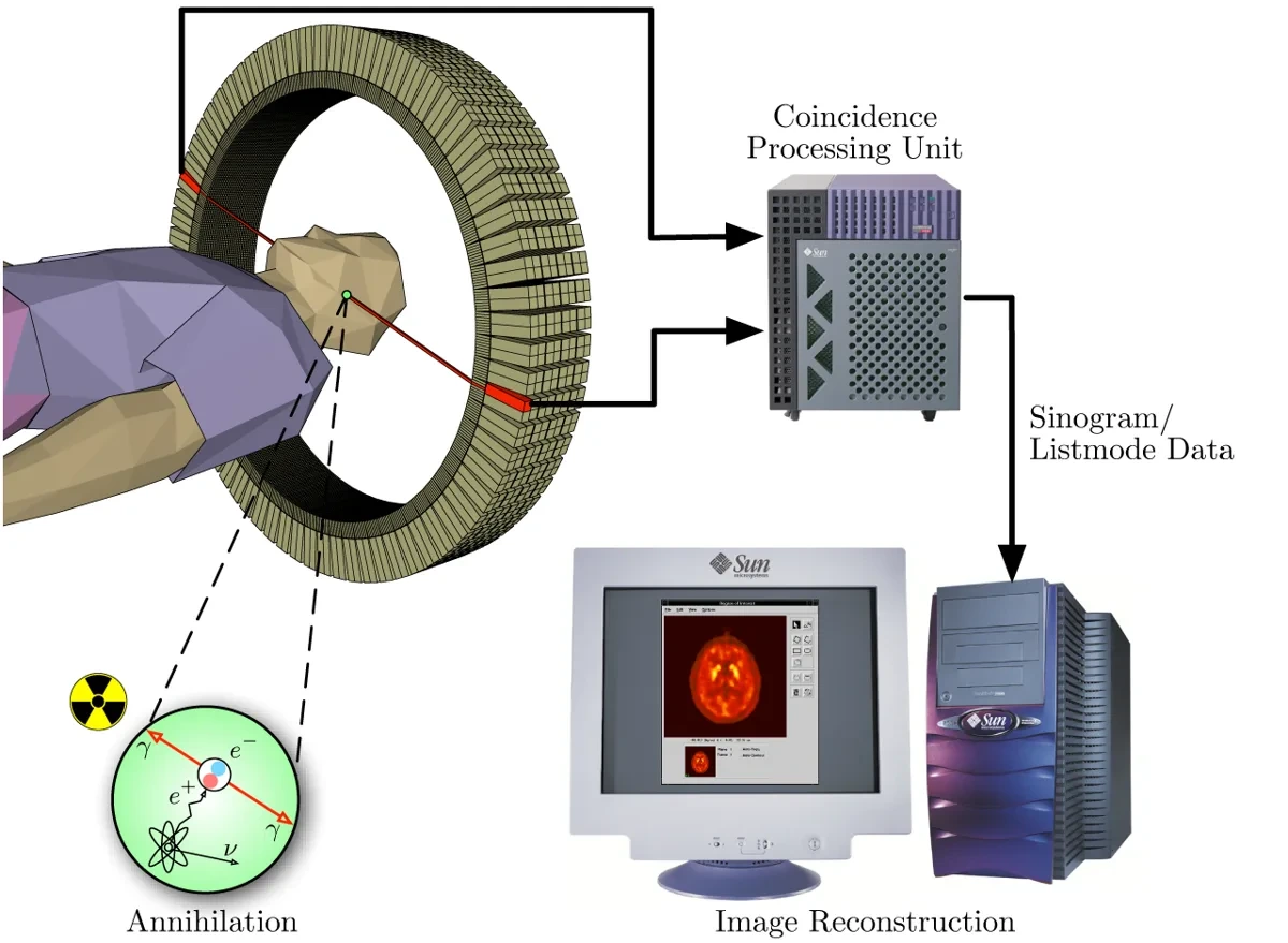
CT と単純 X線撮影の違い:どちらも体の外から X線を当てて、通り抜けた量を測る(透過型)。単純撮影は厚み全体の影が1枚に重なるが、CT は多方向から測って断面ごとの減弱係数(CT 値)を計算するので、重なりがなく、わずかな濃度差を数値で区別できる。そのぶん被曝は大きい(X線の物理の章)。
PET(positron emission tomography, 陽電子放出断層撮影)は逆に、体の中に入れた放射性薬剤から出る放射線を外で測る(放出型)。CT が「形」を見るのに対し、PET は薬剤が集まる場所=代謝や機能を見る。
PET の仕組み:①β+ 崩壊する核種(代表は 18F、半減期約 110 分)で標識した薬剤を静脈から入れる。②放出された陽電子は組織の中を 1 mm 程度進んで電子と出会い、両方が消えて(対消滅)、511 keV の光子2個がほぼ正反対の向きに飛び出す。③患者を囲むリング状の検出器が、2つの光子を同時にとらえたときだけを数える(同時計数)。④2つの検出器を結ぶ線の上のどこかで崩壊が起きたと分かるので、多数の線から CT と同じように断面の分布を再構成する。
なぜ 511 keV なのか:電子と陽電子の質量がすべてエネルギーに変わる(E = mc2)。電子1個の静止エネルギーがちょうど 511 keV で、運動量が保存されるので2個の光子は反対向きに出る。この「反対向きに2個」が PET の要で、鉛のコリメータで方向を限らなくても、線の上に発生源があると分かる。そのため1個の γ 線を測る SPECT(99mTc の 140 keV など。コリメータが必要)より感度が高い。
FDG が腫瘍に集まる理由:18F-FDG(フルオロデオキシグルコース)はブドウ糖の類似体で、ブドウ糖輸送体で細胞に入り、ヘキソキナーゼでリン酸化される。しかしその先の解糖系には進めず、リン酸化された形で細胞内に閉じこめられる。ブドウ糖を盛んに取りこむ細胞——多くの癌細胞、脳、活動している心筋、炎症細胞——ほど強く光る。
PET は解像度(数 mm)が低く、解剖学的な位置が分かりにくいので、ふつうは CT と一体の PET/CT で撮り、CT で位置を合わせ、体による光子の減弱も CT から補正する。
FDG-PET の主な用途は、がんの病期診断・再発や転移の検索・治療効果の判定である。読むときは、生理的に集まる場所(脳・心筋・尿路。FDG は尿に出る)と、炎症でも集まる(偽陽性)ことを知っておく。検査前に数時間絶食するのは、血糖が高いと血中のブドウ糖と FDG が取り合いになり、腫瘍への集まりが弱くなるためで、糖尿病の患者では血糖の管理が要る。脳では、アルツハイマー病で側頭頭頂葉や後部帯状回の糖代謝が下がる所見が診断の助けになる。
English version
Core
CT versus plain X-ray: both send X-rays through the body from outside and measure how much gets through (transmission). A plain film piles the shadow of the whole thickness into one image, but CT measures from many directions and calculates the attenuation (CT number) for each slice, so there is no overlap and small density differences can be told apart as numbers. The cost is a larger dose (X-ray physics chapter).
PET (positron emission tomography) works the other way: it measures from outside the radiation coming from a radioactive drug placed inside the body (emission). CT shows "form"; PET shows where the drug gathers — metabolism and function.
How PET works: (1) A drug labelled with a β+-decaying nuclide (typically 18F, half-life about 110 minutes) is given into a vein. (2) The emitted positron travels about 1 mm in tissue, meets an electron, and both vanish (annihilation), sending out two 511 keV photons in almost exactly opposite directions. (3) A ring of detectors around the patient counts only events where both photons arrive at the same time (coincidence detection). (4) The decay happened somewhere on the line joining the two detectors, so from many such lines the distribution in each slice is reconstructed, as in CT.
Going further
Why 511 keV? The whole mass of the electron and positron becomes energy (E = mc2). The rest energy of one electron is exactly 511 keV, and momentum is conserved, so the two photons go in opposite directions. This "two photons, opposite ways" is the key to PET: without lead collimators limiting direction, we still know the source lies on the line. So PET is more sensitive than SPECT, which detects single gamma rays (such as 140 keV from 99mTc) and needs a collimator.
Why FDG gathers in tumours: 18F-FDG (fluorodeoxyglucose) is a glucose analogue. It enters cells through glucose transporters and is phosphorylated by hexokinase, but cannot go further down glycolysis, so it is trapped inside the cell in its phosphorylated form. Cells that take up glucose avidly — many cancer cells, the brain, working heart muscle, inflammatory cells — light up most.
PET has low resolution (a few mm) and shows anatomy poorly, so it is usually done as a combined PET/CT: CT gives the location and is also used to correct for photon attenuation by the body.
In the clinic
The main uses of FDG-PET are cancer staging, searching for recurrence or metastasis, and judging treatment response. When reading it, know the sites of normal uptake (brain, heart muscle, urinary tract — FDG is excreted in urine), and that inflammation also takes it up (false positives). Patients fast for some hours before the scan because high blood glucose competes with FDG and weakens uptake in the tumour; in diabetes, glucose control is needed. In the brain, reduced glucose metabolism in the temporoparietal lobes and posterior cingulate in Alzheimer’s disease helps diagnosis.
生物学的影響 — 確定的影響と確率的影響
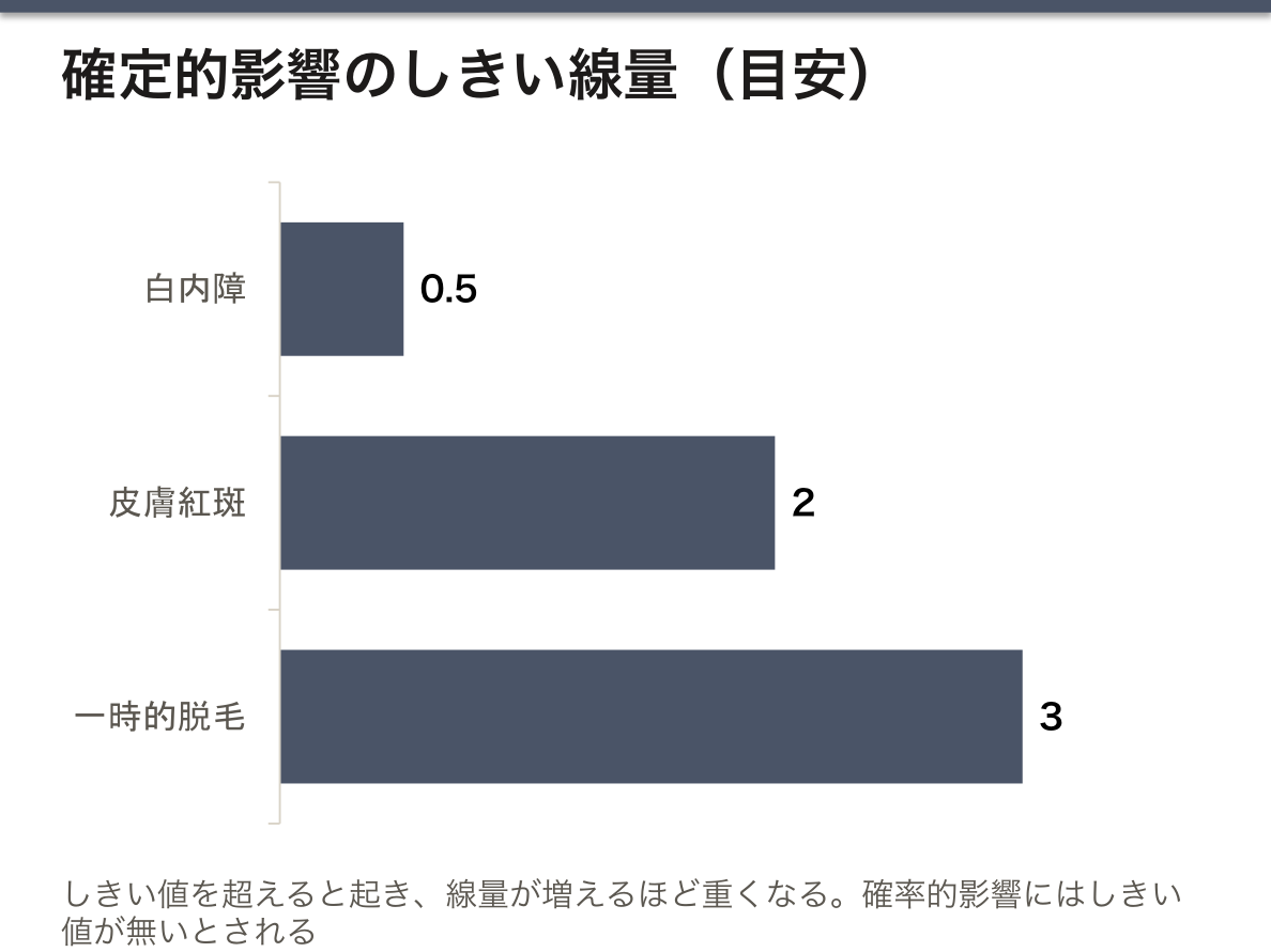
確定的影響(組織反応)——しきい線量があり、それを超えると必ず起き、線量が増えるほど重くなる。細胞が大量に死ぬことによる。例——皮膚紅斑(約2 Gy)、一時的脱毛(約3 Gy)、白内障(約0.5 Gy)、不妊、急性放射線症候群。防護の目標は「しきい値を超えさせないこと」。
確率的影響——しきい線量が無いと仮定し、線量に比例して「起こる確率」が上がる。重症度は線量によらない。DNA の変異による。例——発癌と遺伝的影響。防護の目標は「合理的に達成できる限り低く(ALARA)」。
この2つの区別が、防護体系そのものを決めている——線量限度は確率的影響を抑えるために、しきい値のある組織には別途の限度を置く形で組まれている。
感受性は Bergonié-Tribondeau の法則——分裂が盛んで、分化が低く、将来の分裂回数が多い細胞ほど感受性が高い。骨髄・腸上皮・生殖腺・胎児が高く、神経・筋は低い。
English version
Core
Deterministic effects (tissue reactions) — there is a threshold dose; above it they always occur, and the severity increases with dose. They arise from the death of many cells. Examples — skin erythema (about 2 Gy), temporary epilation (about 3 Gy), cataract (about 0.5 Gy), infertility, acute radiation syndrome. The goal of protection is "do not let the threshold be exceeded".
Stochastic effects — no threshold is assumed, and the probability of occurrence rises in proportion to dose. The severity does not depend on the dose. They arise from mutation of DNA. Examples — carcinogenesis and hereditary effects. The goal of protection is "as low as reasonably achievable (ALARA)".
This distinction decides the whole system of protection — the dose limits are framed to suppress stochastic effects, with separate limits placed on tissues that have a threshold.
Sensitivity follows the law of Bergonié and Tribondeau — the more actively a cell divides, the less differentiated it is, and the more divisions lie ahead of it, the more sensitive it is. Marrow, intestinal epithelium, gonads and the fetus are high; nerve and muscle are low.
まとめ
| 放射線の種類と単位 — Bq・Gy・Sv は別物 | 電離放射線=物質から電子をはじき出せるだけのエネルギーを持つ放射線。 |
|---|---|
| 原子核と核種 — 強い力・弱い力と、安定な核の条件 | 原子は、中心の原子核(陽子と中性子)と、そのまわりの電子からできている。 |
| 放射能と崩壊の法則 — 1個はいつ壊れるか分からないが、全体は指数関数で減る | 放射能(radioactivity)とは、不安定な原子核が外からの刺激なしに自発的に放射線を出して、別の核種に変わっていく性質である。 |
| 自然放射線とバックグラウンド — 私たちは毎年 2〜3 mSv 浴びている | 人は、宇宙・大地・食べ物・空気から、いつも低い線量の放射線を受けている。 |
| 放射線を測る — 電離箱と GM 計数管 | 放射線は見えず、聞こえず、感じない。だから測定器で測る。 |
| 放射線防護 — 3原則は物理から導ける | 外部被曝の3原則:距離・遮蔽・時間。距離——点線源からの線量率は距離の2乗に反比例(逆二乗の法則)。 |
| 放射線は物質とどう相互作用するか | 荷電粒子(α・β)は直接電離する。α は重く電荷が2なので電離密度が高く、飛程が極めて短い(空気数cm、紙で止まる)——体外では無害、体内に入ると最も危険。 |
| 電子の発見と X線スペクトル — 陰極線から X線管へ | 19世紀後半、真空にしたガラス管の両端に高い電圧をかけると、陰極から何かが出て反対側のガラスを光らせることが知られていた(陰極線, cathode rays)。 |
| CT と PET — 外から透かすか、中から光らせるか | CT と単純 X線撮影の違い:どちらも体の外から X線を当てて、通り抜けた量を測る(透過型)。 |
| 生物学的影響 — 確定的影響と確率的影響 | 確定的影響(組織反応)——しきい線量があり、それを超えると必ず起き、線量が増えるほど重くなる。 |
このノートは医学生が授業の内容を自分の言葉でまとめたもので、診療の判断に使うものではありません。誤りに気づいたらお知らせください。