心電図は体表の2点間の電位差を時間の関数として記録したものである。波形の意味(生理学)より前に、なぜ体表で心筋の電気が測れるのかという物理を答えられるようにする。
鍵は「双極子」と「導出=方向を持った窓」の2語である。
- 心臓の電気双極子と体積導体
- 記録の物理 — 増幅・雑音・アーチファクト
- 誘導とは何か — Einthoven の三角形と12誘導
- 皮膚で測れる生体電位 — 心電図・脳波・筋電図
- 膜電位から P・QRS・T まで — 刺激伝導系が波形を作る
- 細動と除細動 — ばらばらの興奮を一度に止める
- ペースメーカ — 電気のパルスで拍動を作る
- 感電 — 交流と直流はどちらが危険か
心臓の電気双極子と体積導体
心筋が興奮すると、興奮した部分(脱分極・細胞外は陰性)と未興奮の部分(細胞外は陽性)が隣り合う。この境界が電気双極子として振る舞い、大きさと向きを持つベクトルで表せる。
体は電解質に満ちた体積導体なので、心臓の双極子が作る電場は体表まで伝わる。心電図は、その電場を体表の2点間の電位差として測っている。
記録される波形の向きは幾何で決まる:興奮が電極に向かってくれば上向き(陽性)、遠ざかれば下向き(陰性)、直交すれば二相性またはほぼ平坦。
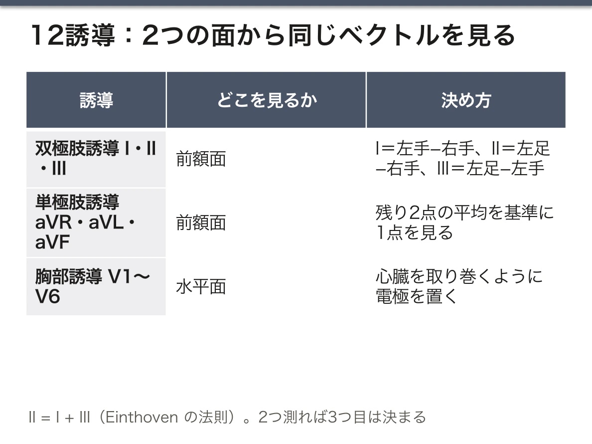
これが12誘導の意味である。誘導とは双極子ベクトルを見る方向で、12方向から同じベクトルを投影している。肢誘導(I・II・III・aVR・aVL・aVF)が前額面、胸部誘導(V1〜V6)が水平面——「1つの立体を2つの平面に投影した6枚ずつの影」である。
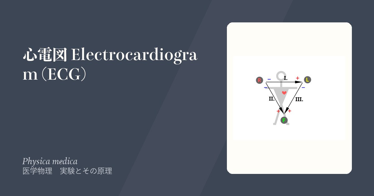
Einthoven の三角形:I + III = II(Einthoven の法則)。3つの肢誘導は独立ではなく、2つ測れば3つ目が決まる。だから12誘導のうち独立な情報は8つしかない。
aVR がほぼ常に陰性なのは、右肩から見ると心臓の脱分極(左下向き)がすべて遠ざかるからである。aVR が陽性なら、電極の付け間違いか、電気軸の異常な回転を疑う——これは物理から直接出る診断規則である。
電気軸は前額面での平均ベクトルの向きで、I 誘導と aVF の陽性/陰性の組み合わせで象限が分かる。正常は −30°〜+90°。
English version
Core
When myocardium is excited, the excited part (depolarised, negative outside the cell) lies next to the unexcited part (positive outside). This boundary behaves as an electric dipole and can be represented by a vector with a magnitude and a direction.
The body is a volume conductor full of electrolyte, so the field created by the cardiac dipole reaches the body surface. The electrocardiogram measures that field as the potential difference between two points on the surface.
The direction of the recorded deflection is set by geometry: if the excitation comes towards the electrode the deflection is upward (positive), if it goes away it is downward (negative), and if it is at right angles it is biphasic or nearly flat.
Going further
This is the meaning of the 12 leads. A lead is a direction from which the dipole vector is viewed, and the same vector is projected from twelve directions. The limb leads (I, II, III, aVR, aVL, aVF) give the frontal plane and the chest leads (V1–V6) the horizontal plane — six shadows each of one solid object projected onto two planes.
Einthoven’s triangle: I + III = II (Einthoven’s law). The three limb leads are not independent; measure two and the third is determined. So only eight of the twelve leads carry independent information.
aVR is almost always negative because, seen from the right shoulder, the depolarisation of the heart (down and to the left) goes away in every case. A positive aVR should raise a misplaced electrode or an abnormal rotation of the electrical axis — a diagnostic rule that comes directly from the physics.
The electrical axis is the direction of the mean vector in the frontal plane, and the quadrant can be found from the combination of positive and negative in lead I and aVF. Normal is −30° to +90°.
記録の物理 — 増幅・雑音・アーチファクト
心電図の信号は0.5〜5 mVと非常に小さいので、増幅が必要になる。記録の標準は1 mV = 10 mm、紙送り 25 mm/s——小さいマス1つが 0.04 秒・0.1 mV、大きいマス1つが 0.2 秒・0.5 mV。
主な雑音源:
- 交流障害(50/60 Hz)— 電源からの誘導。規則正しい細かい振動
- 筋電図(EMG)— 不規則で高周波。振戦や緊張で出る
- 基線の動揺— 呼吸・体動・電極の接触不良。低周波
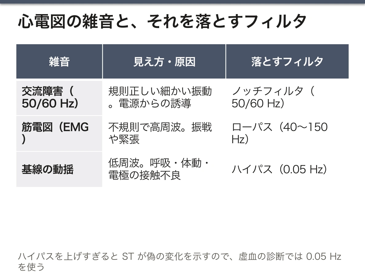
雑音の周波数が違うから、フィルタで分けられる。心電図の主成分は 0.05〜150 Hz。ハイパスフィルタ(0.05 Hz)が基線の動揺を、ローパスフィルタ(40〜150 Hz)が筋電図を、ノッチフィルタ(50/60 Hz)が交流障害を落とす。
ただしフィルタは波形も歪める。ハイパスのカットオフを上げすぎるとST 部分が偽の変化を示す(ST 上昇や低下に見える)。だから虚血の診断では 0.05 Hz を使う。「雑音を消すこと」と「情報を保つこと」は常にトレードオフである。
電極接触インピーダンスを下げることが最も効果的な対策である。皮膚の角質層が抵抗の大半を占めるので、清拭・除毛・ゲルの使用で桁単位に下がる。増幅器の入力インピーダンスは非常に高い(>10 MΩ)ので、電極間のインピーダンス差が大きいと同相除去比(CMRR)が落ち、交流障害が消えなくなる。「フィルタで消す前に、入口で入れないこと」。
電極の付け間違いは最も多いアーチファクトである。右手と左手を入れ替えると I 誘導が反転し、aVR が陽性、aVL と aVR が入れ替わる——物理から予測できる形の異常なので、覚えるのではなく導ける。
English version
Core
The ECG signal is very small, 0.5–5 mV, so amplification is needed. The standard record is 1 mV = 10 mm with paper at 25 mm/s — one small square is 0.04 s and 0.1 mV, one large square 0.2 s and 0.5 mV.
The main sources of noise:
- Mains interference (50/60 Hz) — induction from the supply. A regular fine oscillation.
- Electromyogram (EMG) — irregular and high-frequency. It appears with tremor and tension.
- Baseline wander — respiration, movement, poor electrode contact. Low frequency.
Going further
The frequencies of the noise differ, so filters can separate them. The main components of the ECG are 0.05–150 Hz. A high-pass filter (0.05 Hz) removes baseline wander, a low-pass filter (40–150 Hz) the EMG, and a notch filter (50/60 Hz) mains interference.
But a filter distorts the waveform too. Raise the high-pass cut-off too far and the ST segment shows spurious change (apparent ST elevation or depression). So 0.05 Hz is used when diagnosing ischaemia. "Removing noise" and "preserving information" are always a trade-off.
Lowering the electrode contact impedance is the most effective measure. The stratum corneum accounts for most of the resistance, so cleaning, removing hair and using gel lower it by orders of magnitude. The input impedance of the amplifier is very high (>10 MΩ), so a large difference of impedance between electrodes lowers the common-mode rejection ratio (CMRR) and mains interference cannot be removed. "Keep it out at the entrance rather than filtering it out later."
Misplaced electrodes are the commonest artefact. Swap the right and left arms and lead I inverts, aVR becomes positive, and aVL and aVR are exchanged — an abnormality whose shape can be predicted from the physics, so it is derived rather than memorised.
誘導とは何か — Einthoven の三角形と12誘導
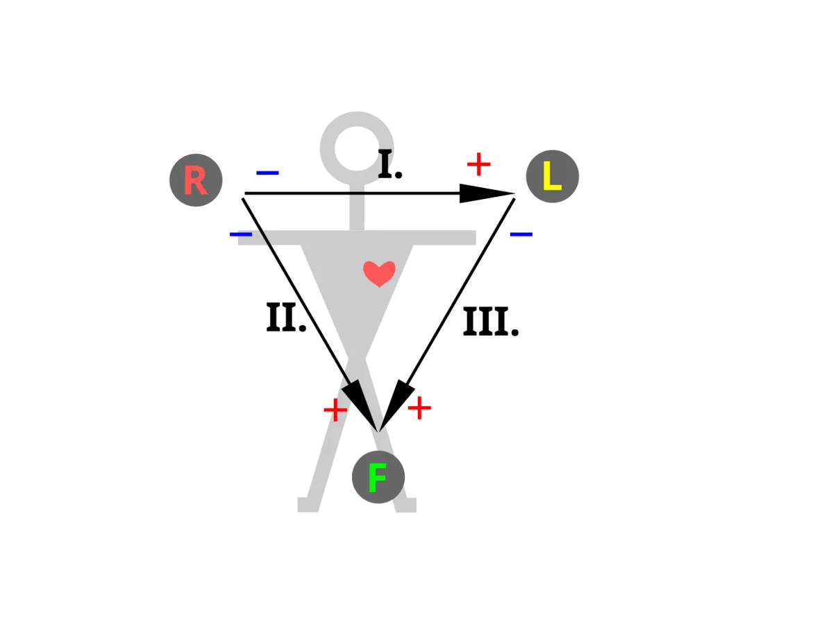
誘導(lead)とは「2点間の電位差を、どの向きで見るか」。心臓の起電力ベクトルを、その向きに射影したものが波形になる。
双極肢誘導(I・II・III)——右手・左手・左足の3点でEinthoven の三角形を作る。I = 左手 − 右手、II = 左足 − 右手、III = 左足 − 左手。よってII = I + III(Einthoven の法則)——これは Kirchhoff の電圧則そのものであり、心電図を「物理として説明する」ときの中心になる部分。
単極肢誘導(aVR・aVL・aVF)——残り2点の平均を基準にして1点を見る。増大(augmented)されるのはそのため。
胸部誘導(V1〜V6)——水平面で心臓を取り巻く。肢誘導が前額面、胸部誘導が水平面を見る——2つの面で3次元のベクトルを捉える。
体は電解質を含む体積導体なので、心臓の電気は体表まで伝わる。ただし減衰し、組織の不均一で歪む。
English version
Core
A lead is "in which direction the potential difference between two points is viewed". The waveform is the projection of the cardiac vector onto that direction.
The bipolar limb leads (I, II, III) — the three points of right arm, left arm and left leg form Einthoven’s triangle. I = left arm − right arm, II = left leg − right arm, III = left leg − left arm. Hence II = I + III (Einthoven’s law) — this is Kirchhoff’s voltage law itself, and the centre of explaining the ECG "as physics".
The augmented limb leads (aVR, aVL, aVF) — one point is viewed against the average of the other two as reference. Hence "augmented".
The chest leads (V1–V6) — they surround the heart in the horizontal plane. The limb leads see the frontal plane and the chest leads the horizontal plane — capturing a three-dimensional vector in two planes.
The body is a volume conductor containing electrolyte, so the electricity of the heart reaches the surface. But it is attenuated and distorted by the inhomogeneity of the tissues.
皮膚で測れる生体電位 — 心電図・脳波・筋電図
細胞の電気活動が作る電場は、体積導体である体を通って皮膚まで届く。体表の2点間の電位差として記録できる代表が3つある。
| 記録 | 何の電気か | 大きさ | 主な周波数 |
|---|---|---|---|
| 心電図 ECG | 心筋の脱分極と再分極 | 約 1 mV | 0.05〜150 Hz |
| 脳波 EEG | 大脳皮質の錐体細胞のシナプス後電位の総和 | 数十 μV | 約 0.5〜数十 Hz |
| 表面筋電図 EMG | 骨格筋線維の活動電位 | 0.1〜数 mV | 約 20〜500 Hz |
ほかに眼球運動(眼電図 EOG)、網膜(網膜電図 ERG)、胃(胃電図 EGG)の電位も同じ原理で測れる。
記録の流れは電極 → 差動増幅器 → フィルタ → A/D 変換 → 表示である。電極は、体の中を流れるイオンの電流を導線の中の電子の電流に変える変換器(transducer)で、銀–塩化銀(Ag/AgCl)電極では Ag + Cl⁻ ⇄ AgCl + e⁻ の反応がこの受け渡しをしている。
なぜ脳波は心電図より桁違いに小さいのか。①発生源の向きがそろいにくい——心臓では心筋全体がほぼ決まった順に興奮して大きな双極子を作るが、脳では無数の神経細胞が別々のタイミングで活動し、互いに打ち消し合う。皮膚まで届くのは、皮質の表面に垂直に並んだ錐体細胞が同期してシナプス後電位を出したぶんだけである。②あいだに電気を通しにくい頭蓋骨があり、電位が弱まりながら広がってしまう。
筋電図は周波数が高いので、心電図と帯域が重なる部分は小さい。だから心電図の記録では筋電図を雑音として扱い、ローパスフィルタで落とせる。逆に筋電図を測りたいときは、心電図のほうが雑音になる。「信号」か「雑音」かは、何を測りたいかで決まる。
脳波はてんかんの診断の中心で、欠神発作の3 Hz 棘徐波複合はその典型である。日本の法的脳死判定でも平坦脳波の確認が要件の1つになっている。筋電図と神経伝導検査は、筋力の低下が神経の病気(ニューロパチー)か、筋肉の病気(ミオパチー)かを分けるのに使う。
English version
Core
The electric field made by the electrical activity of cells reaches the skin through the body, which is a volume conductor. Three main signals can be recorded as a potential difference between two points on the body surface.
| Recording | Source | Size | Main frequencies |
|---|---|---|---|
| ECG | Depolarisation and repolarisation of heart muscle | about 1 mV | 0.05–150 Hz |
| EEG | Sum of postsynaptic potentials of cortical pyramidal cells | tens of μV | about 0.5 to several tens of Hz |
| Surface EMG | Action potentials of skeletal muscle fibres | 0.1 to several mV | about 20–500 Hz |
Potentials of eye movement (EOG), the retina (ERG) and the stomach (EGG) can be measured in the same way.
The recording chain is electrode → differential amplifier → filters → A/D conversion → display. The electrode is a transducer that changes the ionic current in the body into an electronic current in the wire. In a silver/silver-chloride (Ag/AgCl) electrode the reaction Ag + Cl⁻ ⇄ AgCl + e⁻ does this exchange.
Going further
Why is the EEG so much smaller than the ECG? ① The sources do not line up well. In the heart, the whole muscle is excited in a fixed order and forms one large dipole. In the brain, huge numbers of neurons are active at different times and cancel each other. Only the postsynaptic potentials of pyramidal cells that stand perpendicular to the cortical surface and fire in synchrony reach the skin. ② The skull, which conducts poorly, lies in between, so the potential is weakened and spread out.
The EMG has higher frequencies, so its band overlaps only a little with the ECG. That is why, in an ECG recording, the EMG is treated as noise and can be removed with a low-pass filter. When you want to measure the EMG, the ECG becomes the noise. Whether something is “signal” or “noise” depends on what you want to measure.
In the clinic
The EEG is central to the diagnosis of epilepsy; the 3 Hz spike-and-wave complex of absence seizures is the classic example. In Japan, a flat EEG is one of the requirements for the legal determination of brain death. EMG and nerve conduction studies are used to decide whether muscle weakness comes from a nerve disease (neuropathy) or a muscle disease (myopathy).
膜電位から P・QRS・T まで — 刺激伝導系が波形を作る
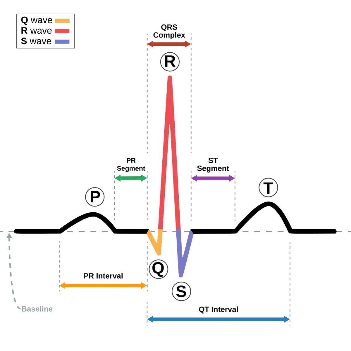
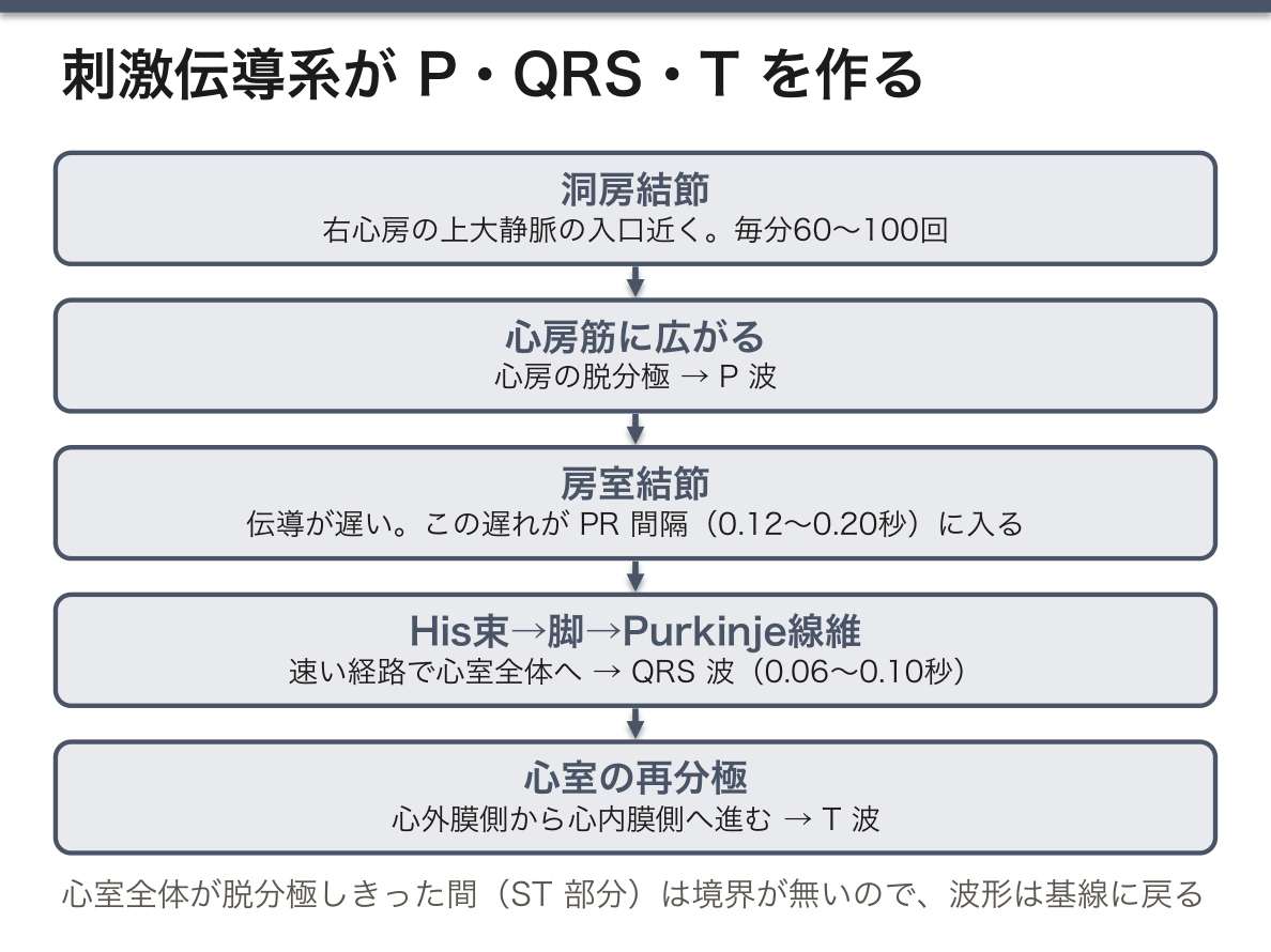
細胞レベルの起源——心筋細胞の内と外ではイオンの濃度が違い、静止時には主に K⁺ が K⁺ チャネルから外へ漏れて、内側が約 −90 mV に保たれている(分極)。細胞膜はこの電荷を蓄えた薄いコンデンサで、膜のどの部分も小さな双極子になっている。興奮すると Na⁺ チャネルが開いて Na⁺ が流れ込み、内側が正になる(脱分極)。Ca²⁺ の流入によるプラトーを経て、K⁺ の流出で元に戻る(再分極)。膜を横切るこうしたイオンの流れと、それに伴って細胞の外を流れる電流が微小電流(microcurrents)で、体表の電位はその合計である。
刺激伝導系——興奮は洞房結節(nodus sinuatrialis。右心房の上大静脈の入口近く、毎分 60〜100 回)で生まれ、心房筋を広がって房室結節(nodus atrioventricularis)に届く。房室結節は伝導が遅く、この遅れが心房の収縮を心室の充満に間に合わせる。その後は His 束 → 右脚・左脚 → Purkinje 線維という速い経路で、心室全体へ一気に広がる。
| 波形 | 心臓で起きていること | 正常の幅 |
|---|---|---|
| P 波 | 心房の脱分極 | — |
| PR 間隔 | 心房の興奮の始まりから心室の興奮の始まりまで(房室結節での遅れを含む) | 0.12〜0.20 秒 |
| QRS 波 | 心室の脱分極 | 0.06〜0.10 秒 |
| ST 部分 | 心室全体が脱分極したまま(プラトー) | — |
| T 波 | 心室の再分極 | — |
平らな部分にも意味がある。体表に電位差が出るのは、興奮した部分と静止した部分の境界が双極子になっているあいだだけである。心室全体が静止しているとき(T 波の後)も、全体が脱分極しきったとき(ST 部分)も、境界が無いので波形は基線に戻る。
T 波はなぜ QRS と同じ向きなのか。再分極は脱分極と電荷の向きが逆なので、同じ順序で進めば逆向きの波になるはずである。ところが心室では心外膜側の活動電位のほうが短く、再分極は心外膜から心内膜へと、脱分極とは逆の順に進む。「電荷の向きが逆」と「進む向きが逆」の2つの逆が打ち消し合い、T 波は QRS と同じ上向きになる。
心房の再分極の波は小さく、時間的に QRS と重なるので、ふつうは見えない。
波形のずれは伝導路の異常として読める。PR 間隔の延長は房室伝導の遅れ(1度房室ブロック)、QRS の幅が広いのは脚ブロックなどで心室内の伝導が遅いこと、ST の偏位は心筋虚血を示す。洞房結節が止まれば房室接合部(毎分 40〜60 回)、さらに心室(毎分 20〜40 回)が補充調律として拍動を引き継ぐ——下位ほど遅い。
English version
Core
Origin at the cell level. Ion concentrations differ inside and outside a heart muscle cell. At rest, mainly K⁺ leaks out through K⁺ channels, and the inside is kept at about −90 mV (polarisation). The membrane is a thin capacitor that stores this charge, and every part of it is a small dipole. When the cell is excited, Na⁺ channels open, Na⁺ flows in and the inside becomes positive (depolarisation). After a plateau caused by Ca²⁺ inflow, K⁺ outflow brings it back (repolarisation). These ion flows across the membrane, and the currents they cause outside the cells, are the microcurrents; the potential on the body surface is their sum.
The conduction system. The impulse starts in the sinoatrial node (nodus sinuatrialis; in the right atrium near the opening of the superior vena cava; 60–100 per minute). It spreads through the atrial muscle and reaches the atrioventricular node (nodus atrioventricularis). The AV node conducts slowly, and this delay lets atrial contraction finish filling the ventricles. Then the impulse spreads quickly through the bundle of His → right and left bundle branches → Purkinje fibres to the whole ventricle.
| Wave | What happens in the heart | Normal duration |
|---|---|---|
| P wave | Atrial depolarisation | — |
| PR interval | From the start of atrial excitation to the start of ventricular excitation (includes the AV-node delay) | 0.12–0.20 s |
| QRS complex | Ventricular depolarisation | 0.06–0.10 s |
| ST segment | The whole ventricle stays depolarised (plateau) | — |
| T wave | Ventricular repolarisation | — |
Going further
The flat parts mean something too. A potential difference appears on the body surface only while the border between excited and resting tissue forms a dipole. When the whole ventricle is at rest (after the T wave) or fully depolarised (the ST segment), there is no border, so the trace returns to the baseline.
Why does the T wave point the same way as the QRS? Repolarisation has the opposite charge direction to depolarisation, so if it went in the same order it would give an opposite wave. But in the ventricle the action potential is shorter on the epicardial side, so repolarisation runs from the epicardium to the endocardium, the reverse of depolarisation. The two reversals (“opposite charge” and “opposite direction”) cancel, and the T wave points upward like the QRS.
The wave of atrial repolarisation is small and falls at the same time as the QRS, so it is usually not seen.
In the clinic
Changes in the waves can be read as faults in the conduction pathway. A long PR interval means slow AV conduction (first-degree AV block), a wide QRS means slow conduction inside the ventricles, as in bundle branch block, and ST deviation points to myocardial ischaemia. If the SA node stops, the AV junction (40–60 per minute) and then the ventricles (20–40 per minute) take over as escape rhythms; the lower the site, the slower the rate.
細動と除細動 — ばらばらの興奮を一度に止める
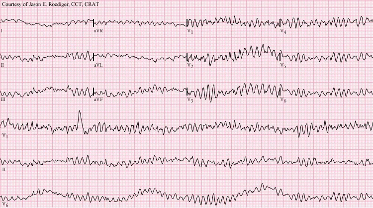
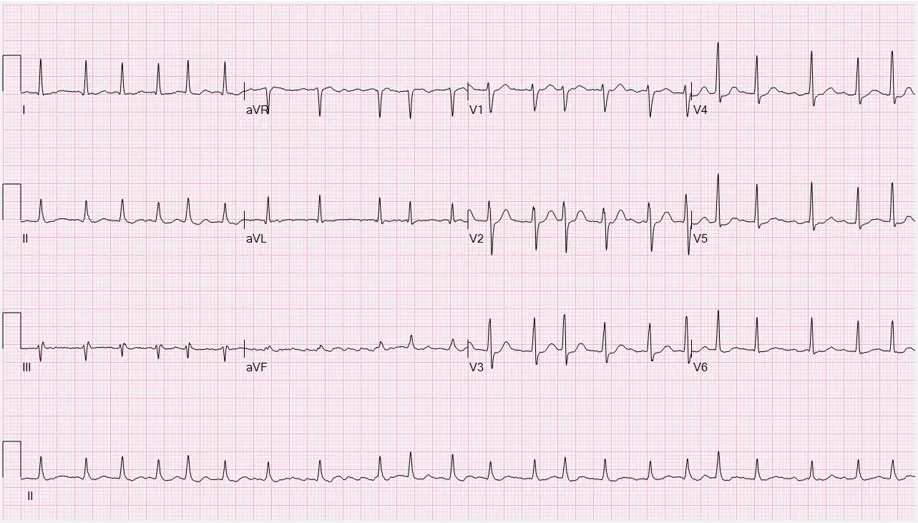
細動(fibrillation)は、心筋の小さな部分がそれぞれ勝手に、非常に速く興奮している状態である。全体としてまとまった収縮にならず、筋がふるえるだけになる。
| 心房細動 AF | 心室細動 VF | |
|---|---|---|
| 心電図 | P 波が消えて細かい f 波。RR 間隔が完全に不規則 | QRS も T も無く、不規則な波だけ |
| 血液の拍出 | 心室は不規則ながら収縮するので保たれる | 0(心停止) |
| 危険 | 左心耳にできた血栓による脳塞栓 | 数分で脳に不可逆な障害。突然死の最大の原因 |
心房細動で心室がまだ働けるのは、房室結節がふるいになって、心房からの無数の興奮の一部しか心室へ通さないからである。心室細動にはこのふるいが無く、ポンプそのものが止まる。
細動の多くは旋回(リエントリー)で続く。興奮の波が回路をぐるりと回って出発点に戻ってきたとき、そこがもう不応期を抜けていれば、もう一度興奮させられる。虚血や線維化で伝導が遅くなったり不応期が短くなったりすると小さな回路でも旋回が成り立ち、小さな渦がたくさんできて細動になる。心室の一部だけが不応期を抜けている T 波の時期(受攻期)に刺激が入ると、この渦が生まれやすい。
除細動(defibrillation)は、コンデンサに蓄えた電気を一気に胸に流し、心筋の大部分を同時に脱分極させる。すべての細胞が一斉に不応期に入ると、渦が進める場所が無くなって消える。その後、最初に興奮を出すのは自動能を持つ洞房結節なので、正常な調律が戻る。つまり除細動は心臓を「動かす」のではなく、一度すべてを止めてやり直させる操作である。だから電気的に何も起きていない心静止には効かない。
除細動器には、AED(自動体外式。心電図を自分で解析し、VF・無脈性心室頻拍のときだけ放電する。日本では一般市民も使える)、医師が波形を読んで使う手動式、心室の頻拍・細動を自ら検出して体内で放電する植込み型除細動器(ICD)、植込みまでのつなぎに着る着用型がある。今の機器は途中で電流の向きを反転させる二相性波形で、単相性(360 J)より少ない 120〜200 J で効く。除細動が1分遅れるごとに成功率は約7〜10%下がるので、時間がすべてである。心房細動を戻すときは放電を R 波に合わせ(同期下カルディオバージョン)、受攻期に当てて VF を起こさないようにする。
English version
Core
Fibrillation is a state in which small parts of the heart muscle are each excited on their own and very fast. There is no coordinated contraction; the muscle only quivers.
| Atrial fibrillation (AF) | Ventricular fibrillation (VF) | |
|---|---|---|
| ECG | No P waves, fine f waves; RR intervals completely irregular | No QRS or T, only irregular waves |
| Cardiac output | Kept, because the ventricles still contract, though irregularly | Zero (cardiac arrest) |
| Danger | Stroke from a clot formed in the left atrial appendage | Irreversible brain damage within minutes; the most common cause of sudden death |
The ventricles still work in AF because the AV node acts as a filter and lets only some of the countless atrial impulses through. In VF there is no such filter, and the pump itself stops.
Going further
Most fibrillation is kept going by re-entry. When an excitation wave travels round a circuit and comes back to its starting point, it can excite that point again if the tissue there has already left its refractory period. When ischaemia or fibrosis slows conduction or shortens the refractory period, re-entry works even in small circuits, many small whirls form, and the result is fibrillation. A stimulus that arrives during the T wave, when only part of the ventricle has recovered (the vulnerable period), easily starts such whirls.
Defibrillation sends the charge stored in a capacitor through the chest in one burst and depolarises most of the heart muscle at the same moment. When all cells become refractory together, the whirls have nowhere to go and die out. The first site to fire again is the SA node, which has automaticity, so normal rhythm returns. In other words, defibrillation does not “start” the heart; it stops everything once so that it can restart. That is why it does not work in asystole, where there is no electrical activity.
In the clinic
Types of defibrillator: the AED (automated external defibrillator; it analyses the ECG itself and shocks only in VF or pulseless ventricular tachycardia; in Japan lay people may use it), the manual defibrillator, used by a doctor who reads the trace, the implantable cardioverter-defibrillator (ICD), which detects ventricular tachycardia or fibrillation and shocks inside the body, and the wearable type used as a bridge before implantation. Modern devices use a biphasic waveform that reverses the current halfway, and work with 120–200 J instead of the 360 J of monophasic shocks. Each minute of delay lowers the success rate by about 7–10%, so time is everything. To convert AF, the shock is timed to the R wave (synchronised cardioversion) so that it does not hit the vulnerable period and cause VF.
ペースメーカ — 電気のパルスで拍動を作る
体の中の自然のペースメーカは洞房結節である。これが働かなくなったり(洞不全症候群)、興奮が心室へ伝わらなくなったり(高度・完全房室ブロック)すると、心拍が遅くなって失神(Adams-Stokes 発作)や心不全を起こす。人工ペースメーカは、電池と回路を収めた本体(ふつう鎖骨の下の皮下に植え込む)と、静脈を通して心内膜に固定したリード(電極)からなるパルス発生器である。
1回の刺激は数 V・1 ms 未満の短い電圧パルスで、リード先端のまわりの心筋を閾値まで脱分極させる。そこから興奮が心筋を伝わり、心室全体が収縮する——除細動が「全部を一度に」脱分極させるのに対し、ペースメーカは「1点に火をつけて広げる」。
| 種類 | リード | 主な使いどころ |
|---|---|---|
| シングルチャンバ | 右心室(または右心房)に1本 | 徐脈を伴う慢性心房細動など |
| デュアルチャンバ | 右心房と右心室に1本ずつ | 房室ブロック・洞不全。心房→心室の順番を保てる |
| 両心室(CRT) | 右心室+冠静脈洞を通して左心室 | 左右の心室の収縮がずれた心不全(左脚ブロックで QRS の幅が広い) |
| リードレス | 本体ごと右心室の中に留置する小さなカプセル | リードの断線や感染を避けたいとき |
今のペースメーカのほとんどはデマンド型で、まず自分の心拍を感知し、決めた時間内に自己の拍動が来なかったときだけ刺激する。いつも一定のリズムで刺激すると、自己の拍動の T 波(受攻期)に刺激が重なって心室細動を起こしうるからである。
働き方は文字のコードで書く。1文字目=刺激する部屋、2文字目=感知する部屋(A 心房・V 心室・D 両方)、3文字目=感知したときの反応(I 抑制・T 同期・D 両方)。VVI は「心室を感知し、自己拍動があれば休み、無ければ心室を刺激する」、DDD は心房と心室の両方で感知と刺激をする。4文字目の R は、加速度センサなどで体の動きを感じて心拍数を上げる機能(rate responsive)である。
刺激に要る強さは電圧とパルス幅の兼ね合いで決まり、パルスが短いほど高い電圧が要る(強さ–時間曲線)。閾値に余裕を持たせつつ出力を小さくして、電池を長持ちさせる。
心電図では刺激のたびに細い縦線(ペーシングスパイク)が入り、右心室からのペーシングでは興奮が刺激伝導系を通らずに広がるので、QRS の幅が広くなる。植込み後は強い電磁場に注意が要る。MRI は対応機種でのみ条件つきで撮れ、手術の電気メスも感知の誤作動の原因になるので、術前に設定を変える。
English version
Core
The body’s natural pacemaker is the SA node. If it fails (sick sinus syndrome), or if the impulse cannot reach the ventricles (high-degree or complete AV block), the heart rate falls, causing fainting (Adams-Stokes attacks) or heart failure. An artificial pacemaker is a pulse generator: a case holding a battery and circuits (usually implanted under the skin below the clavicle) and leads (electrodes) passed through a vein and fixed to the endocardium.
Each stimulus is a short voltage pulse of a few volts lasting less than 1 ms. It depolarises the heart muscle around the lead tip to threshold, and the excitation then spreads through the muscle so that the whole ventricle contracts. A defibrillator depolarises “everything at once”; a pacemaker “lights one point and lets it spread”.
| Type | Leads | Main use |
|---|---|---|
| Single-chamber | One lead in the right ventricle (or right atrium) | Chronic AF with slow heart rate, for example |
| Dual-chamber | One in the right atrium and one in the right ventricle | AV block, sick sinus syndrome; keeps the atrium-then-ventricle order |
| Biventricular (CRT) | Right ventricle + left ventricle through the coronary sinus | Heart failure with unsynchronised ventricles (left bundle branch block, wide QRS) |
| Leadless | A small capsule placed entirely inside the right ventricle | When lead fracture or infection should be avoided |
Going further
Almost all modern pacemakers work on demand. They first sense the heart’s own beats and pace only when no beat comes within a set time. Pacing at a fixed rate all the time could put a stimulus on the T wave (the vulnerable period) of a natural beat and cause VF.
The mode is written as a letter code. The first letter is the chamber paced, the second the chamber sensed (A atrium, V ventricle, D both), and the third the response to a sensed beat (I inhibit, T trigger, D both). VVI means “sense the ventricle, stay quiet if there is a natural beat, otherwise pace the ventricle”; DDD senses and paces both atrium and ventricle. A fourth letter R means rate response: a sensor such as an accelerometer detects body movement and raises the rate.
The stimulus strength needed depends on both voltage and pulse width: the shorter the pulse, the higher the voltage needed (the strength–duration curve). The output is kept safely above threshold but as low as possible, so that the battery lasts longer.
In the clinic
On the ECG each stimulus shows as a thin vertical line (the pacing spike). With right-ventricular pacing the excitation spreads without using the conduction system, so the QRS is wide. After implantation, strong electromagnetic fields need care: MRI is possible only with compatible devices and under set conditions, and electrosurgery can disturb sensing, so the settings are changed before an operation.
感電 — 交流と直流はどちらが危険か
体に害を与えるのは電圧そのものではなく、体を流れる電流である。電流は I = V/R で決まり、抵抗の大部分は皮膚にある。乾いた皮膚の抵抗は大きいが、濡れたり傷があったりすると桁違いに下がる。体の内部は電解質の多い良い導体である。だから同じ電圧でも、濡れた手では危険がずっと大きい。
商用交流(50/60 Hz)が手から手(または足)へ1秒ほど流れたときの目安(マクロショック):
| 電流 | 起こること |
|---|---|
| 約 1 mA | ピリッと感じる(最小感知電流) |
| 約 10 mA | 筋が強直して自分で離せなくなる(離脱限界電流) |
| 約 100 mA | 心室細動 |
電流が皮膚を通らずに心臓へ直接流れる場合(心臓カテーテル、一時ペーシングのリード)は、約 0.1 mA(100 μA)で心室細動が起こる(ミクロショック)。
同じ大きさなら交流のほうが危険である。理由は2つある。①交流は1秒に何十回も向きを変えるので、そのたびに神経と筋を刺激し直し、筋を強直させる。電線を握ってしまうと屈筋が勝って握ったまま離せず、通電の時間が延びる。②刺激が続くので、いずれ心周期の受攻期(T 波)に当たって心室細動を起こす。直流は主に流れ始めと切れる瞬間に刺激するだけなので強直を起こしにくく、心室細動を起こす電流も数倍大きい。ただし直流でも大電流は熱傷と電気分解による組織の障害を起こす。
周波数にも危険の山がある。50/60 Hz は、心筋を最も細動させやすい数十 Hz の帯域にある。周波数が kHz 以上になると、半周期が短すぎて膜が脱分極する前に向きが変わるので、神経も筋も刺激されにくくなる。電気メスが数百 kHz 以上の交流で、感電させずに熱だけで組織を切れるのはこのためである。
電流は大きいほど危険、とも限らない。1 A 程度の電流は心臓の一部だけを強直させて細動を起こすが、ずっと大きな電流は心臓全体を一度に強直させ、電流が止まると拍動が戻ることがある——除細動器と同じ物理である。
病院では、マクロショックには機器の保護接地(3ピンの医用コンセント)で、ミクロショックには心臓に直接触れる装置の漏れ電流を 10 μA 以下(細動を起こす値の10分の1)に抑える規格(CF 形装着部)で備えている。電撃傷では皮膚の傷が小さくても、電流の通り道の筋が壊れて横紋筋融解(ミオグロビン尿 → 急性腎障害)を起こしうるので、見た目で軽症と決めない。
English version
Core
What harms the body is not the voltage itself but the current flowing through the body. The current is set by I = V/R, and most of the resistance is in the skin. Dry skin has a high resistance, but when it is wet or broken the resistance falls by orders of magnitude. The inside of the body, full of electrolytes, is a good conductor. So the same voltage is far more dangerous with wet hands.
Typical effects of mains AC (50/60 Hz) flowing from hand to hand (or foot) for about one second (macroshock):
| Current | Effect |
|---|---|
| about 1 mA | Just felt (perception threshold) |
| about 10 mA | Muscles go into tetanus and you cannot let go (let-go limit) |
| about 100 mA | Ventricular fibrillation |
When the current reaches the heart directly without passing the skin (cardiac catheters, temporary pacing wires), about 0.1 mA (100 μA) causes VF (microshock).
Going further
For the same size of current, AC is more dangerous. There are two reasons. ① AC changes direction dozens of times a second, so it stimulates nerves and muscles again and again and causes tetanus. If you grip a wire, the flexors win, you cannot let go, and the shock lasts longer. ② Because the stimulation keeps going, sooner or later it hits the vulnerable period (T wave) of the cardiac cycle and causes VF. DC stimulates mainly when it starts and stops, so it causes tetanus less easily, and the current needed for VF is several times higher. Still, large DC currents cause burns and tissue damage by electrolysis.
There is also a peak of danger in frequency. 50/60 Hz lies in the band of a few tens of hertz in which the heart fibrillates most easily. Above the kilohertz range, each half-cycle is too short for the membrane to depolarise before the current reverses, so nerves and muscles are hardly stimulated. This is why electrosurgery can cut tissue by heat alone with AC of hundreds of kilohertz or more, without giving a shock.
A larger current is not always more dangerous. A current of about 1 A may put only part of the heart into tetanus and cause fibrillation, while a much larger current puts the whole heart into tetanus at once, and the beat may return when it stops. This is the same physics as a defibrillator.
In the clinic
In hospitals, macroshock is prevented by protective earthing of equipment (three-pin medical-grade outlets), and microshock by standards that keep the leakage current of devices that touch the heart at 10 μA or less (one tenth of the VF level; CF-type applied parts). In electrical injury the skin wound may be small while the muscle along the current path is destroyed, causing rhabdomyolysis (myoglobinuria → acute kidney injury), so do not judge the injury as mild by its look.
まとめ
| 心臓の電気双極子と体積導体 | 心筋が興奮すると、興奮した部分(脱分極・細胞外は陰性)と未興奮の部分(細胞外は陽性)が隣り合う。 |
|---|---|
| 記録の物理 — 増幅・雑音・アーチファクト | 心電図の信号は0.5〜5 mVと非常に小さいので、増幅が必要になる。 |
| 誘導とは何か — Einthoven の三角形と12誘導 | 誘導(lead)とは「2点間の電位差を、どの向きで見るか」。 |
| 皮膚で測れる生体電位 — 心電図・脳波・筋電図 | 細胞の電気活動が作る電場は、体積導体である体を通って皮膚まで届く。 |
| 膜電位から P・QRS・T まで — 刺激伝導系が波形を作る | 細胞レベルの起源——心筋細胞の内と外ではイオンの濃度が違い、静止時には主に K⁺ が K⁺ チャネルから外へ漏れて、内側が約 −90 mV に保たれている(分極)。 |
| 細動と除細動 — ばらばらの興奮を一度に止める | 細動(fibrillation)は、心筋の小さな部分がそれぞれ勝手に、非常に速く興奮している状態である。 |
| ペースメーカ — 電気のパルスで拍動を作る | 体の中の自然のペースメーカは洞房結節である。 |
| 感電 — 交流と直流はどちらが危険か | 体に害を与えるのは電圧そのものではなく、体を流れる電流である。 |
このノートは医学生が授業の内容を自分の言葉でまとめたもので、診療の判断に使うものではありません。誤りに気づいたらお知らせください。