ここで扱うのは脳幹の下2つ(延髄・橋)と小脳、そのあいだにできる腔(第四脳室)である。
脳幹の標本の上で構造を示すときは、順番を決めておくと落ちない——①前面(錐体・オリーブ・脳底溝)→ ②そこから出る脳神経を上から数える → ③後面(薄束結節・楔状束結節)→ ④小脳を外して菱形窩 → ⑤3対の小脳脚。
この範囲の芯は「神経が出る場所と、その内側にある核の位置が対応している」ことである。溝の名前を覚えるのが目的ではなく、溝から出る神経を言えることが目的である。
- 延髄 — 外形
- 橋
- 小脳 — 外形と内部
- 菱形窩 — 脳神経核の地図
- 第四脳室 — 壁と3つの孔
- 錐体
- 前外側溝
- オリーブ
- 後外側溝
- 薄束結節
- 楔状束結節
- 脳底溝
- 中小脳脚
- 下小脳脚
- 上小脳脚
- 菱形窩の正中溝
- 内側隆起
- 顔面神経丘
- 前庭野
- 第四脳室髄条
- 舌下神経三角
- 迷走神経三角
- 小脳半球
- 虫部
- 水平裂
- 小脳の裂
- 小脳回(葉片)
- 小脳活樹(生命の樹)
- 延髄 — 外形Medulla oblongata
- 橋Pons
- 小脳 — 外形と内部Cerebellum
- 菱形窩 — 脳神経核の地図Fossa rhomboidea
- 第四脳室 — 壁と3つの孔Ventriculus quartus
- 錐体Pyramis
- 前外側溝Sulcus anterolateralis
- オリーブOliva
- 後外側溝Sulcus posterolateralis
- 薄束結節Tuberculum gracile
- 楔状束結節Tuberculum cuneatum
- 脳底溝Sulcus basilaris
- 中小脳脚Pedunculi cerebellares medii
- 下小脳脚Pedunculi cerebellares inferiores
- 上小脳脚Pedunculi cerebellares superiores
- 菱形窩の正中溝Sulcus medianus
- 内側隆起Eminentia medialis
- 顔面神経丘Colliculus facialis
- 前庭野Area vestibularis
- 第四脳室髄条Striae medullares ventriculi quarti
- 舌下神経三角Trigonum nervi hypoglossi
- 迷走神経三角Trigonum nervi vagi
- 小脳半球Hemispheria cerebelli
- 虫部Vermis cerebelli
- 水平裂Fissura horizontalis cerebelli
- 小脳の裂Fissurae cerebelli
- 小脳回(葉片)Folia cerebelli
- 小脳活樹(生命の樹)Arbor vitae
- まとめ
延髄 — 外形Medulla oblongata
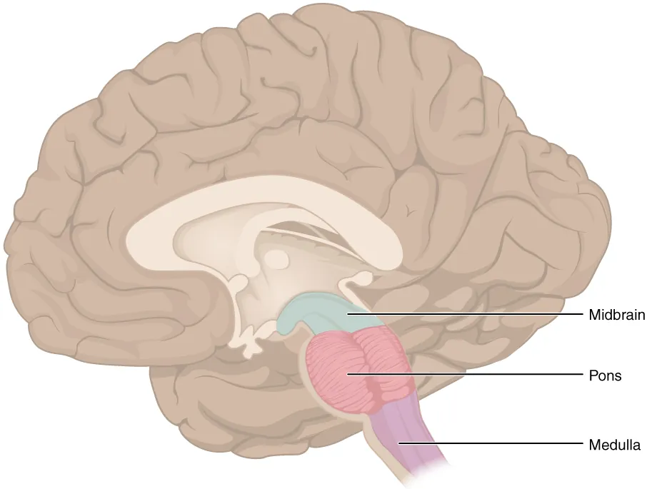
脊髄と橋のあいだ。下端は大後頭孔(第1頸神経の出る高さ)、上端は橋との境の溝 sulcus bulbopontinus。円錐台の形で、上へ行くほど太い。
前面:正中裂の両側に pyramis(錐体、皮質脊髄路の隆起)、その外側に oliva(オリーブ、下オリーブ核の隆起)。錐体とオリーブのあいだが sulcus anterolateralis で、XII 舌下神経が出る。オリーブの外側の sulcus posterolateralis から IX・X・XI が出る。
後面:下部に tuberculum gracile(内側)と tuberculum cuneatum(外側)——薄束核・楔状束核の隆起。上部は開いて菱形窩の下半分になる。
錐体交叉 decussatio pyramidum は延髄の下端、前正中裂の中で起きる。皮質脊髄路の約 75〜90% がここで反対側へ渡って側索へ入り、残りが前索を同側で下る。「運動が反対側になるのは延髄の下端」と場所まで言えるようにする。
内側毛帯交叉 decussatio lemniscorum はその少し上。薄束核・楔状束核で乗り換えた2次ニューロンが交叉して lemniscus medialis を作り、視床へ上る。2つの交叉は場所も高さも別で、これが延髄内側症候群の所見(対側の片麻痺+対側の深部感覚障害+同側の舌の偏位)を決める。
延髄外側症候群(Wallenberg 症候群)——椎骨動脈または後下小脳動脈(PICA)の閉塞で延髄外側部が梗塞になる。延髄の外形と内部構造を覚える最大の理由がこれである。
落ちるものを、外側から内側へ並べる。下小脳脚→ 同側の小脳失調(患側へ倒れる)。三叉神経脊髄路核→ 同側顔面の温痛覚消失・角膜反射消失。外側脊髄視床路→ 反対側の首から下の温痛覚障害。疑核→ 嗄声・嚥下障害・カーテン徴候。孤束核→ 味覚障害。前庭神経核→ 回転性めまい・眼振・悪心嘔吐。下行する交感神経路(網様体)→ 同側の Horner 症候群。
錐体路は障害されないので、四肢の麻痺は出ない。また深部感覚は残る(後索‐内側毛帯路は内側にあるため)。「顔は同側、体は反対側の温痛覚がなくなり、麻痺はない」——これが押さえるべき形である。
出典:イラスト解剖学 誌面659頁「延髄外側症候群」
English version
Core
Between the spinal cord and the pons. The lower end is the foramen magnum (the level at which the first cervical nerve leaves), the upper end the sulcus bulbopontinus at the border with the pons. Shaped like a truncated cone, thicker the higher you go.
Anterior surface: on either side of the median fissure the pyramis (the ridge of the corticospinal tract), and lateral to it the oliva (the ridge of the inferior olivary nucleus). Between pyramid and olive is the sulcus anterolateralis, from which XII, the hypoglossal, emerges. From the sulcus posterolateralis, lateral to the olive, emerge IX, X and XI.
Posterior surface: below, the tuberculum gracile (medial) and tuberculum cuneatum (lateral) — the ridges of the gracile and cuneate nuclei. Above, it opens out as the lower half of the rhomboid fossa.
Going further
The decussatio pyramidum occurs at the lower end of the medulla, within the anterior median fissure. About 75–90 per cent of the corticospinal fibres cross here into the contralateral lateral funiculus, the rest descending ipsilaterally in the anterior funiculus. Be able to say not only that movement crosses but where: at the lower end of the medulla.
The decussatio lemniscorum is a little higher. The second neurones that relayed in the gracile and cuneate nuclei cross to form the lemniscus medialis and ascend to the thalamus.
The two decussations are at different places and different levels, and this decides the findings of the medial medullary syndrome (contralateral hemiplegia + contralateral loss of deep sensation + ipsilateral deviation of the tongue).
In the clinic
Lateral medullary (Wallenberg) syndrome — infarction of the lateral medulla from occlusion of the vertebral artery or the posterior inferior cerebellar artery (PICA). This is the single greatest reason for learning the external form and internal structure of the medulla.
List what is lost, from lateral to medial.
- Inferior cerebellar peduncle → ipsilateral cerebellar ataxia (falling to the affected side).
- Spinal nucleus of the trigeminal → loss of pain and temperature on the ipsilateral face; loss of the corneal reflex.
- Lateral spinothalamic tract → loss of pain and temperature on the contralateral body below the neck.
- Nucleus ambiguus → hoarseness, dysphagia, the curtain sign.
- Nucleus of the solitary tract → disturbance of taste.
- Vestibular nuclei → rotatory vertigo, nystagmus, nausea and vomiting.
- The descending sympathetic pathway (reticular formation) → ipsilateral Horner’s syndrome.
The pyramidal tract is not involved, so there is no limb paralysis, and deep sensation is preserved (the posterior column–medial lemniscus pathway lies medially). "Loss of pain and temperature on the same side of the face and the opposite side of the body, with no paralysis" — this is the form to remember.
橋Pons
延髄と中脳のあいだの、前へ強く張り出した部分。前面の正中に sulcus basilaris(脳底動脈が走る溝)。外側で細くなって pedunculus cerebellaris medius となり小脳へ入る。
要点は神経が出る場所である。V 三叉神経は橋の外側(中小脳脚との境)から。VI・VII・VIII は sulcus bulbopontinus(橋と延髄の境)から、内側から外側へ VI → VII → VIII の順に並ぶ。
内部は前後2層。Pars basilaris(腹側)=下行路と橋核・橋横線維。Pars dorsalis / tegmentum(背側)=脳神経核・網様体・内側毛帯。
橋核 nuclei pontis は、大脳皮質から来た線維(皮質橋路)を受け、反対側の中小脳脚を通って小脳へ渡す中継点である。これが「大脳の運動計画を小脳へ知らせる」経路(皮質→橋→小脳路)で、橋が前へ張り出しているのはこの核と線維の量そのものである。
橋の断面で腹側と背側を見分ける決め手は横に走る線維の帯(fibrae pontis transversae)があるかどうか。あれば腹側、無ければ被蓋である。
橋には V〜VIII の脳神経核があり、そこを錐体路が通り抜ける。だから片側の病変では同側の脳神経麻痺+反対側の片麻痺という組み合わせになる。これを交叉性片麻痺という。
Millard–Gubler 症候群(橋下部内側)——同側の外転神経麻痺・顔面神経麻痺と反対側の片麻痺。中脳の Weber 症候群(同側動眼神経+対側片麻痺)と対にして覚える。
MLF 症候群(核間性眼筋麻痺)——内側縦束が切れると、患側の眼が内転できず、反対側の眼に外転時の眼振が出る。若年で両側なら多発性硬化症を、高齢で片側なら脳幹梗塞をまず考える。
出典:イラスト解剖学 誌面739頁「交叉性片麻痺」
English version
Core
The part between medulla and midbrain, bulging strongly forwards. In the midline of the anterior surface is the sulcus basilaris (in which the basilar artery runs). Laterally it narrows into the pedunculus cerebellaris medius and enters the cerebellum.
The point is where the nerves emerge. V, the trigeminal, from the lateral aspect of the pons (at the junction with the middle cerebellar peduncle). VI, VII and VIII from the sulcus bulbopontinus (the pontomedullary junction), arranged from medial to lateral as VI → VII → VIII.
The interior is in two layers. Pars basilaris (ventral) = the descending tracts, the pontine nuclei and the transverse pontine fibres. Pars dorsalis / tegmentum = the cranial nerve nuclei, the reticular formation and the medial lemniscus.
Going further
The nuclei pontis receive the fibres coming from the cerebral cortex (the corticopontine tract) and hand them across the contralateral middle cerebellar peduncle to the cerebellum. This is the route by which the cerebrum informs the cerebellum of its motor plan (cortico-ponto-cerebellar), and the forward bulge of the pons is precisely the mass of these nuclei and fibres.
On a section of the pons, what distinguishes ventral from dorsal is whether the band of transversely running fibres (fibrae pontis transversae) is present. Present = basilar part; absent = tegmentum.
In the clinic
The pons contains the nuclei of cranial nerves V–VIII, and the pyramidal tract passes through it. So a unilateral lesion gives the combination of an ipsilateral cranial nerve palsy with a contralateral hemiplegia — a crossed hemiplegia.
Millard–Gubler syndrome (lower medial pons) — ipsilateral abducens and facial palsy with contralateral hemiplegia. Learn it as a pair with Weber’s syndrome of the midbrain (ipsilateral oculomotor palsy + contralateral hemiplegia).
MLF syndrome (internuclear ophthalmoplegia) — when the medial longitudinal fasciculus is interrupted, the eye on the affected side cannot adduct and the other eye shows nystagmus on abduction. Bilateral in a young person, think first of multiple sclerosis; unilateral in an older person, of brainstem infarction.
小脳 — 外形と内部Cerebellum
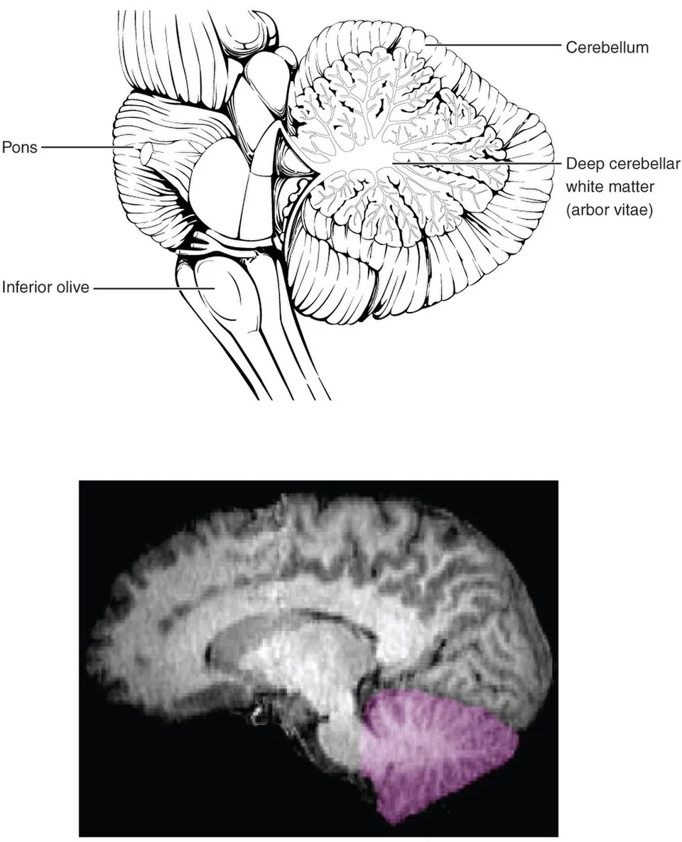
後頭蓋窩、脳幹の背側。中央の vermis(虫部)と左右の hemispheria cerebelli からなる。表面は細かい葉 folia に刻まれ、深い fissura horizontalis が上面と下面を分ける。
矢状断で白質が枝分かれして見えるのが arbor vitae(生命の樹)。この形は矢状断で示す。
3対の脚 pedunculi cerebellares で脳幹に繋がる。
| 脚 | 繋ぐ相手 | 向き |
|---|---|---|
| Superior(上小脳脚) | 中脳 | 主に出る(歯状核→赤核・視床) |
| Medius(中小脳脚) | 橋 | 入る(橋核から。最も太い) |
| Inferior(下小脳脚) | 延髄 | 入る(オリーブ・脊髄小脳路) |
白質の中に4対の核が並ぶ(外側から内側へ):nucleus dentatus → emboliformis → globosus → fastigii。「歯状・栓状・球状・室頂」の順で、外側ほど新しく、内側ほど古い。出力はほぼ全て上小脳脚から出る(例外は室頂核で、下小脳脚から前庭核へ)。
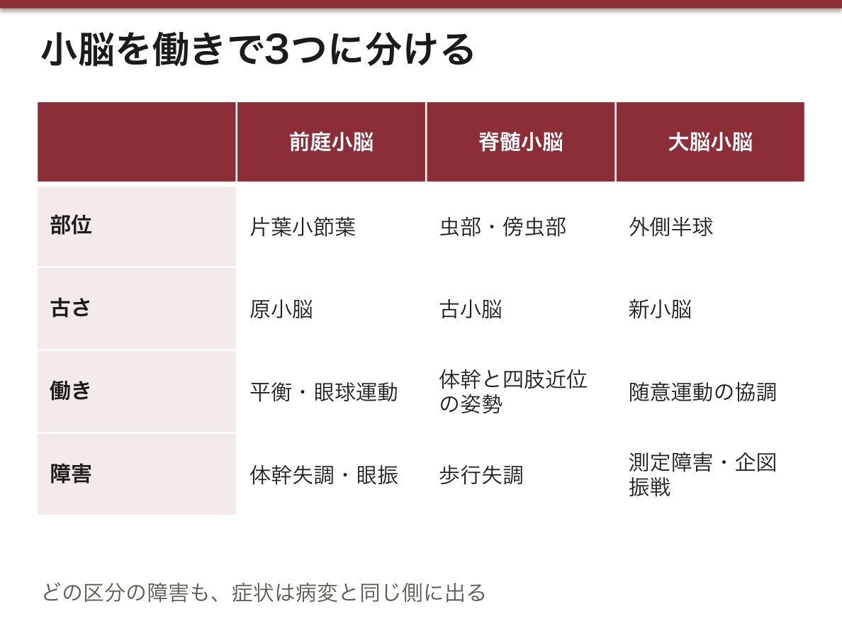
機能で3分すると症状が読める。①前庭小脳(片葉小節葉)=平衡・眼球運動 → 体幹失調・眼振。②脊髄小脳(虫部・傍虫部)=体幹と四肢近位の姿勢 → 歩行失調。③大脳小脳(外側半球)=随意運動の協調 → 四肢の測定障害・企図振戦・変換運動障害。
小脳の障害は同側に出る。出力が上小脳脚で交叉し、そのあと皮質脊髄路が延髄でもう一度交叉するため、2回交叉して元に戻るからである。
小脳の障害は同側に出る。出力が2回交叉して戻るためで、「右の小脳半球が傷めば右手が狂う」。大脳と正反対なので必ず押さえる。
原小脳症候群(片葉小節葉)——前庭系の障害。平衡障害と眼球運動障害。
古小脳症候群(虫部)——体幹の協調が崩れる。体幹失調(座位で前後に揺れる)・失調性歩行(千鳥足)。小児では虫部の髄芽腫で起こることが多い。
新小脳症候群(小脳半球)——同側の 筋緊張低下・協調運動障害(測定障害 dysmetria・構音障害)・企図振戦。指鼻試験で目標の手前や先で震えるのが測定障害である。
出典:イラスト解剖学 誌面674頁「小脳障害の部位診断」
English version
Core
In the posterior cranial fossa, dorsal to the brainstem. It consists of the median vermis and the two hemispheria cerebelli. The surface is cut into fine folia, and the deep fissura horizontalis divides the upper from the lower surface. On sagittal section the branching white matter appears as the arbor vitae. This appearance is shown on a sagittal section.
Three pairs of peduncles connect it to the brainstem.
| Peduncle | Connects with | Direction |
|---|---|---|
| Superior | Midbrain | Chiefly out (dentate nucleus → red nucleus, thalamus) |
| Middle | Pons | In (from the pontine nuclei; the thickest) |
| Inferior | Medulla | In (olive, spinocerebellar tracts) |
Going further
Four pairs of nuclei lie in the white matter, from lateral to medial: nucleus dentatus → emboliformis → globosus → fastigii. The more lateral the newer, the more medial the older. Output leaves almost entirely by the superior peduncle (the exception is the fastigial nucleus, which goes by the inferior peduncle to the vestibular nuclei).
Divide it functionally into three and the symptoms can be read.
- Vestibulocerebellum (flocculonodular lobe) = balance and eye movement → truncal ataxia, nystagmus.
- Spinocerebellum (vermis and paravermis) = posture of trunk and proximal limbs → ataxic gait.
- Cerebrocerebellum (lateral hemisphere) = coordination of voluntary movement → dysmetria, intention tremor, dysdiadochokinesia.
Cerebellar signs appear ipsilaterally, because the output decussates in the superior peduncle and the corticospinal tract then decussates again in the medulla — two crossings return it to the same side.
In the clinic
Cerebellar lesions appear on the same side, the output crossing twice and returning: "damage the right cerebellar hemisphere and the right hand goes wrong". It is the exact opposite of the cerebrum, so fix it firmly.
Archicerebellar syndrome (flocculonodular lobe) — a disorder of the vestibular system: disturbance of balance and of eye movement.
Palaeocerebellar syndrome (vermis) — the coordination of the trunk breaks down: truncal ataxia (swaying back and forth when sitting), ataxic gait (staggering). In children it is often caused by a medulloblastoma of the vermis.
Neocerebellar syndrome (cerebellar hemisphere) — ipsilateral hypotonia, incoordination (dysmetria, dysarthria) and intention tremor. Trembling short of or beyond the target on the finger–nose test is dysmetria.
菱形窩 — 脳神経核の地図Fossa rhomboidea
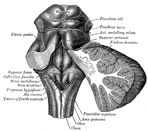
第四脳室の床。上半分は橋、下半分は延髄が作る。小脳を外すと現れる菱形の面で、ここに脳神経核の位置が投影されている。
目印を順に:正中の sulcus medianus → その両側の隆起 eminentia medialis → 上部でその中の丸い高まりが colliculus facialis(顔面神経膝が外転神経核を回る所)→ 外側の三角が area vestibularis(前庭蝸牛神経核)→ 横に走る白い線条が striae medullares ventriculi quarti(上下の境) → 下部内側の trigonum n. hypoglossi、その外側の trigonum n. vagi。
核の並びには規則がある。神経管の sulcus limitans を境に、内側が運動性(基板由来)・外側が感覚性(翼板由来)。さらに内側から外側へ4列:
| 列 | 性質 | 核の例 |
|---|---|---|
| 1(最内側) | 体性運動 | III・IV・VI・XII |
| 2 | 鰓弓性運動 | V・VII・IX・X・XI(疑核) |
| 3 | 副交感(内臓運動) | Edinger-Westphal・上下唾液核・迷走神経背側核 |
| 4(最外側) | 感覚 | 三叉神経核・孤束核・前庭蝸牛神経核 |
colliculus facialis は「顔面神経核」ではない。顔面神経の線維が外転神経核をぐるりと回る(genu n. facialis)ためにできる隆起で、隆起の中身は外転神経核である。
顔面神経丘(colliculus facialis)は、外転神経核を顔面神経の膝が回り込んで作る隆起である。この解剖から、橋下部・菱形窩の病変では外転神経麻痺と顔面神経麻痺が一緒に出ることが説明できる。
また第四脳室底に近い病変は、迷走神経背側核・孤束核・最後野に届く。最後野(area postrema)は血液脳関門を欠く化学受容器引金帯なので、ここが刺激されると難治性の嘔吐が起こる。視神経脊髄炎(NMOSD)の「難治性吃逆・嘔吐」はこの部位の病変による。
English version
Core
The floor of the fourth ventricle. The upper half is formed by the pons, the lower by the medulla. It is the rhomboid surface revealed when the cerebellum is removed, and the positions of the cranial nerve nuclei are projected onto it.
Take the landmarks in order: the midline sulcus medianus → on either side the ridge, the eminentia medialis → within it above, the rounded elevation, the colliculus facialis (where the genu of the facial nerve loops round the abducens nucleus) → laterally the triangle, the area vestibularis (the vestibulocochlear nuclei) → the white bands running transversely, the striae medullares ventriculi quarti (the boundary between upper and lower) → below and medially the trigonum n. hypoglossi, and lateral to it the trigonum n. vagi.
Going further
The arrangement of the nuclei follows a rule. With the sulcus limitans of the neural tube as the boundary, medial is motor (from the basal plate) and lateral is sensory (from the alar plate). And from medial to lateral there are four columns:
| Column | Nature | Examples |
|---|---|---|
| 1 (most medial) | Somatic motor | III, IV, VI, XII |
| 2 | Branchial motor | V, VII, IX, X, XI (nucleus ambiguus) |
| 3 | Parasympathetic (visceral motor) | Edinger–Westphal, superior and inferior salivatory, dorsal nucleus of the vagus |
| 4 (most lateral) | Sensory | Trigeminal nuclei, nucleus of the solitary tract, vestibulocochlear nuclei |
The colliculus facialis is not the facial nucleus. It is the elevation produced by the fibres of the facial nerve looping around the abducens nucleus (genu n. facialis), and what lies within the elevation is the abducens nucleus.
In the clinic
The facial colliculus is the elevation made by the genu of the facial nerve looping round the abducens nucleus. From this anatomy it follows that a lesion of the lower pons and the rhomboid fossa gives an abducens palsy and a facial palsy together.
A lesion near the floor of the fourth ventricle also reaches the dorsal nucleus of the vagus, the nucleus of the solitary tract and the area postrema. The area postrema is a chemoreceptor trigger zone lacking a blood–brain barrier, so irritation there causes intractable vomiting. The intractable hiccups and vomiting of neuromyelitis optica (NMOSD) are due to a lesion at this site.
第四脳室 — 壁と3つの孔Ventriculus quartus
橋・延髄と小脳のあいだのテント状の腔。上は中脳水道で第三脳室に、下は脊髄中心管に続く。
- 床 — 菱形窩
- 天井 — 上半分は velum medullare superius(上小脳脚のあいだ)、下半分は velum medullare inferius と tela choroidea
- 外側角 — recessus lateralis が左右に伸びる
孔は3つ。ここから髄液がくも膜下腔へ出る。Apertura mediana(Magendie 孔)が1つ、aperturae laterales(Luschka 孔)が左右に2つ。
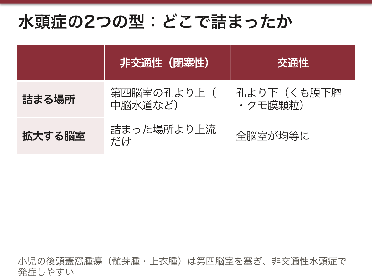
髄液の道は一本道である。側脳室 →(室間孔 Monro)→ 第三脳室 →(中脳水道 Sylvius)→ 第四脳室 →(Magendie・Luschka)→ くも膜下腔 →(クモ膜顆粒)→ 上矢状静脈洞。この順に指をなぞって言えるようにしておく。
どこで詰まるかで水頭症の型が決まる。孔より上(水道狭窄など)=非交通性で、詰まった上流の脳室だけが拡大する。孔より下(くも膜下腔・顆粒の障害)=交通性で、全脳室が均等に拡大する。第四脳室の3孔が閉じる Dandy-Walker 奇形は、第四脳室が嚢状に拡大し小脳虫部が低形成になる。
第四脳室は髄液路のいちばん細い出口で、ここが詰まると非交通性(閉塞性)水頭症になる。中脳水道・Luschka 孔・Magendie 孔のどこが塞がっても、塞がった場所より上流だけが拡大する。だから CT・MRI の脳室拡大の分布を見れば、閉塞点が読める。
小児の後頭蓋窩腫瘍(髄芽腫・上衣腫)は、第四脳室を塞いで水頭症で発症することが多い。朝の頭痛・嘔吐・うっ血乳頭という頭蓋内圧亢進の3徴で気づかれる。
Chiari 奇形では小脳扁桃が大後頭孔へ落ち込み、第四脳室からの流出が妨げられて脊髄空洞症を合併する。
出典:イラスト解剖学 誌面587頁「水頭症って?」
English version
Core
The tent-shaped cavity between the pons and medulla and the cerebellum. Above it continues into the third ventricle through the cerebral aqueduct, below into the central canal of the cord.
- Floor — the rhomboid fossa.
- Roof — above, the velum medullare superius (between the superior cerebellar peduncles); below, the velum medullare inferius and the tela choroidea.
- Lateral angles — the recessus laterales extend to each side.
There are three apertures, through which CSF leaves into the subarachnoid space: one apertura mediana (of Magendie) and two aperturae laterales (of Luschka).
Going further
The path of the CSF is a single line. Lateral ventricle → (interventricular foramen of Monro) → third ventricle → (cerebral aqueduct of Sylvius) → fourth ventricle → (Magendie and Luschka) → subarachnoid space → (arachnoid granulations) → superior sagittal sinus. Be able to trace it with a finger and say it in that order.
Where it blocks decides the type of hydrocephalus. Above the apertures (aqueduct stenosis and the like) = non-communicating, and only the ventricles upstream of the block dilate. Below the apertures (disease of the subarachnoid space or the granulations) = communicating, and all the ventricles dilate evenly.
In the Dandy–Walker malformation, in which the three apertures are closed, the fourth ventricle dilates as a cyst and the cerebellar vermis is hypoplastic.
In the clinic
The fourth ventricle is the narrowest outlet of the CSF pathway, and a block here gives non-communicating (obstructive) hydrocephalus. Whether the aqueduct, the foramina of Luschka or Magendie is closed, only what lies upstream of the block dilates. So the distribution of ventricular dilatation on CT or MRI reads out the point of obstruction.
Posterior fossa tumours in children (medulloblastoma, ependymoma) often present with hydrocephalus from blocking the fourth ventricle, noticed by the triad of raised intracranial pressure — morning headache, vomiting and papilloedema.
In Chiari malformation the cerebellar tonsils descend into the foramen magnum, outflow from the fourth ventricle is obstructed, and syringomyelia is associated.
錐体Pyramis
延髄の前面、前正中裂のすぐ両側にある縦に走る隆起。中を皮質脊髄路(錐体路)が下る。
下端で線維の約75〜90%が反対側へ渡る——これが錐体交叉 decussatio pyramidum で、延髄と脊髄の境の目印になる。「運動が左右で入れ替わる場所」として指せること。
前正中裂と前外側溝のあいだの縦長の隆起。中身は tractus pyramidalis(錐体路)の下行線維そのもので、名前は形(三角錐)に由来する。
延髄の下端で decussatio pyramidum(錐体交叉)が起こり、fibrae corticospinales の約80%が交叉して tractus corticospinalis lateralis に、残り約20%は交叉せず tractus corticospinalis anterior になる。錐体を示せたら、続けて「ここで何が起こるか」まで言えるようにする。
錐体交叉があるから、片側の大脳の病変で反対側が麻痺する。内包や中心前回の梗塞で対側片麻痺になる根拠がここにある。
逆に錐体交叉より下(頸髄)の病変では同側の麻痺になる。「交叉の場所より上か下か」で左右が決まる——脳幹病変の局在診断はこの一点から始まる。
English version
Core
The longitudinal ridge on the anterior surface of the medulla, immediately beside the anterior median fissure. The corticospinal (pyramidal) tract descends within it.
At its lower end about 75–90 per cent of the fibres cross to the other side — the decussatio pyramidum, which marks the boundary between medulla and spinal cord. Be able to point to it as "the place where movement changes sides".
前外側溝Sulcus anterolateralis
延髄前面で、錐体とオリーブの間を縦に走る溝。舌下神経(XII)の根糸がここから並んで出る。
「錐体とオリーブの谷間=XII」と、溝と神経を一組で覚える。
錐体とオリーブのあいだの溝。ここから出るのは XII(n. hypoglossus)だけである。
脊髄の前外側溝が前根を出していたのと同じ位置関係で、運動性の神経が腹側から出るという原則が延髄でも保たれている。
XII の障害=舌下神経麻痺。舌を出すと麻痺側へ偏位する(健側の genioglossus が押し出すため)。
延髄内側症候群では、錐体(対側片麻痺)・内側毛帯(対側の深部感覚障害)・XII(同側の舌偏位)が同時に出る。3つとも前外側溝の近くに並んでいるから一緒に落ちる——構造の隣接がそのまま症候群になる例。
English version
Core
The longitudinal groove on the anterior surface of the medulla, between the pyramid and the olive. The rootlets of the hypoglossal nerve (XII) emerge in a row from it.
Learn the sulcus and the nerve as a pair: the valley between pyramid and olive = XII.
オリーブOliva
錐体の外側にある卵形のふくらみ。中身は下オリーブ核 nucleus olivaris inferior で、小脳へ登上線維を送る。
延髄前面では錐体・オリーブ・その外側の溝の3つが並ぶ。この並び順が延髄を見分ける決め手である。
前外側溝と後外側溝のあいだの楕円形の膨らみ。中身は nucleus olivaris(下オリーブ核)。
ここから tractus olivospinalis(↓)と tractus olivocerebellaris(↑)が出る。後者は交叉して下小脳脚から小脳皮質へ入り、平衡と運動学習に関わる。
下オリーブ核は小脳へ登上線維(climbing fiber)を送る唯一の起始核で、運動の誤差信号を伝えると考えられている。
症候性口蓋ミオクローヌスは、赤核・下オリーブ核・歯状核を結ぶ Guillain–Mollaret の三角の障害で起こり、下オリーブ核の肥大性変性を伴う。オリーブが「大きくなる変性」という珍しい形をとる。
English version
Core
The oval swelling lateral to the pyramid. Within it is the nucleus olivaris inferior, which sends climbing fibres to the cerebellum.
On the anterior surface of the medulla, pyramid, olive and the sulcus lateral to it lie in a row. That order is the decisive way of recognising the medulla.
後外側溝Sulcus posterolateralis
オリーブの後ろを縦に走る溝。舌咽神経(IX)・迷走神経(X)・副神経(XI)の根糸が、上から順に一列に出る。
前外側溝が XII だけなのに対し、後外側溝は3本まとめて。左右の溝で出る神経が違うことを言えるようにする。
オリーブの外側の溝。IX(n. glossopharyngeus)・X(n. vagus)・XI(n. accessorius)が上から順にここから出る。
前外側溝=XII、後外側溝=IX・X・XI。この対比を必ず押さえる。
この3本はいずれも nucleus ambiguus を共有しており、まとめて障害されると嚥下障害・嗄声・カーテン徴候が出る(球麻痺)。
延髄外側症候群(Wallenberg)では後外側溝の深部が虚血に陥り、nucleus ambiguus(嚥下・発声)と nucleus spinalis n. trigemini(同側顔面の温痛覚)が同時にやられる。神経が出る場所と、その内側の核が対応しているという原則の典型例。
English version
Core
The longitudinal groove behind the olive. The rootlets of the glossopharyngeal (IX), vagus (X) and accessory (XI) nerves emerge from it in a single row, in that order from above down.
Where the anterolateral sulcus gives only XII, the posterolateral gives three together. Be able to say which nerves leave which of the two sulci.
薄束結節Tuberculum gracile
延髄の後面、後正中溝のすぐ両側にある小さなふくらみ。中に薄束核 nucleus gracilis があり、下半身からの深部感覚をここで乗り換える。
延髄背側面の下部、後正中溝と後中間溝のあいだの膨らみ。中身は nucleus gracilis。
下肢からの意識的固有感覚・触覚を運ぶ fasciculus gracilis の終点=第2ニューロンがここにある。
ここを出た線維は fibrae arcuatae internae として交叉(decussatio lemnisci medialis)し、lemniscus medialis となって視床へ上る。
後索路は延髄で交叉、脊髄視床路は脊髄内で交叉——この交叉部位の違いが、脊髄半側切断(Brown–Séquard)で「深部感覚は同側・温痛覚は対側」という解離を生む。
English version
Core
The small swelling on the posterior surface of the medulla, immediately beside the posterior median sulcus. Within it is the nucleus gracilis, where deep sensation from the lower half of the body relays.
楔状束結節Tuberculum cuneatum
薄束結節の外側のふくらみ。中に楔状束核 nucleus cuneatus があり、上半身からの深部感覚を中継する。
内側が下半身、外側が上半身——後索の体部位対応がそのまま結節の並びに出ている。この一組で覚える。
後中間溝の外側の膨らみ。中身は nucleus cuneatus。
上肢からの意識的固有感覚・触覚を運ぶ fasciculus cuneatus の第2ニューロン。内側が下肢(gracilis)、外側が上肢(cuneatus)という体部位配列を覚える。
後索の体部位配列は「内側ほど下位」——仙髄が最内側、頸髄が最外側。脊髄で下から順に線維が外側へ積み重なるためである。
亜急性連合性脊髄変性症(ビタミンB12欠乏)では後索が選択的に侵され、深部感覚障害と Romberg 徴候が出る。侵されるのが gracilis 側から始まるため、下肢の症状が先に出る。
English version
Core
The swelling lateral to the gracile tubercle. Within it is the nucleus cuneatus, relaying deep sensation from the upper half of the body.
Medial is the lower body, lateral the upper — the somatotopy of the posterior columns appears directly in the arrangement of the tubercles. Learn them as a pair.
脳底溝Sulcus basilaris
橋の腹側の正中を縦に走る浅い溝。ここに脳底動脈 a. basilaris が乗る。
橋の腹面を見せられたら、まずこの溝で正中を決める。
橋の腹側面の正中を縦に走る浅い溝。ここに a. basilaris(脳底動脈)が乗る。
橋と延髄の境は sulcus pontobasilaris、外側へ続くのが pedunculi cerebellares medii。
脳底動脈は左右の椎骨動脈が合流してできる。ここが閉塞すると橋が広範に虚血になり、閉じ込め症候群(locked-in syndrome)——意識と垂直眼球運動だけ残り、四肢と球麻痺で動けない状態——を起こす。
溝の名前を覚える意味は、そこに何が走るかを言えることにある。
English version
Core
The shallow groove running longitudinally in the midline of the ventral pons. The basilar artery lies in it.
Shown the ventral surface of the pons, use this groove first to establish the midline.
中小脳脚Pedunculi cerebellares medii
橋と小脳をつなぐ、3対のうち最も太い脚。橋核から出て交叉した線維(横橋線維)が入る。
大脳皮質 → 橋核 → 反対側の小脳、という皮質橋小脳路の通り道。太さで一目で分かる。
3対の小脳脚のうち最も太い。橋(pons)と小脳を結ぶ。
通るのは tractus pontocerebellaris——nuclei pontis から出て交叉して対側の小脳皮質へ入る線維。
大脳皮質 → nuclei pontis → 交叉 → 中小脳脚 → 小脳皮質、という tractus corticopontocerebellaris の後半にあたる。大脳が小脳に「これから動く」と予告する経路である。
交叉するため、大脳半球は対側の身体を、小脳半球は同側の身体を担当するという「二重交叉」が成立する。小脳症状が同側に出る理由。
English version
Core
The peduncle joining pons to cerebellum, the thickest of the three pairs. The fibres that leave the pontine nuclei and cross (the transverse pontine fibres) enter through it.
The route of the cortico-ponto-cerebellar pathway — cerebral cortex → pontine nuclei → contralateral cerebellum. Recognisable at a glance by its thickness.
下小脳脚Pedunculi cerebellares inferiores
延髄と小脳をつなぐ脚(索状体 corpus restiforme)。後脊髄小脳路とオリーブ小脳線維が入る、ほぼ入力専用。
延髄(medulla oblongata)と小脳を結ぶ。延髄背側面の外側の肥厚として見える。
通るのは tractus spinocerebellaris posterior(無意識の固有感覚)・tractus olivocerebellaris・tractus vestibulocerebellaris。
「後ろは下、前は上」——後脊髄小脳路は下小脳脚、前脊髄小脳路は上小脳脚を通る。必ず対にして覚える。
前庭小脳路がここを通るため、下小脳脚の障害では平衡障害・眼振が前景に出る。Wallenberg 症候群でめまいと眼振が出るのは、下小脳脚と前庭神経核が同じ領域にあるため。
English version
Core
The peduncle joining medulla to cerebellum (the corpus restiforme). The posterior spinocerebellar tract and the olivocerebellar fibres enter through it — almost purely an input.
上小脳脚Pedunculi cerebellares superiores
中脳と小脳をつなぐ脚。歯状核からの出力が通る、ほぼ出力専用。上小脳脚交叉を経て赤核・視床へ向かう。
3対はまとめて「下=入る(延髄)/中=入る(橋・最も太い)/上=出る(中脳)」で押さえる。
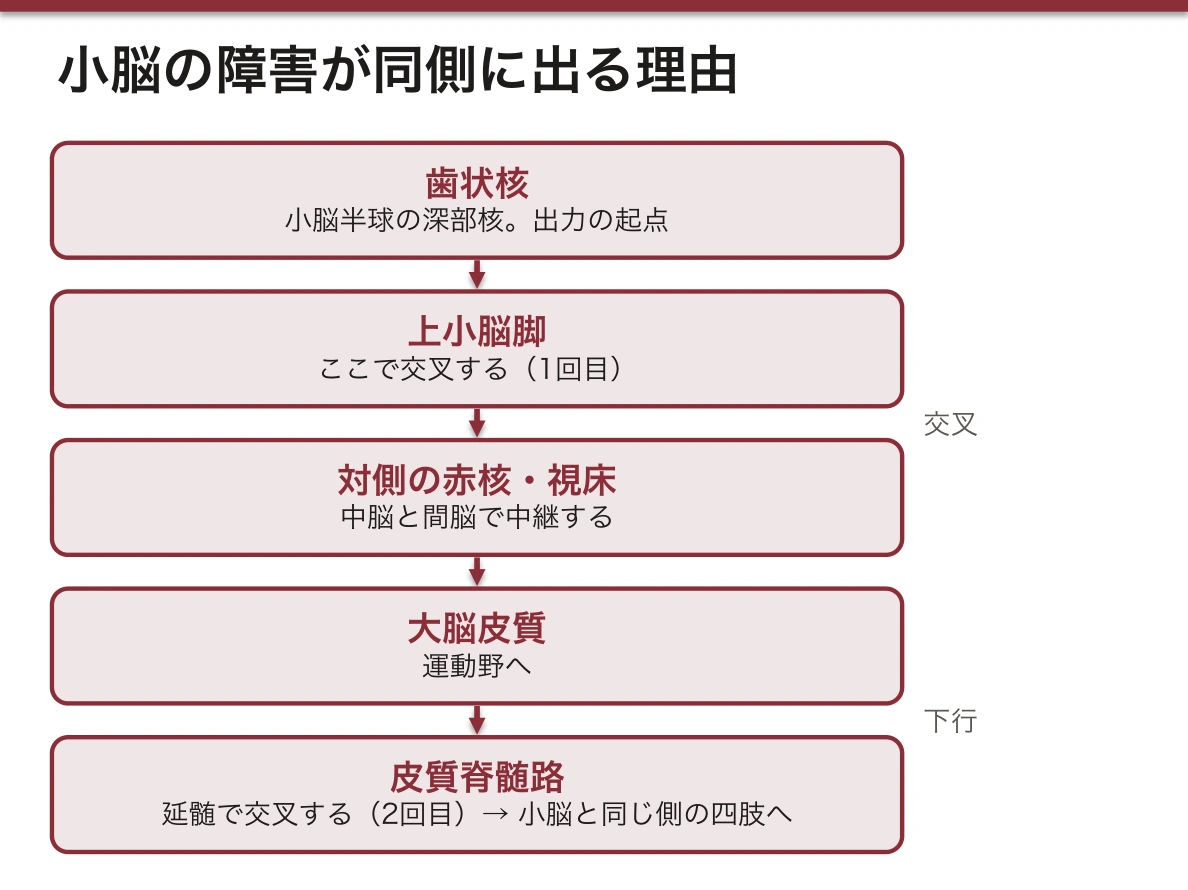
中脳(mesencephalon)と小脳を結ぶ。菱形窩の上縁をなす。
通るのは tractus spinocerebellaris anterior(入力)と tractus cerebellorubralis(出力)。後者は nucleus dentatus から nucleus ruber へ向かう。
上小脳脚は小脳の主要な「出口」である。歯状核 → 上小脳脚 →(交叉)→ 対側赤核・視床 → 大脳皮質、という dentato-rubro-thalamo-cortical 路。
この経路の障害で企図振戦・測定異常が出る。小脳の入力は下・中小脳脚、出力は上小脳脚と整理すると、症状の説明がそのまま経路の説明になる。
English version
Core
The peduncle joining midbrain to cerebellum. The output from the dentate nucleus passes through it — almost purely an output, running through the decussation of the superior cerebellar peduncles to the red nucleus and the thalamus.
Fix the three together as: inferior = in (medulla) / middle = in (pons, the thickest) / superior = out (midbrain).
菱形窩の正中溝Sulcus medianus
菱形窩(第四脳室の床)の正中を縦に走る溝。窩を左右に分ける基準線で、他の構造はすべてこの溝からの距離で言う。
菱形窩の縦の正中線をなす溝。ここを基準に左右対称の構造を探す。
外側に sulcus limitans があり、両溝のあいだが eminentia medialis。
sulcus limitans は発生学的な境界である。神経管の alar plate(翼板=感覚性)と basal plate(基板=運動性)の境がそのまま残ったもの。
だから正中側(basal 由来)に運動核、外側(alar 由来)に感覚核という配置になる。「内側=運動・外側=感覚」は暗記事項ではなく発生の帰結——ここを説明できると強い。
English version
Core
The groove running longitudinally in the midline of the rhomboid fossa (the floor of the fourth ventricle). It is the reference line dividing the fossa into right and left, and every other structure is described by its distance from it.
内側隆起Eminentia medialis
正中溝のすぐ両側に伸びる、縦に細長い隆起。その外側は境界溝で区切られる。
この隆起の上に、次の顔面神経丘が乗る。
sulcus medianus と sulcus limitans のあいだの細長い隆起。
髄条より上では colliculus facialis をなし、下では細くなって trigonum nervi hypoglossi になる。
正中隆起は基板由来=運動核の並ぶ列である。上から VI(外転神経核)、下へ XII(舌下神経核)と、体節性の運動核が縦一列に並ぶ。
この「列」を意識すると、菱形窩の核の位置が丸暗記でなくなる。同じ列の中で上下に並んでいるだけだと分かる。
English version
Core
The long, narrow longitudinal ridge running immediately on either side of the median sulcus, bounded laterally by the sulcus limitans. The facial colliculus that follows sits on this ridge.
顔面神経丘Colliculus facialis
内側隆起の上部(橋のあたり)にある丸いふくらみ。
名前に反して、中身は顔面神経核ではない。丘を作っているのは外転神経核で、その上を顔面神経の膝(内膝)が回り込んでいる。この「名前と中身のずれ」が要点である。
髄条の上、正中隆起の膨らみ。中にあるのは VI(nucleus n. abducentis)である。
名前は「顔面」だが中身は外転神経核——顔面神経の線維(genu n. facialis)が外転神経核の周りを回り込むため、外から見て膨らんで見える。最頻出の引っかけ。
VII の運動核は colliculus facialis の外側かつ深部にあり、その軸索が背内側へ上って外転神経核を巻き(internal genu)、再び腹外側へ出る。
この解剖の帰結が one-and-a-half 症候群や、橋病変での「同側の外転麻痺+同側の末梢性顔面神経麻痺」の併発である。2つの核が物理的に絡んでいるから一緒に落ちる。
English version
Core
The rounded elevation in the upper part of the medial eminence (at the level of the pons). Despite the name, what lies within it is not the facial nucleus.
What makes the elevation is the abducens nucleus, around which the genu of the facial nerve loops. This discrepancy between the name and the contents is the point.
前庭野Area vestibularis
菱形窩の外側角にある領域。下に前庭神経核が広がる。
菱形窩の外側角。VIII(n. vestibulocochlearis)の核が入る。
nuclei vestibulares(4個)は延髄側、nuclei cochleares(背側・腹側)は橋側に位置する。
前庭神経核は最も外側=alar plate 由来の感覚核の列にある。ここから tractus vestibulospinalis(姿勢)・fasciculus longitudinalis medialis(眼球と頭位の協調)・tractus vestibulocerebellaris が出る。
前庭核は「感覚核なのに出力が多い」のが特徴で、めまいに眼振・姿勢異常・嘔吐が伴うのはこの出力の広さによる。
English version
Core
The area at the lateral angle of the rhomboid fossa. The vestibular nuclei lie beneath it.
第四脳室髄条Striae medullares ventriculi quarti
菱形窩を横切って走る白い線維束。弓状核から出て正中へ向かう。
この線が菱形窩を上下に分ける——上が橋の部分、下が延髄の部分。窩の中で唯一の「横向きの目印」なので、標本ですぐ見つかる。
菱形窩の横の正中線をなす線維束。これを境に上下で核の配置が変わる。
髄条より上= nucleus salivatorius superior(VII)/髄条より下= nucleus tractus solitarii・nucleus salivatorius inferior(IX)・nucleus dorsalis n. vagi。
髄条は nuclei arcuati から出て下小脳脚へ向かう線維で、聴覚路とは無関係(名前が似た corpus trapezoideum と混同しない)。
臨床的な意味は「高さの目安」——病変が髄条より上か下かで、唾液分泌障害の側(耳下腺 vs 顎下腺)や味覚障害の範囲が変わる。「この線より上の核」を挙げられるようにしておく。
English version
Core
The white bundles of fibres crossing the rhomboid fossa transversely, arising from the arcuate nuclei and running towards the midline.
This line divides the fossa into upper and lower — above is the pontine part, below the medullary. It is the only transverse landmark in the fossa, and so is found at once on a specimen.
舌下神経三角Trigonum nervi hypoglossi
菱形窩の下部、正中溝のすぐ外側にある細長い三角。下に舌下神経核がある。
髄条の下、正中隆起が細くなった三角形の部分。中にあるのは XII(nucleus n. hypoglossi)。
その外側が trigonum nervi vagi。内側から XII → X の順に並ぶ。
舌下神経核は最も正中側の運動核で、脊髄前角の運動細胞が上へ続いたものと考えられる(体節性運動核の列の最下端)。
核性(延髄内)の障害では舌の萎縮と線維束攣縮が出るのに対し、核上性(皮質延髄路)の障害では萎縮を伴わない偏位になる。萎縮の有無で核の上下を切り分けるのが神経診察の型。
English version
Core
The long narrow triangle in the lower part of the rhomboid fossa, immediately lateral to the median sulcus. The hypoglossal nucleus lies beneath it.
迷走神経三角Trigonum nervi vagi
舌下神経三角の外側の三角。下に迷走神経背側核(副交感)がある。
2つの三角は「内側が XII、外側が X」。菱形窩の下半分は、この順序で読む。
trigonum nervi hypoglossi の外側の三角。中にあるのは X の副交感核=nucleus dorsalis n. vagi。
運動核(内側)と感覚核(外側)のあいだに自律核が入る——菱形窩の3層構造の中間層にあたる。
迷走神経背側核は胸腹部臓器への副交感性出力を担う。心拍の抑制、気管支収縮、消化管運動と分泌の促進。
同じ X でも、咽頭・喉頭の随意筋は nucleus ambiguus(運動核)、内臓の副交感は nucleus dorsalis(自律核)と担当が分かれている。「1本の神経に複数の核」という脳神経の原則が最もはっきり出る例。
English version
Core
The triangle lateral to the hypoglossal trigone. The dorsal nucleus of the vagus (parasympathetic) lies beneath it.
The two triangles are "XII medial, X lateral". Read the lower half of the rhomboid fossa in that order.
小脳半球Hemispheria cerebelli
虫部の左右に広がる大きな部分。外側ほど新しく、上肢下肢の巧緻な運動(協調)に関わる。
障害は同側に出る——大脳と違って交叉しないので、右半球の障害は右手の失調になる。
小脳の左右の半球。上面(facies superior)と下面(facies inferior)を fissura horizontalis cerebelli が分ける。
半球は四肢の協調を担当し、対応する深部核は nucleus dentatus。
小脳症状は同側に出る——小脳からの出力は上小脳脚で交叉し、皮質脊髄路が延髄の錐体交叉でもう一度交叉するため(二重交叉)。
半球の障害では測定異常・企図振戦・変換運動障害・協調運動障害が同側の四肢に出る。指鼻試験・回内回外試験は半球の機能を見ている。虫部の障害(体幹失調)と切り分けるのが診察の目的。
English version
Core
The large parts spreading to either side of the vermis. The more lateral the newer, concerned with the skilled (coordinated) movement of the limbs.
Lesions appear ipsilaterally — unlike the cerebrum there is no net crossing, so a lesion of the right hemisphere gives ataxia of the right hand.
虫部Vermis cerebelli
左右の半球にはさまれた正中の細い部分。名のとおり虫のように節がある。
体幹の平衡と姿勢を受け持つので、障害で体幹失調(座れない・歩けない)が出る。半球の障害=四肢の失調と対にして覚える。
左右の半球を繋ぐ正中の細い部分。上部は上面に溶け込み、下部は溝で半球と分かれる。
体幹と姿勢の制御を担当し、対応する深部核は nucleus fastigii。
虫部は前庭系との結びつきが強い(原小脳・前庭小脳に相当)。fastigii 核から前庭神経核・網様体へ出力する。
虫部の障害=体幹失調。座位や立位が保てず、開脚歩行(wide-based gait)になる。四肢の協調は保たれることがあるのが半球障害との違い。小児の髄芽腫は虫部に好発し、体幹失調と水頭症で発症する。
English version
Core
The narrow median part between the two hemispheres, segmented like a worm as its name says. It is responsible for the balance and posture of the trunk, so a lesion gives truncal ataxia (unable to sit, unable to walk).
Learn it as a pair with hemispheric lesions = ataxia of the limbs.
水平裂Fissura horizontalis cerebelli
小脳の後外側を水平に一周する、最も深く目立つ裂。中小脳脚のあたりから始まる。
小脳の上面と下面を分ける境として使う。標本で「まずどこを見るか」と言われたらこれ。
小脳の上面と下面を分ける水平の深い裂。標本を手に取ったとき、まずこの裂で上下を決める。
前縁(margo anterior)・後縁(margo posterior)とあわせて、小脳の向きを決める目印になる。
水平裂は発生学的な区分とは一致しない——機能的な区分は前葉・後葉・片葉小節葉で、境界は fissura prima と fissura posterolateralis である。
「見た目の目印」と「機能的な区分」を混同しないこと。水平裂は標本の向きを決めるための目印として使い、機能を語るときは別の裂を挙げる。
English version
Core
The deepest and most conspicuous fissure, running horizontally right round the posterolateral cerebellum and beginning near the middle cerebellar peduncle. Use it as the boundary between the upper and lower surfaces of the cerebellum.
Asked "what do you look at first" on a specimen, this is it.
小脳の裂Fissurae cerebelli
小脳の表面を横に走る多数の深い溝の総称。これで皮質が細かく折り畳まれ、表面積が稼がれている。
裂は小脳を小葉 lobuli に区切る。大脳の溝が不規則なのに対し、小脳の裂はほぼ平行に走るのが見分けどころ。
小脳の表面を横に走る多数の裂。半球と虫部の全面を覆う。
裂と裂のあいだの薄い板が folia cerebelli。
裂が深く密なのは皮質の表面積を稼ぐため。小脳の表面積は展開すると大脳の約75%に達し、ニューロン数では脳全体の半分以上を小脳が占める。
「小脳は小さいのに細胞が多い」——顆粒細胞が極端に密に詰まっているため。組織学で小脳皮質を見るとき、顆粒層が濃青に見える理由でもある。
English version
Core
The collective name for the many deep transverse grooves on the surface of the cerebellum. They fold the cortex finely and so gain surface area, and divide the cerebellum into lobuli.
Where the sulci of the cerebrum are irregular, the fissures of the cerebellum run almost parallel — this is how they are told apart.
小脳回(葉片)Folia cerebelli
裂と裂の間の薄い葉状のひだ。大脳の回にあたるが、はるかに薄く細かい。
裂と裂のあいだの薄い葉状の隆起。小脳皮質はこの表面に乗っている。
矢状断で見ると、白質が葉の中へ枝を伸ばし、全体として arbor vitae の像になる。
1枚の葉の断面に、小脳皮質の3層(分子層・Purkinje 細胞層・顆粒層)がそのまま現れる。組織学の標本はこの断面を見ている。
Purkinje 細胞は小脳皮質の唯一の出力細胞で、すべて抑制性(GABA)。皮質から深部核への出力が抑制性であることが、小脳の「ブレーキとしての働き」の基礎になる。
English version
Core
The thin leaf-like folds between the fissures. They correspond to the gyri of the cerebrum but are far thinner and finer.
小脳活樹(生命の樹)Arbor vitae
小脳を正中矢状断にしたときに見える、白質が枝分かれした樹の形。中心の白質から皮質へ枝が伸びる像を指す。
断面でしか見えないので、標本で示すときは切った標本を選んで指す。
虫部の矢状断で白質が樹枝状に分岐して見える像。「生命の樹」の名のとおりの形。
白質は虫部では薄い層、半球では厚い層をなす。
白質の中に nuclei cerebelli(外→内に dentatus・emboliformis・globosus・fastigii)が埋まっている。枝の根元に核があると覚えると位置関係が崩れない。
画像診断では、正中矢状断 MRI で arbor vitae の形が保たれているかが小脳虫部の萎縮の指標になる。脊髄小脳変性症やアルコール性小脳変性で、葉が細く裂が広がる像を示す。
English version
Core
The tree-like branching of white matter seen when the cerebellum is cut in the median sagittal plane — the image of branches extending from the central white matter to the cortex.
It can only be seen on a section, so to show it you must choose a cut specimen and point to it.
まとめ
| 延髄 — 外形 | 脊髄と橋のあいだ。下端は大後頭孔(第1頸神経の出る高さ)、上端は橋との境の溝 sulcus bulbopontinus。 |
|---|---|
| 橋 | 延髄と中脳のあいだの、前へ強く張り出した部分。 |
| 小脳 — 外形と内部 | 後頭蓋窩、脳幹の背側。中央の vermis(虫部)と左右の hemispheria cerebelli からなる。 |
| 菱形窩 — 脳神経核の地図 | 第四脳室の床。上半分は橋、下半分は延髄が作る。 |
| 第四脳室 — 壁と3つの孔 | 橋・延髄と小脳のあいだのテント状の腔。上は中脳水道で第三脳室に、下は脊髄中心管に続く。 |
| 錐体 | 延髄の前面、前正中裂のすぐ両側にある縦に走る隆起。 |
| 前外側溝 | 延髄前面で、錐体とオリーブの間を縦に走る溝。 |
| オリーブ | 錐体の外側にある卵形のふくらみ。中身は下オリーブ核 nucleus olivaris inferior で、小脳へ登上線維を送る。 |
| 後外側溝 | オリーブの後ろを縦に走る溝。舌咽神経(IX)・迷走神経(X)・副神経(XI)の根糸が、上から順に一列に出る。 |
| 薄束結節 | 延髄の後面、後正中溝のすぐ両側にある小さなふくらみ。 |
| 楔状束結節 | 薄束結節の外側のふくらみ。中に楔状束核 nucleus cuneatus があり、上半身からの深部感覚を中継する。 |
| 脳底溝 | 橋の腹側の正中を縦に走る浅い溝。ここに脳底動脈 a. basilaris が乗る。 |
| 中小脳脚 | 橋と小脳をつなぐ、3対のうち最も太い脚。 |
| 下小脳脚 | 延髄と小脳をつなぐ脚(索状体 corpus restiforme)。 |
| 上小脳脚 | 中脳と小脳をつなぐ脚。歯状核からの出力が通る、ほぼ出力専用。 |
| 菱形窩の正中溝 | 菱形窩(第四脳室の床)の正中を縦に走る溝。 |
| 内側隆起 | 正中溝のすぐ両側に伸びる、縦に細長い隆起。 |
| 顔面神経丘 | 内側隆起の上部(橋のあたり)にある丸いふくらみ。 |
| 前庭野 | 菱形窩の外側角にある領域。下に前庭神経核が広がる。 |
| 第四脳室髄条 | 菱形窩を横切って走る白い線維束。弓状核から出て正中へ向かう。 |
| 舌下神経三角 | 菱形窩の下部、正中溝のすぐ外側にある細長い三角。 |
| 迷走神経三角 | 舌下神経三角の外側の三角。下に迷走神経背側核(副交感)がある。 |
| 小脳半球 | 虫部の左右に広がる大きな部分。外側ほど新しく、上肢下肢の巧緻な運動(協調)に関わる。 |
| 虫部 | 左右の半球にはさまれた正中の細い部分。名のとおり虫のように節がある。 |
| 水平裂 | 小脳の後外側を水平に一周する、最も深く目立つ裂。 |
| 小脳の裂 | 小脳の表面を横に走る多数の深い溝の総称。 |
| 小脳回(葉片) | 裂と裂の間の薄い葉状のひだ。大脳の回にあたるが、はるかに薄く細かい。 |
| 小脳活樹(生命の樹) | 小脳を正中矢状断にしたときに見える、白質が枝分かれした樹の形。 |
このノートは医学生が授業の内容を自分の言葉でまとめたもので、診療の判断に使うものではありません。誤りに気づいたらお知らせください。