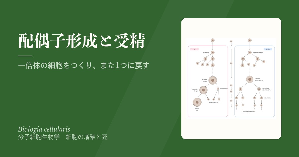
配偶子形成は、二倍体の個体の中で一倍体の細胞、つまり卵子と精子をつくる過程である。仕組みの中心は減数分裂だが、卵と精子では始まる時期、止まる場所、分け方、できる数がまるでちがう。卵は胎児のうちに減数分裂を始めて何十年も途中で止まり、細胞質を1つの細胞に集める。精子は思春期から一生作られ続け、細胞質を捨てて泳ぐための形に作り変えられる。
この章では、生殖細胞の起源と性の決定、生殖細胞で起きるエピジェネティックな「初期化」、卵子形成と卵胞の発育、精子形成、そして受精の分子の流れを扱う。なぜ両者はこれほどちがう設計になっているのかを考えながら読むと、不妊症や染色体異常の話が自然につながる。
- 始原生殖細胞 — 卵と精子のもとは、体の外側から旅してくる
- 性の決定 — SRYが短い時間だけSOX9を押し上げる
- 生殖細胞のエピジェネティクス — 親の印を消して、自分の性の印を付け直す
- 卵子形成 — 胎児期に始まり、2回止まり、受精で終わる
- 卵胞の発育 — 卵を育てる部屋が、1か月で大きく変わる
- 精子形成 — つながったまま分裂し、細胞質を捨てて完成する
- 受精の分子機構 — 1つの精子だけを入れて、扉を閉じる
始原生殖細胞 — 卵と精子のもとは、体の外側から旅してくるCellula germinalis primordialis
卵子と精子のもとになる細胞は、生殖腺の中で生まれるのではない。妊娠およそ4週に、卵黄嚢の壁に始原生殖細胞(PGC)が現れる。PGCはアルカリホスファターゼの活性が高く、これが目印になる。
PGCは4〜6週にかけて体の中を移動し、将来の生殖腺である生殖隆起にたどり着く。ここはまだ男女どちらにもなれる未分化の生殖腺である。移動の初期には、周りの中胚葉が発現するタンパク質(IFITM1・IFITM3)が道案内として必要になる。生殖隆起に入ったPGCは、精巣では精祖細胞(2n)、卵巣では卵祖細胞(2n)になる。
配偶子形成がいつ行われるかは男女で大きくちがう。男性では思春期に始まり、成人期を通じて一生続く。女性では胎児期に始まり、個々の卵にとっては受精して初めて終わる。
なぜ生殖細胞は、わざわざ体の外側でつくられてから戻ってくるのか。発生の早い時期に体細胞の運命づけから離れた場所でPGCを確保しておくことで、体をつくるための分化の合図やエピジェネティックな印から生殖細胞を守り、次の世代に渡す「まっさらな」状態を保ちやすくなると考えられている。PGCはこのあと、自分のゲノムの印を大きく消し去る(後の項目)。
移動の途中で道をそれたPGCが残ると、性腺外胚細胞腫瘍の原因になると考えられている。胚細胞腫瘍が仙尾部、縦隔、松果体部など体の正中線に沿って生じやすいのは、PGCの通り道と重なるためである。
English version
Core
The cells that become eggs and sperm are not born inside the gonads. At about week 4 of pregnancy, primordial germ cells (PGCs) appear in the wall of the yolk sac. PGCs have high alkaline phosphatase activity, which serves as a marker.
During weeks 4–6, PGCs travel through the body to the genital ridge, the future gonad. At this point it is an undifferentiated gonad that can still become either sex. Early migration needs guiding proteins (IFITM1 and IFITM3) expressed by the surrounding mesoderm. In the gonad, PGCs become spermatogonia (2n) in the testis or oogonia (2n) in the ovary.
Going further
When gametogenesis happens differs greatly between the sexes. In males it starts at puberty and continues for life. In females it starts before birth and, for each egg, ends only at fertilisation.
Why are germ cells made outside the body proper and then brought back? Setting PGCs aside early, away from the signals that decide somatic fates, probably protects them from differentiation signals and epigenetic marks used to build the body. This helps keep them in a “clean” state for the next generation. PGCs then erase most of the marks on their genome (see a later item).
In the clinic
PGCs that stray from their route and survive are thought to cause extragonadal germ cell tumours. These tumours tend to arise along the midline of the body (sacrococcygeal region, mediastinum, pineal region) because this matches the path of the PGCs.
性の決定 — SRYが短い時間だけSOX9を押し上げる
未分化の生殖腺は、皮質と髄質をもち、どちらの性にもなれる。精巣になるか卵巣になるかを決める最初のスイッチが、Y染色体の短腕にあるSRY(Y染色体性決定領域、精巣決定因子)である。
SRYの主な役目は、ごく短い決定的な時間帯に、常染色体の遺伝子SOX9の発現を引き上げることである。SOX9はSertoli細胞の分化を決める親玉の転写因子で、HMGボックスというDNA結合部分をもつ。SOX9はもともと男女どちらの未分化生殖腺でも、SF1の調節のもとで少しだけ発現している。SRYの発現が山を越えたあとも、精巣のSertoli細胞でだけSOX9が発現し続ける。
Sertoli細胞ができると、それが周りを精巣の構造へと導く。SRYが無ければSOX9は上がらず、生殖腺は卵巣へ進む。X染色体のDAX1など、精巣化に逆らう方向の因子もかかわる。
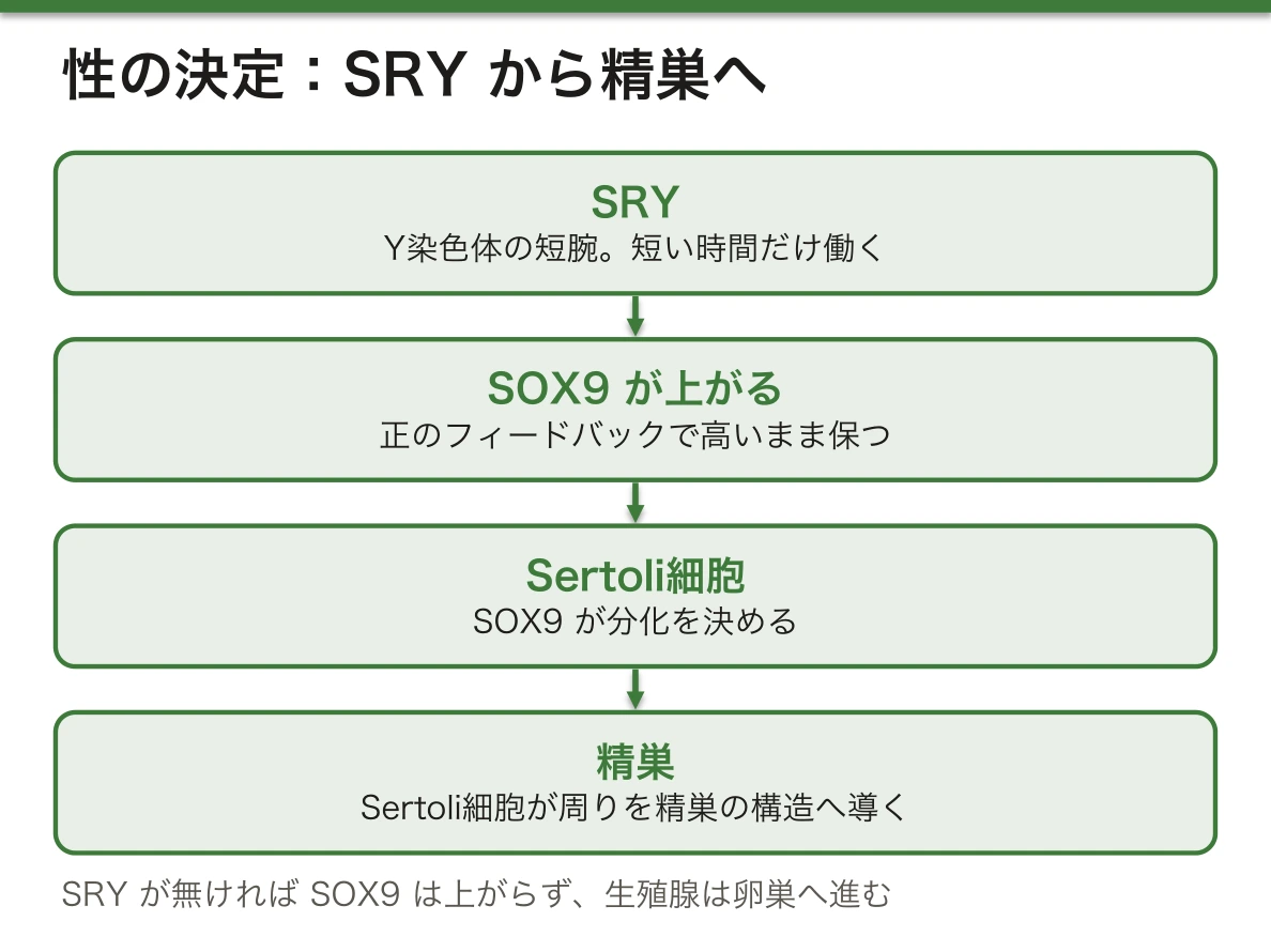
なぜSRYは短い時間だけ働けば足りるのか。いったん高くなったSOX9は、自分自身の発現を保つ正のフィードバックの回路(SOX9が自身や増殖因子を介して自らの発現を支える)に入る。スイッチは「押し続ける」必要がなく、一度倒せばその位置に留まる。逆にいえば、決定的な時間帯にSOX9が十分に上がらなければ、後から挽回するのは難しい。発生の多くの決定はこのように、短い合図を安定した状態に変える回路で作られている。
父親の減数分裂の交叉でSRYを含む部分がX染色体に移ると、46,XXの男性が生まれる。逆にSRYの欠失や変異があると、核型は46,XYでも精巣ができず、性腺形成不全になる。性分化疾患の診療で、核型だけでなくSRYの有無を調べるのはこのためである。
English version
Core
The undifferentiated gonad has a cortex and a medulla and can become either sex. The first switch that decides testis or ovary is SRY (sex-determining region on the Y chromosome; testis-determining factor), on the short arm of the Y chromosome.
The main job of SRY is to raise the expression of the autosomal gene SOX9 during a very short, critical time window. SOX9 is the master transcription factor for Sertoli cell differentiation and has a DNA-binding part called the HMG box. SOX9 is already expressed at a low level, under SF1 control, in the undifferentiated gonads of both sexes. After SRY expression has peaked, SOX9 stays on only in the Sertoli cells of the testis.
Once Sertoli cells form, they guide the surrounding tissue into a testis. Without SRY, SOX9 does not rise and the gonad becomes an ovary. Factors that work against testis formation, such as DAX1 on the X chromosome, are also involved.
Going further
Why is a short burst of SRY enough? Once SOX9 is high, it enters a positive feedback loop that keeps its own expression up (SOX9 supports its own expression directly and through growth factors). The switch does not need to be held down; once flipped, it stays. On the other hand, if SOX9 does not rise enough in the critical window, it is hard to catch up later. Many decisions in development work this way, turning a short signal into a stable state.
In the clinic
If the part containing SRY moves to the X chromosome during crossing over in the father’s meiosis, a 46,XX male is born. If SRY is deleted or altered, no testis forms even with a 46,XY karyotype, and gonadal dysgenesis results. This is why SRY is tested, not just the karyotype, in differences of sex development.
生殖細胞のエピジェネティクス — 親の印を消して、自分の性の印を付け直す
体細胞では、受精卵から受け継いだインプリンティング(親由来で決まる遺伝子の印)は一生保たれなければならない。一方、生殖細胞の遺伝子だけが次の世代に渡る。そのため生殖細胞では、①受精卵にあった印を消し、②胎児の性に合った印を付け直す必要がある。
PGCは移動のころからゲノム全体の脱メチル化を受け、印のほとんど無い「基底状態」に近づく。生殖腺に着いたあと2回目の脱メチル化が起き、インプリント遺伝子の印まで消される。その後、胎児期のうちに生殖細胞のゲノムは再びメチル化され、男なら父型、女なら母型の印が付けられる。
女性のPGCでは、体細胞で眠らされていたX染色体が再び活性化され、2本とも働く状態になる。XIST RNAの量が下がることがその合図である。
減数分裂そのものにもエピジェネティックな印がかかわる。ヒストンメチル基転移酵素PRDM9はH3K4にメチル基をつけ、染色体の特定の場所を開いて組換えが起きやすい場所(ホットスポット)を決める。G9a(EHMT2)はH3K9の抑制的なメチル化をつくり、相同染色体の対合に必要である。ヒストンのアセチル化も、染色体を正しく分けるのに大切である。
なぜ印を一度すべて消す必要があるのか。インプリンティングは「この遺伝子は父から来たので働かせる、母から来たので眠らせる」という印である。女性の卵巣の中の生殖細胞には父由来の染色体も含まれるが、その子どもにとっては、それらはすべて母由来になる。だから性別に合わせて印を書き換えなければ、次の世代で親の区別が狂ってしまう。
この消去と付け直しがうまくいかないと、インプリンティング疾患(15番染色体が関わるPrader-Willi症候群とAngelman症候群、11番染色体が関わるBeckwith-Wiedemann症候群など)の原因の1つになる。生殖補助医療とインプリンティング異常との関係も調べられている。
English version
Core
In somatic cells, imprinting (gene marks set by the parent of origin) inherited from the zygote must be kept for life. But only germ cell genes pass to the next generation. So germ cells must (1) erase the marks present in the zygote and (2) set new marks that match the sex of the embryo.
From the time of migration, PGCs undergo genome-wide demethylation and approach a “ground state” with almost no marks. After they reach the gonad, a second wave of demethylation also erases the marks on imprinted genes. Later in fetal life, the germ cell genome is methylated again, with paternal marks in males and maternal marks in females.
In female PGCs, the X chromosome that was silenced is reactivated, so both X chromosomes are active. A fall in XIST RNA is the sign of this.
Going further
Epigenetic marks also take part in meiosis itself. The histone methyltransferase PRDM9 adds methyl groups to H3K4, opening certain sites of the chromosome and deciding where recombination tends to happen (hotspots). G9a (EHMT2) makes repressive H3K9 methylation and is needed for homologous chromosome pairing. Histone acetylation is also important for correct chromosome separation.
Why must all the marks be erased once? Imprinting is a mark that says “this gene came from the father, so use it” or “it came from the mother, so silence it”. Germ cells in a woman’s ovary contain chromosomes from her father too, but for her child, all of them will be maternal. So the marks must be rewritten to match her sex; otherwise the parent-of-origin labels would be wrong in the next generation.
In the clinic
Failure of this erasing and resetting is one cause of imprinting disorders (Prader-Willi, Angelman and Beckwith-Wiedemann syndromes, among others). Possible links between assisted reproduction and imprinting errors are also being studied.
卵子形成 — 胎児期に始まり、2回止まり、受精で終わるOogenesis
卵子形成は増殖期・成長期・成熟期に分けられる。
増殖期(胎生3か月ごろ)にPGCは卵祖細胞になり、体細胞分裂で数を増やす。成長期(胎生4〜6か月)に卵祖細胞は一次卵母細胞になり、5か月ごろに約700万個の山を迎えたあと、退化(閉鎖)が始まる。成熟期は胎生7〜8か月に減数分裂Iとともに始まる。開始の合図はレチノイン酸の上昇である。一次卵母細胞は前期Iの複糸期(ディプロテン期)で止まり、周りを卵胞細胞が囲んで原始卵胞になる。
出生時には約70万個、思春期には約40万個に減る。思春期以降、毎月いくつかが成熟を始めるが、排卵まで進むのはふつう1つで、一生で排卵される卵は約400〜500個である。排卵の直前に減数分裂Iが終わり、二次卵母細胞と第一極体ができる。二次卵母細胞は減数分裂IIの中期で再び止まり、受精して初めて減数分裂IIを終えて第二極体を出す。
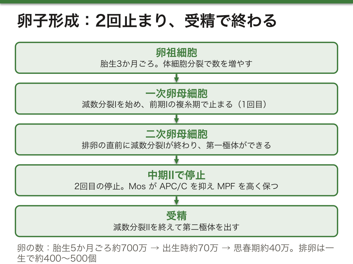
卵の細胞質分裂は不等である。アクチンとミオシンの収縮環でくびれるが、片方は大きな卵母細胞、もう片方は小さな極体になる。極体の役目は染色体だけを捨てて、細胞質を卵に残すことである。受精後の初期発生は、卵に蓄えられた細胞質の物資でまかなわれるからである。
止まる仕組みにはMPF(CDK1–サイクリンB)がかかわる。MPFが高まると染色体の凝縮、核膜の崩壊、紡錘体の形成が起き、減数分裂が進む。中期IIで止まり続けるには、MPFを高いまま保ち、サイクリンBの分解を防ぐ必要がある。ここで働くのがMosというキナーゼで、MEK・ERK・Rskを介してAPC/Cを抑え、後期への移行を止めている。
なぜ母親の年齢が上がると、子の染色体の数の異常(21トリソミーなど)が増えるのか。卵は前期Iで止まったまま、数十年も相同染色体どうしを交叉の部分(キアズマ)とコヒーシンでつなぎ止めておかなければならない。この結合は年とともに弱まり、ようやく分裂するときに染色体が正しく分かれない(不分離)危険が高くなる。21トリソミーの余分な染色体の多くが母親由来であることと合う。
English version
Core
Oogenesis has a proliferation period, a growth period and a maturation period.
In the proliferation period (around fetal month 3), PGCs become oogonia and multiply by mitosis. In the growth period (fetal months 4–6), oogonia become primary oocytes; their number peaks at about 7 million around month 5, and then degeneration (atresia) begins. The maturation period starts with meiosis I at fetal months 7–8, triggered by a rise in retinoic acid. Primary oocytes stop at the diplotene stage of prophase I, and follicular cells surround them to form primordial follicles.
About 700,000 remain at birth and about 400,000 at puberty. From puberty, several start to mature each month, but usually only one reaches ovulation; about 400–500 eggs are ovulated in a lifetime. Meiosis I ends just before ovulation, giving a secondary oocyte and the first polar body. The secondary oocyte stops again at metaphase II and finishes meiosis II, releasing the second polar body, only after fertilisation.
Going further
Cytokinesis in the egg is unequal. An actin–myosin contractile ring pinches the cell, but one side becomes a large oocyte and the other a small polar body. The polar body’s job is to throw away chromosomes while leaving the cytoplasm in the egg, because early development after fertilisation runs on materials stored in the egg cytoplasm.
The arrests involve MPF (CDK1–cyclin B). When MPF rises, chromosomes condense, the nuclear envelope breaks down, the spindle forms, and meiosis moves on. To stay arrested at metaphase II, MPF must stay high and cyclin B must not be destroyed. The kinase Mos does this: through MEK, ERK and Rsk it holds back APC/C and blocks the move to anaphase.
In the clinic
Why do chromosome number abnormalities in children (such as trisomy 21) increase with the mother’s age? The egg stays in prophase I for decades and must keep homologous chromosomes joined by crossover points (chiasmata) and cohesin all that time. These links weaken with age, so when the cell finally divides, the risk that chromosomes fail to separate correctly (nondisjunction) is higher. This fits with the fact that the extra chromosome 21 is usually from the mother.
卵胞の発育 — 卵を育てる部屋が、1か月で大きく変わるFolliculus ovaricus
卵母細胞は単独ではなく、周りの細胞と卵胞をつくって育つ。
| 段階 | つくり |
|---|---|
| 原始卵胞 | 一次卵母細胞を扁平な卵胞細胞が1層囲む(胎児期からある) |
| 一次卵胞 | 一次卵母細胞、透明帯、1層の卵胞細胞 |
| 二次卵胞(発育卵胞) | 卵胞細胞が数層に増え、液のたまる卵胞腔が広がり始める |
| 成熟卵胞(Graaf卵胞) | 卵胞腔がさらに大きくなり、卵を囲む放線冠と、卵を壁につなぐ卵丘ができる |
思春期以降、毎月5〜15個ほどの卵胞が発育を始めるが、成熟して排卵するのはふつう1つだけである。排卵しなかったものは退化する。排卵後の卵胞は黄体になり、ホルモンを分泌する。
卵胞の細胞は2種類に分かれる。顆粒膜細胞は性ホルモン(エストロゲン、のちにプロゲステロン)を作り、FSHやLHに反応し、いろいろな増殖因子を出す。外側の莢膜細胞のうち、内莢膜はアンドロゲンとプロゲステロンを作り、外莢膜は結合組織として排卵に働く。
内莢膜が作るアンドロゲンを、顆粒膜細胞がアロマターゼでエストロゲンに変える。この2つの細胞の分業が卵巣のエストロゲン合成の基本である。
なぜ毎月たくさん育ち始めるのに、1つしか残らないのか。完全にはわかっていない。発育の進んだ卵胞ほどFSHへの感受性が高くなり、FSHが下がっても生き残れる、という説明がある。下垂体と卵巣のホルモン、増殖因子の複雑なフィードバックで選ばれると考えられている。
不妊治療の卵巣刺激では、FSHを外から与えて、本来なら退化するはずの卵胞もまとめて育てる。そのため卵が多く採れる反面、卵巣が腫れて腹水がたまる卵巣過剰刺激症候群が起きることがある。
English version
Core
The oocyte does not grow alone; it grows inside a follicle with the cells around it.
| Stage | Structure |
|---|---|
| Primordial follicle | A primary oocyte surrounded by one layer of flat follicular cells (present from fetal life) |
| Primary follicle | Primary oocyte, zona pellucida, one layer of follicular cells |
| Secondary (growing) follicle | Several layers of follicular cells; a fluid-filled antrum begins to form |
| Mature (Graafian) follicle | A larger antrum; the corona radiata around the egg and the cumulus oophorus joining it to the wall |
From puberty, about 5–15 follicles start to grow each month, but usually only one matures and ovulates. The rest degenerate. After ovulation, the follicle becomes the corpus luteum, which secretes hormones.
Going further
Follicle cells are of two kinds. Granulosa cells make sex hormones (oestrogen, later progesterone), respond to FSH and LH, and release various growth factors. Of the outer theca cells, the theca interna makes androgens and progesterone, and the theca externa is connective tissue that helps with ovulation.
Granulosa cells convert the androgens from the theca interna into oestrogen with aromatase. This division of labour between two cell types is the basis of oestrogen production in the ovary.
Why do many start to grow each month, but only one remains? This is not fully known. One explanation is that more advanced follicles become more sensitive to FSH and can survive when FSH falls. The choice is thought to be made by complex feedback between pituitary and ovarian hormones and growth factors.
In the clinic
In ovarian stimulation for infertility treatment, FSH is given from outside so that follicles that would normally degenerate also grow. Many eggs can be collected, but ovarian hyperstimulation syndrome, with swollen ovaries and fluid in the abdomen, can occur.
精子形成 — つながったまま分裂し、細胞質を捨てて完成するSpermatogenesis
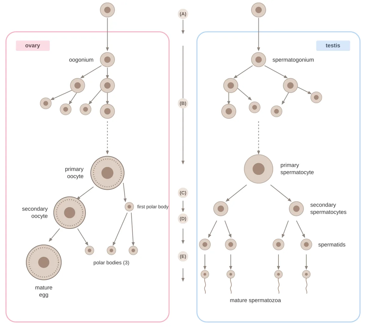
精子形成は精巣の精細管で、思春期から一生続く。1回の精子形成には64〜72日かかる。管の外側(基底側)から中心(内腔)に向かって進み、増殖期・成長期・成熟期・精子完成期に分けられる。
増殖期では、A型精祖細胞(2n)が体細胞分裂をくり返してB型精祖細胞になる。成長期(思春期から)にB型精祖細胞が少し大きくなって一次精母細胞になるが、卵に比べると成長はわずかである。成熟期で2回の減数分裂(約22〜24日)を経て精子細胞ができる。最後の精子完成(精子形成の仕上げ)では、ゴルジ装置から先体ができ(ヒアルロニダーゼやアクロシンなどの分解酵素を含む)、核が凝縮して形が変わり、遠位の中心小体から鞭毛の微小管が伸び、ミトコンドリアが中片部にらせん状に巻きつき、余分な細胞質が捨てられる。
精細管のSertoli細胞は、密着結合で血液精巣関門をつくり、栄養を与え、液やタンパク質を分泌し、精子形成の進行をそろえる。
精子のもとの細胞は細胞質分裂を最後まで終えない。同じ大きさの細胞に分かれるが、細胞質の橋でつながった合胞体のまま進み、精子完成の終わりにようやく切り離される。なぜつながっている必要があるのか。減数分裂の後、精子細胞の半分はX染色体を、半分はY染色体をもつ。X染色体には精子に必須の遺伝子(尾の形成に要るAKAP82など)がある。橋で細胞質を共有していれば、Y染色体をもつ精子細胞にもXの遺伝子の産物が届く。
胎児の精巣で生殖細胞が減数分裂に入らない理由も、マウスではわかってきた。周りの組織が作る減数分裂の誘導物質レチノイン酸を、Sertoli細胞の酵素CYP26B1が分解し、NANOS2も減数分裂を抑える。ヒトでの仕組みはまだはっきりしない。
| 卵子形成 | 精子形成 | |
|---|---|---|
| 期間 | 出生前に始まり、閉経で終わる | 思春期に始まり、一生続く |
| 時期の数 | 増殖・成長・成熟の3つ | 精子完成を加えた4つ |
| 1個の母細胞から | 卵1個と極体 | 精子4個 |
| 細胞質分裂 | 不等だが完全 | 等分だが不完全(合胞体) |
| 数 | 毎月ふつう1個 | 一度に膨大な数 |
男性不妊には、精液に精子が無い無精子症、数が少ない乏精子症(1 mLあたり2,000万未満とされてきた)、精細管にSertoli細胞しか無いSertoli細胞単独症候群などがある。Kartagener症候群は線毛のダイニン腕の遺伝子の変異による原発性線毛運動不全症の一部で、気道の感染をくり返し、内臓逆位を伴い、精子の鞭毛が動かずに不妊になる。
English version
Core
Spermatogenesis takes place in the seminiferous tubules of the testis and continues from puberty for life. One round takes 64–72 days. It moves from the outer (basal) side of the tubule towards the lumen and has a proliferation, growth, maturation and spermiogenesis period.
In the proliferation period, type A spermatogonia (2n) divide by mitosis and give type B spermatogonia. In the growth period (from puberty), type B spermatogonia grow a little into primary spermatocytes, much less than eggs grow. In the maturation period, two meiotic divisions (about 22–24 days) give spermatids. In the final step, spermiogenesis, the acrosome forms from the Golgi apparatus (it contains enzymes such as hyaluronidase and acrosin), the nucleus condenses and changes shape, microtubules of the flagellum grow from the distal centriole, mitochondria wind in a spiral around the midpiece, and extra cytoplasm is shed.
Sertoli cells in the tubule form the blood–testis barrier with tight junctions, give nutrition, secrete fluid and proteins, and keep spermatogenesis in step.
Going further
The cells that make sperm do not finish cytokinesis. They split into cells of equal size but stay joined as a syncytium by cytoplasmic bridges, which are cut only at the end of spermiogenesis. Why must they stay connected? After meiosis, half the spermatids carry an X chromosome and half carry a Y. The X carries genes essential for sperm (such as AKAP82, needed for the tail). By sharing cytoplasm through bridges, spermatids with a Y chromosome also receive the products of X genes.
Why germ cells in the fetal testis do not enter meiosis is now partly known in mice. The meiosis-inducing signal retinoic acid, made by nearby tissues, is broken down by the Sertoli cell enzyme CYP26B1, and NANOS2 also blocks meiosis. The mechanism in humans is still unclear.
| Oogenesis | Spermatogenesis | |
|---|---|---|
| Time span | Starts before birth, ends at menopause | Starts at puberty, lasts for life |
| Periods | Three: proliferation, growth, maturation | Four, adding spermiogenesis |
| From one mother cell | One egg and polar bodies | Four sperm |
| Cytokinesis | Unequal but complete | Equal but incomplete (syncytium) |
| Number | Usually one per month | Huge numbers at a time |
In the clinic
Male infertility includes azoospermia (no sperm in semen), oligozoospermia (low count; traditionally under 20 million per mL), and Sertoli cell-only syndrome (only Sertoli cells in the tubules). Kartagener syndrome is part of primary ciliary dyskinesia, caused by variants in genes for ciliary dynein arms. It brings repeated airway infections and situs inversus, and the sperm flagellum cannot move, causing infertility.
受精の分子機構 — 1つの精子だけを入れて、扉を閉じるFertilisatio
受精は決まった順番で進む。
①認識と結合:精子は卵を囲む放線冠の細胞の間を通り抜け、卵の透明帯のタンパク質に結合する。透明帯はZP1〜ZP4などの糖タンパク質でできており、マウスの研究ではZP3が最初の結合に、ZP2がその後の結合にかかわり、ZP1は透明帯の構造を保つとされてきた。ヒトではZP2の役割が重視されている。
②先体反応:先体が開口分泌を起こし、分解酵素を透明帯に放出して、精子が透明帯を掘り進む。
③膜の融合:精子の膜のタンパク質(IZUMO1)が卵の膜の受容体(JUNO)に結合し、2つの細胞膜が融合して精子が卵に入る。
④卵の活性化:融合をきっかけに卵の細胞質でCa2+濃度が上昇する。これがAPC/Cを働かせて中期IIで止まっていた減数分裂を終わらせ、同時に表層粒の開口分泌(表層反応)を起こす。放出された酵素が透明帯を硬く変え、ほかの精子が入れなくなる。
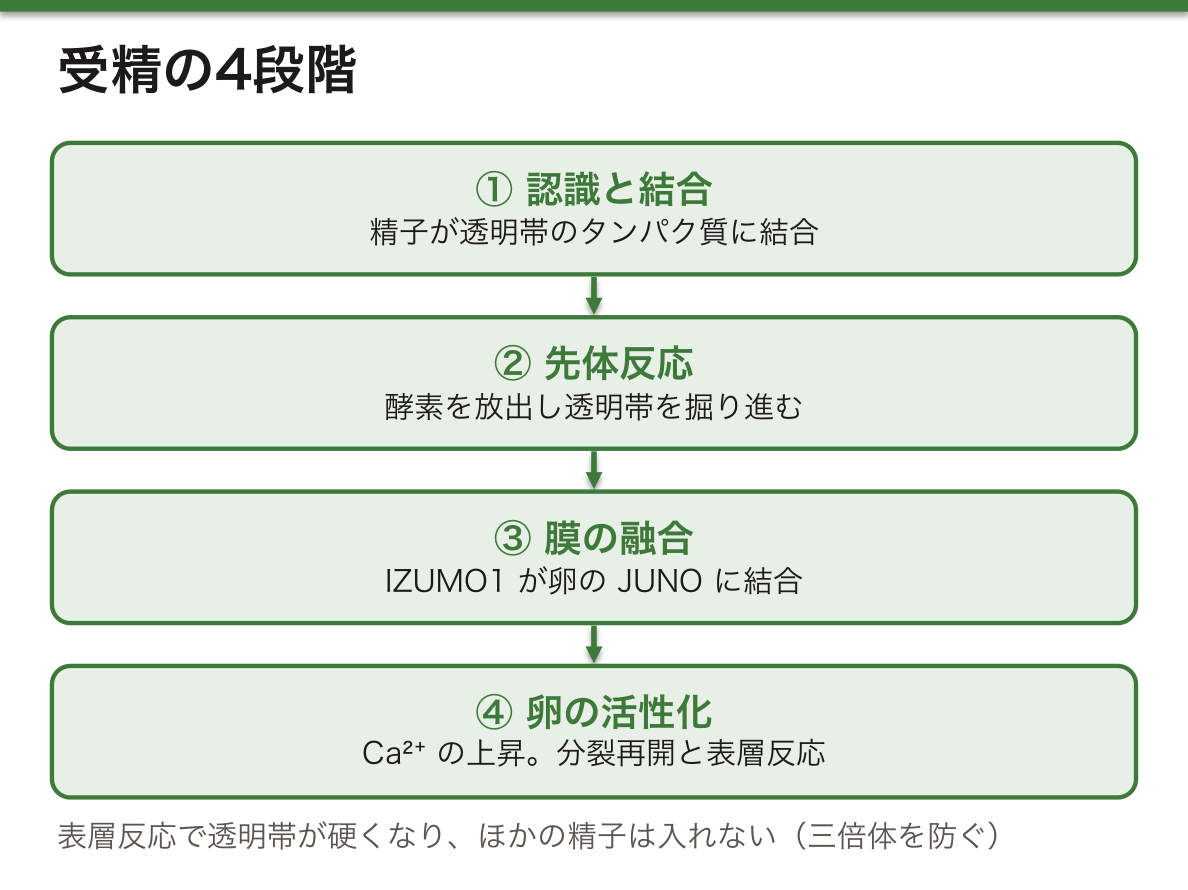
なぜ精子は1つしか入ってはいけないのか。2つ入れば染色体が3組になり、三倍体(69本)になって正常に発生できない。Ca2+の波は、1つ目の精子が入った瞬間に「扉を閉じる」合図と「止まっていた分裂を再開する」合図を同時に出す、うまい仕組みである。
精子が入ったあと、精子の鞭毛の根元の基底小体が分かれて受精卵の中心小体になる(ヒトでは中心体は父親から来る)。精子と卵の核はそれぞれ前核になって近づき、合わさる。そこから卵割(細胞の大きさを増やさずにくり返す体細胞分裂)が始まる。受精の意味は、①卵の細胞質を変化させて発生を始めさせる、②二倍体の染色体数を回復する、③父と母の染色体を混ぜ合わせる、の3つにまとめられる。
卵細胞質内精子注入法(ICSI)は、1つの精子を針で直接卵の中に入れる方法で、透明帯への結合や先体反応の段階を飛ばせる。精子の数や運動性がきわめて低い男性不妊で使われる。逆に、自然の受精で2つの精子が入る二精子受精は、ヒトの三倍体の最も多い原因である。
English version
Core
Fertilisation goes in a fixed order.
(1) Recognition and binding: the sperm passes between the cells of the corona radiata and binds proteins of the egg’s zona pellucida. The zona is made of glycoproteins such as ZP1–ZP4. Studies in mice suggested that ZP3 handles the first binding, ZP2 later binding, and ZP1 keeps the zona’s structure. In humans, the role of ZP2 is now stressed.
(2) Acrosome reaction: the acrosome undergoes exocytosis and releases its enzymes into the zona, so the sperm can dig through.
(3) Membrane fusion: a sperm membrane protein (IZUMO1) binds a receptor on the egg membrane (JUNO); the two membranes fuse and the sperm enters the egg.
(4) Egg activation: fusion causes a rise in Ca2+ in the egg cytoplasm. This activates APC/C so that meiosis, paused at metaphase II, is completed, and at the same time causes exocytosis of cortical granules (the cortical reaction). The released enzymes harden the zona so that no other sperm can enter.
Going further
Why must only one sperm enter? If two enter, there are three sets of chromosomes, a triploid (69 chromosomes), and the embryo cannot develop normally. The Ca2+ wave is a clever system: the moment the first sperm enters, it gives both the signal to “close the door” and the signal to “restart the paused division”.
After entry, the basal body at the base of the sperm flagellum divides and becomes the centrioles of the zygote (in humans, the centrosome comes from the father). The nuclei of sperm and egg each become a pronucleus; they move together and combine. Then cleavage begins: repeated mitotic divisions without growth in total size. Fertilisation does three things: (1) changes the egg cytoplasm and starts development, (2) restores the diploid chromosome number, and (3) mixes the father’s and mother’s chromosomes.
In the clinic
Intracytoplasmic sperm injection (ICSI) puts a single sperm directly into the egg with a needle, skipping zona binding and the acrosome reaction. It is used in male infertility with very low sperm count or motility. On the other hand, dispermy, entry of two sperm in natural fertilisation, is the most common cause of triploidy in humans.
まとめ
| 始原生殖細胞 — 卵と精子のもとは、体の外側から旅してくる | 卵子と精子のもとになる細胞は、生殖腺の中で生まれるのではない。 |
|---|---|
| 性の決定 — SRYが短い時間だけSOX9を押し上げる | 未分化の生殖腺は、皮質と髄質をもち、どちらの性にもなれる。 |
| 生殖細胞のエピジェネティクス — 親の印を消して、自分の性の印を付け直す | 体細胞では、受精卵から受け継いだインプリンティング(親由来で決まる遺伝子の印)は一生保たれなければならない。 |
| 卵子形成 — 胎児期に始まり、2回止まり、受精で終わる | 卵子形成は増殖期・成長期・成熟期に分けられる。 |
| 卵胞の発育 — 卵を育てる部屋が、1か月で大きく変わる | 卵母細胞は単独ではなく、周りの細胞と卵胞をつくって育つ。 |
| 精子形成 — つながったまま分裂し、細胞質を捨てて完成する | 精子形成は精巣の精細管で、思春期から一生続く。 |
| 受精の分子機構 — 1つの精子だけを入れて、扉を閉じる | 受精は決まった順番で進む。①認識と結合:精子は卵を囲む放線冠の細胞の間を通り抜け、卵の透明帯のタンパク質に結合する。 |
このノートは医学生が授業の内容を自分の言葉でまとめたもので、診療の判断に使うものではありません。誤りに気づいたらお知らせください。