内分泌腺は導管を持たない腺である。発生の途中で上皮が落ち込み、導管が退化して分泌物を血中へ直接出すようになった。だから組織像には共通の特徴がある——細胞の塊が毛細血管(多くは有窓性)に密に取り巻かれている。
読むときの手順は3つ。
- 細胞がどう並ぶか——索状か、濾胞(袋)を作るか、島状か
- 何を貯めるか——甲状腺だけがホルモンを細胞の外(コロイド)に大量に貯めるという例外的な作りをしている
- 何から発生したか——同じ器官の中に由来の違う組織が同居していることがある(副腎、下垂体、甲状腺)
由来が違えば病気も違う。副腎皮質の腫瘍と髄質の腫瘍がまったく別の病気である理由は、そもそも別の器官が合体しているからである。
- 下垂体
- 甲状腺
- 副甲状腺 — 4つの小さな腺と咽頭嚢の交叉
- 副腎
- 膵島(Langerhans 島)
- 松果体と下垂体後葉 — 神経がそのままホルモンを出す2つの器官
- 濾胞の形を見れば、甲状腺が働いているかどうかが分かる
下垂体Hypophysis
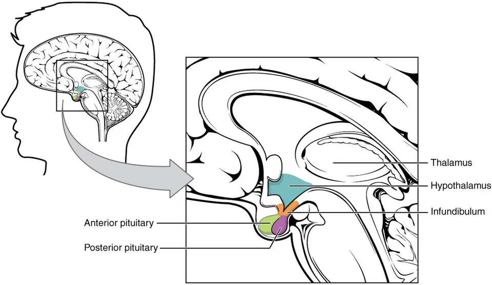
由来の異なる2つの器官が合体している。
- 前葉(腺性下垂体)——口腔外胚葉(Rathke 嚢)由来。索状に並ぶ内分泌細胞と類洞。染色性で酸好性細胞(成長ホルモン・プロラクチン)、塩基好性細胞(甲状腺刺激ホルモン・副腎皮質刺激ホルモン・性腺刺激ホルモン)、嫌色素性細胞に分かれる
- 後葉(神経性下垂体)——間脳底部由来。細胞体は視索上核・室傍核にあり、後葉にあるのは軸索の末端とグリア(下垂体細胞)だけ
前葉は下垂体門脈系で、後葉は神経で視床下部に支配される。
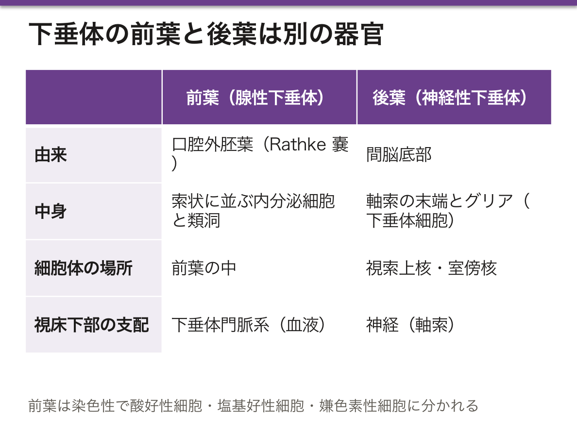
支配の様式の違いが、そのまま病態の違いになる。
前葉は視床下部で作られた放出ホルモンが、正中隆起の一次毛細血管網から下垂体門脈を通って前葉の二次毛細血管網に届くという遠回りをする。2度毛細血管を通るため、微量のホルモンを薄めずに届けられる一方で、血流が途絶えると前葉だけが壊死する。分娩時大量出血による Sheehan 症候群がこれである。
後葉のホルモン(オキシトシン・バソプレシン)は視床下部の神経細胞が作り、軸索を流れて後葉で放出される(神経分泌)。だから下垂体柄が切れると、切断部より上の細胞は生きていて、一時的な尿崩症のあと回復することがある。
「前葉は血で、後葉は神経で繋がっている」——この一行から、下垂体茎断裂・Sheehan 症候群・中枢性尿崩症の全部が導ける。
下垂体腺腫で最多はプロラクチノーマ。無月経・乳汁漏出・男性では性腺機能低下を起こす。手術より先にドパミン作動薬(カベルゴリン)を使うのは、プロラクチン分泌がドパミンによる持続的な抑制で制御されている唯一のホルモンだからである。
同じ理由で、下垂体柄が腫瘍に圧迫されるとプロラクチンだけが上がる(抑制が届かなくなる)。他のホルモンは下がり、プロラクチンだけ上がるという組み合わせを見たら茎の障害を疑う。
腫瘍が上方へ伸びると視交叉を圧迫し、鼻側網膜からの交叉線維が障害されて両耳側半盲になる。
English version
Core
Two organs of different origin fused together.
- Anterior lobe (adenohypophysis) — from oral ectoderm (Rathke’s pouch). Endocrine cells arranged in cords with sinusoids. By staining they divide into acidophils (growth hormone, prolactin), basophils (TSH, ACTH, gonadotrophins) and chromophobes.
- Posterior lobe (neurohypophysis) — from the floor of the diencephalon. The cell bodies lie in the supraoptic and paraventricular nuclei; the posterior lobe contains only the axon terminals and glia (pituicytes).
The anterior lobe is governed by the hypophyseal portal system, the posterior lobe by nerves.
Going further
The difference in the mode of control becomes directly the difference in the disease.
For the anterior lobe, releasing hormones made in the hypothalamus take a roundabout route from the primary capillary plexus of the median eminence, through the portal veins, to the secondary plexus of the anterior lobe. Passing through capillaries twice allows minute amounts to be delivered undiluted; but if the blood supply is interrupted, the anterior lobe alone infarcts. This is Sheehan’s syndrome after massive obstetric haemorrhage.
The posterior hormones (oxytocin, vasopressin) are made by hypothalamic neurones and flow down the axons to be released in the posterior lobe (neurosecretion). So when the pituitary stalk is severed, the cells above the cut are alive, and a transient diabetes insipidus may recover.
The anterior lobe is joined by blood, the posterior by nerve — from that one line, stalk transection, Sheehan’s syndrome and central diabetes insipidus can all be derived.
In the clinic
The commonest pituitary adenoma is a prolactinoma, causing amenorrhoea and galactorrhoea, and hypogonadism in men. A dopamine agonist (cabergoline) is used before surgery, because prolactin is the only hormone controlled by continuous inhibition — by dopamine.
For the same reason, when the stalk is compressed by a tumour, prolactin alone rises (the inhibition no longer arrives). Seeing the combination of every other hormone falling while prolactin rises, suspect a lesion of the stalk.
Extending upwards, the tumour compresses the optic chiasm, damaging the crossing fibres from the nasal retinae and giving bitemporal hemianopia.
甲状腺Glandula thyroidea
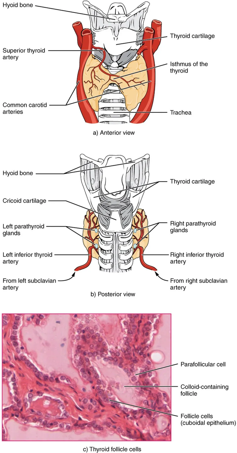
濾胞という袋を作る、内分泌腺のなかで例外的な構造を持つ。
- 濾胞上皮細胞——単層立方。サイログロブリンを作って内腔へ出す
- コロイド——濾胞の内腔を満たす。ホルモンの貯蔵庫。ここに数か月分が貯まる
- 傍濾胞細胞(C細胞)——濾胞の外側、間質側にある。カルシトニンを出す。神経堤由来で、他とは出自が違う
ホルモンを細胞の外に貯めるのは甲状腺だけである。
濾胞上皮の高さが機能を映す。刺激されている腺では上皮が丈が高く(円柱状に)なり、コロイドを取り込むため周辺に吸収空胞が並ぶ。逆に休んでいる腺では上皮が扁平になり、コロイドが充満する。顕微鏡で見た形が、そのまま機能状態の報告になっている。
作り方も独特である。ヨウ素をナトリウム・ヨウ素共輸送体で取り込み、ペルオキシダーゼでサイログロブリンのチロシンに結合させ、いったんコロイドとして貯蔵し、必要な分だけ細胞が飲み戻して分解しT4・T3 を出す。いったん外に出してから回収するという二度手間が、大量貯蔵を可能にしている。
この経路がそのまま治療標的になる——抗甲状腺薬(チアマゾール)はペルオキシダーゼを阻害し、放射性ヨウ素は取り込み機構を使って濾胞細胞だけを内部から照射する。組織の作りが、薬の選択そのものである。
由来の違いが腫瘍の性格を決める。
- 乳頭癌(約90%)——濾胞上皮由来。すりガラス状核・核内細胞質封入体・砂粒体。リンパ行性に転移するが予後良好
- 濾胞癌——濾胞上皮由来。血行性に肺・骨へ。被膜浸潤の有無でしか腺腫と区別できないため、細胞診では確定できない
- 髄様癌——C細胞由来。カルシトニンとCEAが腫瘍マーカー、アミロイド沈着を伴う。RET 遺伝子変異による多発性内分泌腫瘍症2型で家族性に出る
髄様癌だけがまったく別の細胞から出るので、マーカーも遺伝形式も転移様式も違う。「甲状腺の癌」と一括りにできないのは、同じ器官に別の由来の細胞が同居しているからである。
English version
Core
It forms sacs — follicles — a structure exceptional among endocrine glands.
- Follicular epithelial cell — simple cuboidal. Makes thyroglobulin and releases it into the lumen.
- Colloid — fills the lumen. The store of hormone: some months’ worth is kept here.
- Parafollicular (C) cell — outside the follicle, on the stromal side. Secretes calcitonin. Neural crest derived, of different origin from the rest.
The thyroid is the only gland that stores its hormone outside the cell.
Going further
The height of the follicular epithelium mirrors the function. In a stimulated gland the epithelium becomes tall (columnar), and resorption vacuoles line the periphery as the colloid is taken up. In a resting gland the epithelium is flattened and the colloid is abundant. The shape seen down the microscope is itself a report of the functional state.
The way it is made is also peculiar. Iodide is taken up by the sodium–iodide symporter, bound by peroxidase to the tyrosines of thyroglobulin, stored for a time as colloid, and then only as much as is needed is drunk back into the cell, broken down, and released as T4 and T3. The double handling — putting it outside and then retrieving it — is what makes large-scale storage possible.
That pathway is itself the target of treatment — antithyroid drugs (thiamazole) inhibit the peroxidase, and radioactive iodine uses the uptake mechanism to irradiate the follicular cells alone from within. The architecture of the tissue is the choice of drug.
In the clinic
The difference in origin decides the character of the tumour.
- Papillary carcinoma (about 90 per cent) — from follicular epithelium. Ground-glass nuclei, intranuclear cytoplasmic inclusions, psammoma bodies. Metastasises by lymphatics but has a good prognosis.
- Follicular carcinoma — from follicular epithelium. Haematogenous spread to lung and bone. It can be distinguished from an adenoma only by capsular invasion, so cytology cannot make the diagnosis.
- Medullary carcinoma — from the C cell. Calcitonin and CEA are the markers, with amyloid deposition. Occurs familially in MEN2 from RET mutations.
Only the medullary carcinoma comes from an entirely different cell, so its markers, its inheritance and its route of metastasis all differ. "Thyroid cancer" cannot be treated as one thing, because cells of different origin live together in the one organ.
副甲状腺 — 4つの小さな腺と咽頭嚢の交叉
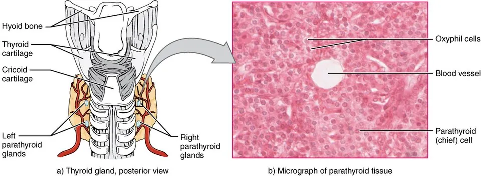
甲状腺の背面に上下2対(計4個)。米粒大。
- 主細胞——小型で明るい。副甲状腺ホルモンを分泌する主役
- 酸好性細胞——大型で好酸性。ミトコンドリアに富むが機能は不明瞭。思春期以降に増える
- 加齢とともに脂肪細胞が増える
発生は咽頭嚢から。下副甲状腺が第3咽頭嚢、上副甲状腺が第4咽頭嚢という逆転が起こる。
下が第3、上が第4という食い違いは、第3咽頭嚢由来の組織が胸腺と一緒に下へ引きずられるために起きる。上下が入れ替わるのはこのためで、丸暗記ではなく移動の副産物として理解できる。
この移動は臨床に直結する。下副甲状腺は位置の変異が大きく、縦隔まで下がって異所性に存在しうる。原発性副甲状腺機能亢進症で手術しても腫大腺が見つからない場合、縦隔を探すのはこのためである。
副甲状腺ホルモンの制御は負のフィードバックではなく、カルシウム受容体による直接感知である。主細胞の表面にあるカルシウム感知受容体が血中カルシウムを直に測り、下がれば分泌が増える。シナカルセトはこの受容体を感作させて「カルシウムが足りている」と錯覚させる薬で、二次性副甲状腺機能亢進症を手術せずに抑えられるのはこの仕組みによる。
甲状腺全摘の合併症としての低カルシウム血症が現実的な問題になる。副甲状腺は小さく色も甲状腺に似るため、一緒に取ってしまう/血流を絶ってしまうことが起こりうる。術後の口周囲のしびれ・手指のこわばり・テタニー(Chvostek 徴候、Trousseau 徴候)を見張る。
反対に原発性副甲状腺機能亢進症では高カルシウム血症により骨(線維性骨炎)・結石・腹部症状・精神症状が出る。整形外科の観点では骨吸収の亢進と骨嚢胞(褐色腫)が問題になり、骨密度低下の原因として内分泌を疑う入口になる。
English version
Core
Two pairs (four in all) on the back of the thyroid, the size of a grain of rice.
- Chief cell — small and pale. The one that secretes parathyroid hormone.
- Oxyphil cell — large and eosinophilic, rich in mitochondria, function unclear. Increases after puberty.
- Adipocytes increase with age.
They develop from the pharyngeal pouches, with the inversion that the inferior parathyroid comes from the third pouch and the superior from the fourth.
Going further
The discrepancy — inferior from the third, superior from the fourth — arises because the tissue from the third pouch is dragged downwards together with the thymus. That is why they exchange positions: it can be understood as a by-product of migration rather than learnt by rote.
That migration bears directly on the clinic. The inferior parathyroid varies greatly in position and may descend ectopically as far as the mediastinum. When surgery for primary hyperparathyroidism fails to find an enlarged gland, this is why the mediastinum is searched.
The control of parathyroid hormone is not a negative feedback but direct sensing by a calcium receptor. The calcium-sensing receptor on the surface of the chief cell measures blood calcium directly, and secretion rises when it falls. Cinacalcet sensitises that receptor so the cell is deceived into thinking calcium is sufficient, which is how secondary hyperparathyroidism can be suppressed without surgery.
In the clinic
Hypocalcaemia as a complication of total thyroidectomy is the practical problem. The parathyroids are small and similar in colour to the thyroid, so they may be removed with it or have their blood supply cut. Watch for perioral numbness, stiffness of the fingers and tetany (Chvostek’s and Trousseau’s signs) after the operation.
Conversely, in primary hyperparathyroidism, hypercalcaemia gives bone disease (osteitis fibrosa), stones, abdominal symptoms and psychiatric symptoms. From an orthopaedic point of view, increased bone resorption and bone cysts (brown tumours) are the problem, and this is the entry point for suspecting endocrine disease as a cause of low bone density.
副腎Glandula suprarenalis
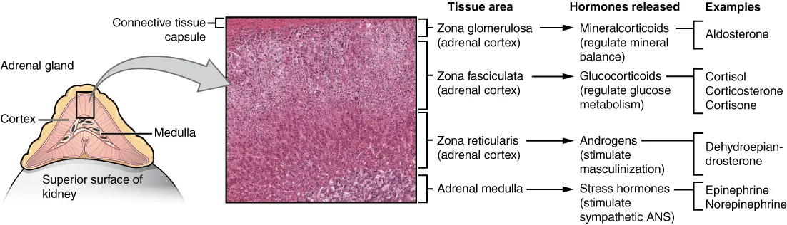
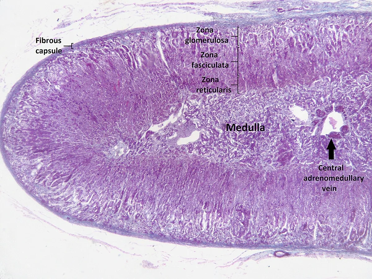
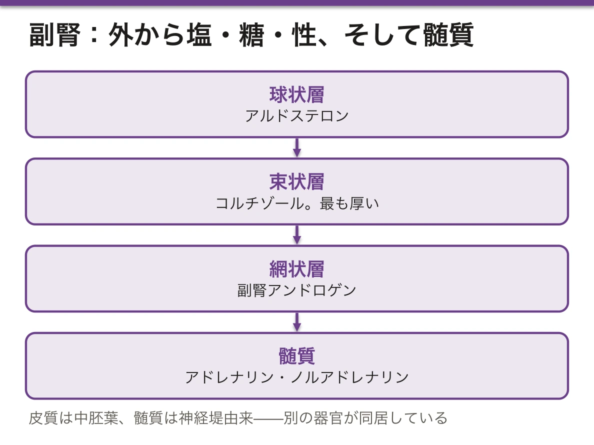
皮質(中胚葉由来)と髄質(神経堤由来)という、まったく別の器官が同居している。
皮質は外から3層。
- 球状層——アルドステロン(電解質コルチコイド)
- 束状層——コルチゾール。最も厚く、脂質を蓄えて明るく見える
- 網状層——副腎アンドロゲン
髄質はクロム親和性細胞がアドレナリン・ノルアドレナリンを出す。
外側から塩・糖・性の順と覚える。
髄質は交感神経節後細胞が変形したものである。軸索を伸ばす代わりにホルモンを血中へ出す細胞になったので、節前線維が直接シナプスする(内臓神経が副腎髄質に節を作らず入るのはこのため)。「副腎髄質は血管の中へ放出する交感神経節」と捉えると、ストレス応答が神経と内分泌で二重に走る理由が分かる。
皮質と髄質が隣り合うことにも意味がある。皮質を流れた高濃度のコルチゾールを含む血液が髄質へ流れ込み、ノルアドレナリンをアドレナリンに変える酵素(PNMT)を誘導する。アドレナリンを作れるのは副腎髄質だけである理由がこの位置関係にある。合体は偶然ではなく機能的な必然である。
層ごとの制御も違う。束状層と網状層は副腎皮質刺激ホルモンが、球状層はレニン・アンジオテンシン系とカリウムが支配する。だから下垂体不全では糖質コルチコイドは落ちるがアルドステロンは保たれ、原発性副腎不全(Addison 病)と違って高カリウム血症と色素沈着が出ない。
先天性副腎皮質過形成(多くは21水酸化酵素欠損症)は、この層構造から症状が読める。コルチゾールとアルドステロンが作れず、行き場を失った前駆体がアンドロゲンへ流れる。結果として女児の外性器男性化+塩喪失(低ナトリウム・高カリウム・脱水・ショック)が新生児期に出る。負のフィードバックが外れて副腎皮質刺激ホルモンが上がり続けるため過形成になる。
褐色細胞腫は髄質の腫瘍で、発作性の高血圧・頭痛・動悸・発汗。手術前に必ずα遮断を先に行う(β遮断を先にすると血管拡張が失われて血圧が急上昇する)。皮質と髄質でまったく別の病気になることが、そのまま二重起源の証明である。
English version
Core
Two entirely different organs living together — the cortex (mesodermal) and the medulla (neural crest).
The cortex has three zones from outside in.
- Zona glomerulosa — aldosterone (mineralocorticoid)
- Zona fasciculata — cortisol. The thickest; pale from stored lipid.
- Zona reticularis — adrenal androgens
The medulla contains chromaffin cells secreting adrenaline and noradrenaline.
Remember from outside in: salt, sugar, sex.
Going further
The medulla is a modified postganglionic sympathetic cell. Instead of extending an axon it became a cell that releases hormone into the blood, so the preganglionic fibres synapse on it directly (which is why the splanchnic nerves enter the adrenal medulla without forming a ganglion). Taking the adrenal medulla as a sympathetic ganglion that discharges into a blood vessel explains why the stress response runs twice over, by nerve and by hormone.
That cortex and medulla are adjacent also has meaning. Blood carrying a high concentration of cortisol from the cortex flows into the medulla and induces the enzyme that converts noradrenaline to adrenaline (PNMT). The reason only the adrenal medulla can make adrenaline lies in this positional relationship — the fusion is not accidental but functionally necessary.
The zones are also controlled differently. The fasciculata and reticularis are governed by ACTH, the glomerulosa by the renin–angiotensin system and by potassium. So in pituitary failure the glucocorticoids fall but aldosterone is preserved, and unlike primary adrenal failure (Addison’s disease) there is neither hyperkalaemia nor pigmentation.
In the clinic
In congenital adrenal hyperplasia (usually 21-hydroxylase deficiency) the symptoms can be read from this layered structure. Cortisol and aldosterone cannot be made, and the precursors with nowhere to go are diverted into androgens. The result in the newborn period is virilisation of the external genitalia in a girl together with salt wasting (hyponatraemia, hyperkalaemia, dehydration, shock). Negative feedback is lost so ACTH rises continuously, hence the hyperplasia.
Phaeochromocytoma is a tumour of the medulla, with paroxysmal hypertension, headache, palpitation and sweating. Alpha blockade must always come first before surgery (beta blockade first removes the vasodilatation and the pressure rises sharply).
That cortex and medulla give entirely different diseases is itself the proof of the dual origin.
膵島(Langerhans 島)Insula pancreatica
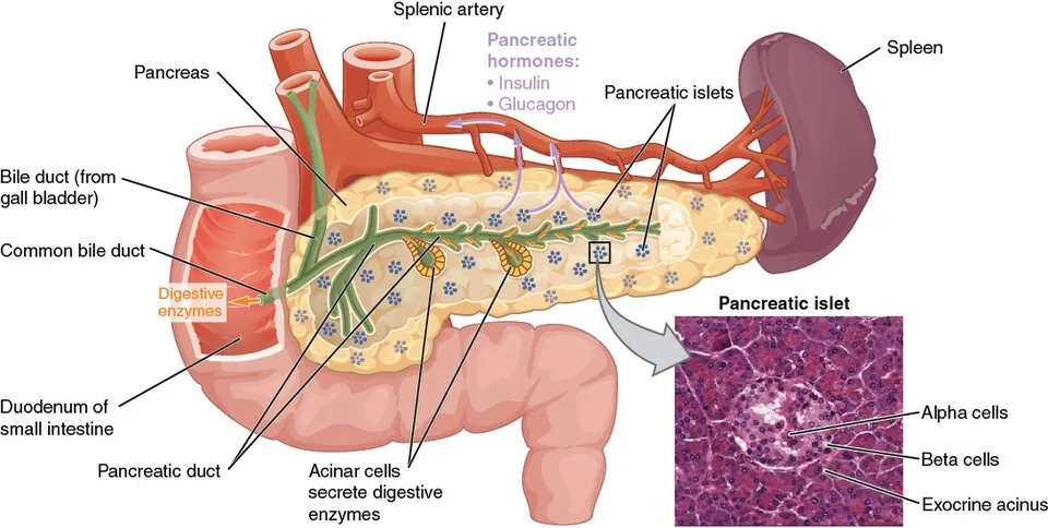
外分泌部(腺房)のなかに島のように散らばる内分泌組織。膵全体の1〜2%にすぎないが、血流は不釣り合いに多い。尾部に多い。
- B(β)細胞——約70%。インスリン。島の中心に多い
- A(α)細胞——約20%。グルカゴン。島の周辺
- D(δ)細胞——ソマトスタチン。両者を抑える
- PP細胞——膵ポリペプチド
周囲を有窓性毛細血管が密に取り巻く。
中心にB細胞、周辺にA細胞という配置は偶然ではない。島内の血流は中心から周辺へ流れるので、B細胞が出したインスリンが、下流にあるA細胞を浴びる。インスリンはグルカゴン分泌を抑えるため、血糖が上がれば自動的にグルカゴンも止まるという調節が、血流の向きだけで実現している。
この仕組みが壊れると説明がつく現象がある。1型糖尿病ではB細胞が失われるため、A細胞への抑制も同時に失われ、グルカゴンが不適切に出続けてケトアシドーシスを起こしやすい。インスリンが無いことだけでなく、グルカゴンが止まらないことが病態の半分を占める。
組織像も両型で異なる。1型では発症早期に膵島炎(リンパ球浸潤)が見られ、やがて島がB細胞を失って縮む。2型では島にアミリン由来のアミロイド沈着が蓄積し、B細胞が徐々に減る。同じ「糖尿病」でも顕微鏡の下では別の病気である。
インスリノーマは低血糖発作を起こす。Whipple の三徴(低血糖症状・血糖低値・糖投与で改善)に加え、インスリンと同時に C ペプチドが高いことが決め手になる。
C ペプチドはプロインスリンが切断されるときに等モルで出る副産物なので、自前で作ったインスリンにだけ付いてくる。外から打ったインスリンには含まれない。だからインスリン自己注射による低血糖(詐病・事故)ではCペプチドが低い——合成される過程を知っていることが、そのまま鑑別になる。
English version
Core
Endocrine tissue scattered like islands within the exocrine (acinar) tissue. Though only 1–2 per cent of the pancreas, its blood supply is disproportionately large. Commoner in the tail.
- B (β) cell — about 70 per cent. Insulin. Mostly at the centre of the islet.
- A (α) cell — about 20 per cent. Glucagon. At the periphery.
- D (δ) cell — somatostatin, which inhibits both.
- PP cell — pancreatic polypeptide.
Fenestrated capillaries surround it densely.
Going further
B cells at the centre and A cells at the periphery is not accidental. Blood within the islet flows from centre to periphery, so the insulin released by the B cells bathes the A cells downstream. Insulin inhibits glucagon secretion, so a rise in blood glucose automatically stops glucagon too — a regulation achieved by the direction of blood flow alone.
There is a phenomenon explained by that mechanism breaking. In type 1 diabetes the B cells are lost, so the inhibition of the A cells is lost with them; glucagon continues to be released inappropriately and ketoacidosis occurs readily. Not only the absence of insulin, but the failure of glucagon to stop, accounts for half the pathology.
The histology also differs between the two types. In type 1, insulitis (lymphocytic infiltration) is seen early and the islet then shrinks as it loses its B cells. In type 2, amyloid derived from amylin accumulates in the islet and the B cells fall gradually. The same "diabetes" is a different disease under the microscope.
In the clinic
An insulinoma causes attacks of hypoglycaemia. Beyond Whipple’s triad (symptoms of hypoglycaemia, a low blood glucose, relief with glucose), the decisive point is that C-peptide is high at the same time as insulin.
C-peptide is a by-product released in equimolar amount when proinsulin is cleaved, so it accompanies only insulin the body has made itself. Injected insulin contains none. So in hypoglycaemia from self-administered insulin (factitious or accidental) the C-peptide is low — knowing how the molecule is synthesised is itself the differential diagnosis.
松果体と下垂体後葉 — 神経がそのままホルモンを出す2つの器官Corpus pineale
内分泌腺の多くは上皮性の腺だが、この2つは神経組織そのものという点で例外になる。
| 松果体 | 下垂体後葉(神経下垂体) | |
|---|---|---|
| 由来 | 間脳の天井(神経外胚葉) | 間脳の底が下垂した神経外胚葉 |
| 細胞 | 松果体細胞+グリア(星状膠細胞様) | 神経分泌線維の終末+下垂体細胞〈pituicyte〉(グリア) |
| ホルモン | メラトニン | バソプレシン・オキシトシン |
| 作る場所 | 松果体細胞 | 視床下部(視索上核・室傍核)で作られ、軸索を流れて後葉で放出される |
| 特徴的所見 | 脳砂〈corpora arenacea〉——加齢とともに増える石灰化。X線・CTで正中の目印になる | Herring 小体——軸索内のホルモン貯蔵の膨大部 |
後葉は「腺」ではなく「終末」である——ホルモンを作っているのは視床下部の神経細胞で、後葉はその軸索が集まって放出する場所にすぎない。だから下垂体茎が切れると後葉ホルモンが出なくなり、中枢性尿崩症になる。前葉とは支配の仕組みがまったく違う。
| 前葉(腺下垂体) | 後葉(神経下垂体) | |
|---|---|---|
| 由来 | Rathke 嚢(口腔外胚葉)——上へ伸びる | 神経外胚葉——下へ伸びる |
| 支配 | 下垂体門脈系(視床下部ホルモンが血流で届く) | 神経(軸索が直接来る) |
| 遺残からできる腫瘍 | 頭蓋咽頭腫(Rathke嚢の遺残。小児・石灰化・嚢胞) | — |
メラトニンは「暗さ」で分泌される——網膜(内因性光感受性網膜神経節細胞)→ 視交叉上核〈体内時計〉→ 上頸神経節 → 交感神経を経て松果体に届く。光が当たると抑制されるので、夜間の照明とブルーライトが分泌を妨げる。時差ぼけ・交代勤務・非24時間睡眠覚醒障害にメラトニン受容体作動薬(ラメルテオン)が用いられる根拠。
小児では松果体腫瘍が中脳水道を圧迫して水頭症を起こし、上丘の圧迫で Parinaud 症候群(上方注視麻痺・対光近見反射解離)を来す。hCG産生腫瘍なら思春期早発症も伴う。
下垂体後葉は神経そのものなので、断たれれば分泌が止まる。
中枢性尿崩症 — 視索上核・室傍核から後葉へ届く軸索の障害。多尿・多飲・低比重尿で、デスモプレシンで反応することが腎性尿崩症との鑑別になる。下垂体茎より上の障害ほど重い——茎だけの離断なら断端から漏れ続けるので軽い。
ADH 不適合分泌症候群(SIADH) — 低ナトリウム血症・尿浸透圧が高い・volume は保たれる。小細胞肺癌の異所性産生が代表。補正が速すぎると浸透圧性脱髄症候群になるので1日 8〜10mEq/L 以内で直す。
松果体はメラトニンで概日リズムを作る。松果体部腫瘍は中脳上丘を圧迫して Parinaud 症候群(上方注視麻痺)を起こし、思春期早発症を伴うことがある。
English version
Core
Most endocrine glands are epithelial, but these two are exceptions in being nervous tissue itself.
| Pineal gland | Posterior pituitary (neurohypophysis) | |
|---|---|---|
| Origin | Roof of the diencephalon (neuroectoderm) | Floor of the diencephalon descending — neuroectoderm |
| Cells | Pinealocytes + glia (astrocyte-like) | Terminals of neurosecretory fibres + pituicytes (glia) |
| Hormone | Melatonin | Vasopressin, oxytocin |
| Made where | In the pinealocyte | In the hypothalamus (supraoptic and paraventricular nuclei); flows down the axon and is released in the posterior lobe |
| Characteristic finding | Brain sand (corpora arenacea) — calcification increasing with age; a midline marker on X-ray and CT | Herring bodies — axonal dilatations storing hormone |
Going further
The posterior lobe is not a gland but a terminal — the hormone is made by hypothalamic neurones and the posterior lobe is merely where their axons gather and release it. So if the stalk is cut, the posterior hormones cease and central diabetes insipidus follows.
The mechanism of control is entirely different from the anterior lobe.
| Anterior (adenohypophysis) | Posterior (neurohypophysis) | |
|---|---|---|
| Origin | Rathke’s pouch (oral ectoderm) — growing up | Neuroectoderm — growing down |
| Control | Hypophyseal portal system (hypothalamic hormones arrive by blood) | Nerve (axons arrive directly) |
| Tumour from a remnant | Craniopharyngioma (remnant of Rathke’s pouch; children, calcification, cysts) | — |
Melatonin is secreted in response to darkness — retina (intrinsically photosensitive retinal ganglion cells) → suprachiasmatic nucleus (the body clock) → superior cervical ganglion → sympathetic fibres to the pineal. Light inhibits it, so night-time lighting and blue light suppress secretion. This is the rationale for melatonin receptor agonists (ramelteon) in jet lag, shift work and non-24-hour sleep–wake disorder.
In children a pineal tumour compresses the cerebral aqueduct and causes hydrocephalus, and compression of the superior colliculi gives Parinaud’s syndrome (upgaze palsy, light–near dissociation). If it secretes hCG, precocious puberty accompanies it.
In the clinic
The posterior pituitary is nerve itself, so if it is cut off, secretion stops.
Central diabetes insipidus — damage to the axons running from the supraoptic and paraventricular nuclei to the posterior lobe. Polyuria, polydipsia, dilute urine; a response to desmopressin distinguishes it from the nephrogenic form. The higher above the stalk the lesion, the more severe — a transection of the stalk alone is mild, because hormone continues to leak from the cut end.
SIADH — hyponatraemia, a high urine osmolality, and preserved volume. Ectopic production by small cell lung cancer is the classic cause. Correcting too quickly causes osmotic demyelination, so keep within 8–10 mEq/L per day.
The pineal creates the circadian rhythm with melatonin. A pineal region tumour compresses the superior colliculi and gives Parinaud’s syndrome (upgaze palsy), and may be accompanied by precocious puberty.
濾胞の形を見れば、甲状腺が働いているかどうかが分かる
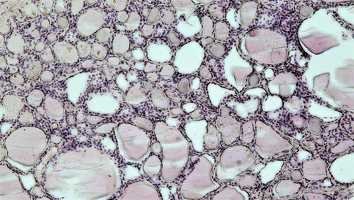
甲状腺は組織像がそのまま機能を語る数少ない内分泌腺である。コロイドを溜めておいて必要なときに取り込む、という仕組みのためである。
| 機能が低い/休んでいる | 機能が高い/刺激されている | |
| 濾胞上皮 | 扁平 | 円柱状に背が高くなる |
| コロイド | 濾胞いっぱいに充満し、濃く染まる | 減り、辺縁に吸収空胞(扇形空胞)が並ぶ |
| 濾胞の大きさ | 大きい | 小さい・不整 |
| 上皮の乳頭状突出 | 無い | あり(Basedow 病) |
「上皮が高く、コロイドが減り、空胞が並ぶ」=刺激されていると覚える。刺激の出どころが TSH でも TSH 受容体抗体でも、像は同じになる。
他の内分泌腺でも、同じ読み方ができる。
- 副腎皮質——ACTH で刺激されると束状帯の細胞から脂質が抜けて、淡明な細胞が緻密な好酸性の細胞に変わる。逆に外からステロイドを入れ続ければ萎縮する
- 副甲状腺——刺激されると脂肪細胞が減り、主細胞が増える。正常な副甲状腺は脂肪を多く含む
- 下垂体——標的腺が機能低下すると、その刺激ホルモンを作る細胞が肥大する(原発性甲状腺機能低下症で下垂体が腫れる)
⚠️ 「ホルモンを溜めておけるかどうか」が、内分泌腺の中で甲状腺だけ特殊である。ペプチドホルモンは顆粒として細胞内に、ステロイドは前駆体として置かれるが、甲状腺ホルモンだけは細胞の外(濾胞腔)に、数週間分が貯蔵されている。
だから抗甲状腺薬を始めても、効果が出るまで数週間かかる——既に作られて溜まっている分が使われるためである。組織の構造が、そのまま薬の効き始めの遅さを説明している。
甲状腺機能亢進症で、症状が改善するまで数週間かかることをあらかじめ伝える。その間はβ遮断薬で症状を抑える。
English version
Core
The thyroid is one of the few endocrine glands whose histology states its function directly, because of the arrangement by which colloid is stored and taken up when needed.
| Underactive / resting | Overactive / stimulated | |
|---|---|---|
| Follicular epithelium | Flattened | Tall, columnar |
| Colloid | Fills the follicle, stains densely | Reduced, with resorption (scalloped) vacuoles at the margin |
| Size of the follicles | Large | Small and irregular |
| Papillary infoldings of epithelium | Absent | Present (Graves’ disease) |
Remember: tall epithelium, reduced colloid, vacuoles in a row = it is being stimulated. Whether the stimulus is TSH or TSH receptor antibody, the picture is the same.
Going further
The same reading applies to other endocrine glands.
- Adrenal cortex — stimulated by ACTH, the fasciculata cells lose their lipid and the pale cells become compact and eosinophilic. Conversely, continued exogenous steroid causes atrophy.
- Parathyroid — when stimulated the adipocytes decrease and the chief cells increase. A normal parathyroid contains much fat.
- Pituitary — when a target gland fails, the cells making its trophic hormone hypertrophy (the pituitary enlarges in primary hypothyroidism).
⚠️ Whether the hormone can be stored is what makes the thyroid unique among endocrine glands. Peptide hormones are held within the cell as granules and steroids as precursors, but thyroid hormone alone is stored outside the cell, in the follicular lumen, in an amount sufficient for weeks.
So after starting an antithyroid drug it takes weeks for an effect, because what has already been made and stored is used up first. The architecture of the tissue explains directly why the drug is slow to begin working.
In the clinic
In hyperthyroidism, tell the patient in advance that it will take some weeks before the symptoms improve. Meanwhile use a beta blocker to control them.
まとめ
| 下垂体 | 由来の異なる2つの器官が合体している。前葉(腺性下垂体)——口腔外胚葉(Rathke 嚢)由来。 |
|---|---|
| 甲状腺 | 濾胞という袋を作る、内分泌腺のなかで例外的な構造を持つ。 |
| 副甲状腺 — 4つの小さな腺と咽頭嚢の交叉 | 甲状腺の背面に上下2対(計4個)。米粒大。 |
| 副腎 | 皮質(中胚葉由来)と髄質(神経堤由来)という、まったく別の器官が同居している。 |
| 膵島(Langerhans 島) | 外分泌部(腺房)のなかに島のように散らばる内分泌組織。 |
| 松果体と下垂体後葉 — 神経がそのままホルモンを出す2つの器官 | 内分泌腺の多くは上皮性の腺だが、この2つは神経組織そのものという点で例外になる。 |
| 濾胞の形を見れば、甲状腺が働いているかどうかが分かる | 甲状腺は組織像がそのまま機能を語る数少ない内分泌腺である。 |
このノートは医学生が授業の内容を自分の言葉でまとめたもので、診療の判断に使うものではありません。誤りに気づいたらお知らせください。