骨は石灰化した結合組織である。有機成分(I型コラーゲンが約90%)が引っ張りに耐え、無機成分(ハイドロキシアパタイト)が圧縮に耐える。鉄筋コンクリートに喩えられるのは正確で、鉄筋=コラーゲン、コンクリート=アパタイト。どちらが欠けても骨は弱くなるが、弱くなり方が違う。
ここを押さえると疾患が整理できる。両方が同時に減るのが骨粗鬆症(量の病気)、石灰化だけができないのが骨軟化症・くる病(質の病気)、コラーゲンが作れないのが骨形成不全症。骨密度・ALP・Ca・P の動き方がそれぞれ違うのは、壊れている場所が違うからである。
そして骨は一生作り替えられ続けている。「硬い=変わらない」ではない。破骨細胞が壊し、骨芽細胞が作る——この均衡の傾き方が、骨粗鬆症からPaget病、骨転移までを説明する。
- 骨(組織としての)
- 骨芽細胞
- 破骨細胞
- 骨細胞
- 緻密骨(皮質骨)
- 海綿骨(骨梁)
- 骨膜
- 膜内骨化
- 軟骨内骨化
- 骨リモデリング — 壊すと作るの均衡
- 骨折の治癒 — 組織が入れ替わる順序
- 骨は、標識を打てば「速さ」まで測れる
骨(組織としての)Os
基質が石灰化した結合組織。組成は有機35%(I型コラーゲン+オステオカルシンなど)と無機65%(ハイドロキシアパタイト Ca₁₀(PO₄)₆(OH)₂)。
働きは支持・保護・運動の梃子・造血の場・Ca と P の貯蔵庫。体内Caの約99%が骨にある。
軟骨と違い、骨には血管が豊富に入る。だから骨折は治る。
「有機と無機のどちらが壊れたか」で骨の弱り方が変わる。
- 無機だけ抜ける(脱灰)→ 曲がるがしなやか。骨軟化症で骨が変形するのはこれ
- 有機だけ抜ける(加熱した骨)→ 硬いが脆く砕ける。骨形成不全症が易骨折を示すのはこちら
- 両方が減る→ 量が減るだけで質は正常。骨粗鬆症
骨がCa の貯蔵庫である点も重要で、血清Caは骨を犠牲にしてでも一定に保たれる。血清Caが正常だからといって骨が無事とは限らない——副甲状腺機能亢進症では血清Caを保つために骨が溶け続ける。
有機と無機の比率が、そのまま骨の壊れ方になる。
有機(コラーゲン)の異常=骨形成不全症。しなやかさを失って折れる。青色強膜・難聴・易骨折性。
無機(石灰化)の不足=くる病・骨軟化症。類骨は作られるが石灰化しないので軟らかく変形する。ビタミン D 欠乏が主因で、小児では O 脚・肋骨念珠・頭蓋癆、成人では偽骨折(Looser 帯)。ALP 高値・低リン・低〜正常 Ca。
量そのものの減少=骨粗鬆症。石灰化した骨が量的に減るだけで質は正常なので、血液検査(Ca・P・ALP)は正常である——骨軟化症との決定的な違いで、骨密度で診断する。
骨は Ca の貯蔵庫でもある。血清 Ca は骨を犠牲にしてでも保たれる。
English version
Core
Connective tissue whose matrix is calcified. Composition: 35 per cent organic (type I collagen with osteocalcin and others) and 65 per cent inorganic (hydroxyapatite, Ca₁₀(PO₄)₆(OH)₂).
Functions: support, protection, levers for movement, the site of haemopoiesis, and the store of calcium and phosphate — about 99 per cent of body calcium is in bone.
Unlike cartilage, bone is richly supplied with vessels. That is why fractures heal.
Going further
Which component is lost changes how the bone fails.
- Only the mineral is removed (decalcified) → it bends but remains pliable. This is why bone deforms in osteomalacia.
- Only the organic component is lost (as in heated bone) → hard but brittle, and it shatters. This is why osteogenesis imperfecta presents with fractures.
- Both fall together → the quantity is reduced but the quality is normal. Osteoporosis.
That bone is the store of calcium matters too: the serum calcium is held constant even at the expense of the skeleton. A normal serum calcium does not mean the bone is intact — in hyperparathyroidism bone continues to dissolve in order to keep the serum level.
In the clinic
The ratio of organic to inorganic is directly the way the bone breaks.
Abnormal organic component (collagen) = osteogenesis imperfecta. It loses its pliability and snaps. Blue sclerae, deafness, fragility.
Insufficient mineral = rickets and osteomalacia. Osteoid is made but not mineralised, so the bone is soft and deforms. Vitamin D deficiency is the main cause: in children, bow legs, rachitic rosary and craniotabes; in adults, pseudofractures (Looser zones). High ALP, low phosphate, low to normal calcium.
Reduced quantity alone = osteoporosis. Mineralised bone is simply reduced in amount while its quality is normal, so the blood tests (Ca, P, ALP) are normal — the decisive difference from osteomalacia, and the reason it is diagnosed by bone density.
Bone is also the calcium store, and the serum calcium is maintained at the expense of the skeleton.
骨芽細胞Osteoblastus
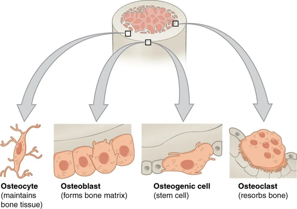
骨を作る細胞。間葉系幹細胞に由来する。骨の表面に立方形で並び、類骨(osteoid:まだ石灰化していない有機基質)を分泌したのち石灰化させる。
粗面小胞体が発達して細胞質が好塩基性。ALP(アルカリホスファターゼ)を強く発現するので、活動が上がると血清ALPが上昇する。
基質に埋め込まれると骨細胞 osteocytus になり、表面で休止すると骨被覆細胞になる。
血清ALPは骨形成の指標であり、この解釈が国試で繰り返し問われる。
- 上がる——骨形成が盛んなとき。成長期(生理的)・骨折治癒期・骨軟化症/くる病・Paget病・副甲状腺機能亢進症・造骨性の骨転移(前立腺癌)
- 上がらない——骨形成を伴わない骨破壊。多発性骨髄腫(骨芽細胞が抑制される)
骨粗鬆症では原則としてALPは正常。ここが骨軟化症との重要な鑑別点になる(骨軟化症は石灰化できない類骨が溜まり、骨芽細胞が代償的に働き続けるためALPが上がる)。
骨芽細胞は破骨細胞の司令塔でもある。RANKL を出して破骨細胞を活性化し、オステオプロテゲリン(OPG) を出してそれを抑える。骨吸収の量は骨芽細胞側が決めている——この一見逆説的な関係が、後述の薬物治療の標的になっている。
骨芽細胞の活動は ALP で測れる。骨芽細胞は ALP を出すので、骨形成が盛んな状態で ALP が上がる——成長期の小児・骨折の治癒期・骨転移(造骨性)・Paget 病・くる病。肝胆道疾患との鑑別にはγ-GTP を併せて見る。
骨肉腫 — 骨芽細胞由来の悪性腫瘍。10代の膝周囲(大腿骨遠位・脛骨近位)に好発する。腫瘍細胞が類骨を作ることが診断の要件で、X 線ではCodman 三角・日光放射状陰影。肺転移を来しやすい。
骨形成を促す薬 — テリパラチド(PTH 製剤)は間欠投与すると骨芽細胞を優位に働かせる。持続的に高い PTH(副甲状腺機能亢進症)では逆に骨吸収が進む——同じホルモンが、与え方で逆に働く。
English version
Core
The cell that makes bone, derived from mesenchymal stem cells. It lines the bone surface as a cuboidal layer, secretes osteoid (organic matrix not yet mineralised) and then mineralises it.
Rough ER is well developed, so the cytoplasm is basophilic. It strongly expresses alkaline phosphatase (ALP), so serum ALP rises when its activity increases.
Embedded in matrix it becomes an osteocyte; resting on the surface it becomes a bone lining cell.
Going further
Serum ALP is the index of bone formation, and its interpretation is asked repeatedly.
- Raised — when formation is active: growth (physiological), fracture healing, osteomalacia and rickets, Paget disease, hyperparathyroidism, osteoblastic metastases (prostate cancer).
- Not raised — bone destruction without accompanying formation: multiple myeloma, in which osteoblasts are suppressed.
In osteoporosis ALP is normal as a rule — the important point of distinction from osteomalacia, where unmineralisable osteoid accumulates and osteoblasts go on working in compensation, raising the ALP.
The osteoblast is also the commander of the osteoclast. It releases RANKL to activate osteoclasts and osteoprotegerin (OPG) to restrain them. The amount of resorption is decided from the osteoblast’s side — an apparently paradoxical relationship that is the target of the drug treatments described below.
In the clinic
The activity of osteoblasts can be measured as ALP. It rises whenever formation is active — childhood growth, fracture healing, osteoblastic metastases, Paget disease, rickets. Check γ-GT alongside to exclude a hepatobiliary source.
Osteosarcoma — a malignant tumour of osteoblastic origin, arising around the knee (distal femur, proximal tibia) in teenagers. That the tumour cells produce osteoid is a requirement for the diagnosis; radiographically, Codman triangle and a sunburst pattern. It metastasises readily to the lung.
Drugs that promote formation — teriparatide (a PTH preparation) favours the osteoblast when given intermittently. A persistently high PTH (hyperparathyroidism) does the opposite and drives resorption — the same hormone acting in opposite directions according to how it is given.
破骨細胞Osteoclastus
骨を壊す細胞。単球・マクロファージ系の細胞が融合してできる多核巨細胞。骨表面のくぼみ(Howship 窩)に座る。
骨に接する面に波状縁を作り、そこを封鎖帯で密閉して外界から隔てた小部屋を作る。そこへ
- H⁺(プロトンポンプ)→ 酸でアパタイトを溶かす
- カテプシンK → コラーゲンを分解する
を放出する。無機と有機を別々の道具で壊すのが要点。
破骨細胞の分化には骨芽細胞が出す RANKL と、マクロファージ由来の M-CSF が要る。PTH・活性型ビタミンD・IL-6・TNF-α は骨芽細胞のRANKL発現を上げることで間接的に骨吸収を増やす。破骨細胞にPTH受容体は無い——ここは頻出の落とし穴である。
この経路がそのまま治療標的になっている。
- ビスホスホネート——アパタイトに結合して骨に沈着し、吸収時に破骨細胞に取り込まれてアポトーシスを誘導。顎骨壊死・非定型大腿骨骨折が長期使用の問題
- デノスマブ——抗RANKL抗体。RANKLを中和する。中止すると反跳性に骨吸収が亢進する
- ロモソズマブ——抗スクレロスチン抗体。骨形成を上げ吸収を下げる
閉経後骨粗鬆症の本態はエストロゲン減少によるRANKL抑制の解除で、破骨細胞が増えて骨代謝回転が上がる。閉経後10年ほどで海綿骨が急速に失われるのはこのためである。
大理石骨病(骨硬化症)——破骨細胞の機能不全(炭酸脱水酵素II欠損やプロトンポンプ異常)。骨が吸収されず異常に硬くなるが、リモデリングできないので脆く、易骨折。骨髄腔が骨で埋まるため汎血球減少・髄外造血(肝脾腫)、頭蓋孔の狭窄で脳神経麻痺(視神経・顔面神経)。「硬い=丈夫」ではないことを示す代表例である。
English version
Core
The cell that destroys bone: a multinucleate giant cell formed by fusion of monocyte–macrophage lineage cells. It sits in a depression on the bone surface, a Howship lacuna.
On the surface facing bone it forms a ruffled border, sealed off from the exterior by a sealing zone to create an enclosed compartment, into which it releases:
- H⁺ (a proton pump) → acid dissolves the apatite.
- Cathepsin K → degrades the collagen.
The point is that mineral and organic matrix are destroyed by two separate tools.
Going further
Differentiation of the osteoclast requires RANKL from the osteoblast and M-CSF from macrophages. PTH, calcitriol, IL-6 and TNF-α increase resorption indirectly, by raising RANKL expression in the osteoblast. The osteoclast has no PTH receptor — a frequently examined trap.
This pathway is directly the target of treatment.
- Bisphosphonates — bind to apatite, deposit in bone, are taken into the osteoclast during resorption and induce apoptosis. Osteonecrosis of the jaw and atypical femoral fracture are the problems of long use.
- Denosumab — an anti-RANKL antibody. Stopping it causes rebound resorption.
- Romosozumab — an anti-sclerostin antibody; raises formation and lowers resorption.
Postmenopausal osteoporosis is essentially the release of RANKL from oestrogen restraint: osteoclasts increase and turnover rises. This is why cancellous bone is lost rapidly for about ten years after the menopause.
In the clinic
Osteopetrosis (marble bone disease) — failure of osteoclast function (carbonic anhydrase II deficiency, or a defective proton pump).
Bone is not resorbed and becomes abnormally dense, but because it cannot be remodelled it is brittle and fractures easily. The marrow cavity fills with bone, giving pancytopenia and extramedullary haemopoiesis (hepatosplenomegaly), and narrowing of the cranial foramina causes cranial nerve palsies (optic, facial).
The classic demonstration that hard does not mean strong.
骨細胞Osteocytus
骨基質に埋め込まれた骨芽細胞のなれの果て。骨小腔 lacuna に収まり、骨細管 canaliculus を通して細い突起を伸ばし、隣の骨細胞とギャップ結合で繋がる。
骨の細胞のうち最も数が多く(90%以上)、最も長生き。かつては「休眠した細胞」と見られていたが、現在は骨の力学センサーと理解されている。
骨細胞は骨小腔内の液の流れから力学的負荷を感知し、それに応じてリモデリングを指令する。負荷が減るとスクレロスチンを出して骨形成を抑える。
これがWolff の法則(骨は加わる力に応じて構造を変える)の細胞レベルの実体である。臨床上の帰結は大きい。
- 不動・臥床・無重力→ 荷重が消える → スクレロスチン増 → 廃用性骨萎縮。宇宙飛行士の骨量減少や長期臥床の骨折リスクはこれ
- 人工関節周囲の stress shielding——金属ステムが荷重を肩代わりすると、その周囲の骨が「要らない」と判断されて痩せる。インプラント設計上の中心問題であり、人工関節を扱うなら必ず出会う
ロモソズマブがスクレロスチンを標的にするのは、この経路を薬で押し戻すためである。
骨細胞が「荷重を感じる」センサーである。骨細管の中の液体の流れで力学的な刺激を感知し、スクレロスチンを出して骨形成を調節する。
だから使わない骨は減る。臥床・ギプス固定・無重力で廃用性骨萎縮が起こる。骨粗鬆症の予防に荷重運動が要るのは、骨細胞に「使っている」と伝える必要があるからである。
抗スクレロスチン抗体(ロモソズマブ)はこの仕組みを利用した骨粗鬆症治療薬である。
骨細胞が死ぬと骨は壊死する。大腿骨頭壊死では骨小腔が空になる(empty lacunae)ことが組織学的な決め手になる。ステロイド・アルコール・外傷が主な原因で、大腿骨頭は血流が終末的なぶん起こりやすい。
English version
Core
The osteoblast after it has been buried in matrix. It occupies a lacuna and extends fine processes through canaliculi, connected to neighbouring osteocytes by gap junctions.
It is the most numerous of the bone cells (over 90 per cent) and the longest lived. Once regarded as a dormant cell, it is now understood as the mechanosensor of bone.
Going further
The osteocyte senses mechanical load from the flow of fluid within its lacuna and directs remodelling accordingly. When load falls it releases sclerostin, which suppresses bone formation. This is the cellular substance of Wolff’s law — that bone adapts its structure to the forces applied to it.
The clinical consequences are large.
- Immobility, bed rest, weightlessness → load disappears → sclerostin rises → disuse osteopenia. The bone loss of astronauts and the fracture risk of prolonged bed rest.
- Stress shielding around a prosthesis — when a metal stem takes over the load, the surrounding bone judges itself unnecessary and thins. A central problem in implant design, and one that anyone dealing with joint replacement will meet.
Romosozumab targets sclerostin in order to push this pathway back with a drug.
In the clinic
The osteocyte is the sensor that feels load. It detects mechanical stimulus through fluid flow in the canaliculi and regulates formation by releasing sclerostin.
So bone that is not used diminishes. Bed rest, casting and weightlessness cause disuse osteopenia. Weight-bearing exercise is needed to prevent osteoporosis because the osteocyte has to be told that the bone is being used.
The anti-sclerostin antibody romosozumab exploits this mechanism.
When osteocytes die, the bone is necrotic. In osteonecrosis of the femoral head, empty lacunae are the histological hallmark. Corticosteroids, alcohol and trauma are the main causes, and the femoral head is susceptible because its blood supply is end-arterial.
緻密骨(皮質骨)Substantia compacta
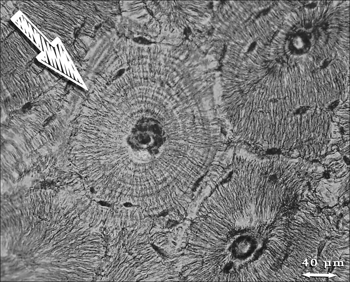
骨の外層をなす密な骨。基本単位が骨単位(オステオン、Havers 系)で、中心のHavers 管(血管・神経が通る)を同心円状の層板が取り巻く。
層板のあいだに骨小腔が並び、骨細管が中心管へ向かって放射状に伸びる——栄養が中心の血管から拡散で届く距離に、骨単位の太さが決まっている。
Volkmann 管が Havers 管どうしを横に繋ぎ、骨膜側・骨髄側と連絡する。
皮質骨は骨量の約80%を占めるが、表面積が小さいため代謝回転は遅い。逆に海綿骨は量が20%でも表面積が大きく回転が速い。
この差が骨粗鬆症で先に折れる場所を決める。代謝回転が速い海綿骨の多い部位——椎体・大腿骨頸部・橈骨遠位端が早期から脆くなり、これがそのまま脆弱性骨折の好発部位になる。「なぜ背骨と手首と股関節なのか」の答えは組織構成にある。
骨単位はリモデリングのたびに作り替えられる。古い骨単位が破骨細胞に削られ(cutting cone)、そのあとを骨芽細胞が埋める。削り残された古い層板が介在層板として骨単位のあいだに残り、標本ではこれが年輪のように見える。
皮質骨と海綿骨で、減り方も折れ方も違う。
海綿骨は表面積が大きく回転が速いので、骨粗鬆症では海綿骨から先に減る。だから椎体圧迫骨折・大腿骨頸部骨折・橈骨遠位端骨折という海綿骨の多い場所が好発部位になる。
骨密度は腰椎と大腿骨近位部で測る——変化が早く出る場所だからである。
Havers 管は血管の通り道なので、骨髄炎では管を通じて感染が広がる。血流が絶たれた骨片が腐骨(sequestrum)として残り、抗菌薬が届かないため難治になる。掻爬(デブリードマン)が必要なのはこのためである。
皮質骨は緻密で薬も届きにくい——骨髄炎の治療期間が長い(6週以上)根拠になっている。
English version
Core
The dense bone of the outer shell. Its basic unit is the osteon (Haversian system): concentric lamellae around a central Haversian canal carrying vessels and nerves.
Lacunae lie between the lamellae and canaliculi radiate towards the central canal — the diameter of an osteon is set by the distance over which nutrition can diffuse from the central vessel.
Volkmann canals connect Haversian canals transversely and communicate with the periosteal and medullary surfaces.
Going further
Cortical bone is about 80 per cent of bone mass but has a small surface area, so turnover is slow. Cancellous bone is only 20 per cent of the mass but has a large surface area and turns over rapidly.
That difference decides where fractures occur first in osteoporosis. The sites rich in rapidly turning-over cancellous bone — vertebral bodies, femoral neck, distal radius — become fragile early, and these are precisely the sites of fragility fracture. The answer to "why the spine, the wrist and the hip?" lies in the tissue composition.
Osteons are rebuilt at each remodelling cycle: an old osteon is cut away by osteoclasts (a cutting cone) and refilled by osteoblasts behind them. The remnants of older lamellae persist between osteons as interstitial lamellae, appearing on a slide like growth rings.
In the clinic
Cortical and cancellous bone differ in how they are lost and how they break.
Cancellous bone has a large surface area and rapid turnover, so it is lost first in osteoporosis. Hence the sites of predilection are those rich in cancellous bone: vertebral compression fracture, femoral neck fracture, distal radial fracture. Bone density is measured at the lumbar spine and proximal femur because that is where change appears earliest.
The Haversian canal is a route for vessels, so in osteomyelitis infection spreads through the canals. A fragment cut off from its blood supply remains as a sequestrum, which antibiotics cannot reach — which is why debridement is required. Cortical bone is dense and poorly penetrated by drugs, the reason osteomyelitis is treated for six weeks or more.
海綿骨(骨梁)Substantia spongiosa
骨の内部にある、骨梁 trabecula が三次元の網を作った構造。すきまは骨髄で満たされる。骨単位は作らず、層板が板状に走る。
骨端・椎体・扁平骨に多い。軽さと強さを両立させ、荷重を分散する。
骨梁は無作為に並んでいるのではなく、主応力の方向に沿って配列している。大腿骨近位部の骨梁が圧縮群と張力群に分かれて弓なりに走るのはその典型で、Wolff の法則の最も分かりやすい実例である。
骨粗鬆症では骨梁が細くなるだけでなく、繋がりが切れる(連結性の喪失)。1本切れると隣の骨梁に荷重が集中し、連鎖的に破綻する。骨密度が同じでも構造が壊れていれば折れやすい——これが「骨質」という概念であり、骨密度検査だけで骨折リスクを測りきれない理由である。
大腿骨近位部では骨梁の疎な領域(Ward 三角)が生じ、骨脆弱性の指標になる。
椎体骨折は海綿骨の破綻そのもので、約2/3は無症候のまま進む。気づかれずに身長低下・円背として現れ、後彎が進むと呼吸機能低下・逆流性食道炎・転倒リスク増につながる。1つ椎体骨折が起きると次の骨折リスクが数倍になるため、続発を防ぐ薬物治療の開始点になる。
English version
Core
The internal structure in which trabeculae form a three-dimensional network, the spaces filled with marrow. It forms no osteons; the lamellae run as plates.
Abundant in the epiphyses, vertebral bodies and flat bones. It combines lightness with strength and distributes load.
Going further
Trabeculae are not arranged at random but along the lines of principal stress. The trabeculae of the proximal femur, arching in separate compressive and tensile groups, are the clearest illustration of Wolff’s law.
In osteoporosis the trabeculae do not merely thin — the connections between them are broken (loss of connectivity). When one strut fails the load concentrates on its neighbours and failure propagates. Bone of the same density is more fragile if the architecture is destroyed — this is the concept of bone quality, and the reason bone densitometry alone cannot measure fracture risk.
In the proximal femur an area of sparse trabeculae appears, Ward triangle, used as an index of fragility.
In the clinic
A vertebral fracture is the failure of cancellous bone itself, and about two-thirds are asymptomatic.
They pass unnoticed and present as loss of height and kyphosis; as the kyphosis progresses it leads to impaired respiratory function, reflux oesophagitis and an increased risk of falling.
Once one vertebral fracture has occurred, the risk of the next is several times higher, which is the point at which drug treatment is begun to prevent further fractures.
骨膜Periosteum
骨の外表面を覆う膜(関節面を除く)。2層からなる。
- 外層(線維層)——密性結合組織。Sharpey 線維で骨に食い込んで固定される
- 内層(形成層)——骨芽細胞になれる前駆細胞を含む。骨を太くする(付加性成長)のはここ
血管と神経に富む。骨髄腔側を覆う薄い膜が骨内膜 endosteum。
骨膜に神経が豊富であることが、臨床所見の多くを作る。
- 骨折の激痛は骨膜由来。骨基質そのものはほとんど痛まない
- 骨腫瘍の夜間痛——骨膜の伸展・挙上による
- 骨転移の痛みも骨膜刺激が主体
X線での骨膜反応は骨膜が持ち上げられた証拠であり、持ち上げ方の速さがパターンを決める。ゆっくりなら層状(onion skin)や連続性の骨膜反応(良性・骨髄炎・Ewing 肉腫)、急速なら骨膜が追いつけずCodman 三角や日光放射状(sunburst)になる(骨肉腫)。画像は組織の時間経過を写している。
小児の骨膜は厚く強靭で、骨から剥がれにくい。そのため小児骨折では骨膜がヒンジとして残り(若木骨折)、転位が少なく治癒が速く、自家矯正(remodelling)も期待できる。
骨膜が骨折の治癒を担う。内層(形成層)の前駆細胞が骨芽細胞になって仮骨を作る。小児は骨膜が厚く血流が豊富なので治りが速く、変形も自家矯正される。骨膜が保たれる若木骨折が小児に特有なのもこの性質による。
骨膜には痛覚がある。骨そのものではなく骨膜が痛むので、骨転移・骨髄炎・骨折の痛みは骨膜の刺激と伸展による。シンスプリント(脛骨過労性骨膜炎)もこれである。
骨膜反応が X 線に出る。腫瘍が骨膜を押し上げると Codman 三角、急速に進めば日光放射状、緩徐なら層状(onion skin:Ewing 肉腫)——骨膜の反応の仕方が、病変の速さを教える。
手術で骨膜を剥がしすぎると血流が絶たれ、癒合が遅れる。
English version
Core
The membrane covering the outer surface of bone, except at articular surfaces. Two layers:
- Outer (fibrous) layer — dense connective tissue, anchored into the bone by Sharpey fibres.
- Inner (cambium) layer — contains precursors able to become osteoblasts. This is what thickens the bone (appositional growth).
Rich in vessels and nerves. The thin membrane lining the marrow cavity is the endosteum.
Going further
That the periosteum is richly innervated creates many of the clinical findings.
- The severe pain of a fracture is periosteal; the bone matrix itself scarcely hurts.
- The night pain of a bone tumour comes from stretching and elevation of the periosteum.
- The pain of bone metastasis is likewise chiefly periosteal irritation.
A periosteal reaction on radiography is evidence that the periosteum has been lifted, and the speed of lifting determines the pattern. Slowly, and it is lamellated (onion skin) or a continuous reaction (benign lesions, osteomyelitis, Ewing sarcoma); rapidly, and the periosteum cannot keep up, giving Codman triangle or a sunburst pattern (osteosarcoma). The image records the time course of the tissue.
In children the periosteum is thick, tough and not easily stripped. So in a paediatric fracture it remains as a hinge (a greenstick fracture), displacement is limited, healing is quick, and spontaneous remodelling can be expected.
In the clinic
The periosteum carries out fracture healing. Precursors in the cambium layer become osteoblasts and make callus. Children heal quickly and remodel their deformity because the periosteum is thick and well supplied — and the greenstick fracture, peculiar to children, arises from the same property.
The periosteum has pain fibres. Since it is the periosteum and not the bone that hurts, the pain of metastasis, osteomyelitis and fracture is periosteal irritation and stretch. Shin splints (tibial periostitis) are the same.
Periosteal reactions appear on radiographs. A tumour lifting the periosteum gives a Codman triangle; rapid growth gives a sunburst; slow growth gives onion skin layering (Ewing sarcoma) — the manner of the reaction tells you the speed of the lesion.
Stripping too much periosteum at operation cuts off the blood supply and delays union.
膜内骨化Ossificatio desmalis
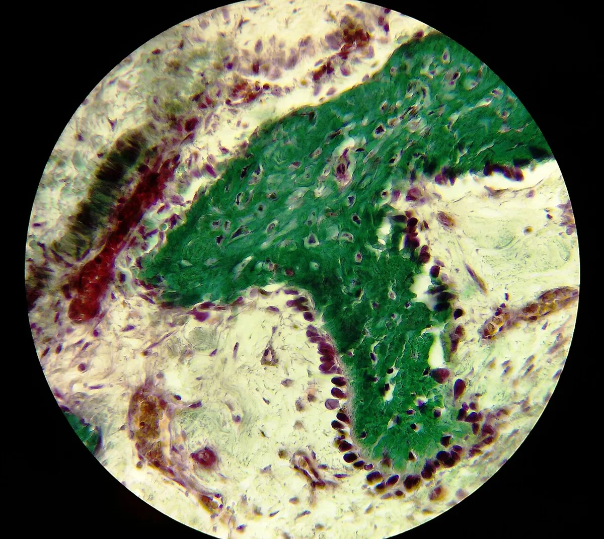
間葉が直接、骨になる骨化。軟骨の段階を経ない。
間葉細胞が集まって骨芽細胞に分化 → 類骨を分泌 → 石灰化 → 骨梁が形成され融合。
この様式でできる骨:頭蓋冠の扁平骨・顔面骨・鎖骨(の大部分)。またすべての骨の太さの成長(骨膜からの付加)もこの様式である。
頭蓋が膜内骨化であることが、新生児の泉門と骨縫合を生む。骨化の中心から放射状に広がり、まだ骨化していない膜性の部分が泉門として残る。大泉門は生後1年半〜2年で閉鎖し、閉鎖の遅れはくる病・甲状腺機能低下症・水頭症などの手がかりになる。
骨縫合が早期に閉じるのが頭蓋縫合早期癒合症で、閉じた縫合と直交する方向へ代償性に伸びるため特徴的な頭形になる(矢状縫合の早期癒合=舟状頭)。FGFR の変異による症候群性のもの(Crouzon・Apert)もあり、軟骨無形成症と同じ FGFR ファミリーである点は覚えやすい対比になる。
膜性骨化する骨だけが揃って侵される病気がある。
鎖骨頭蓋異形成症 — 鎖骨の低形成(肩を前で合わせられる)・大泉門の閉鎖遅延・過剰歯。RUNX2 の異常で、膜性骨化する骨に限って症状が出る。
頭蓋縫合早期癒合症 — 縫合が早く閉じると、その縫合に垂直な方向へ頭が伸びられない。矢状縫合が閉じれば長頭(舟状頭)になる。頭蓋内圧亢進を来せば手術の適応。
大泉門は生後 1歳半頃までに閉じる。膨隆=頭蓋内圧亢進(髄膜炎・水頭症)、陥凹=脱水——乳児の診察で最初に触れるところである。閉鎖の遅れはくる病・甲状腺機能低下症・Down 症候群を示唆する。
English version
Core
Ossification in which mesenchyme becomes bone directly, without a cartilaginous stage.
Mesenchymal cells condense and differentiate into osteoblasts → secrete osteoid → mineralise → trabeculae form and fuse.
Bones formed this way: the flat bones of the cranial vault, the facial bones, most of the clavicle. Growth in thickness of every bone (apposition from the periosteum) also uses this mode.
Going further
That the skull ossifies in membrane produces the fontanelles and sutures of the newborn. Ossification spreads radially from centres, and the membranous areas not yet ossified remain as fontanelles. The anterior fontanelle closes at 18 months to 2 years, and delayed closure is a clue to rickets, hypothyroidism or hydrocephalus.
Premature closure of the sutures is craniosynostosis; the head compensates by growing perpendicular to the fused suture, giving a characteristic shape (sagittal synostosis = scaphocephaly).
There are syndromic forms caused by FGFR mutations (Crouzon, Apert) — the same FGFR family as achondroplasia, which makes a memorable contrast.
In the clinic
There are diseases in which only the bones that ossify in membrane are affected.
Cleidocranial dysplasia — hypoplastic clavicles (the shoulders can be brought together in front), delayed closure of the anterior fontanelle, supernumerary teeth. Caused by a defect of RUNX2, with symptoms confined to the membranous bones.
Craniosynostosis — if a suture closes early the head cannot grow perpendicular to it. Closure of the sagittal suture gives a long, boat-shaped head. Surgery is indicated if intracranial pressure rises.
The anterior fontanelle closes by about 18 months. Bulging = raised intracranial pressure (meningitis, hydrocephalus); sunken = dehydration — the first thing palpated when examining an infant. Delayed closure suggests rickets, hypothyroidism or Down syndrome.
軟骨内骨化Ossificatio chondralis
硝子軟骨の型が先にでき、それが骨に置き換わる骨化。体の骨の大半がこの様式でできる。
順序:軟骨の型 → 軟骨膜に骨頸(bone collar)ができる → 内部の軟骨が肥大・石灰化して細胞が死ぬ → 血管が侵入(栄養動脈)→ 一次骨化中心(骨幹)→ 出生後に骨端で二次骨化中心 → あいだに成長板が残る。
これでできる骨:四肢の長管骨・椎骨・骨盤・頭蓋底。
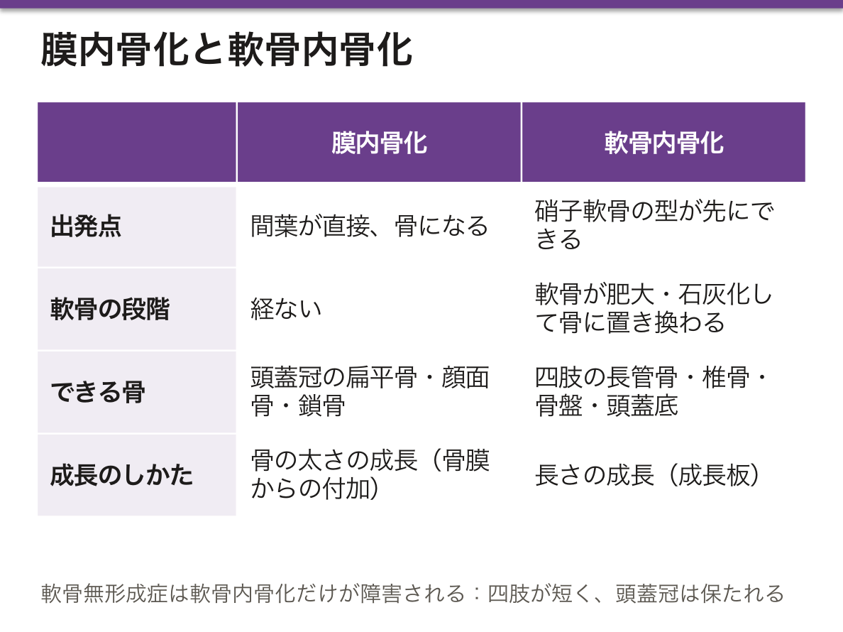
骨化様式の違いは疾患で侵される骨の分布を決める。軟骨無形成症では軟骨内骨化だけが障害されるので、四肢は短いのに頭蓋冠と鎖骨(膜内骨化)は正常という体型になる。逆に鎖骨頭蓋異形成症(RUNX2 変異)では膜内骨化が障害され、鎖骨の低形成と泉門の閉鎖遅延が起きる。
血管侵入のタイミングは骨端の血行にも関わる。骨端の血流が成長板によって骨幹から遮られる時期があり、この終動脈的な血行が阻まれると骨端が壊死する。小児のPerthes 病(大腿骨頭壊死)や、大腿骨頭すべり症後の壊死はこの脆弱性による。
成長板は小児の骨の弱点である。靱帯より先に成長板が壊れるので、捻挫に見えても骨端離開を疑う。Salter–Harris 分類で評価し、成長板を横切る型(III〜V 型)は成長障害・変形を残す。
骨年齢は手根骨の化骨核で評価する。成長ホルモン分泌不全・甲状腺機能低下症では骨年齢が遅れる。
成長板はエストロゲンで閉じる。思春期早発症では最終身長が低くなるのは、早く伸びて早く閉じるからである。アロマターゼ欠損の男性は成長板が閉じず、背が伸び続ける——閉鎖の引き金がエストロゲンであることの証明になっている。
成長板は血流が乏しく、細菌が停留しやすい。小児の急性血行性骨髄炎が長管骨の骨幹端に好発するのはこのためである。
English version
Core
Ossification in which a hyaline cartilage model is formed first and then replaced by bone. Most bones of the body form this way.
Sequence: cartilage model → a bone collar forms from the perichondrium → the cartilage within hypertrophies, calcifies and the cells die → vessels invade (the nutrient artery) → primary ossification centre in the diaphysis → after birth, secondary centres in the epiphyses → the growth plate remains between them.
Bones formed this way: the long bones of the limbs, the vertebrae, the pelvis, the base of the skull.
Going further
The mode of ossification determines which bones a disease affects. In achondroplasia only endochondral ossification is impaired, so the limbs are short while the cranial vault and clavicle (membranous) are normal. Conversely in cleidocranial dysplasia (RUNX2) membranous ossification is impaired, giving hypoplastic clavicles and delayed fontanelle closure.
The timing of vascular invasion also bears on the epiphysial circulation. There is a period when the epiphysial blood supply is cut off from the diaphysis by the growth plate, and if this end-arterial supply is obstructed the epiphysis undergoes necrosis. Perthes disease in children and necrosis after slipped capital femoral epiphysis arise from this vulnerability.
In the clinic
The growth plate is the weak point of the child’s skeleton. Because the physis fails before the ligament, suspect a physeal separation even when the injury looks like a sprain. Assess by the Salter–Harris classification; types III–V cross the physis and leave growth disturbance and deformity.
Bone age is assessed from the ossification centres of the carpus; it is delayed in growth hormone deficiency and hypothyroidism.
The growth plate is closed by oestrogen. Final height is reduced in precocious puberty because growth starts early and stops early. A man with aromatase deficiency never closes his physes and keeps growing — proof that oestrogen is the trigger for closure.
The growth plate has a sluggish blood supply where bacteria lodge, which is why acute haematogenous osteomyelitis in children favours the metaphysis of long bones.
骨リモデリング — 壊すと作るの均衡
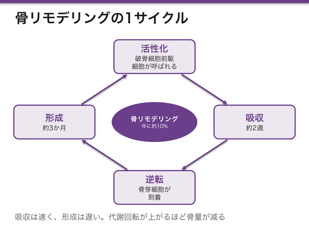
成人の骨も年に約10%が作り替えられている。1サイクルは決まった順序で進む。
- 活性化——骨被覆細胞が退き、破骨細胞前駆細胞が呼ばれる
- 吸収(約2週)——破骨細胞が Howship 窩を掘る
- 逆転——破骨細胞がアポトーシス、骨芽細胞が到着
- 形成(約3か月)——類骨を置き、石灰化する
吸収は速く、形成は遅い。この非対称が、代謝回転が上がるほど骨量が減る理由である。
疾患はこの均衡がどちらへ、どう傾いたかで整理できる。血液検査の組み合わせが鑑別になる。
- 骨粗鬆症——吸収>形成。骨量は減るが質は正常。Ca・P・ALP すべて正常
- 骨軟化症・くる病——石灰化ができず類骨が溜まる。ビタミンD欠乏や低リン血症が原因。Ca低め・P低下・ALP上昇・PTH上昇。小児では成長板が侵されO脚・肋骨念珠
- 原発性副甲状腺機能亢進症——PTH過剰で吸収亢進。Ca上昇・P低下・ALP上昇。「骨・結石・腹痛・精神症状」
- Paget 病——吸収も形成も異常に速い。無秩序なモザイク状骨ができ、脆く変形する。ALPが著明高値なのにCa・Pは正常
- 骨形成不全症——I型コラーゲンの異常。青色強膜・易骨折・難聴
ALPが上がるか、Ca/Pが動くか——この2軸で並べると、紛らわしい代謝性骨疾患がほぼ切り分けられる。
骨転移も同じ枠組みで読める。溶骨性(乳癌・肺癌・腎癌・多発性骨髄腫)は破骨細胞を活性化し、高Ca血症・病的骨折を起こす。造骨性(前立腺癌)は骨芽細胞を活性化し、ALP著明上昇・骨シンチで強い集積を示す。多発性骨髄腫が骨シンチで写りにくいのは、骨形成を伴わない純粋な溶骨だからである。
English version
Core
About 10 per cent of adult bone is replaced each year, and one cycle proceeds in a fixed order.
- Activation — lining cells retract and osteoclast precursors are recruited.
- Resorption (about 2 weeks) — osteoclasts excavate a Howship lacuna.
- Reversal — osteoclasts undergo apoptosis and osteoblasts arrive.
- Formation (about 3 months) — osteoid is laid down and mineralised.
Resorption is fast and formation is slow. This asymmetry is why bone mass falls whenever turnover rises.
Going further
The diseases can be organised by which way, and how far, this balance has tipped — and the combination of blood tests makes the differential.
- Osteoporosis — resorption > formation. Mass falls, quality normal. Ca, P and ALP all normal.
- Osteomalacia / rickets — mineralisation fails and osteoid accumulates; from vitamin D deficiency or hypophosphataemia. Ca low-normal, P low, ALP raised, PTH raised. In children the growth plate is affected, giving bow legs and a rachitic rosary.
- Primary hyperparathyroidism — excess PTH drives resorption. Ca raised, P low, ALP raised. "Bones, stones, abdominal groans and psychic moans."
- Paget disease — both resorption and formation abnormally rapid, producing disorganised mosaic bone that is brittle and deformed. ALP markedly raised with normal Ca and P.
- Osteogenesis imperfecta — abnormal type I collagen. Blue sclerae, fragility, deafness.
Whether the ALP rises and whether Ca and P move — arranging the diseases on these two axes separates almost all of the confusing metabolic bone diseases.
In the clinic
Bone metastasis can be read in the same framework.
Osteolytic (breast, lung, renal, myeloma) activates osteoclasts, causing hypercalcaemia and pathological fracture. Osteoblastic (prostate) activates osteoblasts, giving a markedly raised ALP and intense uptake on bone scintigraphy.
Multiple myeloma is poorly seen on bone scan because it is purely lytic, without accompanying bone formation.
骨折の治癒 — 組織が入れ替わる順序
骨は瘢痕を残さず元の組織に戻れる数少ない器官である。二次性(自然)治癒は4段階で進む。
- 炎症期(〜1週)——血腫形成。マクロファージ・サイトカインが集まる
- 軟性仮骨期(〜3週)——線維組織と軟骨ができる。まだX線に写らない
- 硬性仮骨期(〜3か月)——軟骨内骨化で仮骨が骨に置き換わる。X線に写る
- リモデリング期(数か月〜数年)——荷重に応じて余分な骨が削られ、元の形に戻る
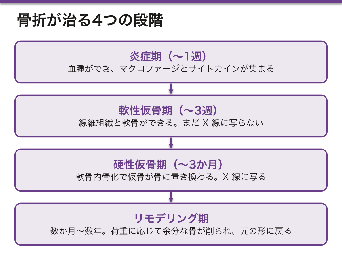
治癒の様式は固定の仕方で変わる——ここが整形外科の核心である。
- 二次性(間接)治癒——骨折端にわずかな動きがあると仮骨ができる。ギプス・髄内釘・創外固定。仮骨が見える
- 一次性(直接)治癒——プレートで解剖学的に整復し強固に固定すると、仮骨を作らずに骨単位が骨折線を越えて直接伸びる(cutting cone)。仮骨が見えない
だから「プレート固定後に仮骨が見えない」のは正常であり、逆に仮骨が見えたら固定が不安定という読み方になる。X線で仮骨の有無を評価する意味が、固定法によって正反対になる。
癒合しない場合を偽関節という。原因は血流不足・不安定な固定・感染・骨欠損。血流の乏しい部位(舟状骨近位・大腿骨頸部・距骨・脛骨遠位1/3)は逆行性の血行に頼るため、骨折で血流が断たれ偽関節・骨壊死を起こしやすい。解剖の血行を知らないと治療方針が立たない典型例である。
喫煙・NSAIDs・ステロイド・糖尿病・放射線照射はいずれも骨癒合を遅らせる。とくに喫煙は偽関節の独立した危険因子で、血管収縮と骨芽細胞機能の抑制による。手術前の禁煙指導に根拠がある。
English version
Core
Bone is one of the few organs that can return to its original tissue without leaving a scar. Secondary (natural) healing proceeds in four stages.
- Inflammatory (to 1 week) — haematoma forms; macrophages and cytokines gather.
- Soft callus (to 3 weeks) — fibrous tissue and cartilage form. Not yet visible radiographically.
- Hard callus (to 3 months) — the callus is replaced by bone through endochondral ossification. Now visible on radiographs.
- Remodelling (months to years) — surplus bone is removed according to load and the original shape returns.
Going further
The mode of healing changes with the method of fixation — and this is the core of orthopaedic practice.
- Secondary (indirect) healing — with slight movement at the fracture ends, callus forms. Cast, intramedullary nail, external fixation. Callus is visible.
- Primary (direct) healing — with anatomical reduction and rigid plate fixation, osteons extend directly across the fracture line (a cutting cone) without forming callus. No callus is visible.
So the absence of callus after plate fixation is normal, and the appearance of callus is read as instability of the fixation. The meaning of looking for callus on a radiograph is reversed depending on the method of fixation.
Failure to unite is non-union, caused by poor blood supply, unstable fixation, infection or bone loss. Sites with a poor blood supply — the proximal scaphoid, the femoral neck, the talus, the distal third of the tibia — depend on a retrograde supply, so a fracture interrupts it and causes non-union and necrosis. A classic example of a treatment plan that cannot be made without knowing the anatomy of the blood supply.
In the clinic
Smoking, NSAIDs, corticosteroids, diabetes and irradiation all delay union. Smoking in particular is an independent risk factor for non-union, through vasoconstriction and suppression of osteoblast function. There is a real basis for advising cessation before surgery.
骨は、標識を打てば「速さ」まで測れる
骨の生検には、他の臓器に無い特徴がある。あらかじめテトラサイクリンを飲ませておくと、その時点で石灰化していた前線に取り込まれて蛍光を発する。
期間をあけて 2 回飲ませれば、2 本の標識線ができる。2 本の間隔を、あいだの日数で割れば、1 日あたり何 μm 石灰化が進んだかが出る。組織から「速度」を測れる、きわめて珍しい方法である。
- 類骨の幅と面積——石灰化していない基質がどれだけ溜まっているか
- 石灰化速度・骨形成速度——二重標識から計算する
- 破骨細胞の数と吸収窩——壊す側の活動
採取は腸骨稜から、骨を脱灰せずに樹脂に包埋して薄切する——脱灰すると、石灰化している部分と類骨の区別が付かなくなるからである。
「作る速さ」と「石灰化する速さ」を分けて測れることが、診断を変える。
| 骨軟化症 | 類骨が厚く広い+石灰化が遅い | ビタミン D 欠乏・低リン血症。基質は作れているが固まらない |
| 骨粗鬆症 | 類骨は正常+骨の量が少ない | 作った骨の質ではなく、量の問題 |
| 線維性骨炎 | 破骨細胞が多く、線維化を伴う | 副甲状腺機能亢進症。回転が速い |
| 無形成骨 | 標識がほとんど付かない | 骨の代謝が止まっている。透析患者で問題になる |
無形成骨は、治療によって作られることがある。慢性腎臓病で副甲状腺ホルモンを下げすぎたり、カルシウムを負荷しすぎたりすると、骨の回転が止まる。骨がカルシウムを受け取れなくなるので、行き場を失ったカルシウムが血管へ沈着する——骨と血管の石灰化が、逆向きに進むという現象が起きる。
⚠️ 「骨密度が下がっているから薬」では足りない場面がある。骨軟化症にビスホスホネートを使えば、石灰化をさらに妨げて悪化させる。透析患者の骨病変は型が複数あり、型によって治療が逆になるので、骨生検が今も意味を持つ数少ない領域である。
この考え方は骨代謝マーカー(血液で回転の速さを測る)にも受け継がれている。生検の代わりに、非侵襲的に「速さ」を推定している。
骨密度の数値だけで治療を決めない。腎機能・ビタミン D・カルシウム・リン・PTH を揃えてから、型を考える。
English version
Core
Bone biopsy has a property no other organ has. If tetracycline is given beforehand it is taken up at the mineralisation front active at that moment and fluoresces. Give it twice with an interval and two labelled lines appear.
Divide the distance between the lines by the number of days and you have how many micrometres a day mineralisation advanced — a remarkably rare method for measuring a rate from histology.
- Osteoid width and area — how much unmineralised matrix has accumulated.
- Mineral apposition rate and bone formation rate — calculated from the double label.
- Osteoclast number and resorption lacunae — the activity of the destroying side.
The specimen is taken from the iliac crest and embedded in resin without decalcification — because decalcifying makes mineralised bone indistinguishable from osteoid.
Going further
Being able to measure "the rate of making" and "the rate of mineralising" separately changes the diagnosis.
| Condition | Findings | Meaning |
|---|---|---|
| Osteomalacia | Osteoid thick and extensive; mineralisation slow | Vitamin D deficiency, hypophosphataemia. The matrix can be made but will not set |
| Osteoporosis | Osteoid normal; the amount of bone is low | A problem of quantity, not of the quality of what was made |
| Osteitis fibrosa | Many osteoclasts, with fibrosis | Hyperparathyroidism; rapid turnover |
| Adynamic bone | Almost no label is taken up | Bone metabolism has stopped. A problem in dialysis patients |
Adynamic bone can be created by treatment. Suppressing PTH too far in chronic kidney disease, or loading with calcium, stops turnover. The bone can no longer accept calcium, so calcium with nowhere to go is deposited in the vessels — bone and vessel calcifying in opposite directions.
⚠️ "The density is low, so give a drug" is not always enough. A bisphosphonate given in osteomalacia impairs mineralisation further and makes it worse. The bone disease of dialysis has several types and the treatment is opposite between them, which is why bone biopsy still has a place. The same thinking is carried into the bone turnover markers, which estimate rate non-invasively in place of biopsy.
In the clinic
Do not decide treatment on the bone density figure alone. Assemble renal function, vitamin D, calcium, phosphate and PTH first, and then consider the type.
まとめ
| 骨(組織としての) | 基質が石灰化した結合組織。組成は有機35%(I型コラーゲン+オステオカルシンなど)と無機65%(ハイドロキシアパタイト Ca₁₀(PO₄)₆(OH)₂)。 |
|---|---|
| 骨芽細胞 | 骨を作る細胞。間葉系幹細胞に由来する。 |
| 破骨細胞 | 骨を壊す細胞。単球・マクロファージ系の細胞が融合してできる多核巨細胞。 |
| 骨細胞 | 骨基質に埋め込まれた骨芽細胞のなれの果て。 |
| 緻密骨(皮質骨) | 骨の外層をなす密な骨。基本単位が骨単位(オステオン、Havers 系)で、中心のHavers 管(血管・神経が通る)を同心円状の層板が取り巻く。 |
| 海綿骨(骨梁) | 骨の内部にある、骨梁 trabecula が三次元の網を作った構造。 |
| 骨膜 | 骨の外表面を覆う膜(関節面を除く)。2層からなる。 |
| 膜内骨化 | 間葉が直接、骨になる骨化。軟骨の段階を経ない。 |
| 軟骨内骨化 | 硝子軟骨の型が先にでき、それが骨に置き換わる骨化。 |
| 骨リモデリング — 壊すと作るの均衡 | 成人の骨も年に約10%が作り替えられている。 |
| 骨折の治癒 — 組織が入れ替わる順序 | 骨は瘢痕を残さず元の組織に戻れる数少ない器官である。 |
| 骨は、標識を打てば「速さ」まで測れる | 骨の生検には、他の臓器に無い特徴がある。 |
このノートは医学生が授業の内容を自分の言葉でまとめたもので、診療の判断に使うものではありません。誤りに気づいたらお知らせください。