顕微鏡で見えているものの半分は、作る過程が作っている。組織学ではスライドを見て言い当てることが学習の中心になる。
だから「本当は何があったか」と「処理で何が起きたか」を分ける訓練が要る——「空胞に見えるものは、溶けて出ていったものの跡かもしれない」。
- 脂肪——アルコールとキシレンで溶ける。だから普通の標本では空白。見たければ凍結切片+Sudan III
- グリコーゲン——水に溶ける。固定が遅れると消える。PAS で染める
- 石灰——脱灰しないと切れない。脱灰すると染まりが落ちる
そして染色は「何を見たいか」で選ぶ。HE で分からないときに次を選べることが、実力になる。
- 標本ができるまで — 6つの工程と、そこで失われるもの
- 染色の選び方 — HEで分からないときに何を追加するか
- 免疫組織化学と電子顕微鏡 — 由来を決める、微細構造を見る
- 凍結切片 — 速さと引き換えに、何を失うか
- 人工産物 — 標本の傷を、病変と読み違えない
- 切り出しと標本の向き — 断端が陰性かどうかは、ここで決まる
- その場で遺伝子を見る — 組織の上に、DNA と RNA を光らせる
- スライドを画像として扱う — デジタル病理
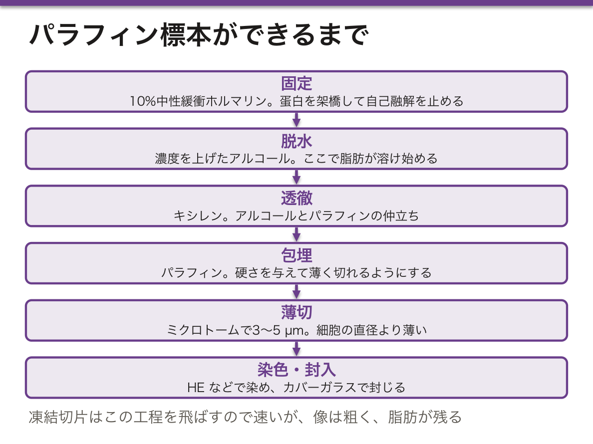
標本ができるまで — 6つの工程と、そこで失われるものPraeparatum histologicum
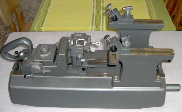
固定 → 脱水 → 透徹 → 包埋 → 薄切 → 染色 → 封入。
- 固定——10%中性緩衝ホルマリン。蛋白を架橋して自己融解と腐敗を止める。電子顕微鏡ではグルタルアルデヒド+四酸化オスミウム
- 脱水——濃度を上げたアルコール。ここで脂肪が溶け始める
- 透徹——キシレン。アルコールとパラフィンの両方に混ざる仲立ち
- 包埋——パラフィン。硬さを与えて薄く切れるようにする
- 薄切——ミクロトームで 3〜5 µm。細胞の直径より薄い
凍結切片——術中迅速診断に使う。10分程度で出せるが、像は粗い。脂肪が残る。
細胞診——組織の構築は見えないが、細胞の異型は見える。Papanicolaou 染色。
「切片の厚さが細胞より薄い」ことが、像の読み違えを生む最大の原因になる。核を通らない断面では、核の無い細胞に見える(切片は断面であるの章)——「無いことの証明に、1枚の切片は使えない」。
術中迅速診断が凍結切片なのは、パラフィン包埋に一晩かかるからになる。手術中に断端と転移の有無を返す必要がある(術中迅速診断の章)——「速さと引き換えに、像の質を落としている」。
だから凍結の結果は暫定で、永久標本で覆ることがある。その前提を外科側と共有しておく(情報の受け渡しの章)——「暫定であることを言い添えるところまでが、報告になる」。
ホルマリン固定は、遺伝子検査の質を左右する。長すぎる固定と酸性のホルマリンが核酸を壊す(分子病理の章)——「標的薬を選ぶ検査の成否が、検体を瓶に入れた瞬間に決まっている」。
だから採取から固定までの時間も記録する。虚血時間が長いと蛋白の抗原性が落ちる——「検査室に着く前の時間が、結果の一部になっている」。
細胞診と組織診の違いは、構築が見えるかどうかになる。浸潤の有無は構築の話なので、細胞診では原則として言えない(子宮頸部細胞診の章)——「細胞診で上皮内か浸潤かを決めない」。
工程のどこで何が失われるかを知っていないと、標本を読み違える。
脂肪はアルコールとキシレンに溶けるので、通常のパラフィン切片では脂肪滴が「空胞」として抜けて見える。脂肪肝・脂肪塞栓・脂肪肉腫を証明したいなら凍結切片+脂溶性色素(Sudan III・オイルレッド O)が要る。
グリコーゲンは水に溶けるので、アルコール固定にしてPAS で染める。
脱灰 — 骨と石灰化した組織は酸で脱灰しないと切れないが、脱灰は抗原と核酸を壊す。骨転移の検体で免疫染色や遺伝子検査が必要なら、脱灰の方法をあらかじめ相談する——後から取り返しがつかない。
固定の遅れ(阻血時間)はホルモン受容体・HER2 の判定を狂わせる。乳癌の検体は摘出後すみやかに固定することが治療選択に直結する。
English version
Core
Fixation → dehydration → clearing → embedding → sectioning → staining → mounting.
- Fixation — 10% neutral buffered formalin. Cross-links protein and stops autolysis and putrefaction. For electron microscopy, glutaraldehyde plus osmium tetroxide.
- Dehydration — alcohols of rising concentration. Fat begins to dissolve here.
- Clearing — xylene, the intermediary miscible with both alcohol and paraffin.
- Embedding — paraffin, giving the hardness to cut thinly.
- Sectioning — 3–5 µm on the microtome. Thinner than the diameter of a cell.
Frozen section — used for intraoperative diagnosis. Available in about ten minutes but the image is coarse; fat is retained.
Cytology — the architecture is not seen but cellular atypia is. Papanicolaou stain.
Going further
That the section is thinner than a cell is the greatest cause of misreading. A plane that misses the nucleus shows a cell that appears to have none — one section cannot be used to prove an absence.
Intraoperative diagnosis uses frozen sections because paraffin embedding takes a night, and the margin and the presence of metastasis must be reported during the operation — the quality of the image is sacrificed for speed. So a frozen result is provisional and may be overturned on the permanent sections. Share that premise with the surgeon — saying that it is provisional is part of the report.
Formalin fixation governs the quality of genetic testing. Fixation that is too long, and acidic formalin, destroy nucleic acid — the success of the test that chooses a targeted drug is decided the moment the specimen goes into the jar. So the time from removal to fixation is recorded too: a long ischaemic time reduces the antigenicity of proteins — the time before it reaches the laboratory is part of the result.
The difference between cytology and histology is whether the architecture is visible. Invasion is a matter of architecture, so as a rule cytology cannot speak to it — do not decide in situ versus invasive on cytology.
In the clinic
Without knowing what is lost at which step, the slide is misread.
Fat dissolves in alcohol and xylene, so on an ordinary paraffin section fat droplets appear as empty vacuoles. To demonstrate fatty liver, fat embolism or liposarcoma, a frozen section with a lipid-soluble dye (Sudan III, oil red O) is required.
Glycogen dissolves in water, so alcohol fixation is used and it is stained with PAS.
Decalcification — bone and calcified tissue cannot be cut without acid decalcification, but decalcification destroys antigens and nucleic acid. If immunostaining or genetic testing will be needed on a bone metastasis, discuss the method of decalcification in advance — it cannot be undone afterwards.
Delay in fixation (ischaemic time) distorts the assessment of hormone receptors and HER2. Fixing a breast specimen promptly after removal bears directly on the choice of treatment.
染色の選び方 — HEで分からないときに何を追加するか
| 染色 | 染めるもの | 色 | 使う場面 |
|---|---|---|---|
| HE | 核/細胞質 | 青紫/赤 | 基本 |
| PAS | 糖(グリコーゲン・基底膜・粘液・真菌) | 赤紫 | 腎生検・真菌 |
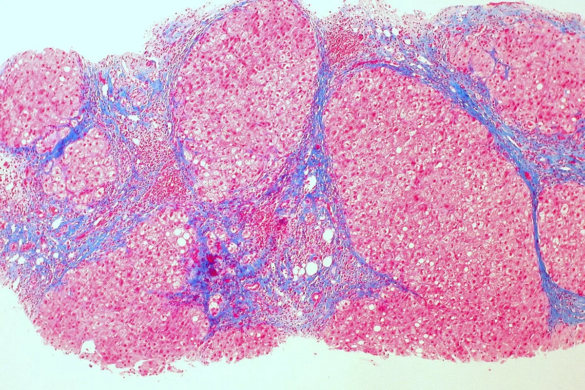
| Masson トリクローム | 膠原線維 | 青(緑) | 線維化の評価 |
| Elastica van Gieson | 弾性線維/膠原線維 | 黒紫/赤 | 血管・肺 |
| 鍍銀(渡銀) | 細網線維・基底膜 | 黒 | 肝・リンパ節・腎 |
| Congo red | アミロイド | 橙赤・偏光で緑 | アミロイドーシス |
| Berlin blue(Prussian blue) | 三価鉄(ヘモジデリン) | 青 | 鉄過剰・出血の跡 |
| Sudan III/Oil red O | 中性脂肪 | 赤橙 | 凍結切片のみ |
| トルイジンブルー | 肥満細胞の顆粒 | 異染性の赤紫 | 肥満細胞症 |
| Ziehl-Neelsen | 抗酸菌 | 赤 | 結核・らい |
| Grocott | 真菌 | 黒 | ニューモシスチス |
Congo red は、偏光で緑色複屈折を確認して初めて陽性になる。
この表は「所見から染色を逆算する」ために使う。間質が広い→トリクローム、基底膜が厚い→PASと鍍銀、褐色の顆粒→鉄染色——「HEで気になった1点に対して、1つ足す」。
褐色の色素を見たら、少なくとも3つを区別する。ヘモジデリン(鉄染色陽性)・メラニン(Fontana-Masson)・リポフスチン(どちらも陰性)(色素沈着の章)——「同じ茶色が、出血の跡・色素細胞・加齢のどれでもありうる」。
アミロイドの診断は、偏光顕微鏡まで行って完結する。Congo red の赤だけでは弱い(アミロイドーシスの章)——「緑色複屈折を見たか、が報告の要点になる」。
そして型(AL・AA・ATTR)は免疫染色か質量分析で決める。治療がまったく違うからになる(アミロイドーシスの型の章)——「あることの確認と、何であるかの確認は別の検査になる」。
腎生検で PAS と鍍銀を必ず加えるのは、基底膜の厚さと二重化を見るためになる(腎生検の章)——「糸球体の病変は、基底膜の形で分類されている」。
脂肪染色が凍結切片限定であることは、依頼の段階で効いてくる。脂肪肝や脂肪塞栓を証明したければ、あらかじめ凍結を頼んでおく——「後から染めればよい、が通用しない染色がある」。
「HE で分からない」と思った時点で、次の1手が決まっている。
PAS — 糖。腎生検の基底膜、真菌(カンジダ・アスペルギルス)、グリコーゲン、粘液。PAS 陽性・ジアスターゼ消化で消える=グリコーゲン、消えない=糖蛋白——1手間で区別できる。
Masson トリクローム — 線維化の量。肝生検で線維化のステージを決める。
Elastica van Gieson — 弾性線維。血管の内弾性板を見て、癌が血管に浸潤しているか(脈管侵襲)を判定する——予後と追加治療を左右する。
Congo red — アミロイド。偏光下でリンゴ緑色の複屈折が決め手。
鍍銀(Grocott) — 真菌とニューモシスチス。Ziehl–Neelsen — 抗酸菌。疑う病原体を先に決めてから染色を注文するのが実務である。
English version
Core
| Stain | Stains | Colour | Used for |
|---|---|---|---|
| H&E | Nucleus / cytoplasm | Blue-purple / red | The basic stain |
| PAS | Carbohydrate (glycogen, basement membrane, mucin, fungi) | Magenta | Renal biopsy, fungi |
| Masson trichrome | Collagen | Blue (green) | Assessment of fibrosis |
| Elastica van Gieson | Elastic fibres / collagen | Black-purple / red | Vessels, lung |
| Silver impregnation | Reticulin fibres, basement membrane | Black | Liver, lymph node, kidney |
| Congo red | Amyloid | Orange-red; green under polarised light | Amyloidosis |
| Prussian (Berlin) blue | Ferric iron (haemosiderin) | Blue | Iron overload, old haemorrhage |
| Sudan III / oil red O | Neutral fat | Red-orange | Frozen sections only |
| Toluidine blue | Mast cell granules | Metachromatic red-purple | Mastocytosis |
| Ziehl–Neelsen | Acid-fast bacilli | Red | Tuberculosis, leprosy |
| Grocott | Fungi | Black | Pneumocystis |
Congo red is positive only when green birefringence under polarised light has been confirmed.
Going further
Use this table to work backwards from the finding to the stain. Wide stroma → trichrome; thickened basement membrane → PAS and silver; brown granules → iron stain — one stain added for the one thing that caught your eye on H&E.
On seeing a brown pigment, distinguish at least three — haemosiderin (iron positive), melanin (Fontana–Masson), lipofuscin (negative for both) — the same brown may be old haemorrhage, a pigment cell, or ageing.
The diagnosis of amyloid is complete only when the polarising microscope has been used. The red of Congo red alone is weak — whether green birefringence was seen is the point of the report. And the type (AL, AA, ATTR) is decided by immunostaining or mass spectrometry, because the treatments are entirely different — confirming that it is there and confirming what it is are two different investigations.
PAS and silver are always added to a renal biopsy in order to see the thickness and the double contours of the basement membrane — glomerular lesions are classified by the shape of the basement membrane.
That fat stains are limited to frozen sections tells at the moment of requesting. To demonstrate fatty liver or fat embolism, ask for a frozen section in advance — there are stains for which "we can stain it later" does not hold.
In the clinic
The moment you think "H&E does not answer this", the next step is already decided.
PAS — carbohydrate. The basement membrane in a renal biopsy, fungi (candida, aspergillus), glycogen, mucin. PAS positive and abolished by diastase = glycogen; not abolished = glycoprotein — distinguished in one extra step.
Masson trichrome — the amount of fibrosis. Used to stage fibrosis on liver biopsy.
Elastica van Gieson — elastic fibres. The internal elastic lamina is examined to judge whether a cancer has invaded a vessel (lymphovascular invasion) — which governs prognosis and adjuvant treatment.
Congo red — amyloid. Apple-green birefringence under polarised light is decisive.
Grocott silver — fungi and Pneumocystis. Ziehl–Neelsen — acid-fast bacilli.
In practice, decide which organism you suspect before ordering the stain.
免疫組織化学と電子顕微鏡 — 由来を決める、微細構造を見るImmunohistochemia
免疫組織化学——抗体で特定の蛋白を検出し、酵素反応で発色させる。細胞の由来と分化を示す。
- 上皮——サイトケラチン、EMA
- 間葉——ビメンチン。筋——デスミン、平滑筋アクチン
- リンパ球——CD3(T)、CD20(B)。形質細胞——CD138
- 神経・グリア——S-100、GFAP、シナプトフィジン、クロモグラニンA
- 造血・血管内皮——CD34、CD31
- 治療に直結するもの——ER・PgR・HER2(乳癌)、PD-L1、ALK、Ki-67(増殖能)
電子顕微鏡——透過型で内部構造、走査型で表面。今も必須なのは腎生検・繊毛病・一部の腫瘍。
蛍光抗体法——凍結切片に用いる。腎生検の免疫グロブリンと補体の沈着。
免疫組織化学は、形態では決められない2つの問いに答える。①どこから来た細胞か ②治療の標的があるか——「診断のためだけでなく、薬を選ぶために染めている」。
乳癌の ER・PgR・HER2 が、そのまま治療方針になる(乳癌の章)——「病理報告書が、処方箋の前段になっている」。
Ki-67 は増殖している細胞の割合を示す。神経内分泌腫瘍の分類では、この数値が悪性度そのものになる(神経内分泌腫瘍の章)——「見た目の異型より、増える速さで分ける腫瘍がある」。
腎生検が3つの方法を全部使う唯一の領域になる。光学顕微鏡(構築)・蛍光抗体法(沈着した免疫複合体)・電子顕微鏡(沈着の位置)(腎生検の章)——「3つが揃って初めて糸球体腎炎の型が決まる」。
特に電子顕微鏡は、沈着が上皮下か内皮下かメサンギウムかを教える。これが分類の軸になっている(糸球体腎炎の分類の章)——「同じ沈着物でも、置かれた場所で病名が変わる」。
そして免疫染色は陰性の解釈が難しい。固定と抗原賦活の条件で偽陰性が出る——「陽性対照が染まっているかを、必ず一緒に見る」。
免疫組織化学は「この細胞は何者か」に答える道具である。
原発不明癌で最も力を発揮する。サイトケラチン(上皮)/ビメンチン(間葉)/CD45(造血系)/S-100・HMB-45(メラノーマ)で大分類を決め、TTF-1(肺・甲状腺)・CDX2(消化管)・GATA3(乳腺・尿路)・PSA(前立腺)で臓器を絞る。
治療そのものを決める染色がある。乳癌の ER・PgR・HER2(内分泌療法と分子標的薬の適応)、PD-L1(免疫チェックポイント阻害薬)、消化管間質腫瘍の KIT(CD117)。「診断のため」から「治療を選ぶため」へ役割が移っている。
増殖能は Ki-67 で見る。神経内分泌腫瘍の grade はこれで決まる。
English version
Core
Immunohistochemistry — a specific protein is detected with an antibody and revealed by an enzyme reaction. It shows the origin and differentiation of the cell.
- Epithelium — cytokeratins, EMA
- Mesenchyme — vimentin. Muscle — desmin, smooth muscle actin
- Lymphocytes — CD3 (T), CD20 (B). Plasma cells — CD138
- Nerve and glia — S-100, GFAP, synaptophysin, chromogranin A
- Haematopoietic and endothelium — CD34, CD31
- Those that bear on treatment — ER, PgR, HER2 (breast), PD-L1, ALK, Ki-67 (proliferation)
Electron microscopy — transmission for internal structure, scanning for surfaces. Still indispensable in renal biopsy, ciliopathies and some tumours.
Immunofluorescence — used on frozen sections; deposition of immunoglobulin and complement in renal biopsy.
Going further
Immunohistochemistry answers two questions morphology cannot settle — ① where did this cell come from ② is there a target for treatment — staining not only for diagnosis but to choose a drug.
ER, PgR and HER2 in breast cancer are themselves the management — the pathology report has become the stage before the prescription.
Ki-67 shows the proportion of proliferating cells. In the classification of neuroendocrine tumours that figure is the grade itself — there are tumours divided by the speed of proliferation rather than by the atypia of appearance.
Renal biopsy is the only field that uses all three methods — light microscopy (architecture), immunofluorescence (deposited immune complexes), electron microscopy (the position of the deposits) — only with all three is the type of glomerulonephritis settled. Electron microscopy in particular tells whether the deposits are subepithelial, subendothelial or mesangial, and that is the axis of the classification — the same deposit changes its disease name according to where it sits.
And a negative immunostain is hard to interpret. False negatives arise from the conditions of fixation and antigen retrieval — always look at whether the positive control has stained.
In the clinic
Immunohistochemistry is the tool that answers "what is this cell?". It is most powerful in carcinoma of unknown primary. Cytokeratin (epithelial) / vimentin (mesenchymal) / CD45 (haematopoietic) / S-100 and HMB-45 (melanoma) settle the broad class, then TTF-1 (lung, thyroid), CDX2 (gut), GATA3 (breast, urothelium) and PSA (prostate) narrow the organ.
Some stains decide the treatment itself — ER, PgR and HER2 in breast cancer (endocrine therapy and targeted agents), PD-L1 (checkpoint inhibitors), KIT (CD117) in gastrointestinal stromal tumour. The role has shifted from "for the diagnosis" to "to choose the treatment".
Proliferation is assessed by Ki-67, which determines the grade of a neuroendocrine tumour.
凍結切片 — 速さと引き換えに、何を失うかSectio congelata
組織を凍らせて固め、そのまま薄く切る。固定・脱水・パラフィン包埋を全部飛ばすので、10〜20分で診断できる。
| 凍結切片 | パラフィン切片 | |
|---|---|---|
| 所要時間 | 10〜20分 | 1〜2日 |
| 切片の厚さ | 5〜10 μm(厚く不均一) | 3〜4 μm(均一) |
| 核の像 | 氷晶で歪む | 鮮明 |
| 脂肪 | 残る(Sudan 染色ができる) | 溶け出す |
| 用途 | 術中迅速診断・脂肪染色・一部の蛍光抗体法 | 確定診断 |
術中迅速診断で答えられるのは3つだけ——①悪性か否か ②断端に腫瘍があるか ③リンパ節転移があるか。
細かい組織型や分化度は、後日のパラフィン切片で決める。
凍結切片の限界は「水が凍る」という一点から全部出てくる。細胞内の水が氷の結晶になり、そこが穴になって核が歪む——「速さのために、像の質を捨てている」。
だから聞ける問いが限られる。手術中の判断を変える問いにだけ使い、変えない問いには使わない(病理診断の実際の章)——「その答えで手術の続きが変わるか、を先に決める」。
脂肪が残ることは、逆に凍結でしかできない検査を作っている。脂肪はパラフィン処理の有機溶媒で溶け出すので、脂肪染色は凍結切片でしか成立しない(脂質蓄積症の章)——「見たいものが溶ける処理を、避けるという選び方」。
センチネルリンパ節生検は、この技術があって初めて成立した。転移が無ければ郭清をやめられるので、同じ手術のうちに答えが要る(広がりの病理の章)——「10分で出る答えが、手術の範囲を決める」。
凍結切片は手術中の意思決定のために存在する。
断端が陰性か(切り足すかどうか)、センチネルリンパ節に転移があるか(郭清するかどうか)、そもそも悪性か(術式を変えるかどうか)——患者が麻酔下にあるあいだに答える。
速さと引き換えに精度を失うことを、依頼する側が理解しておく必要がある。切片が厚く不均一で、氷晶による人工産物が出るため、細かい核所見の判定には向かない。甲状腺の濾胞性腫瘍のように「被膜浸潤の全周を見なければ決まらない」病変は、原理的に凍結切片で診断できない。
脂肪を含む組織は凍っても硬くならず切りにくい。最終診断は必ずパラフィン切片で確定する——凍結の結果は暫定である。
English version
Core
The tissue is frozen to harden it and cut thin as it is. Fixation, dehydration and paraffin embedding are all skipped, so a diagnosis is possible in 10–20 minutes.
| Frozen section | Paraffin section | |
|---|---|---|
| Time required | 10–20 minutes | 1–2 days |
| Thickness | 5–10 µm (thick and uneven) | 3–4 µm (even) |
| Nuclear detail | Distorted by ice crystals | Sharp |
| Fat | Retained (Sudan staining possible) | Dissolved out |
| Use | Intraoperative diagnosis, fat stains, some immunofluorescence | Definitive diagnosis |
Only three questions can be answered intraoperatively — ① is it malignant ② is there tumour at the margin ③ is there nodal metastasis. The precise histological type and grade are settled later on the paraffin sections.
Going further
Every limitation of the frozen section follows from the single fact that water freezes. The water in the cell becomes ice crystals, leaving holes that distort the nuclei — the quality of the image is discarded for speed.
So the questions that can be asked are limited. Use it only for questions that will change the operation, and not for those that will not — decide first whether the answer changes what happens next.
That fat is retained creates, conversely, tests that only a frozen section can do. Fat is dissolved by the organic solvents of paraffin processing, so a fat stain is possible only on a frozen section — choosing to avoid a process that dissolves what you want to see.
Sentinel node biopsy only became possible because of this technique. If there is no metastasis the dissection can be abandoned, so the answer is needed within the same operation — an answer given in ten minutes deciding the extent of surgery.
In the clinic
The frozen section exists for decisions during an operation. Is the margin clear (do we cut more)? Is there metastasis in the sentinel node (do we dissect)? Is it malignant at all (do we change the operation)? — answered while the patient is anaesthetised.
The requesting side must understand that accuracy is traded for speed. The sections are thick and uneven with ice-crystal artefact, so they are unsuited to fine nuclear assessment.
A lesion such as a follicular neoplasm of the thyroid, which cannot be settled without examining the whole circumference of the capsule for invasion, cannot in principle be diagnosed on a frozen section.
Tissue containing fat does not harden even when frozen and is hard to cut. The final diagnosis is always made on paraffin sections — the frozen result is provisional.
人工産物 — 標本の傷を、病変と読み違えないArtefactum
生体には無かったのに、標本を作る過程で生じた像を人工産物という。これを病変と読み誤らないことが、標本を見るときの要点になる。
| 像 | いつ生じるか | 見分け |
|---|---|---|
| 組織の収縮(実質と間質の隙間) | 固定・脱水 | 器官全体に一様に出る |
| ナイフの傷(一直線の裂け) | 薄切 | 切片を貫いて直線に走る |
| しわ・重なり(濃く見える帯) | 伸展・貼り付け | 焦点が合わない |
| 気泡 | 封入 | 縁が黒く光る円 |
| ホルマリン色素(褐色の粒) | 酸性のホルマリン+血液 | 出血の多い組織に散在。偏光する |
| 挫滅(核が引き伸ばされる) | 鉗子・生検 | 辺縁に帯状 |
自家融解(オートリシス)は人工産物ではないが、同じく「死後に生じた変化」として区別する——核が薄くなり、細胞境界が失われる。
人工産物を知ることは、標本の作り方を逆から理解することになる。収縮は水を抜いたから、裂けは刃が引っかかったから、色素は液が酸性に傾いたから起きる——「像の傷は、工程のどこで何をしたかを教えている」。
実質と間質のあいだの隙間は、最も多い読み違えになる。本物の浮腫や剥離は分布に偏りがあり、人工の収縮は器官全体に一様に出る——「病変は偏り、人工物は一様になる」。
挫滅は、検体を採る側の問題として返ってくる。鉗子で強く掴んだ縁は診断に使えないので、生検は必要な大きさを、掴まずに採る(病理診断の実際の章)——「診断の質の一部は、採った人が決めている」。
そして自家融解は、時間の記録になる。固定が遅れるほど核の像が失われ、免疫染色も抗原が壊れて出なくなる(剖検と分子病理の章)——「検体は、採った瞬間から劣化が始まっている」。
人工産物を病変と読むのが、標本を読むときに最も多い誤りである。
一様に出ていれば人工産物、局所に限れば病変——これが基本の見分け方になる。収縮による実質と間質の隙間が器官全体に均等なら固定の影響だが、腫瘍の周りだけなら浸潤に伴う変化かもしれない。
気泡・皺・ナイフ傷(一直線に走る)・染色ムラ・異物の混入は組織の構造と無関係な向きに走るので見分けられる。
臨床でも同じ問題が起こる。圧挫標本(crush artifact)は生検鉗子で潰れた細胞が濃く伸びて見えるもので、小細胞癌と紛らわしい。内視鏡生検では「潰さずに採る」ことが診断の前提になる。
English version
Core
An appearance not present in life but produced in making the slide is an artefact. Not mistaking one for a lesion is a key point in reading a slide.
| Appearance | When it arises | How to recognise it |
|---|---|---|
| Shrinkage (a gap between parenchyma and stroma) | Fixation, dehydration | Uniform throughout the organ |
| Knife marks (a straight tear) | Sectioning | Runs straight across the whole section |
| Folds and overlaps (a dark band) | Floating out, mounting | Will not come into focus |
| Air bubbles | Mounting | Circles with a shining black edge |
| Formalin pigment (brown granules) | Acid formalin + blood | Scattered in bloody tissue. Birefringent |
| Crush (nuclei drawn out) | Forceps, biopsy | A band at the edge |
Autolysis is not an artefact but is distinguished in the same way, as a change arising after death — the nuclei fade and the cell boundaries are lost.
Going further
Knowing the artefacts is understanding the making of the slide backwards. Shrinkage because water was removed, tears because the blade caught, pigment because the fluid turned acid — a flaw in the image tells you what was done, and at which step.
The gap between parenchyma and stroma is the commonest misreading. Genuine oedema or separation is unevenly distributed; artefactual shrinkage is uniform throughout the organ — a lesion is uneven, an artefact is uniform.
Crush comes back as a problem for whoever took the specimen. An edge gripped hard with forceps cannot be used for diagnosis, so take a biopsy of the size required, without gripping it — part of the quality of the diagnosis is decided by the person who took it.
And autolysis is a record of time. The longer fixation is delayed, the more nuclear detail is lost, and immunostaining fails as the antigens are destroyed — a specimen begins to deteriorate from the moment it is taken.
In the clinic
Reading an artefact as a lesion is the commonest error in reading a slide. Uniform means artefact, localised means lesion — this is the basic distinction.
If the gap between parenchyma and stroma is even throughout the organ it is the effect of fixation, but if it is only around a tumour it may be a change accompanying invasion.
Bubbles, folds, knife marks (running dead straight), uneven staining and contaminants run in directions unrelated to the architecture of the tissue, and so can be recognised.
The same problem occurs clinically. Crush artefact — cells flattened and drawn out by the biopsy forceps — is easily confused with small cell carcinoma. At endoscopic biopsy, taking the sample without crushing it is the premise of the diagnosis.
切り出しと標本の向き — 断端が陰性かどうかは、ここで決まる
手術で採ったものは、そのまま薄切できない。切り出しという工程で、どの面をどの向きで包埋するかを人が決める。ここが病理診断の質を左右する。
- 断端に墨(インク)を塗る——外科医が切った面を色で標識しておく。顕微鏡で「腫瘍が墨に接しているか」を見るのが断端評価である
- 向きを固定する——外科医が糸や切れ込みで上下左右を示す。陽性断端が見つかったとき、どちらの方向を追加切除すべきかを言えるようにするため
- 面出しの方向を選ぶ——垂直断(腫瘍と断端の距離が測れる)と、水平断(断端の面積を広く見られるが距離が測れない)を目的で使い分ける
顕微鏡で見えるのは、切り出した面だけである。これが病理報告を読むときの決定的な前提になる。
直径 2 cm の腫瘍を 5 mm 間隔で切り出せば、実際に顕微鏡で見ているのは腫瘍全体の 数%にすぎない。「断端陰性」は見た切片では腫瘍が墨に触れていなかったという意味であって、切片と切片のあいだに腫瘍が残っていないことの証明ではない。
だから報告書には断端までの距離(mm)が書かれる。0.1 mm と 5 mm は同じ「陰性」でも意味がまるで違い、追加切除や放射線の判断が変わる。
リンパ節も同じで、1個のリンパ節を1枚しか切らなければ、微小転移は見落とされる。センチネルリンパ節を細かく刻み、さらにサイトケラチン免疫染色まで行うのはこのためである。
病理報告で「断端陰性」とだけ書かれていたら、距離が書かれているかを探す。距離の記載が無ければ、切り出しの粗さも含めて臨床側で解釈しなければならない。
English version
Core
What is removed at operation cannot be sectioned as it is. At grossing, a person decides which face is embedded in which orientation. This governs the quality of the pathological diagnosis.
- Ink the margins — the surface the surgeon cut is marked with colour. Assessment of the margin is looking down the microscope at whether tumour touches the ink.
- Fix the orientation — the surgeon marks superior, inferior, left and right with sutures or notches, so that when a positive margin is found it can be said in which direction to excise further.
- Choose the direction of the cut face — perpendicular sections (the distance from tumour to margin can be measured) and en-face sections (a wide area of margin is seen but no distance can be measured) are used according to the purpose.
Going further
What is seen down the microscope is only the face that was cut. This is the decisive premise for reading a pathology report.
If a 2 cm tumour is grossed at 5 mm intervals, what is actually examined microscopically is only a few per cent of the tumour. "Margin negative" means that on the sections examined the tumour did not touch the ink; it is not proof that no tumour remains between one section and the next.
This is why the report states the distance to the margin in millimetres. 0.1 mm and 5 mm are both "negative" but mean entirely different things, and change the decision on further excision or radiotherapy.
The same holds for lymph nodes: cut one node once and a micrometastasis is missed. This is why a sentinel node is sliced finely and cytokeratin immunostaining is performed as well.
In the clinic
If a pathology report says only "margin negative", look for whether a distance is given. If no distance is stated, the clinician must interpret it including the coarseness of the grossing.
その場で遺伝子を見る — 組織の上に、DNA と RNA を光らせる
免疫染色は蛋白を見る方法だった。核酸を直接見る方法もあり、標識した相補鎖を切片に載せて結合させる(in situ hybridization)。蛍光で見るものを FISH と呼ぶ。
組織の形を保ったまま、どの細胞が持っているかが分かるのが利点である。すり潰して測る PCR では「検体のどこかに在る」ことしか言えない。
- 遺伝子増幅——乳癌の HER2、神経芽腫の MYCN。信号の点の数を数える
- 転座——2色のプローブが融合するか、離れるかで見る(ブレイクアパート法)。肉腫や白血病の診断に使う
- ウイルス——EBV の EBER(RNA)、高リスク HPV。癌細胞の核の中に在ることを示せる
免疫染色と組み合わせて段階的に使うのが実際の運用である。乳癌の HER2 を例にとる。
| まず免疫染色 | 膜の染まり方を 0/1+/2+/3+ で判定 | 安価・全例に実施できる |
| 2+ のときだけ FISH | HER2 遺伝子のコピー数を CEP17 と比で数える | 判定の曖昧な例だけを、遺伝子の側から決着させる |
この「安い検査でふるい、曖昧なものだけ高い検査へ回す」という組み立ては、検査全般に共通する設計であり、感度の高い検査を先に、特異度の高い検査を後にという原則そのものである。
EBER の in situ hybridization は上咽頭癌や EBV 関連胃癌の診断で使う。血清の EBV 抗体では「感染したことがある」しか言えないが、癌細胞の核が光れば、その癌が EBV によるものだと言える。
コンパニオン診断(薬を使うかどうかを決める検査)の多くがこの形をとる。薬の適応が検査結果で定義されているので、検査の判定基準が変わると適応も変わる。
English version
Core
Immunostaining was a way of seeing protein. There is also a way of seeing nucleic acid directly: a labelled complementary strand is applied to the section and allowed to bind (in situ hybridization). Seen with fluorescence it is called FISH.
The advantage is that, with the architecture preserved, one can see which cells carry it. PCR on ground-up tissue can say only that it is somewhere in the specimen.
- Gene amplification — HER2 in breast cancer, MYCN in neuroblastoma. The signal dots are counted.
- Translocation — seen by whether two coloured probes fuse or separate (the break-apart method). Used in the diagnosis of sarcomas and leukaemias.
- Virus — EBER (RNA) of EBV, high-risk HPV. It can be shown to be inside the nucleus of the cancer cell.
Going further
In practice it is used in stages in combination with immunostaining. Take HER2 in breast cancer.
| Step | What is done | Why |
|---|---|---|
| Immunostaining first | Membrane staining scored 0 / 1+ / 2+ / 3+ | Cheap; can be done on every case |
| FISH only when 2+ | The HER2 copy number is counted as a ratio to CEP17 | Only the equivocal cases are settled from the side of the gene |
This arrangement — screen with the cheap test, send only the equivocal ones to the expensive one — is a design common to testing in general, and is the principle of the sensitive test first and the specific test after.
In situ hybridization for EBER is used in the diagnosis of nasopharyngeal carcinoma and EBV-associated gastric cancer. Serum EBV antibodies can say only that the person has been infected, but if the nuclei of the cancer cells light up, it can be said that this cancer is due to EBV.
In the clinic
Most companion diagnostics (tests that decide whether a drug is used) take this form. The indication for the drug is defined by the test result, so when the criteria of the test change, the indication changes with them.
スライドを画像として扱う — デジタル病理
ガラス標本を高解像度で丸ごと取り込み(全スライドイメージング)、画面上で顕微鏡と同じように拡大・移動して観察する方法が実用化されている。
- 同時に複数の人が見られる——カンファレンス・遠隔診断・専門施設への相談が、標本を郵送せずにできる
- 過去の症例と並べられる——同じ画面で比較できる
- 定量できる——Ki-67 の陽性率・免疫染色の強度・腫瘍の面積を、目分量ではなく数値で出せる
- 教育に向く——同じ標本を全員が同時に見ることができる。実習で「隣の人と違うスライドを見ている」という問題が消える
⚠️ 画像になった時点で失われるものがある。
- 焦点は 1 面しか記録されないのが基本である。顕微鏡では微動ねじを回して厚みの中を上下に見るが、これができない。細菌・微細な結晶・核の立体的な重なりの判定が難しくなる(複数の焦点面を記録する方式もあるが、容量が跳ね上がる)
- 色が装置と画面で変わる——HE の色調は判定に使われるので、色の較正が要る
- 容量が大きい——1 枚で数百 MB から数 GB。保管の設計が要る
AI による支援も実用化が進んでいる。前立腺生検の癌の検出・リンパ節の微小転移の拾い上げ・有糸分裂像の計数で、見落としを減らす方向で使われている。
ただし「AI が診断する」のではなく「人が見るべき場所を示す」という位置づけが現在の実態である。学習に使われた集団と違う施設・違う染色条件では性能が落ちることが知られており、導入した施設で性能を確かめる作業が要る。
組織学を学ぶ側にとっての含意は明確で、「像を覚える」ことの価値は下がらない——示された場所が本当に病変かを判断するのは、結局その知識である。
専門的な判断が要る標本は、抱え込まずに相談する。デジタル化で、相談の敷居は下がっている。
English version
Core
Glass slides are captured whole at high resolution (whole slide imaging) and viewed on screen, magnified and moved as under a microscope.
- Several people can look at once — conferences, remote diagnosis and referral to a specialist centre without posting the slide.
- Past cases can be placed beside it — compared on the same screen.
- It can be quantified — the Ki-67 index, the intensity of an immunostain and the area of a tumour can be given as numbers rather than by eye.
- It suits teaching — everyone can look at the same slide at the same time. The problem of "the person next to me has a different slide" in the practical disappears.
Going further
⚠️ Something is lost the moment it becomes an image.
- Only one plane of focus is recorded, as a rule. Under a microscope the fine adjustment is turned to look up and down through the thickness, and this cannot be done. Bacteria, fine crystals and the three-dimensional overlapping of nuclei become hard to judge (systems recording multiple focal planes exist, but the file size leaps).
- Colour varies with the scanner and the display — the tones of H&E are used in the assessment, so calibration is needed.
- The files are large — hundreds of megabytes to gigabytes per slide. Storage must be planned.
AI support is also coming into use, reducing oversight in the detection of cancer on prostate biopsies, the picking up of nodal micrometastases, and the counting of mitoses. But the present reality is not that the AI diagnoses; it shows where a person should look. Performance is known to fall in a centre or with staining conditions different from those of the training set, so the performance must be verified where it is introduced.
The implication for anyone learning histology is clear: the value of knowing the appearances does not fall — judging whether the place indicated really is a lesion is, in the end, that knowledge.
In the clinic
Do not keep a slide requiring specialist judgement to yourself — ask. Digitisation has lowered the threshold for asking.
まとめ
| 標本ができるまで — 6つの工程と、そこで失われるもの | 固定 → 脱水 → 透徹 → 包埋 → 薄切 → 染色 → 封入。 |
|---|---|
| 染色の選び方 — HEで分からないときに何を追加するか | 染色染めるもの色使う場面HE核/細胞質青紫/赤基本PAS糖(グリコーゲン・基底膜・粘液・真菌)赤紫腎生検・真菌Masson トリクローム膠原線維青(緑)線維化の評価Elastica… |
| 免疫組織化学と電子顕微鏡 — 由来を決める、微細構造を見る | 免疫組織化学——抗体で特定の蛋白を検出し、酵素反応で発色させる。 |
| 凍結切片 — 速さと引き換えに、何を失うか | 組織を凍らせて固め、そのまま薄く切る。固定・脱水・パラフィン包埋を全部飛ばすので、10〜20分で診断できる。 |
| 人工産物 — 標本の傷を、病変と読み違えない | 生体には無かったのに、標本を作る過程で生じた像を人工産物という。 |
| 切り出しと標本の向き — 断端が陰性かどうかは、ここで決まる | 手術で採ったものは、そのまま薄切できない。 |
| その場で遺伝子を見る — 組織の上に、DNA と RNA を光らせる | 免疫染色は蛋白を見る方法だった。核酸を直接見る方法もあり、標識した相補鎖を切片に載せて結合させる(in situ hybridization)。 |
| スライドを画像として扱う — デジタル病理 | ガラス標本を高解像度で丸ごと取り込み(全スライドイメージング)、画面上で顕微鏡と同じように拡大・移動して観察する方法が実用化されている。 |
このノートは医学生が授業の内容を自分の言葉でまとめたもので、診療の判断に使うものではありません。誤りに気づいたらお知らせください。