免疫は細胞の性質だけで決まるのではなく、どこで会うかで決まる。リンパ球は血中を漂っているだけでは働けず、抗原と出会うための建物が要る。リンパ性器官はその建物である。
器官は2つに分かれる。
- 一次(中枢)リンパ器官——骨髄と胸腺。リンパ球が作られ、選別される場所。抗原に会う前の段階
- 二次(末梢)リンパ器官——リンパ節・脾臓・粘膜関連リンパ組織。抗原と出会って反応する場所
二次リンパ器官はどの経路の抗原を捕まえるかで分業している。リンパ節はリンパ行性、脾臓は血行性、粘膜関連リンパ組織は粘膜面。この分業を知っていると、感染の広がり方から腫れる場所が予測できるし、逆に腫れている場所から入口が推定できる。
どの器官も共通の作りを持つ——B細胞は濾胞に丸く集まり、T細胞はその周りに帯状に広がる。この配置さえ掴めば、リンパ節も脾臓もPeyer板も同じ図として読める。
- 胸腺
- リンパ節
- 脾臓
- 粘膜関連リンパ組織 — 被膜を持たない免疫
- 扁桃 — 上皮が陥入して免疫が待ち構える場所
- 高内皮細静脈 — リンパ球が血管から出る専用の門
- 胸腺での選択 — 使えるものだけを残し、危ないものを消す
- 胸腺の上皮性細網細胞 — I〜VI型と、血液胸腺関門
- 胸腺のホルモンと、ホルモンによる調節 — 胸腺はなぜ縮むのか
- 胚中心 — 抗体が「鍛えられる」場所
- リンパ球の種類 — 見た目は同じ、働きは違う
- B リンパ球 — 抗体を作る細胞
- T リンパ球 — 亜群と分化
- 液性免疫と細胞性免疫 — どちらが何を相手にするか
- 胸腺Thymus
- リンパ節Nodus lymphoideus
- 脾臓Splen
- 粘膜関連リンパ組織 — 被膜を持たない免疫
- 扁桃 — 上皮が陥入して免疫が待ち構える場所Tonsilla palatina
- 高内皮細静脈 — リンパ球が血管から出る専用の門Venula postcapillaris
- 胸腺での選択 — 使えるものだけを残し、危ないものを消すSelectio thymica
- 胸腺の上皮性細網細胞 — I〜VI型と、血液胸腺関門Epitheliocytus reticularis
- 胸腺のホルモンと、ホルモンによる調節 — 胸腺はなぜ縮むのかInvolutio thymi
- 胚中心 — 抗体が「鍛えられる」場所Centrum germinativum
- リンパ球の種類 — 見た目は同じ、働きは違うLymphocytus
- B リンパ球 — 抗体を作る細胞
- T リンパ球 — 亜群と分化
- 液性免疫と細胞性免疫 — どちらが何を相手にするか
- まとめ
胸腺Thymus
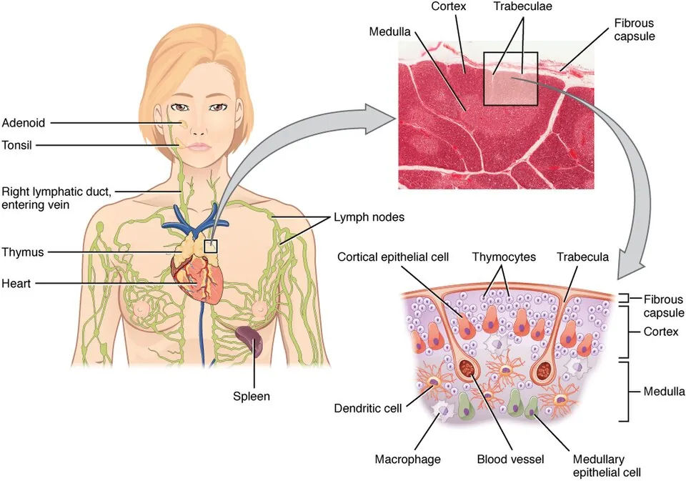
T細胞が教育を受ける一次リンパ器官。前縦隔にあり、小葉に分かれる。
- 皮質——未熟な胸腺細胞が密集し、暗く見える
- 髄質——細胞密度が低く明るい。Hassall 小体(胸腺小体)があり、髄質の目印になる
支持するのは間葉ではなく上皮性細網細胞である(他のリンパ組織は細網線維が骨組み)。
発生は第3咽頭嚢。思春期以降は脂肪に置き換わって退縮する。
胸腺の仕事は2段階の選別である。
- 正の選択(皮質)——自己MHCにほどよく結合できるものだけ生き残す。結合できないものは役に立たないので死ぬ
- 負の選択(髄質)——自己抗原に強く結合しすぎるものを除く。ここで自己反応性のT細胞が消える
負の選択が成立するのは、髄質の上皮細胞がAIRE という転写因子によって本来そこにないはずの臓器の蛋白(インスリンなど)まで発現してみせるためである。体じゅうの自己を胸腺の中で予習させている。AIRE が壊れると多腺性自己免疫症候群1型となり、副甲状腺機能低下症・副腎不全・皮膚粘膜カンジダ症が揃う。
結果として胸腺は免疫寛容の起点になる。「自己を攻撃しない」は生まれつきの性質ではなく、胸腺で作られた結果である。
DiGeorge 症候群(22q11.2欠失)は第3・第4咽頭嚢の発生障害で、胸腺低形成(T細胞不全)+副甲状腺低形成(低カルシウム血症によるテタニー・けいれん)+円錐動脈幹の心奇形が同時に起こる。3つが揃うのは偶然ではなく、同じ場所から発生するからである。
胸腺腫は重症筋無力症を高率に合併する(胸腺腫の30〜50%に筋無力症、筋無力症の約15%に胸腺腫)。アセチルコリン受容体に対する自己抗体が作られる背景に、負の選択が行われる場所そのものの異常がある。胸腺摘除が治療になりうるのはこのためである。
English version
Core
The primary lymphoid organ where T cells are educated. It lies in the anterior mediastinum and is divided into lobules.
- Cortex — densely packed immature thymocytes; appears dark.
- Medulla — lower cell density and paler, containing Hassall corpuscles, the landmark of the medulla.
Its support is epithelial reticular cells, not mesenchyme (other lymphoid tissues are framed by reticular fibres). It develops from the third pharyngeal pouch and involutes into fat after puberty.
Going further
The work of the thymus is selection in two stages.
- Positive selection (cortex) — only those able to bind self MHC with moderate affinity survive; those that cannot bind are useless and die.
- Negative selection (medulla) — those binding self antigen too strongly are removed. Self-reactive T cells are eliminated here.
Negative selection is possible because the medullary epithelial cells, under the transcription factor AIRE, express proteins of organs that have no business being there — insulin, for example. The self of the whole body is rehearsed inside the thymus. When AIRE fails, autoimmune polyendocrine syndrome type 1 results, with hypoparathyroidism, adrenal failure and mucocutaneous candidiasis together.
The consequence is that the thymus is the origin of immune tolerance. "Not attacking the self" is not an innate property but a result manufactured in the thymus.
In the clinic
DiGeorge syndrome (22q11.2 deletion) is a developmental failure of the third and fourth pharyngeal pouches, producing thymic hypoplasia (T cell deficiency), parathyroid hypoplasia (hypocalcaemic tetany and seizures) and conotruncal cardiac malformation together. The three coincide not by chance but because they arise from the same place.
Thymoma is frequently associated with myasthenia gravis (30–50 per cent of thymomas have myasthenia; about 15 per cent of myasthenics have a thymoma). Behind the production of autoantibodies to the acetylcholine receptor lies an abnormality of the very place where negative selection is carried out. This is why thymectomy can be a treatment.
リンパ節Nodus lymphoideus
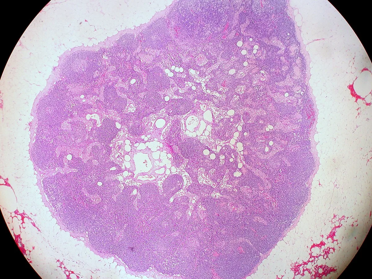
リンパ行性の抗原を捕まえる二次リンパ器官。リンパの流れる順に読む。
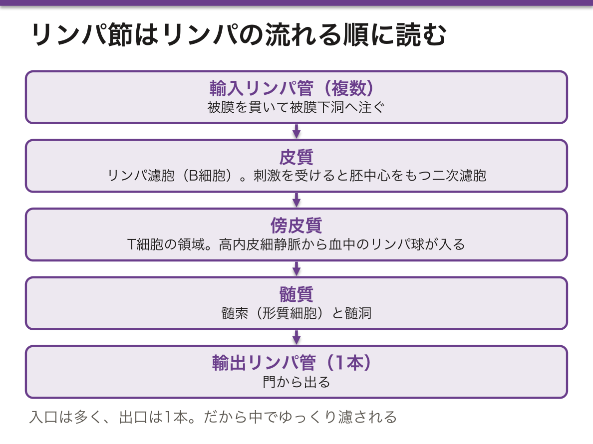
- 輸入リンパ管(複数)→ 被膜下洞
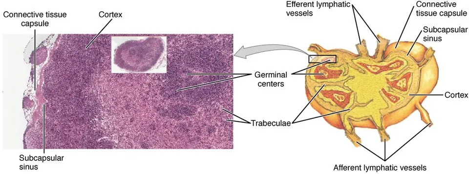
- 皮質——リンパ濾胞(B細胞)。刺激を受けると胚中心を持つ二次濾胞になる
- 傍皮質——T細胞の領域。高内皮細静脈があり、ここからリンパ球が血中より入る
- 髄質——髄索(形質細胞)と髄洞
- 輸出リンパ管(門より1本)
入口は多く、出口は1本。だから中でゆっくり濾される。
胚中心は工場ではなく試験場である。抗原刺激を受けたB細胞はここで分裂しながら体細胞高頻度変異で抗体の可変部を書き換え、抗原によく付くものだけが生き残る(親和性成熟)。同時にクラススイッチでIgM から IgG・IgA・IgE へ持ち替える。どちらも濾胞ヘルパーT細胞からの CD40L の助けが要る。
だから胚中心が見える=抗原に反応した証拠であり、リンパ節の反応性腫大は「働いている」所見である。無痛性で硬く癒着する腫大(転移・リンパ腫)と、有痛性で可動性のある腫大(反応性)が分かれるのはこの違いによる。
この分裂の激しさは代償を伴う。濾胞性リンパ腫は t(14;18) により BCL2 が免疫グロブリン重鎖のプロモーターの支配下に入り、本来なら試験に落ちて死ぬはずのB細胞が死ななくなる病態である。Burkitt リンパ腫の t(8;14)(MYC)も同じ場所で起きる事故である。
センチネルリンパ節——腫瘍から最初にリンパが流れ込む1個。出口が1本で順序が決まっているという構造があるからこそ、そこを調べれば郭清の要否が判断でき、乳癌手術で腋窩郭清を省略できる。
リンパ節の腫れは「どこが増えているか」で読む。
濾胞の過形成(胚中心が目立つ)=B 細胞の反応。感染・関節リウマチ。傍皮質の拡大=T 細胞の反応——ウイルス感染・薬剤性。洞の拡大(洞組織球症)=腫瘍の所属リンパ節で見られる。
正常構造が保たれているか消えているかが反応性か腫瘍性かを分ける最大の手がかりである。悪性リンパ腫では濾胞構造が壊れ、単調な細胞で置き換わる。
触診で悪性を疑う所見 — 硬い・可動性が無い(癒着)・無痛性・進行性に増大・大きさ 2cm 以上・鎖骨上窩。左鎖骨上窩(Virchow リンパ節)は胸管が注ぐ場所なので腹部悪性腫瘍の転移を示す。
診断は摘出生検が原則である——構造を見る必要があるので、針生検では足りないことが多い。
English version
Core
The secondary lymphoid organ that catches antigen arriving by lymph. Read it in the order the lymph flows.
Afferent lymphatics (several) → subcapsular sinus
- Cortex — lymphoid follicles (B cells). On stimulation they become secondary follicles with germinal centres.
- Paracortex — the T cell zone, containing the high endothelial venules through which lymphocytes enter from the blood.
- Medulla — medullary cords (plasma cells) and medullary sinuses.
→ a single efferent lymphatic at the hilum.
Many entrances, one exit — so the lymph is filtered slowly within.
Going further
The germinal centre is not a factory but an examination hall. A B cell stimulated by antigen divides here while rewriting the variable region of its antibody by somatic hypermutation, and only those that bind the antigen well survive (affinity maturation). At the same time class switching exchanges IgM for IgG, IgA or IgE. Both require help from the follicular helper T cell through CD40L.
So a visible germinal centre is evidence of a response to antigen, and reactive lymphadenopathy is a finding that the node is working. This is why painless, hard, fixed enlargement (metastasis, lymphoma) separates from tender, mobile enlargement (reactive).
The intensity of that division has a price. Follicular lymphoma arises when t(14;18) places BCL2 under the immunoglobulin heavy chain promoter, so that a B cell which ought to fail the examination and die no longer dies. The t(8;14) of Burkitt lymphoma (MYC) is an accident in the same place.
Sentinel node — the first node into which lymph drains from a tumour. Because the structure has one exit and a fixed order, examining it decides whether dissection is needed, and allows axillary dissection to be omitted in breast surgery.
In the clinic
Read an enlarged node by what has increased.
- Follicular hyperplasia (prominent germinal centres) = a B cell response. Infection, rheumatoid arthritis.
- Paracortical expansion = a T cell response — viral infection, drug reaction.
- Sinus histiocytosis = seen in nodes draining a tumour.
Whether the normal architecture is preserved or effaced is the greatest single clue to reactive versus neoplastic. In malignant lymphoma the follicular architecture is destroyed and replaced by a monotonous population.
Findings on palpation suggesting malignancy — hard, immobile (fixed), painless, progressively enlarging, over 2 cm, supraclavicular. The left supraclavicular node (Virchow) indicates metastasis from an abdominal malignancy, because the thoracic duct drains there.
Excision biopsy is the rule — the architecture must be seen, and a needle biopsy is often insufficient.
脾臓Splen
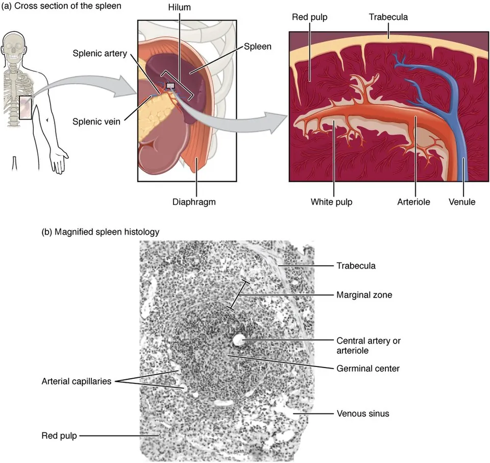
血行性の抗原を捕まえ、同時に血液を濾す。輸入リンパ管を持たないのがリンパ節との決定的な違いである。
- 白脾髄——中心動脈を取り巻く動脈周囲リンパ鞘(T細胞)と、そこに付くリンパ濾胞(B細胞)。免疫の場
- 辺縁帯——白脾髄と赤脾髄の境。血流中の抗原を最初に捕まえる
- 赤脾髄——脾洞と脾索(Billroth 索)。濾過の場
肉眼でも白い斑点(白脾髄)が赤い地(赤脾髄)に散る像として見える。
濾過の仕掛けは開放循環にある。血液はいったん脾索へ放り出され、脾洞の内皮細胞(棒状に並び、隙間がある)の細い隙間を自力ですり抜けて戻る。
つまり変形能を失った赤血球は通れずに残り、その場でマクロファージに食われる。脾臓は赤血球の柔らかさを試す関所である。だから遺伝性球状赤血球症では球状になった赤血球が通り抜けられず脾臓で壊され、脾摘が有効な治療になる。
同じ仕組みが核の遺残物を抜き取る働きも担う。脾臓が無い/働いていないと Howell-Jolly 小体を持つ赤血球が末梢血に現れる——血液像1枚で脾臓の機能が分かる。
脾摘後重症感染症が最重要である。辺縁帯のマクロファージは莢膜を持つ細菌を捕らえて処理する主役なので、脾臓を失うと肺炎球菌・インフルエンザ桿菌b型・髄膜炎菌による電撃的な敗血症のリスクが生涯続く。だから脾摘前(できれば2週間前まで)にワクチンを打つ。
外傷での脾損傷は出血性ショックの原因になるが、可能なら温存する方向へ動いてきたのはこの免疫機能のためである。鎌状赤血球症では繰り返す梗塞で幼少期に脾臓が機能を失い(自己脾摘)、同じリスクを負う。
逆に脾機能亢進症では、肝硬変などによる門脈圧亢進で脾臓がうっ血・腫大し、血球を壊しすぎて汎血球減少になる。「濾しすぎ」が病気になる。
English version
Core
It catches antigen arriving by blood and at the same time filters the blood. Having no afferent lymphatics is the decisive difference from a lymph node.
- White pulp — the periarteriolar lymphoid sheath (T cells) around the central artery, with lymphoid follicles (B cells) attached. The immune compartment.
- Marginal zone — the boundary between white and red pulp; the first to capture antigen from the bloodstream.
- Red pulp — splenic sinuses and cords (of Billroth). The filtering compartment.
Even to the naked eye it appears as white dots (white pulp) scattered on a red ground.
Going further
The mechanism of filtration lies in the open circulation. Blood is discharged into the cords and must find its own way back through the narrow gaps between the rod-shaped endothelial cells of the sinus. So a red cell that has lost its deformability cannot pass, remains behind and is eaten on the spot by macrophages. The spleen is a checkpoint that tests the softness of red cells.
Hence in hereditary spherocytosis the spherical cells cannot pass and are destroyed in the spleen, and splenectomy is an effective treatment.
The same mechanism plucks out nuclear remnants. When the spleen is absent or not working, red cells containing Howell–Jolly bodies appear in the peripheral blood — the function of the spleen read from a single blood film.
In the clinic
Overwhelming post-splenectomy infection is the most important consideration. The macrophages of the marginal zone are the principal cells that trap and process encapsulated bacteria, so losing the spleen carries a lifelong risk of fulminant sepsis from pneumococcus, Haemophilus influenzae type b and meningococcus. Vaccinate before splenectomy, ideally two weeks in advance.
Splenic injury in trauma causes haemorrhagic shock, but practice has moved towards preservation where possible, because of this immune function.
In sickle cell disease repeated infarction destroys splenic function in childhood (autosplenectomy), carrying the same risk.
Conversely in hypersplenism, portal hypertension from cirrhosis congests and enlarges the spleen, which destroys too many cells and causes pancytopenia. Filtering too much becomes a disease.
粘膜関連リンパ組織 — 被膜を持たない免疫
粘膜の直下に散在するリンパ組織。被膜を持たないのが特徴で、上皮のすぐ裏で待ち構える。
- 扁桃——口蓋・咽頭・舌の扁桃が咽頭の入口を輪状に囲む(Waldeyer 輪)
- Peyer 板——回腸の粘膜下組織にまで及ぶリンパ濾胞の集まり。腸間膜対側にある
- 虫垂・気管支関連リンパ組織など
上皮にはM細胞があり、管腔の抗原をそのまま取り込んで下のリンパ球へ手渡す。
ここで作られる抗体は分泌型IgA(二量体+J鎖+分泌成分)である。
分泌型IgA が二量体である理由は、粘液という過酷な場所で働くためである。分泌成分が蛋白分解酵素から守り、二量体であることで細菌に絡みついて上皮への接着を邪魔する(免疫排除)。補体を活性化せず炎症を起こさないのも重要で、常在菌が絶えず接する粘膜で炎症を起こしていては生きていけない。IgA は排除するが、戦わない。
選択的IgA欠損症が最も多い原発性免疫不全であるのに軽症で済むことが多いのはこのためだが、輸血や免疫グロブリン製剤で抗IgA抗体によるアナフィラキシーを起こしうる点だけは重い。
M細胞は同時に弱点でもある。サルモネラ・赤痢菌・ポリオウイルスはM細胞を侵入口として使う。体が抗原を取り込むために開けた窓を、病原体が入口として使う。
MALT リンパ腫は、本来そこに無いはずのリンパ組織が慢性炎症で作られ、そこから腫瘍が出るという成り立ちを持つ。胃には元来 MALT が乏しく、Helicobacter pylori の持続感染がリンパ組織を呼び寄せる。
だから治療がきわめて特徴的で、限局例では除菌だけで腫瘍が消えることがある。抗菌薬で治る悪性腫瘍という例外であり、「炎症が腫瘍を育てる」という原則の最も分かりやすい実例になっている。
English version
Core
Lymphoid tissue scattered immediately beneath a mucosa. It has no capsule, waiting just behind the epithelium.
- Tonsils — palatine, pharyngeal and lingual, forming a ring around the entrance to the pharynx (Waldeyer ring).
- Peyer patches — aggregated lymphoid follicles reaching into the submucosa of the ileum, on the antimesenteric side.
- Appendix, bronchus-associated lymphoid tissue, and others.
The epithelium contains M cells, which take up antigen from the lumen as it is and hand it to the lymphocytes beneath. The antibody made here is secretory IgA (a dimer with J chain and secretory component).
Going further
Secretory IgA is a dimer in order to work in the harsh environment of mucus. The secretory component protects it from proteases, and being a dimer it entangles bacteria and prevents adhesion to the epithelium (immune exclusion). Equally important, it does not activate complement and causes no inflammation — life would be impossible if the mucosa, in constant contact with commensal flora, were inflamed. IgA excludes, but does not fight.
This is why selective IgA deficiency, the commonest primary immunodeficiency, is usually mild — though anti-IgA antibodies can cause anaphylaxis with transfusion or immunoglobulin, and that alone is serious.
The M cell is also a weak point. Salmonella, Shigella and poliovirus use it as their portal of entry. A window the body opened in order to sample antigen is used by pathogens as a door.
In the clinic
MALT lymphoma has the distinctive origin that lymphoid tissue which ought not to be there is created by chronic inflammation, and a tumour then arises from it. The stomach has little native MALT; persistent Helicobacter pylori infection summons the lymphoid tissue.
So the treatment is highly characteristic: in localised disease, eradication alone can make the tumour disappear. A malignant tumour cured by an antibiotic — an exception, and the clearest illustration of the principle that inflammation nurtures neoplasia.
扁桃 — 上皮が陥入して免疫が待ち構える場所Tonsilla palatina
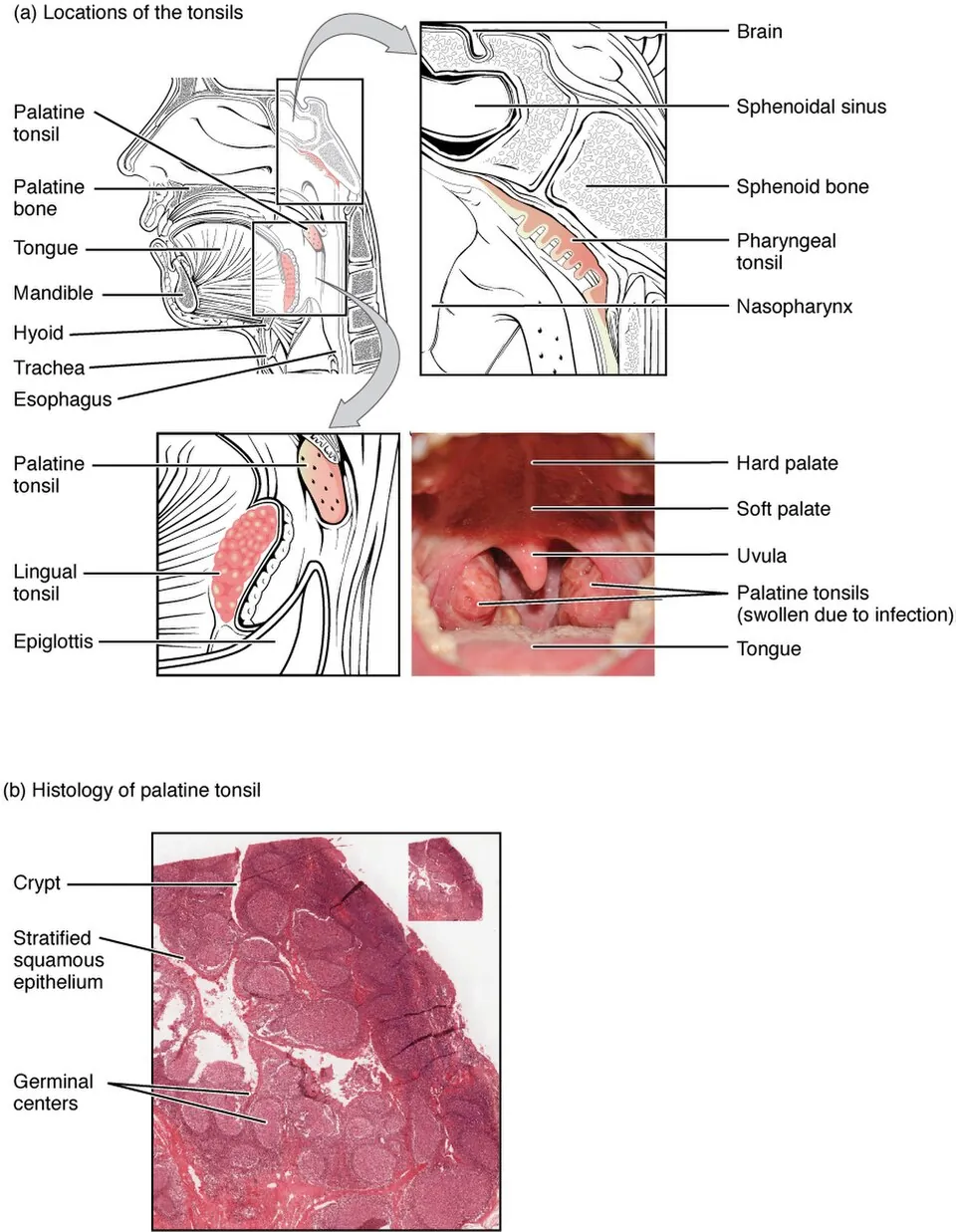
咽頭の入口を取り囲むリンパ組織の輪がWaldeyer 咽頭輪(咽頭扁桃・耳管扁桃・口蓋扁桃・舌扁桃)。空気と食物の両方が通る場所に、最初の免疫の関門を置いている。
| 扁桃 | 覆う上皮 | 陰窩 | 被膜 |
|---|---|---|---|
| 口蓋扁桃 | 重層扁平上皮 | 深く分岐する(10〜20本) | 半被膜(外側のみ) |
| 咽頭扁桃(アデノイド) | 多列線毛円柱上皮 | 浅い(ヒダ状) | 半被膜 |
| 舌扁桃 | 重層扁平上皮 | 単純(分岐しない) | 半被膜 |
リンパ節との決定的な違いは「輸入リンパ管が無い」こと。リンパ節がリンパ液を濾す装置なのに対し、扁桃は上皮の表面から直接抗原を取り込む。陰窩の上皮はところどころ網目状〈reticular epithelium〉になり、リンパ球が上皮の中まで入り込んで抗原に触れる。
内部には胚中心を持つリンパ小節が並ぶ。
陰窩が深いことの代償——口蓋扁桃の陰窩には剥がれた上皮・細菌・食物残渣が溜まる。これが固まったものが膿栓(臭い玉)で、口臭の原因になる。慢性扁桃炎の温床でもあり、扁桃の構造そのものが弱点になっている。
半被膜であることが手術と感染の広がり方を決める。外側にだけ被膜があり内側は上皮に開いているので、炎症は被膜の外へ及びやすい——扁桃周囲膿瘍へ進むと開口障害〈trismus〉・含み声・口蓋垂の偏位・片側の激しい咽頭痛を来す。さらに進めば副咽頭間隙・縦隔へ下る(降下性壊死性縦隔炎)。
扁桃摘出の適応——反復性扁桃炎(年4〜7回以上)、扁桃周囲膿瘍の反復、睡眠時無呼吸を来す扁桃肥大、病巣感染(IgA腎症・掌蹠膿疱症・胸肋鎖骨過形成症——扁桃が離れた臓器の病気の起点になる)。
扁桃は加齢で退縮する——小児期に最も大きく、思春期以降に縮む。成人で片側だけが大きい扁桃は悪性リンパ腫や癌を疑う。伝染性単核症では両側の著明な腫大と白苔を来し、アンピシリンを投与すると高率に皮疹が出るため、溶連菌性咽頭炎との鑑別が実務上重要になる。
陰窩が深く、上皮が薄いから、扁桃は感染しやすい。
扁桃周囲膿瘍 — 片側の強い咽頭痛・開口障害・含み声・口蓋垂の健側偏位。切開排膿が要る。
A 群β溶血性連鎖球菌による急性扁桃炎はリウマチ熱と急性糸球体腎炎を防ぐために抗菌薬(ペニシリン)を10日間投与する。Centor 基準で検査前確率を見積もる。
伝染性単核球症(EBV)との鑑別が重要で、アモキシシリンを投与すると高率に皮疹が出る。脾腫を伴うため、接触競技を一定期間避ける(脾破裂の予防)。
アデノイド増殖症は耳管咽頭口を塞ぎ、滲出性中耳炎と閉塞性睡眠時無呼吸を起こす。小児のいびきと口呼吸を見たら診る。
English version
Core
The ring of lymphoid tissue surrounding the entrance to the pharynx is Waldeyer ring (pharyngeal, tubal, palatine and lingual tonsils) — the first immune checkpoint placed where both air and food pass.
| Tonsil | Covering epithelium | Crypts | Capsule |
|---|---|---|---|
| Palatine | Stratified squamous | Deep and branching (10–20) | Hemicapsule (lateral only) |
| Pharyngeal (adenoid) | Pseudostratified ciliated columnar | Shallow, folded | Hemicapsule |
| Lingual | Stratified squamous | Simple, unbranched | Hemicapsule |
The decisive difference from a lymph node is the absence of afferent lymphatics. A node filters lymph; a tonsil takes antigen directly from the epithelial surface. The crypt epithelium becomes reticular in places, and lymphocytes enter the epithelium itself to meet antigen. Within lie lymphoid nodules with germinal centres.
Going further
The price of deep crypts — desquamated epithelium, bacteria and food debris accumulate in the crypts of the palatine tonsil. Consolidated, these are tonsilloliths, a cause of halitosis, and a nidus of chronic tonsillitis. The structure of the tonsil is itself its weakness.
Being only half-encapsulated determines both surgery and the spread of infection. With a capsule laterally but an open epithelial surface medially, infection readily passes beyond the capsule — a peritonsillar abscess gives trismus, a muffled voice, deviation of the uvula and severe unilateral pain; further spread descends into the parapharyngeal space and mediastinum.
Indications for tonsillectomy — recurrent tonsillitis (4–7 or more episodes a year), recurrent peritonsillar abscess, hypertrophy causing sleep apnoea, and focal infection (IgA nephropathy, palmoplantar pustulosis, SAPHO) — the tonsil as the origin of disease in a distant organ.
Tonsils involute with age, being largest in childhood. A unilaterally enlarged tonsil in an adult raises lymphoma or carcinoma. In infectious mononucleosis both are markedly enlarged with exudate, and ampicillin causes a rash in a high proportion, making the distinction from streptococcal pharyngitis important in practice.
In the clinic
Deep crypts and thin epithelium make the tonsil prone to infection.
Peritonsillar abscess — severe unilateral pain, trismus, a muffled voice, deviation of the uvula away from the affected side. Incision and drainage are required.
Acute tonsillitis due to group A β-haemolytic streptococcus is treated with penicillin for ten days, to prevent rheumatic fever and acute glomerulonephritis. Use the Centor criteria to estimate the pre-test probability.
Distinguishing infectious mononucleosis (EBV) is important, since amoxicillin causes a rash in a high proportion. Splenomegaly accompanies it, so contact sports must be avoided for a period, to prevent splenic rupture.
Adenoid hypertrophy obstructs the pharyngeal opening of the auditory tube and causes otitis media with effusion and obstructive sleep apnoea. Examine it in a snoring, mouth-breathing child.
高内皮細静脈 — リンパ球が血管から出る専用の門Venula postcapillaris
リンパ節の傍皮質(T細胞領域)にある特殊な細静脈。内皮が扁平ではなく立方〜円柱状に背が高いため、H-E標本でも見分けられる。
ここがリンパ球が血液からリンパ組織へ入る唯一の入口になる。他の毛細血管では通れない。
| 段階 | 分子 |
|---|---|
| ①係留と転がり〈rolling〉 | L-セレクチン(リンパ球側)とペリフェラルリンパ節アドレシン〈PNAd〉(HEV側) |
| ②活性化 | ケモカイン(CCL21・CCL19)が内皮上に提示される |
| ③強固な接着 | インテグリン(LFA-1)と ICAM-1 |
| ④遊出〈diapedesis〉 | 内皮の間を通り抜ける |
この4段階は好中球が炎症部位へ出るときとまったく同じ手順で、使う分子だけが違う。
リンパ球の再循環——ナイーブT細胞は血液 → HEV → リンパ節 → 輸出リンパ管 → 胸管 → 血液という輪を1日に何周もする。1つのリンパ節に留まるのは半日ほどで、抗原に出会わなければ次のリンパ節へ移る。
この巡回に意味がある——ある抗原に反応できるナイーブT細胞は全身に数千〜数万個しかいない。抗原がどこから侵入するか分からない以上、細胞のほうが全身のリンパ節を回り続けて、抗原の入ってきた場所を探すしかない。免疫の初動が数日かかるのは、この「出会い」に時間を要するから。
抗原に出会うと、その細胞はリンパ節から出られなくなる——S1P受容体1(S1PR1)の発現が一時的に下がるため。増殖・分化を終えると S1PR1 が戻り、輸出リンパ管から出ていく。
フィンゴリモド(多発性硬化症の薬)はこの仕組みを利用する——S1P受容体を持続的に刺激して受容体を細胞内へ引き込み、リンパ球をリンパ節に閉じ込める。中枢神経へ自己反応性リンパ球が到達できなくなるのが治療効果で、末梢血リンパ球数が著明に減るのは殺しているのではなく出られなくしているだけ。
慢性炎症の場に HEV が新たに作られることがある(三次リンパ組織)——関節リウマチの滑膜、橋本病の甲状腺、慢性移植片拒絶。その場に「リンパ節のような構造」ができて免疫反応が持続する。
高内皮細静脈はリンパ球が組織へ入る「改札口」である。接着分子とケモカインの組み合わせで、行き先が決まる。皮膚へ行くリンパ球、腸へ行くリンパ球は持っている受容体が違う——ホーミングである。
この仕組みが薬の標的になっている。ナタリズマブ(抗 α4 インテグリン)は多発性硬化症と Crohn 病に使われるが、中枢へのリンパ球の移動も止めてしまうため進行性多巣性白質脳症(JC ウイルス)のリスクがある。ベドリズマブは腸管選択的(α4β7)なのでこのリスクが低い——「どの改札を止めるか」で副作用が変わる。
フィンゴリモド(S1P 受容体調節薬)はリンパ球をリンパ節から出られなくする。末梢血リンパ球が著明に減るのは殺しているのではなく閉じ込めているからである。
English version
Core
A specialised venule in the paracortex (T cell zone) of a lymph node. Its endothelium is cuboidal to columnar rather than flattened, so it can be recognised even on H&E. This is the only entrance by which lymphocytes pass from blood into lymphoid tissue; they cannot cross other capillaries.
| Step | Molecules |
|---|---|
| ① Tethering and rolling | L-selectin (lymphocyte) and PNAd (HEV) |
| ② Activation | Chemokines (CCL21, CCL19) presented on the endothelium |
| ③ Firm adhesion | Integrin (LFA-1) and ICAM-1 |
| ④ Diapedesis | Passage between the endothelial cells |
These four steps are exactly the sequence by which a neutrophil leaves a vessel at a site of inflammation; only the molecules differ.
Going further
Lymphocyte recirculation — a naive T cell travels blood → HEV → lymph node → efferent lymphatic → thoracic duct → blood, several times a day. It stays in one node for about half a day and, if it does not meet antigen, moves to the next.
There is a reason for this patrol. There are only thousands to tens of thousands of naive T cells in the whole body able to respond to any given antigen. Since it is not known where the antigen will enter, the cells must go round the nodes of the body looking for the place it entered. The first days of an immune response are taken up with that encounter.
Once it meets antigen the cell can no longer leave the node, because expression of S1P receptor 1 falls temporarily; when proliferation and differentiation are complete S1PR1 returns and it exits by the efferent lymphatic.
Fingolimod (for multiple sclerosis) exploits this: it stimulates the S1P receptor persistently so that the receptor is internalised, confining lymphocytes to the lymph nodes. Self-reactive lymphocytes can no longer reach the CNS, and the marked fall in the blood lymphocyte count is not killing but preventing exit.
HEVs can be newly formed at sites of chronic inflammation (tertiary lymphoid tissue) — the rheumatoid synovium, the thyroid in Hashimoto disease, chronic graft rejection. A structure like a lymph node appears on the spot and the immune response persists.
In the clinic
The high endothelial venule is the ticket gate through which lymphocytes enter tissue, and the combination of adhesion molecules and chemokines decides the destination. Lymphocytes bound for skin and for gut carry different receptors — homing.
This mechanism is a drug target. Natalizumab (anti-α4 integrin) is used in multiple sclerosis and Crohn disease, but it also stops lymphocytes entering the CNS and carries a risk of progressive multifocal leukoencephalopathy (JC virus). Vedolizumab is gut-selective (α4β7) and carries a lower risk — which gate is blocked changes the adverse effects.
Fingolimod prevents lymphocytes leaving the node. The marked fall in circulating lymphocytes is confinement, not killing.
胸腺での選択 — 使えるものだけを残し、危ないものを消すSelectio thymica
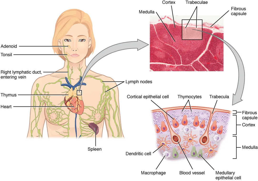
T細胞は胸腺で2段階の試験を受ける。
| 段階 | 場所 | 合格の条件 | 不合格なら |
|---|---|---|---|
| 正の選択 | 皮質 | 自己 MHC に、そこそこ結合できる | 死ぬ(無視による細胞死) |
| 負の選択 | 髄質 | 自己抗原に、強く結合しない | 死ぬ(アポトーシス)/制御性T細胞になる |
生き残るのは 2〜5% 程度になる。
髄質上皮細胞は AIRE という転写因子を使い、本来そこには無いはずの臓器特異的な蛋白(インスリンなど)を発現して見せる。
Hassall 小体は髄質にある同心円状の構造で、制御性T細胞の分化に関わるとされる。
「自己を見せることで、自己を攻撃するものを消す」という逆説が、この器官の核心になる。AIRE が全身の臓器の抗原を胸腺の中で再現している(自己免疫の始まりの章)——「見せなければ、教えられない」。
だから AIRE の異常は、多臓器の自己免疫になる(免疫不全と自己免疫の章)——「自己免疫性多内分泌腺症候群1型(副甲状腺機能低下症・副腎不全・皮膚粘膜カンジダ症)」。
正の選択が MHC 拘束性を作っている(HLA の項)——「自分の MHC の上でしか抗原を認識できない、という性質はここで決まる」。
胸腺が無ければ、T細胞が育たない(咽頭嚢の章)——「DiGeorge 症候群では、第3・第4咽頭嚢の異常で胸腺と副甲状腺が欠ける」。
胸腺は思春期以降に退縮し、脂肪に置き換わる(胸部CTの章)——「成人の前縦隔に胸腺の腫大があれば、胸腺腫やリンパ腫を考える」。
負の選択が破れると自己免疫になる。
AIRE 遺伝子は胸腺髄質上皮に全身の自己抗原を発現させる。ここで自己反応性 T 細胞を消す。AIRE の変異で自己免疫性多内分泌腺症候群 1 型(副甲状腺機能低下症・副腎不全・皮膚粘膜カンジダ症)になる。1つの遺伝子が、全身の自己寛容を担っている。
胸腺腫と重症筋無力症の合併は選択の場が腫瘍化した結果と理解できる。重症筋無力症では胸腺の評価が必須で、胸腺摘出が治療になりうる。
DiGeorge 症候群では胸腺そのものが低形成なのでT 細胞が育たない。生ワクチンが禁忌になる。
胸腺は思春期以降に退縮し、脂肪に置き換わる。成人の前縦隔の胸腺様組織は腫瘍を疑う。
English version
Core
A T cell sits two examinations in the thymus.
| Stage | Site | To pass | If it fails |
|---|---|---|---|
| Positive selection | Cortex | Bind self MHC with moderate affinity | Dies (death by neglect) |
| Negative selection | Medulla | Do not bind self antigen strongly | Dies (apoptosis), or becomes a regulatory T cell |
Only about 2–5 per cent survive.
The medullary epithelial cells use the transcription factor AIRE to display tissue-specific proteins that have no business being there — insulin, for example. Hassall corpuscles are concentric structures in the medulla, thought to be involved in the differentiation of regulatory T cells.
Going further
The paradox at the heart of this organ is that the self is displayed in order to delete what would attack the self. AIRE reproduces the antigens of the whole body inside the thymus — what is not shown cannot be taught.
So a defect of AIRE gives multi-organ autoimmunity — autoimmune polyendocrine syndrome type 1 (hypoparathyroidism, adrenal failure, mucocutaneous candidiasis).
Positive selection creates MHC restriction — the property that antigen can only be recognised on one’s own MHC is settled here.
Without a thymus, T cells do not develop — in DiGeorge syndrome a defect of the third and fourth pharyngeal pouches leaves the thymus and parathyroids absent.
The thymus involutes after puberty and is replaced by fat — so thymic enlargement in the anterior mediastinum of an adult raises thymoma or lymphoma.
In the clinic
When negative selection fails the result is autoimmunity. The AIRE gene makes the thymic medullary epithelium express the self antigens of the whole body, and self-reactive T cells are deleted there. Mutation of AIRE gives autoimmune polyendocrine syndrome type 1. A single gene carries the tolerance of the whole body.
The association of thymoma with myasthenia gravis can be understood as neoplastic transformation of the site of selection itself. Assessment of the thymus is mandatory in myasthenia gravis, and thymectomy may be a treatment.
In DiGeorge syndrome the thymus itself is hypoplastic so T cells do not develop — live vaccines are contraindicated.
The thymus involutes after puberty and is replaced by fat; thymus-like tissue in the anterior mediastinum of an adult raises the suspicion of a tumour.
胸腺の上皮性細網細胞 — I〜VI型と、血液胸腺関門Epitheliocytus reticularis
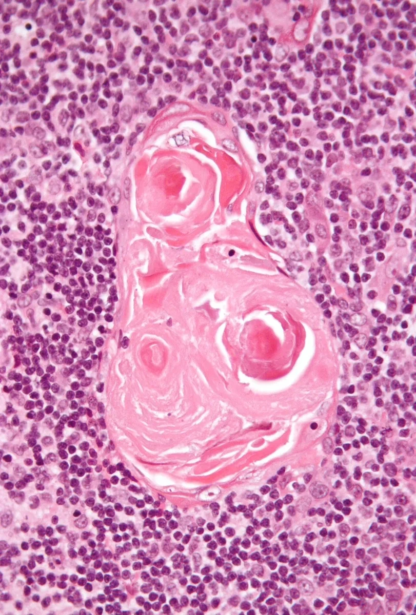
胸腺の骨組みは、ほかのリンパ器官のような細網線維と細網細胞ではない。咽頭嚢の内胚葉に由来する上皮細胞が星形に突起を伸ばして網を作っている。上皮である証拠に、デスモソームとケラチン(中間径フィラメント)を持ち、結合組織に面するところでは基底膜を作る。一方で形は細網細胞に似ているので上皮性細網細胞(偽細網細胞)と呼ぶ。細網線維は作らない。
位置と働きで6つの型に分ける。皮質に3つ、髄質に3つである。
| 型 | 場所 | 形と働き |
|---|---|---|
| I型 | 皮質:被膜・小柱の内側と、皮質の血管の周り | 密着結合で隙間なくつながり、皮質を結合組織から隔てる。血液胸腺関門の一部 |
| II型 | 皮質の中ほど | 長い突起をデスモソームでつないで網を作る。MHC クラス I・II を表出し、正の選択で自己 MHC を見せる |
| III型 | 皮質と髄質の境 | 密着結合で皮質と髄質を仕切る |
| IV型 | 皮髄境界(髄質側) | III型と並んで境界を作る |
| V型 | 髄質 | 髄質の網を作る。自己抗原を見せる(負の選択) |
| VI型 | 髄質 | 大きく、集まって角化しHassall 小体になる |
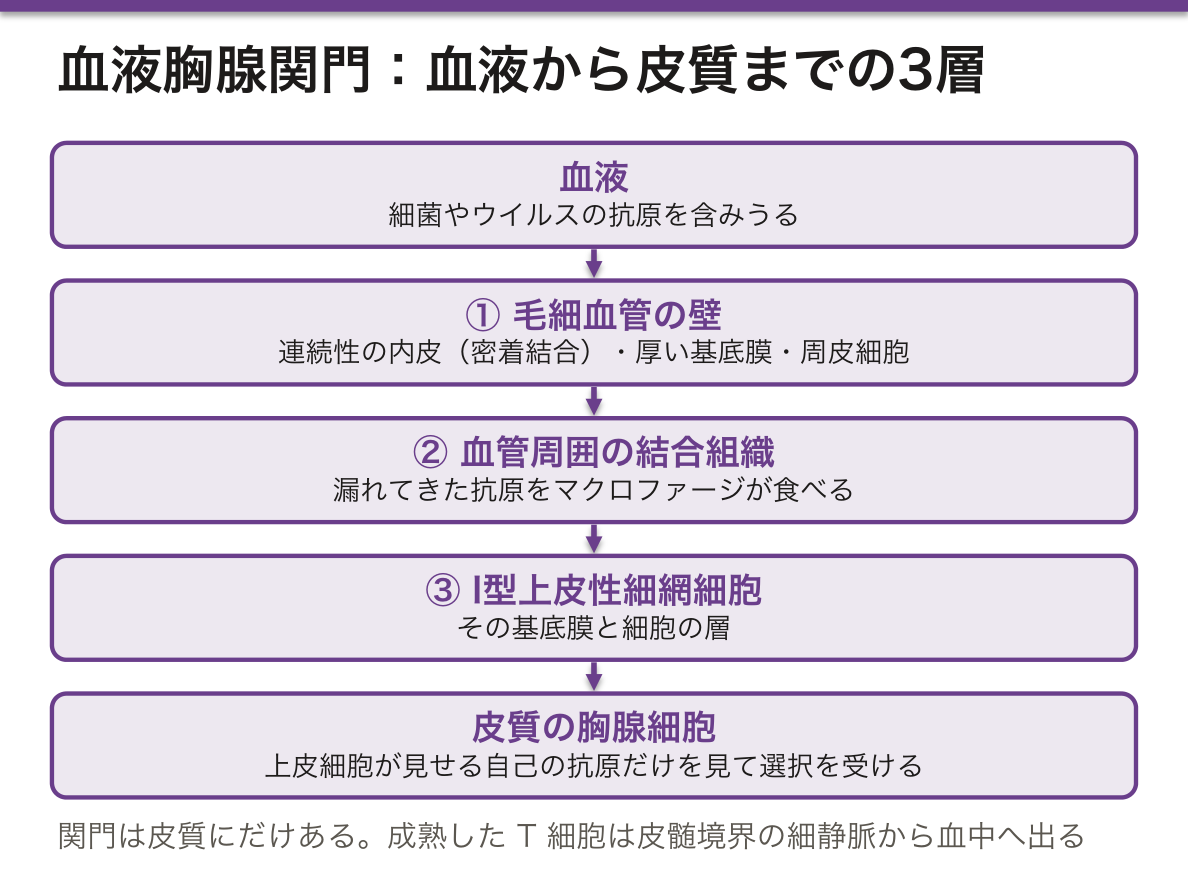
血液胸腺関門は皮質にだけある。血管の中から外へ順に、
- 毛細血管の壁——連続性毛細血管の内皮(密着結合)、厚い基底膜、周皮細胞
- 血管周囲の結合組織——漏れてきた抗原を食べるマクロファージ
- I型上皮性細網細胞の基底膜と細胞の層
の3層である。
なぜ皮質を閉じるのか。皮質で正の選択を受けている胸腺細胞は、上皮細胞が見せる自己の抗原だけを見なければならない。もし血液から細菌やウイルスの抗原が入ってくると、それに反応する細胞まで「自己に反応した」と誤って消されてしまい、本当に戦うべき相手に対する T 細胞が作れなくなる。成熟した T 細胞は皮髄境界の細静脈から血中へ出る。
胸腺腫は、リンパ球ではなく上皮性細網細胞の腫瘍である。WHO 分類は、腫瘍の上皮細胞が紡錘形か類上皮形か、そして一緒にいる未熟な T 細胞がどれだけ多いかで型(A・AB・B1〜B3)を分ける。生検ではサイトケラチンの免疫染色で上皮の網が浮かび上がり、間に TdT 陽性の未熟 T 細胞が詰まる。重症筋無力症を合併しうるのは、腫瘍化した上皮が T 細胞の教育を誤らせるためと考えられている。
DiGeorge 症候群で T 細胞が育たないのは、教育する側=咽頭嚢に由来する上皮が無いからである。リンパ球の前駆細胞は骨髄から来ても、学校が無い。
English version
Core
The framework of the thymus is not reticular fibres and reticular cells, as in other lymphoid organs. It is a net of epithelial cells from the endoderm of the pharyngeal pouch, star-shaped with long processes. They show they are epithelial by having desmosomes and keratin (intermediate filaments), and they make a basement membrane where they face connective tissue. Because their shape resembles reticular cells, they are called epithelioreticular (pseudoreticular) cells. They do not make reticular fibres.
By position and function there are six types: three in the cortex and three in the medulla.
| Type | Site | Form and job |
|---|---|---|
| I | cortex: under the capsule and trabeculae, and around cortical vessels | joined by tight junctions; separate the cortex from connective tissue; part of the blood–thymus barrier |
| II | mid-cortex | long processes joined by desmosomes form a net; express MHC class I and II and show self MHC in positive selection |
| III | corticomedullary border | tight junctions; separate cortex from medulla |
| IV | corticomedullary border (medullary side) | form the border together with type III |
| V | medulla | make the medullary net; show self antigens (negative selection) |
| VI | medulla | large; cluster and keratinise to form Hassall corpuscles |
Going further
The blood–thymus barrier exists only in the cortex. From the vessel lumen outward:
- Capillary wall — continuous endothelium with tight junctions, a thick basement membrane, pericytes.
- Perivascular connective tissue — macrophages that eat any antigen that leaks out.
- The basement membrane and cell layer of type I epithelioreticular cells.
Why close the cortex? Thymocytes in positive selection must see only self antigens shown by epithelial cells. If bacterial or viral antigens came in from the blood, cells reacting to them would be wrongly deleted as "self-reactive", and T cells against real enemies could not be made. Mature T cells leave through venules at the corticomedullary border.
In the clinic
Thymoma is a tumour of epithelioreticular cells, not of lymphocytes. The WHO types (A, AB, B1–B3) depend on whether the tumour epithelial cells are spindle or epithelioid, and on how many immature T cells come with them. In a biopsy, cytokeratin staining shows the epithelial net, with TdT-positive immature T cells packed between. Myasthenia gravis may follow, probably because the tumour epithelium mis-educates T cells.
In DiGeorge syndrome T cells do not develop because the teacher — the epithelium from the pharyngeal pouch — is missing. Precursors still come from the marrow, but there is no school.
胸腺のホルモンと、ホルモンによる調節 — 胸腺はなぜ縮むのかInvolutio thymi
胸腺はリンパ器官であると同時に内分泌腺でもある。上皮性細網細胞が、ホルモンの性質を持つペプチドを出す。
| ホルモン | 要点 |
|---|---|
| サイムリン(thymulin) | 働くのに亜鉛が要る。T 細胞の分化を進める |
| サイモシン(thymosin α1・β4 など) | T 細胞の成熟と免疫応答を高める |
| サイモポエチン | 前駆細胞を T 細胞の系統へ進める |
これらは胸腺の中で胸腺細胞に働くほか、血中に出て末梢の T 細胞の働きにも影響する。
逆に胸腺はほかのホルモンに強く左右される。
- 糖質コルチコイド——皮質の未熟な胸腺細胞(CD4・CD8 ともに陽性)をアポトーシスで大量に死なせる。数日で皮質が薄くなる(急性退縮・ストレス退縮)
- 性ステロイド(アンドロゲン・エストロゲン)——思春期からの加齢退縮を進める。動物では去勢すると胸腺が大きくなる
- 成長ホルモン・甲状腺ホルモン——胸腺を保つ側に働く
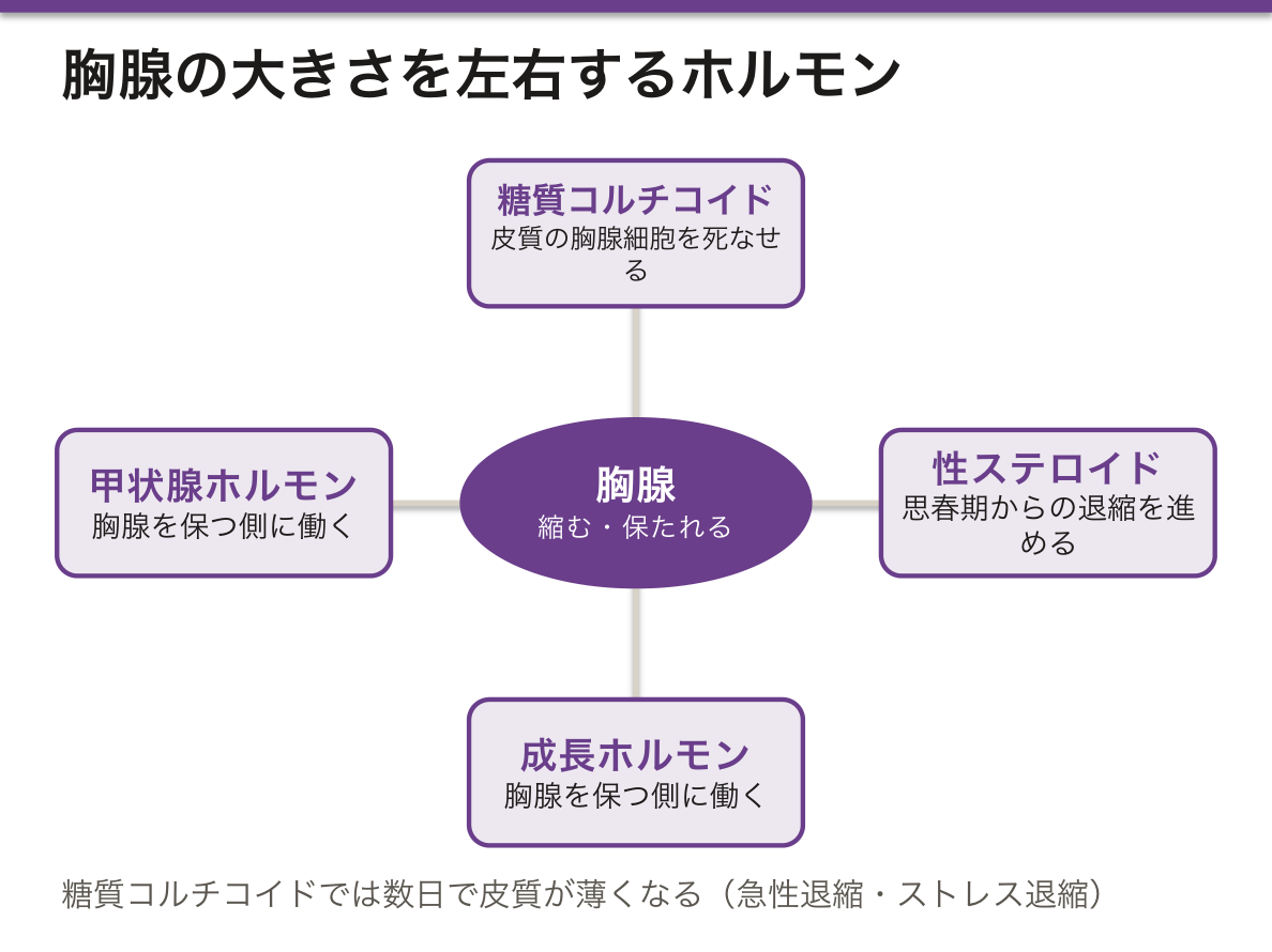
胸腺の実質は小児期に最も多く、重さは新生児で 10〜15 g、思春期に 30〜40 g が目安とされる。その後は皮質と髄質が脂肪組織に置き換わり、高齢では実質がわずかになる(加齢退縮)。標本では、成人の胸腺は脂肪の中に小葉が島のように残って見える。
なぜ早くに縮んでよいのか。T 細胞の品揃え(レパートリー)は小児期にほぼ作り終わる。その後は、長生きする記憶 T 細胞とナイーブ T 細胞が末梢で自分で分裂して数を保つ。だから成人で胸腺を切除しても、すぐには免疫不全にならない。
ステロイドで死にやすいのが皮質の胸腺細胞であることも、選択の仕組みと合う。皮質は選別の途中で、もともと 9 割以上が死ぬ場所である。ストレス退縮では皮質が薄くなり、皮質と髄質の濃淡が逆転して見える。
ステロイドを投与するとリンパ球が減るのは、この感受性による(血管外への移動も加わる)。
化学療法や重い病気から回復したあと、胸腺が一時的に元より大きくなることがある(リバウンド過形成)。リンパ腫の治療後の CT で前縦隔が腫れて見えたとき、再発と区別する必要がある。
成人に造血幹細胞移植をすると、T 細胞の回復が遅い。胸腺が退縮していて新しい T 細胞を作る力が小さいためで、移植後のウイルス感染が長引く一因になる。新生児の重症複合免疫不全症のスクリーニングでは、胸腺から出たばかりの T 細胞にだけある DNA の輪(TREC)を数えて、胸腺の出力を測る。
亜鉛欠乏では胸腺が萎縮し、細胞性免疫が落ちる。サイムリンが亜鉛なしでは働かないことと合う。
English version
Core
The thymus is a lymphoid organ and also an endocrine gland. Its epithelioreticular cells release peptides with hormone-like action.
| Hormone | Key point |
|---|---|
| Thymulin | needs zinc to work; promotes T-cell differentiation |
| Thymosins (thymosin α1, β4 and others) | enhance T-cell maturation and immune responses |
| Thymopoietin | pushes precursors into the T-cell line |
They act on thymocytes inside the thymus, and also enter the blood and affect peripheral T cells.
In the other direction, the thymus is strongly controlled by other hormones.
- Glucocorticoids — make the immature cortical thymocytes (CD4 and CD8 double-positive) die in large numbers by apoptosis. The cortex thins within days (acute or stress involution).
- Sex steroids (androgens, oestrogens) — speed up age involution from puberty. In animals, castration enlarges the thymus.
- Growth hormone and thyroid hormone — help to keep the thymus.
Going further
Thymic tissue is greatest in childhood. Typical weights are 10–15 g at birth and 30–40 g at puberty. After that, cortex and medulla are replaced by fat, and in old age little tissue is left (age involution). On a slide, the adult thymus shows lobules left as islands in fat.
Why can it shrink so early? The T-cell repertoire is almost complete by childhood. After that, long-lived memory and naive T cells divide in the periphery to keep their numbers. So removing the thymus in an adult does not cause immunodeficiency at once.
It fits the selection system that cortical thymocytes are the ones killed by steroids. The cortex is a sorting site where over 90% of cells die anyway. In stress involution the cortex thins and the dark–light pattern of cortex and medulla reverses.
In the clinic
This sensitivity is why steroids lower the lymphocyte count (movement out of the blood adds to it).
After chemotherapy or a severe illness, the thymus may become temporarily larger than before (rebound hyperplasia). When CT after lymphoma treatment shows a swollen anterior mediastinum, this must be told apart from relapse.
After stem-cell transplantation in adults, T-cell recovery is slow. The involuted thymus makes few new T cells, which is one reason viral infections last long after transplant. Newborn screening for severe combined immunodeficiency counts DNA circles (TREC) found only in T cells that have just left the thymus, as a measure of thymic output.
Zinc deficiency shrinks the thymus and weakens cellular immunity, which fits with thymulin not working without zinc.
胚中心 — 抗体が「鍛えられる」場所Centrum germinativum
リンパ濾胞の中心にできる明るい領域で、抗原に出会った後にだけ作られる。ここで B 細胞が選別され、抗体の質が上がる。
- 暗殻——B 細胞が盛んに分裂し、抗体の遺伝子に狙って変異を入れる(体細胞超変異)。細胞が密で核が濃いので暗く見える
- 明殻——濾胞樹状細胞が抗原を提示し、よく結合できる B 細胞だけが生き残る(親和性成熟)。結合できないものはその場でアポトーシスに落ちる
だから胚中心には死んだ細胞が大量に出る。それを食べたマクロファージが散在して見える像を星空像と呼ぶ——暗い背景に明るい点が散る。
胚中心は「あるかないか」だけでなく、形も情報を持つ。
| 目立つ胚中心+星空像 | 反応性の過形成 | 感染・炎症。大きさは不揃いで、明殻と暗殻の極性が保たれる |
| 大きさの揃った濾胞が密に並ぶ | 濾胞性リンパ腫 | 極性が失われ、星空像が消える——選別が働いていない |
| 胚中心が消える | 免疫不全・免疫抑制・高度の消耗 | 濾胞が小さく、萎縮して見える |
星空像が消えることに意味がある——胚中心は本来「大量に死ぬ場所」であり、死んだ細胞を片づけるマクロファージが散在するのが正常な姿である。死ななくなった濾胞は、選別が壊れている。濾胞性リンパ腫で BCL2(アポトーシスを止める蛋白)が転座で過剰になることと、この像は直接に対応している。正常な胚中心の B 細胞は BCL2 が陰性なので、BCL2 の免疫染色が反応性か腫瘍性かを分ける。
⚠️ わざと DNA を切る場所なので、癌が生まれやすい。Burkitt リンパ腫(MYC 転座)は増殖が極めて速く、星空像が最も顕著に見える腫瘍である——腫瘍でありながら大量に死んでいるためで、Ki-67 がほぼ 100% になる。びまん性大細胞型 B 細胞リンパ腫の多くも、胚中心またはその後の細胞に由来する。
濾胞樹状細胞は、名前に反して樹状細胞ではない。間葉系由来で、抗原提示(MHC クラス II による)はしない。免疫複合体を長期間そのまま表面に保持して、B 細胞に触れさせるのが仕事である。骨髄由来の樹状細胞とは別の細胞なので、混同しない。
乳幼児に多糖体ワクチンが効きにくいのは、胚中心を作る仕組みが未熟だからである。だから結合型ワクチンが使われる。
English version
Core
The pale area formed at the centre of a lymphoid follicle, created only after antigen has been met. It is where B cells are selected and the quality of antibody is raised.
- Dark zone — B cells divide vigorously and introduce targeted mutations into the antibody genes (somatic hypermutation). Dense cells with dark nuclei, hence dark.
- Light zone — follicular dendritic cells present antigen and only B cells that bind well survive (affinity maturation). Those that cannot bind undergo apoptosis on the spot.
So a germinal centre produces a great many dead cells, and the macrophages that have eaten them appear scattered against the darker background — the starry sky appearance.
Going further
A germinal centre carries information not only by its presence but by its form.
| Appearance | Meaning |
|---|---|
| Prominent germinal centres with a starry sky | Reactive hyperplasia — infection, inflammation. Sizes are uneven and the polarity of light and dark zones is preserved |
| Uniformly sized follicles packed together | Follicular lymphoma — polarity is lost and the starry sky disappears; selection is not operating |
| Germinal centres absent | Immunodeficiency, immunosuppression, severe wasting |
The loss of the starry sky is meaningful: a germinal centre is by nature a place where a great many cells die, and scattered macrophages clearing them is the normal picture. A follicle in which cells no longer die is one in which selection is broken. That corresponds directly to the overexpression of BCL2 by translocation in follicular lymphoma. Normal germinal centre B cells are BCL2 negative, so BCL2 immunostaining separates reactive from neoplastic.
⚠️ Because DNA is deliberately cut here, cancers arise readily. Burkitt lymphoma (MYC translocation) proliferates extremely fast and shows the most striking starry sky of all — a tumour, yet dying in great numbers — with a Ki-67 of nearly 100 per cent. Most diffuse large B-cell lymphomas also derive from germinal centre or post-germinal centre cells.
The follicular dendritic cell, despite its name, is not a dendritic cell. It is of mesenchymal origin and does not present antigen on MHC class II; its work is to hold immune complexes intact on its surface for long periods so that B cells can touch them. Do not confuse it with the bone marrow-derived dendritic cell.
In the clinic
Polysaccharide vaccines work poorly in infants because the machinery for forming germinal centres is immature. This is why conjugate vaccines are used.
リンパ球の種類 — 見た目は同じ、働きは違うLymphocytus
光学顕微鏡では区別できない——いずれも大きな円い核と、わずかな細胞質。種類を決めるには表面マーカー(免疫染色・フローサイトメトリー)が要る。この「形態では分からない」ことが答えの出発点。
B リンパ球(約 20〜25%)——骨髄で成熟。抗体を作る(形質細胞へ分化)。マーカーは CD19・CD20。
T リンパ球(約 60〜70%)——胸腺で成熟。細胞性免疫を担う。マーカーは CD3。
NK 細胞(約 5〜10%)——大顆粒リンパ球として、唯一やや大きく細胞質に顆粒が見える。抗原提示を受けずに、MHC クラス I を失った細胞(ウイルス感染細胞・腫瘍細胞)を殺す。CD16・CD56 陽性、CD3 陰性。
寿命も違う——多くは数日だが、記憶細胞は数十年生きる。リンパ球は血管とリンパ管の間を再循環する唯一の血球である。
再循環の入口が高内皮細静脈(HEV)。リンパ球はここから血管を出てリンパ節へ入り、輸出リンパ管 → 胸管 → 静脈と戻る。
English version
Core
They cannot be told apart by light microscopy — all have a large round nucleus with scanty cytoplasm. Surface markers (immunostaining, flow cytometry) are needed, and that they cannot be distinguished morphologically is the starting point of the answer.
- B lymphocytes (about 20–25 per cent) — mature in the bone marrow; make antibody (differentiating into plasma cells). Markers CD19, CD20.
- T lymphocytes (about 60–70 per cent) — mature in the thymus; carry out cell-mediated immunity. Marker CD3.
- NK cells (about 5–10 per cent) — as large granular lymphocytes they alone are slightly larger with visible cytoplasmic granules. They kill cells that have lost MHC class I (virus-infected, tumour) without antigen presentation. CD16 and CD56 positive, CD3 negative.
Lifespans differ too — most a few days, but memory cells live for decades. The lymphocyte is the only blood cell that recirculates between blood and lymphatics.
Going further
The entrance to that recirculation is the high endothelial venule. Lymphocytes leave the vessel there, enter the node, and return by efferent lymphatic → thoracic duct → vein.
B リンパ球 — 抗体を作る細胞
発生——骨髄で幹細胞から分化し、そこで抗原受容体(膜結合型 IgM)を組み立てる。自己に強く反応するものはここで除かれる(中枢性寛容)。成熟 B 細胞は末梢のリンパ性器官(リンパ節皮質の一次濾胞、脾の白脾髄、MALT)へ移る。
活性化——抗原が結合し、ヘルパー T 細胞の助けを受けると胚中心を作る。胚中心では体細胞高頻度変異(抗体の親和性を上げる)とクラススイッチ(IgM から IgG・IgA・IgE へ)が起こる。
行き先は2つ——形質細胞(抗体を大量に作る。粗面小胞体が細胞質を埋め、核が車輪のスポーク状——組織で見分けられる)と記憶 B 細胞。
働き——液性免疫の主役。抗体による中和・オプソニン化・補体活性化。抗原提示細胞としても働く。
胚中心の明帯・暗帯の構造と、そこでの選択がhisto.lym.08 の内容。形質細胞は光学顕微鏡で同定できる数少ない免疫細胞である点を押さえておく。
English version
Core
Development — differentiates from a stem cell in the bone marrow, where it assembles its antigen receptor (membrane-bound IgM). Those reacting strongly with self are removed there (central tolerance). The mature B cell moves to the peripheral lymphoid organs — the primary follicles of the nodal cortex, the splenic white pulp, MALT.
Activation — when antigen binds and helper T cell assistance is received, a germinal centre forms, where somatic hypermutation (raising affinity) and class switching (IgM to IgG, IgA, IgE) occur.
Two destinations — the plasma cell (making antibody in quantity; rough ER fills the cytoplasm and the nucleus is spoked like a cartwheel — identifiable in tissue) and the memory B cell.
Functions — the protagonist of humoral immunity: neutralisation, opsonisation and complement activation by antibody. It also acts as an antigen-presenting cell.
Going further
The light and dark zone structure of the germinal centre and the selection that occurs there are covered in the section on the germinal centre. Fix firmly that the plasma cell is one of the few immune cells identifiable by light microscopy.
T リンパ球 — 亜群と分化
発生——骨髄で生まれ、胸腺で成熟する(だから T)。皮質で正の選択(自己 MHC を認識できるものを残す)、髄質で負の選択(自己抗原に強く反応するものを消す)。通過するのは 2〜5% だけ。
亜群——ヘルパー T(CD4+):MHC クラス II を認識し、司令塔として B 細胞・マクロファージ・細胞傷害性 T を動かす。さらに Th1(細胞性免疫)・Th2(液性免疫とアレルギー)・Th17(細胞外細菌と真菌) に分かれる。細胞傷害性 T(CD8+):MHC クラス I を認識し、感染細胞と腫瘍細胞を直接殺す(パーフォリンとグランザイム)。制御性 T(Treg、CD4+CD25+FoxP3+):免疫応答を止める——自己免疫を防ぐ。
覚え方は「クラス I は 8 へ、クラス II は 4 へ」(I×8=II×4=8)。
HIV は CD4+ T 細胞に感染するため、司令塔が失われて液性・細胞性の両方が破綻する。Treg の破綻が自己免疫疾患につながる。
English version
Core
Development — born in the bone marrow, matures in the thymus (hence T). Positive selection in the cortex (retaining those able to recognise self MHC) and negative selection in the medulla (deleting those reacting strongly with self antigen). Only 2–5 per cent pass.
Subpopulations
- Helper T (CD4+) — recognises MHC class II; the commander, directing B cells, macrophages and cytotoxic T cells. Divided further into Th1 (cell-mediated), Th2 (humoral, allergy) and Th17 (extracellular bacteria and fungi).
- Cytotoxic T (CD8+) — recognises MHC class I; kills infected and tumour cells directly with perforin and granzyme.
- Regulatory T (CD4+CD25+FoxP3+) — stops the response; prevents autoimmunity.
Mnemonic: class I goes to 8, class II goes to 4 — I × 8 = II × 4 = 8.
Going further
HIV infects the CD4+ T cell, so the commander is lost and both humoral and cell-mediated immunity collapse. Failure of the regulatory T cell leads to autoimmune disease.
液性免疫と細胞性免疫 — どちらが何を相手にするか
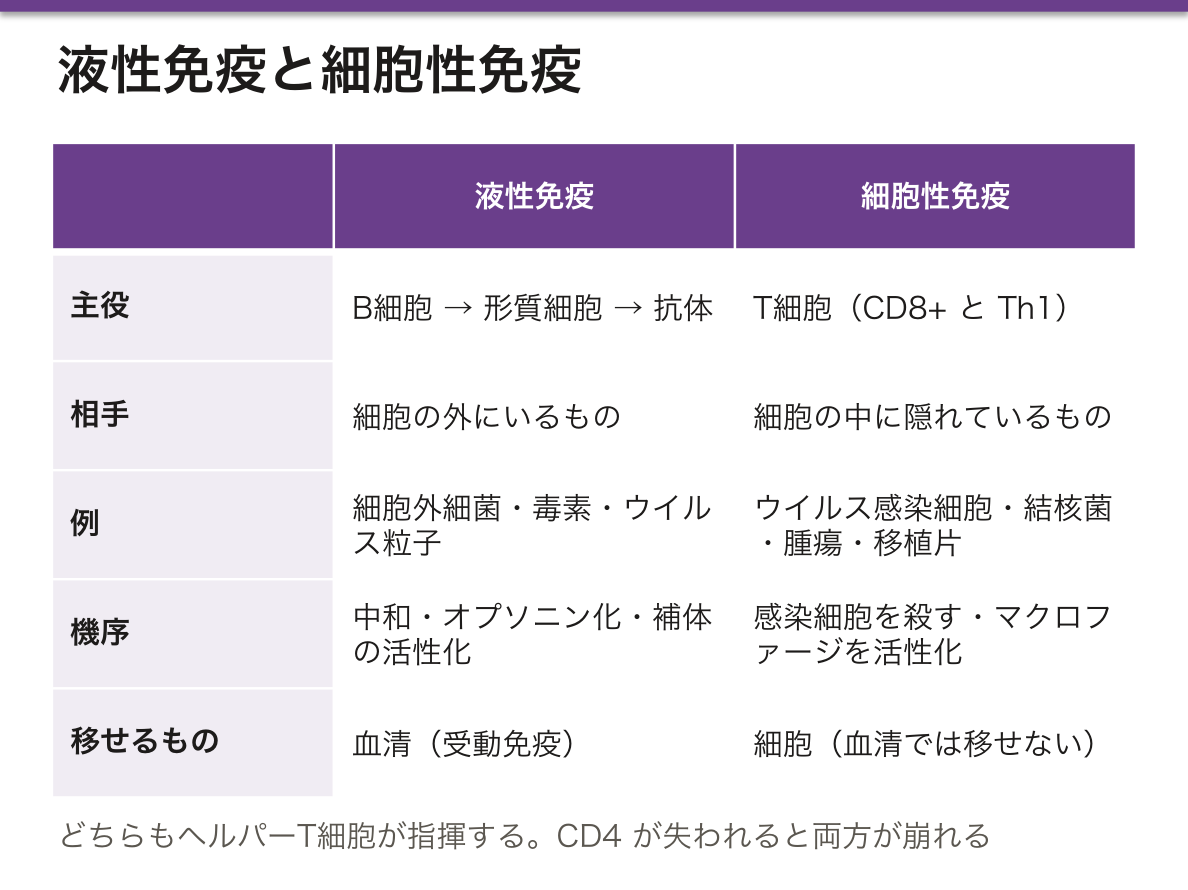
液性免疫 humoral immunity——主役はB 細胞 → 形質細胞 → 抗体。抗体は体液中を巡るので細胞の外にいるものを相手にする——細胞外細菌・毒素・細胞外のウイルス粒子。機序は中和・オプソニン化・補体の活性化。血清を移せば移せる(受動免疫)のが特徴。
細胞性免疫 cell-mediated immunity——主役はT 細胞(CD8+ とマクロファージを活性化する Th1)。相手は細胞の中に隠れているもの——ウイルス感染細胞・結核菌などの細胞内寄生菌・真菌・腫瘍細胞・移植片。血清では移せず、細胞を移す必要がある。
対比の軸は「細胞の外か、中か」。これが決まれば、担い手も機序も自動的に決まる。
両者は独立ではない——ヘルパー T 細胞がどちらも指揮している。だから CD4 が失われると両方が崩れる。
臨床での見分け——ツベルクリン反応と接触性皮膚炎は細胞性(IV 型、遅延型)、アナフィラキシーは液性(I 型、IgE)。移植拒絶は主に細胞性。
English version
Core
Humoral immunity — the protagonists are the B cell → plasma cell → antibody. Antibody circulates in body fluids, so it deals with what is outside cells: extracellular bacteria, toxins, free virus particles. The mechanisms are neutralisation, opsonisation and complement activation. It can be transferred by serum (passive immunity).
Cell-mediated immunity — the protagonists are T cells (CD8+, and the Th1 cells that activate macrophages). The targets are what is hidden inside cells: virus-infected cells, intracellular bacteria such as tuberculosis, fungi, tumour cells and grafts. It cannot be transferred by serum; cells must be transferred.
The axis of the contrast is outside or inside the cell. Settle that, and the effector and the mechanism follow automatically.
The two are not independent — the helper T cell directs both. So when CD4 is lost, both collapse.
Going further
Distinguishing them clinically — the tuberculin reaction and contact dermatitis are cell-mediated (type IV, delayed); anaphylaxis is humoral (type I, IgE). Graft rejection is chiefly cell-mediated.
まとめ
| 胸腺 | T細胞が教育を受ける一次リンパ器官。前縦隔にあり、小葉に分かれる。 |
|---|---|
| リンパ節 | リンパ行性の抗原を捕まえる二次リンパ器官。 |
| 脾臓 | 血行性の抗原を捕まえ、同時に血液を濾す。 |
| 粘膜関連リンパ組織 — 被膜を持たない免疫 | 粘膜の直下に散在するリンパ組織。被膜を持たないのが特徴で、上皮のすぐ裏で待ち構える。 |
| 扁桃 — 上皮が陥入して免疫が待ち構える場所 | 咽頭の入口を取り囲むリンパ組織の輪がWaldeyer 咽頭輪(咽頭扁桃・耳管扁桃・口蓋扁桃・舌扁桃)。 |
| 高内皮細静脈 — リンパ球が血管から出る専用の門 | リンパ節の傍皮質(T細胞領域)にある特殊な細静脈。 |
| 胸腺での選択 — 使えるものだけを残し、危ないものを消す | T細胞は胸腺で2段階の試験を受ける。段階場所合格の条件不合格なら正の選択皮質自己 MHC に、そこそこ結合できる死ぬ(無視による細胞死)負の選択髄質自己抗原に、強く結合しない死ぬ(… |
| 胸腺の上皮性細網細胞 — I〜VI型と、血液胸腺関門 | 胸腺の骨組みは、ほかのリンパ器官のような細網線維と細網細胞ではない。 |
| 胸腺のホルモンと、ホルモンによる調節 — 胸腺はなぜ縮むのか | 胸腺はリンパ器官であると同時に内分泌腺でもある。 |
| 胚中心 — 抗体が「鍛えられる」場所 | リンパ濾胞の中心にできる明るい領域で、抗原に出会った後にだけ作られる。 |
| リンパ球の種類 — 見た目は同じ、働きは違う | 光学顕微鏡では区別できない——いずれも大きな円い核と、わずかな細胞質。 |
| B リンパ球 — 抗体を作る細胞 | 発生——骨髄で幹細胞から分化し、そこで抗原受容体(膜結合型 IgM)を組み立てる。 |
| T リンパ球 — 亜群と分化 | 発生——骨髄で生まれ、胸腺で成熟する(だから T)。 |
| 液性免疫と細胞性免疫 — どちらが何を相手にするか | 液性免疫 humoral immunity——主役はB 細胞 → 形質細胞 → 抗体。 |
このノートは医学生が授業の内容を自分の言葉でまとめたもので、診療の判断に使うものではありません。誤りに気づいたらお知らせください。