上皮が「細胞が主役」の組織なら、結合組織は細胞外基質が主役の組織である。細胞はまばらに散り、あいだを線維と基質が埋める。だから性質を決めるのは細胞ではなく何の線維がどう並んでいるかになる。
覚え方は「線維3種 × 細胞6種 × 並び方」の3軸。線維は膠原(引っ張りに強い)・弾性(伸びて戻る)・細網(網で支える)。並び方が規則的なら腱、不規則なら真皮、まばらなら疎性結合組織。同じ材料の配合と配列が変わるだけで、腱にも皮膚にも骨にもなる。
結合組織は体内で最も分布が広く、そのぶん全身性疾患の舞台になる。膠原病・Marfan 症候群・壊血病はいずれも「線維か基質の異常が全身に出る」病態である。整形外科を志すなら、この章は6年後まで直接効く。
- 結合組織
- 間葉 — すべての結合組織の出発点
- 膠原線維(コラーゲン線維)
- 弾性線維
- 細網線維
- 基質
- 線維芽細胞
- 大食細胞(マクロファージ)
- 肥満細胞
- 形質細胞
- 疎性結合組織
- 密性結合組織
- 腱の組織 — 一次腱束・二次腱束と、腱内膜
- 脂肪組織
- MMP — 細胞外基質を作り替える酵素
- 結合組織Textus connectivus
- 間葉 — すべての結合組織の出発点Mesenchyma
- 膠原線維(コラーゲン線維)Fibra collagena
- 弾性線維Fibra elastica
- 細網線維Fibra reticularis
- 基質Substantia fundamentalis
- 線維芽細胞Fibroblastus
- 大食細胞(マクロファージ)Macrophagus
- 肥満細胞Mastocytus
- 形質細胞Plasmocytus
- 疎性結合組織Textus connectivus laxus
- 密性結合組織Textus connectivus densus
- 腱の組織 — 一次腱束・二次腱束と、腱内膜Tendo
- 脂肪組織Textus adiposus
- MMP — 細胞外基質を作り替える酵素
- まとめ
結合組織Textus connectivus
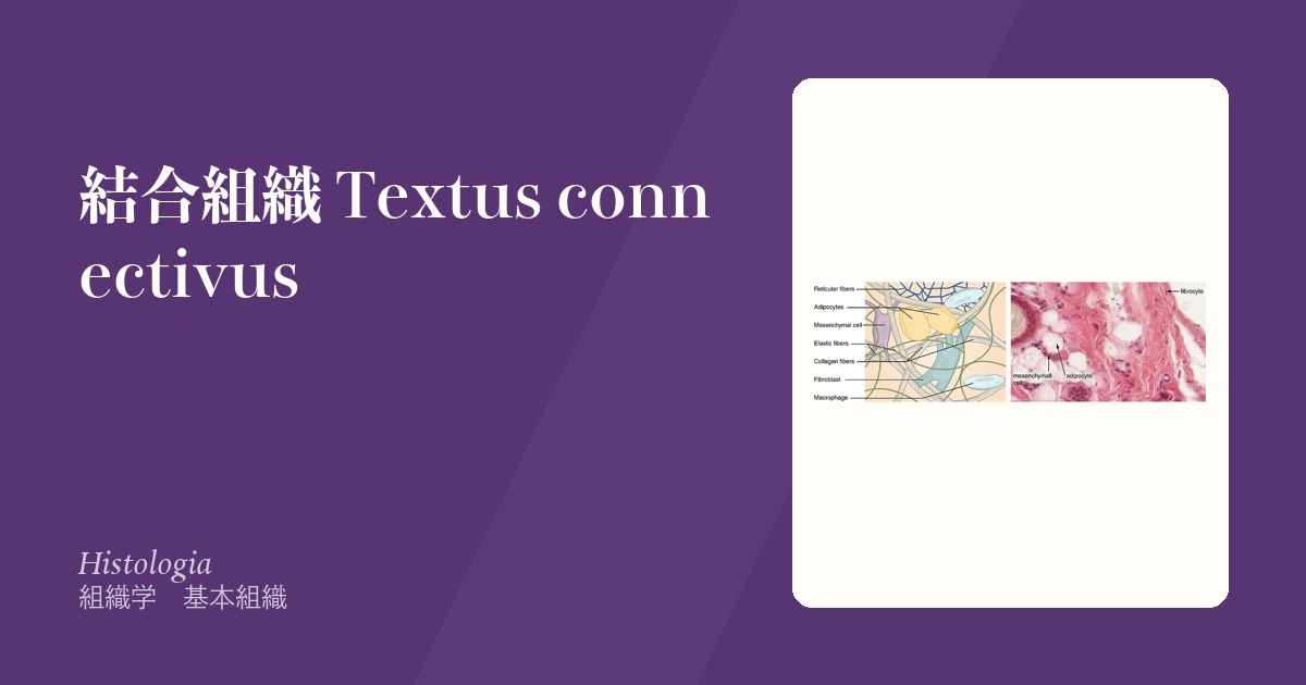
細胞がまばらで、細胞外基質が体積の大半を占める組織。基質は線維成分(膠原線維・弾性線維・細網線維)と基質 substantia fundamentalis(プロテオグリカン・糖タンパク・水)からなる。
働きは支持・結合・栄養の通路・防御・貯蔵。血管とリンパ管が走るのは結合組織の中であり、上皮はここから栄養を受けている。
由来はほぼ中胚葉(間葉)。ただし頭頸部の一部は神経堤に由来する。
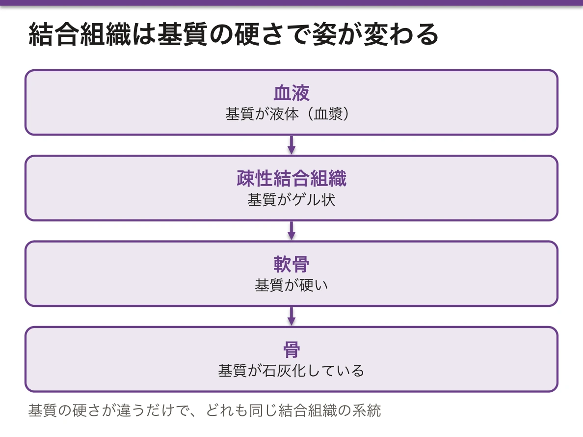
「結合組織」は血液・軟骨・骨まで含む大きな括りである。基質が液体なら血液、ゲル状なら疎性結合組織、硬いなら軟骨、石灰化すれば骨——基質の硬さが違うだけで同じ系統と捉えると、あとの章がつながる。
臨床で「結合組織疾患」と言うとき、実際に壊れているのは線維(Marfan=フィブリリン、Ehlers-Danlos=コラーゲン)か基質を作る過程(壊血病=ビタミンC)か免疫が基質・血管を攻撃する(膠原病)のいずれかである。この3分類で整理すると、症状の分布が説明できる。
結合組織は炎症の舞台である。上皮には血管が無いので、炎症細胞が集まるのも、浮腫が起こるのも、線維化が進むのもすべてここである。
浮腫は基質に水が溜まった状態で、静水圧の上昇(心不全)・膠質浸透圧の低下(低アルブミン)・血管透過性の亢進(炎症)・リンパの閉塞の4つで整理する。
膠原病は全身の結合組織が侵されるからその名がある。複数の臓器に同時に症状が出るのは、同じ組織が全身に分布しているためである。
フィブリノイド壊死は血管壁の結合組織に血漿蛋白が染み込んだもので、血管炎と悪性高血圧の像である。HE で無構造な好酸性物質として見える。
English version
Core
A tissue in which the cells are sparse and the extracellular matrix occupies most of the volume. The matrix consists of fibres (collagen, elastic, reticular) and ground substance (proteoglycans, glycoproteins, water).
Its functions are support, connection, a route for nutrition, defence and storage. Blood and lymphatic vessels run within connective tissue, and the epithelium is nourished from here.
Its origin is almost entirely mesoderm (mesenchyme), though part of the head and neck derives from neural crest.
Going further
"Connective tissue" is a large category that includes blood, cartilage and bone. If the matrix is liquid it is blood, if gel it is loose connective tissue, if firm it is cartilage, and if calcified it is bone — grasp them as one family differing only in the consistency of the matrix and the later chapters connect together.
When the clinic speaks of a connective tissue disease, what is actually broken is one of three things:
- The fibre — Marfan (fibrillin), Ehlers–Danlos (collagen).
- The process of making the matrix — scurvy (vitamin C).
- Immune attack on matrix and vessels — the connective tissue diseases proper.
Organised in these three, the distribution of symptoms can be explained.
In the clinic
Connective tissue is the stage on which inflammation happens. Since epithelium has no vessels, it is here that inflammatory cells gather, that oedema forms and that fibrosis advances.
Oedema is water accumulated in the ground substance, and is organised by four mechanisms: raised hydrostatic pressure (heart failure), reduced colloid osmotic pressure (hypoalbuminaemia), increased vascular permeability (inflammation) and lymphatic obstruction.
The connective tissue diseases are so named because the connective tissue of the whole body is involved; symptoms appear in several organs at once because the same tissue is distributed everywhere.
Fibrinoid necrosis is plasma protein permeating the connective tissue of a vessel wall — the appearance of vasculitis and malignant hypertension, seen on H&E as structureless eosinophilic material.
間葉 — すべての結合組織の出発点Mesenchyma
間葉は胎生期の結合組織で、のちの結合組織すべての出発点になる。標本では次のように見える。
- 星形・不規則な形の細胞が長い細胞質の突起を伸ばし、隣の細胞の突起と触れ合って立体的な網を作る(合胞体のように見えるが、細胞どうしは融合していない)
- 核は大きく楕円形で、クロマチンが細かく、核小体が目立つ。細胞質は少ない(小器官がまだ発達していない)
- 細胞のあいだは、水の多い基質で満たされ、細い細網線維がわずかにある。太い膠原線維の束は無い
由来は主に中胚葉で、頭頸部では神経堤からも生じる(外胚葉性間葉)。
間葉から生まれるもの——線維芽細胞と固有結合組織、軟骨芽細胞、骨芽細胞、脂肪細胞、平滑筋、血管内皮と血球(卵黄嚢の血島)など。臍帯の膠様組織(Wharton のゼリー)は、間葉に近い胎生期の結合組織が出生まで残ったものである。
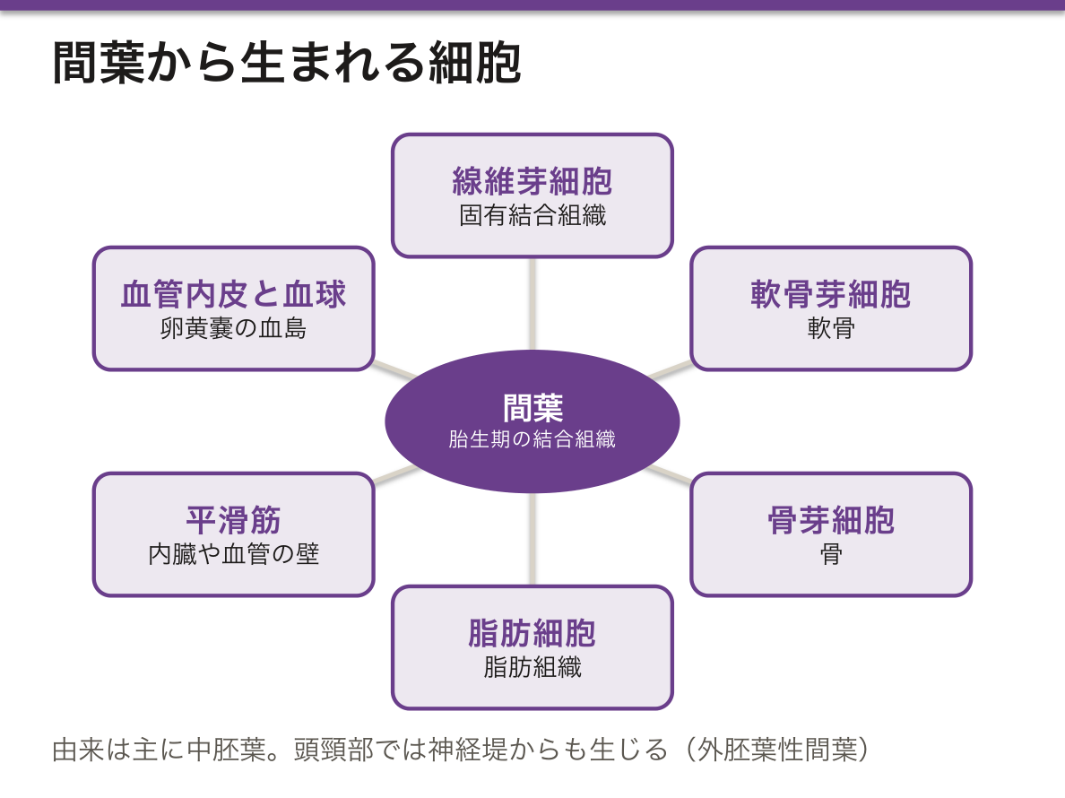
間葉が大事なのは、細胞を供給するだけでなく、上皮に指示を出すからである。歯・腺・毛・腎臓は、上皮と間葉がシグナルをやりとりして形ができる(上皮間葉相互作用、誘導)。毛乳頭が無ければ毛ができないのはその例である。
間葉と上皮は行き来もする。神経堤の細胞は上皮の性質を捨てて間葉になって移動し(上皮間葉転換)、腎臓のネフロンは反対に間葉が上皮に変わって作られる(間葉上皮転換)。
成人にも、小血管の周りに未分化な間葉系の細胞が残っているとされる。骨髄・脂肪・臍帯から取り出せる間葉系幹細胞(間葉系間質細胞)は、骨・軟骨・脂肪に分化でき、組織の修復を支える。
間葉に由来する悪性腫瘍が肉腫で、中間径フィラメントは上皮のケラチンではなくビメンチンを持つ。粘液腫や粘液型脂肪肉腫は、星形の細胞が水の多い基質に浮かび、胎生期の間葉に似た像になる。
間葉系幹細胞は治療にも使われている。日本では、造血幹細胞移植後の急性 GVHD に対する間葉系幹細胞の製剤が承認されている。免疫を抑える働きを利用したものである。
English version
Core
Mesenchyme is embryonic connective tissue and the starting point of all later connective tissues. On a slide it looks like this:
- Star-shaped or irregular cells extend long cytoplasmic processes that touch the processes of neighbours, forming a three-dimensional network (it looks syncytial, but the cells are not fused).
- The nucleus is large and oval, with fine chromatin and clear nucleoli. There is little cytoplasm (organelles are not yet developed).
- The space between cells is filled with a watery ground substance with a few fine reticular fibrils. There are no thick collagen bundles.
It comes mainly from mesoderm; in the head and neck also from the neural crest (ectomesenchyme).
Mesenchyme gives rise to fibroblasts and connective tissue proper, chondroblasts, osteoblasts, adipocytes, smooth muscle, endothelium and blood cells (yolk-sac blood islands), among others. The mucous connective tissue of the umbilical cord (Wharton jelly) is an embryonic connective tissue close to mesenchyme that remains until birth.
Going further
Mesenchyme matters not only as a cell supply but because it gives instructions to epithelium. Teeth, glands, hair and kidneys take shape through signals between epithelium and mesenchyme (epithelial–mesenchymal interaction, induction). Without a hair papilla, no hair forms.
Mesenchyme and epithelium also change into each other. Neural crest cells drop their epithelial features and migrate as mesenchyme (epithelial–mesenchymal transition), while nephrons form when mesenchyme becomes epithelium (mesenchymal–epithelial transition).
Adults are thought to keep undifferentiated mesenchymal cells around small vessels. Mesenchymal stem (stromal) cells taken from marrow, fat or umbilical cord can become bone, cartilage and fat, and support tissue repair.
In the clinic
Malignant tumours from mesenchyme are sarcomas; their intermediate filament is vimentin, not epithelial keratin. Myxoma and myxoid liposarcoma show star-shaped cells floating in a watery matrix, much like embryonic mesenchyme.
Mesenchymal stem cells are also used in treatment. In Japan a mesenchymal stem-cell product is approved for acute GVHD after haemopoietic stem-cell transplantation, using its immunosuppressive action.
膠原線維(コラーゲン線維)Fibra collagena
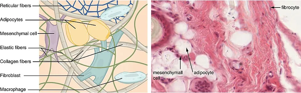
体内で最も多いタンパク質。3本のポリペプチドが右巻きの三重らせんを組む。引っ張りに極端に強く、伸びない。HE ではエオジンで赤に染まる。
型を4つだけ押さえる。
- I型——最多。皮膚・腱・靭帯・骨・線維軟骨
- II型——硝子軟骨・弾性軟骨・硝子体
- III型——細網線維。造血組織・肝・リンパ組織、そして創傷の初期
- IV型——基底膜。三重らせんが網目状に広がる(線維を作らない)
合成の要所はプロリンとリジンの水酸化で、この反応にはビタミンC が補酵素として必須である。水酸化されないと三重らせんが安定せず、細胞内で分解される。
その後リジルオキシダーゼ(銅依存)が架橋を作って強度を出す。だからビタミンC 欠乏でも銅欠乏でもコラーゲンは弱くなる。Menkes 病(銅輸送障害)で血管が脆くなるのはこの経路による。
創傷治癒ではIII型が先に作られ、時間をかけて I型に置き換わる。この置換が瘢痕の成熟であり、術後の創部強度が数か月かけて上がる理由である。置換が過剰に進むとケロイド・肥厚性瘢痕になる。
壊血病——ビタミンC欠乏。水酸化が止まりコラーゲンが作れないため、既存のコラーゲンが更新される場所から順に壊れる。歯肉出血・歯の動揺、毛包周囲の点状出血、創傷治癒の遅延、骨形成障害。「出血」が主症状になるのは血管壁のコラーゲンが保てないためであり、凝固因子はむしろ正常である。
Ehlers-Danlos 症候群——コラーゲンの構造・修飾の異常。皮膚過伸展・関節過可動・易出血。血管型(III型コラーゲン)は動脈・腸管の破裂をきたし予後が悪い。
English version
Core
The most abundant protein in the body. Three polypeptide chains form a right-handed triple helix. Extremely strong in tension and inextensible. Stains red with eosin.
Four types are enough:
- Type I — the commonest. Skin, tendon, ligament, bone, fibrocartilage.
- Type II — hyaline and elastic cartilage, vitreous body.
- Type III — reticular fibres. Haemopoietic tissue, liver, lymphoid tissue, and the early phase of wound healing.
- Type IV — basement membrane. The triple helix spreads as a network and does not form fibrils.
Going further
The critical step in synthesis is the hydroxylation of proline and lysine, for which vitamin C is an essential cofactor. Without hydroxylation the triple helix is unstable and is degraded inside the cell. Lysyl oxidase, which is copper-dependent, then forms the cross-links that give strength. So collagen is weakened by deficiency of either vitamin C or copper — the fragile vessels of Menkes disease (defective copper transport) follow this route.
In wound healing type III is laid down first and is replaced by type I over time. That replacement is the maturation of the scar, and the reason the strength of a surgical wound increases over months. Carried too far it gives keloid and hypertrophic scar.
In the clinic
Scurvy — vitamin C deficiency. Hydroxylation stops and collagen cannot be made, so the tissues that renew their collagen fail first: gingival bleeding and loose teeth, perifollicular petechiae, delayed wound healing and defective bone formation. Bleeding dominates because the collagen of the vessel wall cannot be maintained — the clotting factors are in fact normal.
Ehlers–Danlos syndrome — abnormalities of the structure or modification of collagen: hyperextensible skin, hypermobile joints, easy bruising. The vascular type (type III collagen) causes rupture of arteries and bowel and carries a poor prognosis.
弾性線維Fibra elastica
エラスチンの芯をフィブリリンの微細線維が取り巻く。伸びて元に戻るのが仕事で、コラーゲンとは正反対の性質。
豊富な場所:大動脈などの弾性型動脈・肺・皮膚・弾性軟骨(耳介・喉頭蓋)・項靭帯。
HE ではほとんど染まらないので、エラスチカ・ワンギーソン染色などで確認する。
弾性線維の意味は大動脈で最もよく分かる。心収縮期に大動脈壁が伸びて血液を蓄え、拡張期に縮んで送り出す(Windkessel 効果)。これがあるから拡張期にも臓器への灌流が続く。
加齢で弾性線維が減り膠原線維が増えると、この緩衝が効かなくなる。結果として収縮期血圧だけが上がり拡張期血圧は下がる=脈圧の増大——高齢者の収縮期高血圧の正体である。
エラスチンは再生がほとんど効かない。肺気腫で失われた肺胞壁の弾性が戻らないのはこのためで、α1-アンチトリプシン欠乏症では好中球エラスターゼが抑えられず弾性線維が壊され、若年・非喫煙者でも汎小葉性肺気腫を起こす。
Marfan 症候群——フィブリリン1(FBN1)の変異。常染色体顕性。弾性線維の足場が作れないので、弾性線維に頼る場所すべてに症状が出る。
- 大動脈基部拡張 → 大動脈解離・破裂(生命予後を決める)
- 僧帽弁逸脱
- 水晶体亜脱臼(上方へずれるのが特徴)
- 高身長・くも状指・漏斗胸・側弯
FBN1 は TGF-β を捕まえておく働きも持つため、変異で TGF-β シグナルが過剰になることも病態に関わる。ARB が大動脈拡大を抑えうるのはこの経路への介入である。
English version
Core
A core of elastin surrounded by microfibrils of fibrillin. Its work is to stretch and return — the opposite property to collagen.
Abundant in: elastic arteries such as the aorta, lung, skin, elastic cartilage (auricle, epiglottis) and the ligamentum nuchae.
It hardly stains with H&E, so confirm with elastica–van Gieson or a similar stain.
Going further
The meaning of the elastic fibre is clearest in the aorta. The wall distends in systole and stores blood, then recoils in diastole and drives it onward — the Windkessel effect. It is because of this that perfusion of the organs continues during diastole.
With age the elastic fibres diminish and collagen increases, and this buffering fails. The result is that the systolic pressure alone rises while the diastolic falls — a widened pulse pressure, which is the substance of isolated systolic hypertension in the elderly.
Elastin scarcely regenerates. That is why the elasticity of alveolar walls lost in emphysema does not return; and in α1-antitrypsin deficiency neutrophil elastase is unopposed and destroys elastic fibres, giving panacinar emphysema even in a young non-smoker.
In the clinic
Marfan syndrome — mutation of fibrillin 1 (FBN1), autosomal dominant. The scaffold of the elastic fibre cannot be built, so symptoms appear everywhere that depends on elastic fibres:
- Dilatation of the aortic root → dissection or rupture (this determines the prognosis).
- Mitral valve prolapse.
- Ectopia lentis — characteristically displaced upward.
- Tall stature, arachnodactyly, pectus excavatum, scoliosis.
FBN1 also sequesters TGF-β, so mutation allows excessive TGF-β signalling, which contributes to the disease. An angiotensin receptor blocker may limit aortic dilatation through this pathway.
細網線維Fibra reticularis
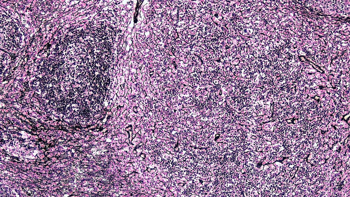
III型コラーゲンの細い線維が網目を作る。細胞を「支える枠」として働き、柔らかい器官の骨組みになる。
場所:骨髄・リンパ節・脾臓・肝臓・脂肪組織・平滑筋のまわり。
HE では見えない。鍍銀染色で黒く染まるので「好銀線維」とも呼ばれる。
細網線維の網は細胞が動きながら働ける足場である。リンパ節では細網線維に沿ってリンパ球が移動し、抗原提示細胞と出会う。骨髄では造血細胞がこの網に支えられて成熟し、成熟したものだけが類洞へ出る。柔らかいまま形を保つという要求に応えている。
肝臓では細網線維の枠が残るかどうかが再生を左右する。急性肝炎で肝細胞が死んでも枠が保たれれば正常に再生するが、枠ごと壊れると結節を作って再生=肝硬変になる。「なぜ肝硬変は不可逆なのか」の答えがここにある。
細網線維の網が壊れたかどうかで、腫瘍と反応が分かれる。鍍銀染色は肝生検で最も役に立つ。肝硬変では小葉の網が崩れて再生結節を取り囲む——「線維化」ではなく「構造の改築」が起きていることを示す。
リンパ節と脾の骨組みも細網線維である。悪性リンパ腫では正常のリンパ濾胞構造が壊れるので、構造が保たれているか(反応性)か、消えているか(腫瘍)が判定の出発点になる。
骨髄線維症では細網線維とコラーゲンが増えて骨髄が線維に置き換わる。骨髄穿刺で吸えない(dry tap)ため生検が必要になり、末梢血に涙滴赤血球と髄外造血による脾腫が出る。
English version
Core
Fine fibres of type III collagen forming a network. They act as a frame that supports cells and provide the skeleton of soft organs.
Sites: bone marrow, lymph node, spleen, liver, adipose tissue, around smooth muscle.
Not visible on H&E. They blacken with silver impregnation and are therefore called argyrophilic fibres.
Going further
The reticular net is a scaffold on which cells can work while moving. In the lymph node, lymphocytes travel along the reticular fibres and meet antigen-presenting cells; in the marrow, haemopoietic cells mature supported by this net and only the mature ones pass into the sinusoids. It answers the requirement to hold a shape while remaining soft.
In the liver, whether the reticular framework survives determines regeneration. If hepatocytes die in acute hepatitis but the frame is preserved, regeneration is orderly; if the frame is destroyed as well, regeneration forms nodules — cirrhosis. Here is the answer to why cirrhosis is irreversible.
In the clinic
Whether the reticular net has been destroyed separates neoplasia from reaction.
Silver impregnation is most useful in liver biopsy. In cirrhosis the lobular net collapses and encircles regenerative nodules — showing that what has occurred is not simply "fibrosis" but architectural remodelling.
The framework of lymph node and spleen is also reticular. In malignant lymphoma the normal follicular architecture is destroyed, so whether the architecture is preserved (reactive) or effaced (neoplastic) is the starting point of assessment.
In myelofibrosis reticular fibres and collagen increase and replace the marrow. Aspiration yields nothing (dry tap) so a trephine biopsy is required, and the blood shows teardrop cells with splenomegaly from extramedullary haemopoiesis.
基質Substantia fundamentalis
線維のあいだを埋める、無定形のゲル。主成分はグリコサミノグリカン(GAG)とそれが結合したプロテオグリカン、そして接着を担う糖タンパク(フィブロネクチン・ラミニン)。
GAG は強い負電荷を持ち、水を大量に引き寄せる。これが圧縮に耐える力と物質拡散の場を生む。
代表的な GAG:ヒアルロン酸(唯一、硫酸化されずタンパクと結合しない)・コンドロイチン硫酸・ヘパラン硫酸。
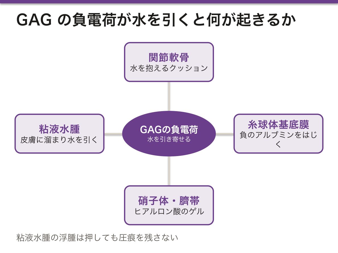
「負電荷が水を引く」という一点から多くが説明できる。
- 関節軟骨——アグリカンが水を抱え、荷重で水が押し出され、除荷で戻る。これが粘弾性のクッション
- 糸球体基底膜——ヘパラン硫酸の負電荷が、負に帯電したアルブミンをはじく(charge barrier)。微小変化型ネフローゼでこの電荷が失われ、選択的にアルブミンだけ漏れる
- 硝子体・臍帯——ヒアルロン酸が大量の水を抱えてゲルを作る
基質は感染の広がり方も決める。ヒアルロニダーゼを産生する細菌(A群溶連菌・ウェルシュ菌)は基質を溶かして急速に組織内を進む——蜂窩織炎や壊死性筋膜炎の進展の速さはここに由来する。
基質の水を保つ力が、そのまま関節と皮膚の状態になる。
変形性関節症では軟骨の GAG(アグリカン)が失われて水を保てなくなり、弾力を失って摩耗する。ヒアルロン酸の関節内注射はこの性質を補う発想である。
ムコ多糖症 — GAG を分解する酵素の欠損。Hurler 症候群・Hunter 症候群。粗な顔貌・角膜混濁・肝脾腫・関節拘縮・骨変形——GAG がある場所すべてに症状が出る。Hunter は X 連鎖で角膜混濁が無いことが Hurler との鑑別点。
粘液水腫 — 甲状腺機能低下で皮膚の基質にグリコサミノグリカンが溜まり、水を引き込む。押しても圧痕を残さない浮腫(non-pitting edema)——通常の浮腫と区別できるのはこの機序による。
English version
Core
The amorphous gel filling the space between the fibres. Its main constituents are glycosaminoglycans (GAGs) and the proteoglycans they attach to, together with the adhesive glycoproteins (fibronectin, laminin).
GAGs carry a strong negative charge and attract large amounts of water, which creates both the resistance to compression and the medium for diffusion.
Principal GAGs: hyaluronic acid (the only one that is not sulphated and not bound to protein), chondroitin sulphate, heparan sulphate.
Going further
Much follows from the single fact that negative charge attracts water.
- Articular cartilage — aggrecan holds water; loading expresses it and unloading draws it back. This is the viscoelastic cushion.
- Glomerular basement membrane — the negative charge of heparan sulphate repels the negatively charged albumin (the charge barrier). In minimal change disease this charge is lost and albumin alone leaks selectively.
- Vitreous body and umbilical cord — hyaluronic acid holds a great deal of water as a gel.
The ground substance also determines how infection spreads. Bacteria that produce hyaluronidase — group A streptococci, Clostridium perfringens — dissolve it and advance rapidly through tissue: the speed of cellulitis and necrotising fasciitis originates here.
In the clinic
The capacity of the matrix to hold water is directly the state of the joints and skin.
In osteoarthritis the GAG (aggrecan) of cartilage is lost, water cannot be held, and the cartilage loses resilience and wears. Intra-articular hyaluronic acid is an attempt to supplement this property.
Mucopolysaccharidoses — deficiency of the enzymes that degrade GAGs. Hurler and Hunter syndromes: coarse facies, corneal clouding, hepatosplenomegaly, joint contracture and skeletal deformity — symptoms appear everywhere GAGs are found. Hunter is X-linked and lacks corneal clouding, which distinguishes it from Hurler.
Myxoedema — in hypothyroidism glycosaminoglycans accumulate in the dermal matrix and draw in water, giving oedema that does not pit on pressure. This mechanism is what distinguishes it from ordinary oedema.
線維芽細胞Fibroblastus
結合組織の常在細胞の主役。線維も基質も、これが作って外に出している。紡錘形で、粗面小胞体が発達するため細胞質が青みを帯びる。
活動が落ち着いた状態を線維細胞 fibrocytus と呼び、核が細く濃くなる。
創傷治癒では線維芽細胞が主役を演じる。損傷部へ遊走し増殖して肉芽組織を作り、一部は筋線維芽細胞に分化して α平滑筋アクチンを発現し、創を収縮させる。
この収縮は本来有益だが、行き過ぎると瘢痕拘縮になる。熱傷後に関節が動かなくなるのはこの機序で、整形外科・形成外科の治療対象となる。
線維化を起こす主要なシグナルは TGF-β。肺線維症・肝硬変・腎の間質線維化・強皮症は臓器こそ違うが、「線維芽細胞が TGF-β で活性化し、I型コラーゲンを過剰に置く」という同じ筋書きを共有している。
創傷治癒の主役であり、線維化の犯人でもある。
肉芽組織は線維芽細胞+新生血管+炎症細胞からなる。筋線維芽細胞が収縮して創を縮めるが、やりすぎると瘢痕拘縮になる。熱傷後の関節拘縮はこれである。
ケロイドと肥厚性瘢痕 — ケロイドは元の創の範囲を越えて広がり、自然に退縮しない。肥厚性瘢痕は範囲内にとどまり、時間とともに軽快する。境界を越えるかどうかで区別する。
治癒を妨げる因子は実務で問われる——感染・血流不足・糖尿病・低栄養(とくにビタミン C と亜鉛)・ステロイド・異物・持続する張力。ビタミン C はコラーゲンの水酸化に必要なので、壊血病では創が開き、古い傷まで再び開く。
English version
Core
The principal resident cell of connective tissue. It makes both the fibres and the ground substance and exports them.
Spindle-shaped, with well-developed rough ER giving the cytoplasm a bluish tint. In its quiescent state it is called a fibrocyte, with a thin, dense nucleus.
Going further
The fibroblast plays the leading role in wound healing. It migrates to the injury, proliferates and forms granulation tissue; some differentiate into myofibroblasts, express α-smooth muscle actin and contract the wound. That contraction is useful, but carried too far it becomes a contracture — the mechanism by which a joint becomes fixed after a burn, and a matter for orthopaedic and plastic surgery.
The principal signal for fibrosis is TGF-β. Pulmonary fibrosis, cirrhosis, renal interstitial fibrosis and scleroderma differ in organ but share one script: the fibroblast is activated by TGF-β and lays down excess type I collagen.
In the clinic
The protagonist of wound healing and the culprit of fibrosis are the same cell.
Granulation tissue consists of fibroblasts, new vessels and inflammatory cells. Myofibroblasts contract and shrink the wound, but excess gives contracture — the fixed joint after a burn.
Keloid and hypertrophic scar — a keloid extends beyond the original wound and does not regress spontaneously; a hypertrophic scar stays within it and improves with time. The distinction is whether the border is crossed.
The factors that impair healing are asked in practice: infection, poor blood supply, diabetes, malnutrition (especially vitamin C and zinc), corticosteroids, foreign material and continued tension. Vitamin C is required for the hydroxylation of collagen, so in scurvy wounds gape and even old scars reopen.
大食細胞(マクロファージ)Macrophagus
血中の単球が組織に入って分化したもの。貪食・抗原提示・サイトカイン産生を担う。
組織ごとに名前が変わるが同じ系統——肝:Kupffer 細胞/肺:肺胞大食細胞/骨:破骨細胞/脳:ミクログリア/皮膚:Langerhans 細胞/骨髄・脾:組織球。
異物が大きすぎて1個で処理できないとき、大食細胞は互いに融合して多核巨細胞を作り、周囲を取り囲む。これが肉芽腫で、「処理できないものを閉じ込める」という戦略である。
- 乾酪壊死を伴う肉芽腫——結核(Langhans 型巨細胞。核が辺縁に馬蹄形)
- 乾酪壊死を伴わない肉芽腫——サルコイドーシス・Crohn 病
- 異物型巨細胞——縫合糸・人工物のまわり(核が散在)
ミクログリアだけは由来が違う点に注意。他が骨髄由来の単球なのに対し、ミクログリアは卵黄嚢由来で胎生早期に脳へ入り、そのまま自己複製で維持される。骨髄移植で置き換わりにくいのはこのためである。
肉芽腫はマクロファージの集団である。類上皮細胞と多核巨細胞からなり、「排除できないものを囲い込む」反応の形である。
乾酪壊死を伴う肉芽腫=結核(Langhans 型巨細胞)。壊死を伴わない肉芽腫=サルコイドーシス。壊死の有無が鑑別の第一歩になる。
組織ごとに名前が変わるが同じ系統という事実が、全身に散る病気を説明する。Langerhans 細胞組織球症では骨・皮膚・下垂体(尿崩症)・肺が同時に侵される。
マクロファージが飲み込めないものを溜め込むと泡沫細胞になる。動脈硬化のプラークの本体はこれで、酸化 LDL を食べたマクロファージである。
TNF-α 阻害薬は肉芽腫を維持できなくさせるため、投与前に潜在性結核のスクリーニングが必須である。
English version
Core
A blood monocyte that has entered the tissue and differentiated. It undertakes phagocytosis, antigen presentation and cytokine production.
The name changes with the tissue but the lineage is one — liver: Kupffer cell; lung: alveolar macrophage; bone: osteoclast; brain: microglia; skin: Langerhans cell; marrow and spleen: histiocyte.
Going further
When the foreign material is too large for one cell to handle, macrophages fuse into multinucleate giant cells and surround it. This is a granuloma, the strategy of walling off what cannot be destroyed.
- Granuloma with caseous necrosis — tuberculosis (Langhans giant cells, nuclei in a peripheral horseshoe).
- Granuloma without caseation — sarcoidosis, Crohn disease.
- Foreign body giant cell — around suture or prosthetic material; nuclei scattered.
Note that microglia alone have a different origin. The others derive from bone marrow monocytes, but microglia come from the yolk sac, enter the brain early in embryonic life and are maintained by self-renewal. This is why they are not readily replaced after bone marrow transplantation.
In the clinic
A granuloma is a collection of macrophages — epithelioid cells and multinucleate giant cells, the form of the reaction that encircles what cannot be eliminated.
Caseating granuloma = tuberculosis (Langhans giant cells); non-caseating granuloma = sarcoidosis. The presence or absence of necrosis is the first step in the differential.
That the name changes by tissue while the lineage is one explains diseases scattered through the body: in Langerhans cell histiocytosis, bone, skin, pituitary (diabetes insipidus) and lung are involved together.
A macrophage that accumulates what it cannot digest becomes a foam cell — the substance of the atherosclerotic plaque is exactly this, a macrophage that has eaten oxidised LDL.
TNF-α inhibitors prevent granulomas from being maintained, so screening for latent tuberculosis is mandatory before treatment.
肥満細胞Mastocytus
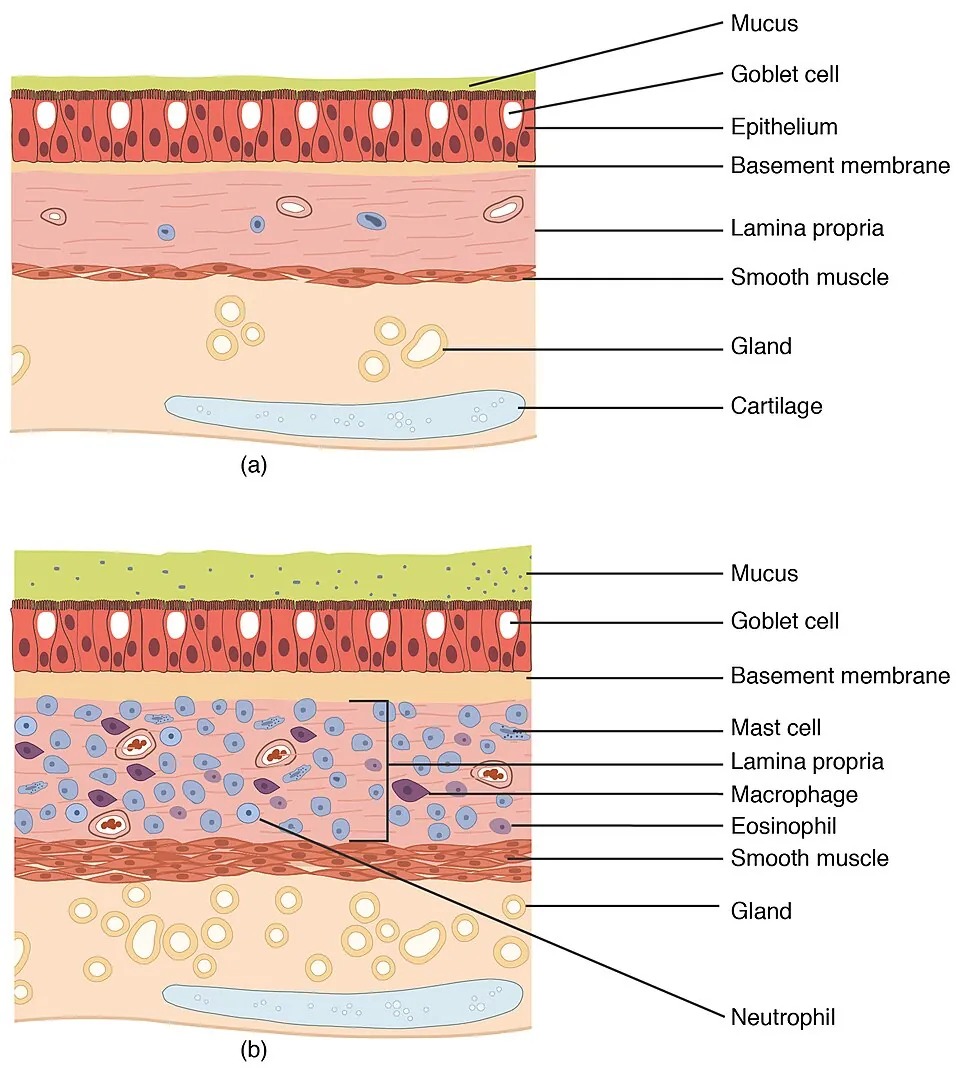
細胞質を好塩基性の顆粒が埋め尽くす細胞。顆粒にはヒスタミン・ヘパリン・ロイコトリエン・プロテアーゼが入る。
血管の周囲・皮膚・気道・消化管粘膜——つまり外界と接する場所に多い。表面の高親和性IgE受容体(FcεRI)に IgE が結合しており、抗原が架橋すると一気に脱顆粒する。
脱顆粒で起きることを一続きに追うと、アナフィラキシーの症状が全部説明できる。
- 血管拡張+透過性亢進 → 血圧低下・血管性浮腫・膨疹
- 気管支平滑筋収縮 → 喘鳴・呼吸困難
- 腸管平滑筋収縮・分泌亢進 → 腹痛・嘔吐・下痢
だから治療も機序で決まる。アドレナリンが第一選択なのは、α1で血管を締め、β2で気管支を広げ、さらに肥満細胞の脱顆粒そのものを抑えるという3方向に同時に効く唯一の薬だからである。抗ヒスタミン薬は皮膚症状にしか効かず、循環と気道は救えない。
「肥満細胞」という名は脂肪とは無関係で、顆粒で膨らんだ見た目に由来する(独 Mastzellen=よく肥えた細胞)。
肥満細胞の由来は骨髄の造血幹細胞だが、成熟は組織に入ってからで、末梢血には現れない。増殖には KIT シグナルが要り、その活性化変異で起きるのが肥満細胞症。皮膚を擦ると膨疹が出る(Darier 徴候)ほか、全身型では反復するアナフィラキシー様発作をきたす。
English version
Core
A cell whose cytoplasm is filled with basophilic granules containing histamine, heparin, leukotrienes and proteases.
Abundant around vessels and in skin, airway and gastrointestinal mucosa — that is, wherever the body meets the outside world.
IgE is bound to the high-affinity receptor (FcεRI) on the surface, and when antigen cross-links it the cell degranulates at once.
Going further
Follow what degranulation does, in sequence, and every symptom of anaphylaxis is accounted for.
- Vasodilatation and increased permeability → hypotension, angio-oedema, weals.
- Bronchial smooth muscle contraction → wheeze and dyspnoea.
- Intestinal smooth muscle contraction and hypersecretion → abdominal pain, vomiting, diarrhoea.
The treatment therefore follows from the mechanism. Adrenaline is first line because it is the only drug that acts in three directions at once: constricting vessels through α1, dilating bronchi through β2, and suppressing mast cell degranulation itself. An antihistamine helps only the skin and can rescue neither the circulation nor the airway.
The name has nothing to do with fat: it comes from the appearance of a cell swollen with granules (German Mastzellen, "well-fed cell").
In the clinic
Mast cells derive from the haemopoietic stem cell but mature only after entering the tissue and do not appear in the blood.
Their proliferation requires KIT signalling, and an activating mutation causes mastocytosis: stroking the skin raises a weal (Darier sign), and the systemic form causes recurrent anaphylaxis-like attacks.
形質細胞Plasmocytus
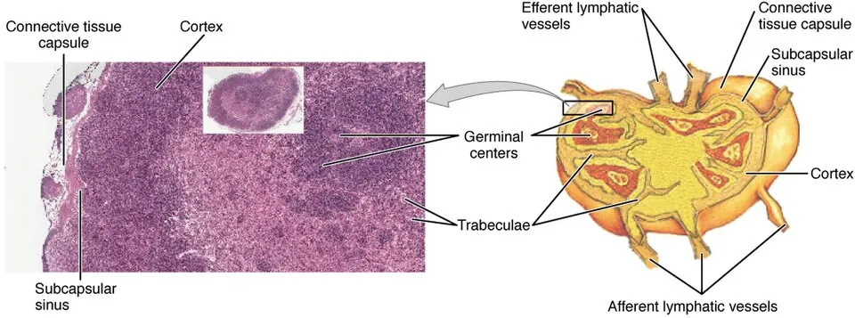
B細胞が最終分化して抗体を作り続ける工場になったもの。粗面小胞体が細胞質を埋めるので細胞質が濃い青紫(好塩基性)に染まる。
核は偏在し、ヘテロクロマチンが放射状に配列して車輪状(cartwheel)に見える。核周囲に Golgi 装置による明るい抜け(核周明庭)がある。
慢性炎症の場——粘膜固有層・慢性炎症巣・骨髄に多い。
形態が機能をそのまま語る好例である。細胞質の青=粗面小胞体=タンパク合成、核周明庭=Golgi=分泌タンパクの糖鎖付加と輸送。この2つを見れば「分泌に特化した細胞」と分かる。
存在そのものが所見になる。組織に形質細胞が多い=慢性の抗原刺激が続いているということで、慢性胃炎(H. pylori)・慢性歯周炎・自己免疫性疾患で目立つ。急性炎症の主役が好中球であるのに対し、形質細胞は時間が経った炎症の指標になる。
多発性骨髄腫——形質細胞の単クローン性増殖。1種類の抗体(M蛋白)だけが大量に作られる。四徴 CRAB:高Ca血症(hyperCalcaemia)・腎障害(Renal)・貧血(Anaemia)・骨病変(Bone lesions)。
骨病変は打ち抜き像(punched-out lesion)が特徴で、骨芽細胞が抑制され破骨細胞が活性化するため骨形成を伴わない。だから骨シンチでは写りにくく、ALP も上がらない——転移性骨腫瘍との鑑別点になる。整形外科で高齢者の病的骨折を診るとき、必ず鑑別に挙がる。
English version
Core
A B lymphocyte in its terminal differentiation — a factory that continuously makes antibody.
Rough ER fills the cytoplasm, so it stains deeply blue-purple (basophilic). The nucleus is eccentric and its heterochromatin is arranged radially, giving the cartwheel appearance. Beside the nucleus is a pale area, the perinuclear hof, created by the Golgi.
Found where chronic inflammation is: lamina propria of mucosa, chronic inflammatory foci, bone marrow.
Going further
A fine example of form declaring function. Blue cytoplasm = rough ER = protein synthesis; the perinuclear hof = Golgi = glycosylation and transport of a secreted protein. Seeing those two, you know it is a cell specialised for secretion.
Its very presence is a finding. Many plasma cells in a tissue means chronic antigenic stimulation is continuing, and they are prominent in chronic gastritis (H. pylori), chronic periodontitis and autoimmune disease. Where the neutrophil is the protagonist of acute inflammation, the plasma cell is the index of inflammation that has lasted.
In the clinic
Multiple myeloma — monoclonal proliferation of plasma cells, producing a single antibody (M protein) in vast quantity.
The tetrad is CRAB: hyperCalcaemia, Renal impairment, Anaemia, Bone lesions.
The bone lesions are characteristically punched-out, because osteoblasts are suppressed while osteoclasts are activated, so there is no accompanying bone formation. Hence they are poorly seen on bone scintigraphy and the alkaline phosphatase is not raised — the point of distinction from metastatic bone disease.
It must always be considered when assessing a pathological fracture in an elderly patient.
疎性結合組織Textus connectivus laxus
線維がまばらで、細胞と基質に富む。体の隙間を埋める汎用の充填材で、血管・神経の通り道でもある。
場所:粘膜固有層・皮下組織・血管や神経の周囲・臓器のあいだ。
ゆるいぶん水が溜まりやすく、感染も広がりやすい。
浮腫が「どこに」出るかは疎性結合組織の分布で決まる。眼瞼・陰嚢・足背が腫れやすいのは、この組織が特に疎で、皮下に空間の余裕があるためである。ネフローゼ症候群の朝の眼瞼浮腫はその典型で、夜間の臥位で顔面に水が移ることによる。
感染では蜂窩織炎の場になる。境界が不明瞭に広がるのは、疎な組織の中を細菌が進むからで、境界明瞭な丹毒(真皮浅層とリンパ管)と対比される。「どの層の炎症か」で境界の見え方が変わる。
ゆるいから、感染も浮腫も広がる。
蜂窩織炎は疎性結合組織(皮下組織)に沿って境界不明瞭に広がる。丹毒が真皮にとどまり境界明瞭で隆起するのと対照的で、深さの違いがそのまま見た目の違いになっている。
頸部の疎性結合組織は縦隔へ連続する。だから咽後膿瘍・歯性感染が降下性壊死性縦隔炎になりうる。「首の感染は胸へ落ちる」——層の連続性が予後を決める。
皮下組織は輸液・薬剤の投与経路でもある(皮下注射・持続皮下輸液)。吸収が緩やかなのは血流が中等度だから。
ヒアルロニダーゼは基質を分解して薬の広がりを助ける。細菌(連鎖球菌)も同じ酵素を使って組織内へ広がる——「広がる仕組み」は病原体も医療も同じものを使っている。
English version
Core
Fibres are sparse and the tissue is rich in cells and ground substance. A general-purpose packing material filling the spaces of the body, and also the route taken by vessels and nerves.
Sites: lamina propria of mucosa, subcutaneous tissue, around vessels and nerves, between organs.
Being loose, it accumulates water readily and infection spreads through it readily.
Going further
Where oedema appears is determined by the distribution of loose connective tissue. The eyelid, scrotum and dorsum of the foot swell easily because the tissue there is especially loose and there is room beneath the skin. The morning periorbital oedema of nephrotic syndrome is the classic example, water having shifted to the face during recumbency.
In infection it is the setting for cellulitis. The border is indistinct because the bacteria advance through loose tissue, in contrast to erysipelas, which involves the superficial dermis and lymphatics and has a sharp, raised edge. Which layer is inflamed changes how the border looks.
In the clinic
Because it is loose, both infection and oedema spread.
Cellulitis spreads with an ill-defined border along the subcutaneous loose connective tissue, in contrast to erysipelas, which is confined to the dermis, sharply demarcated and raised. The difference in depth is directly the difference in appearance.
The loose connective tissue of the neck is continuous with the mediastinum, so a retropharyngeal abscess or an odontogenic infection can become descending necrotising mediastinitis. Infection in the neck falls into the chest — the continuity of the planes decides the prognosis.
The subcutaneous tissue is also a route of administration (subcutaneous injection, hypodermoclysis); absorption is gradual because the blood supply is moderate. Hyaluronidase is used to break down the matrix and help a drug spread — and streptococci use the same enzyme to spread through the tissue. The mechanism of spreading is the same for the pathogen and for medicine.
密性結合組織Textus connectivus densus
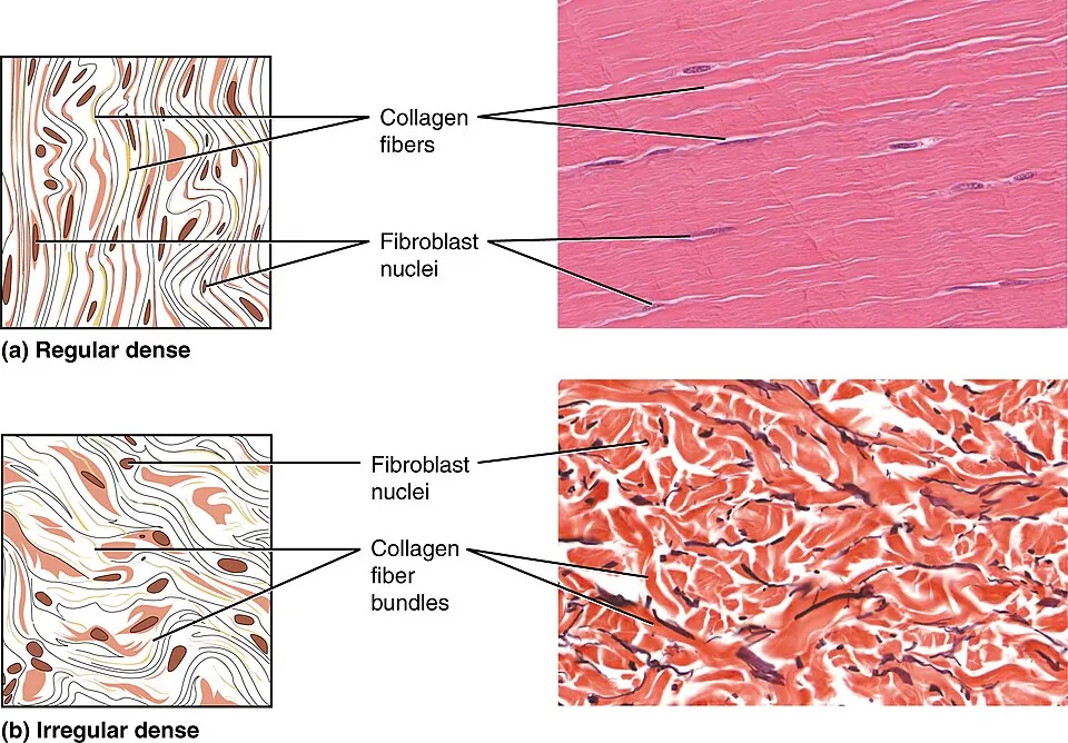
膠原線維がぎっしり詰まり、細胞は少ない。強度に振り切った組織。並び方で2つに分かれる。
- 規則性——線維が一方向に平行。腱・靭帯・腱膜。決まった向きの張力に極めて強い
- 不規則性——線維が様々な向きに交錯。真皮・臓器の被膜・骨膜・関節包。あらゆる向きの力に耐える
腱と靭帯は血流が乏しい。線維芽細胞(腱細胞)がまばらで代謝も低いため、治癒が遅く、治っても元の配列には戻らない。これが整形外科で反復する主題になる。
- アキレス腱断裂——血流の乏しい踵骨付着部から2〜6cm が好発部位
- 前十字靭帯(ACL)断裂——関節内にあり滑液に浸っているため自然治癒がほぼ望めず、再建術の適応になる。関節外の内側側副靭帯(MCL)が保存療法で治りやすいのと対照的
- 腱板断裂——棘上筋腱の血流が乏しい領域(critical zone)に生じる
「関節内か関節外か」「血流があるか」が治療方針を分けるという原則は、この組織の性質から出ている。
強度に振り切った組織は、血流が乏しい。だから腱と靱帯は治りが遅い。
アキレス腱は付着部の 2〜6cm 近位が乏血域で、断裂の好発部位になる。キノロン系抗菌薬とステロイドが腱障害の危険因子である。
腱板(棘上筋腱)にも同様の乏血域があり、加齢で変性・断裂する。整形外科で最も多い肩の疾患の1つ。
不規則性の密性結合組織=真皮である。あらゆる向きの張力に耐えるが、決まった向きの張力線(Langer 線)がある。皮膚切開を Langer 線に沿わせると創が開きにくく瘢痕が目立たない——線維の走行を知っていることが手術手技になる。
English version
Core
Collagen fibres are densely packed and cells are few — a tissue committed entirely to strength. It divides by arrangement:
- Regular — fibres parallel in one direction. Tendon, ligament, aponeurosis. Extremely strong against tension in a fixed direction.
- Irregular — fibres interwoven in many directions. Dermis, organ capsules, periosteum, joint capsule. Withstands force from any direction.
Going further
Tendons and ligaments have a poor blood supply. Fibroblasts (tenocytes) are sparse and metabolism is low, so healing is slow and the original arrangement is never fully restored. This is a recurring theme in orthopaedics.
- Achilles tendon rupture — the usual site is 2–6 cm above the calcaneal insertion, in the poorly vascularised zone.
- Anterior cruciate ligament rupture — being intra-articular and bathed in synovial fluid, spontaneous healing is essentially impossible and reconstruction is indicated. In contrast the extra-articular medial collateral ligament heals well with conservative treatment.
- Rotator cuff tear — occurs in the poorly vascularised critical zone of the supraspinatus tendon.
The principle that "intra-articular or extra-articular" and "is there a blood supply" decide the treatment comes out of the nature of this tissue.
In the clinic
A tissue committed to strength has a poor blood supply, so tendons and ligaments heal slowly. The Achilles tendon has an avascular zone 2–6 cm proximal to its insertion, the site of predilection for rupture. Fluoroquinolones and corticosteroids are risk factors for tendinopathy. The rotator cuff (supraspinatus) has a similar zone and degenerates and tears with age — one of the commonest shoulder disorders in orthopaedics.
Dense irregular connective tissue is the dermis. It resists tension from every direction, yet there are preferred lines of tension — Langer lines. An incision made along a Langer line gapes less and scars less conspicuously — knowing the direction of the fibres is itself a surgical technique.
腱の組織 — 一次腱束・二次腱束と、腱内膜Tendo
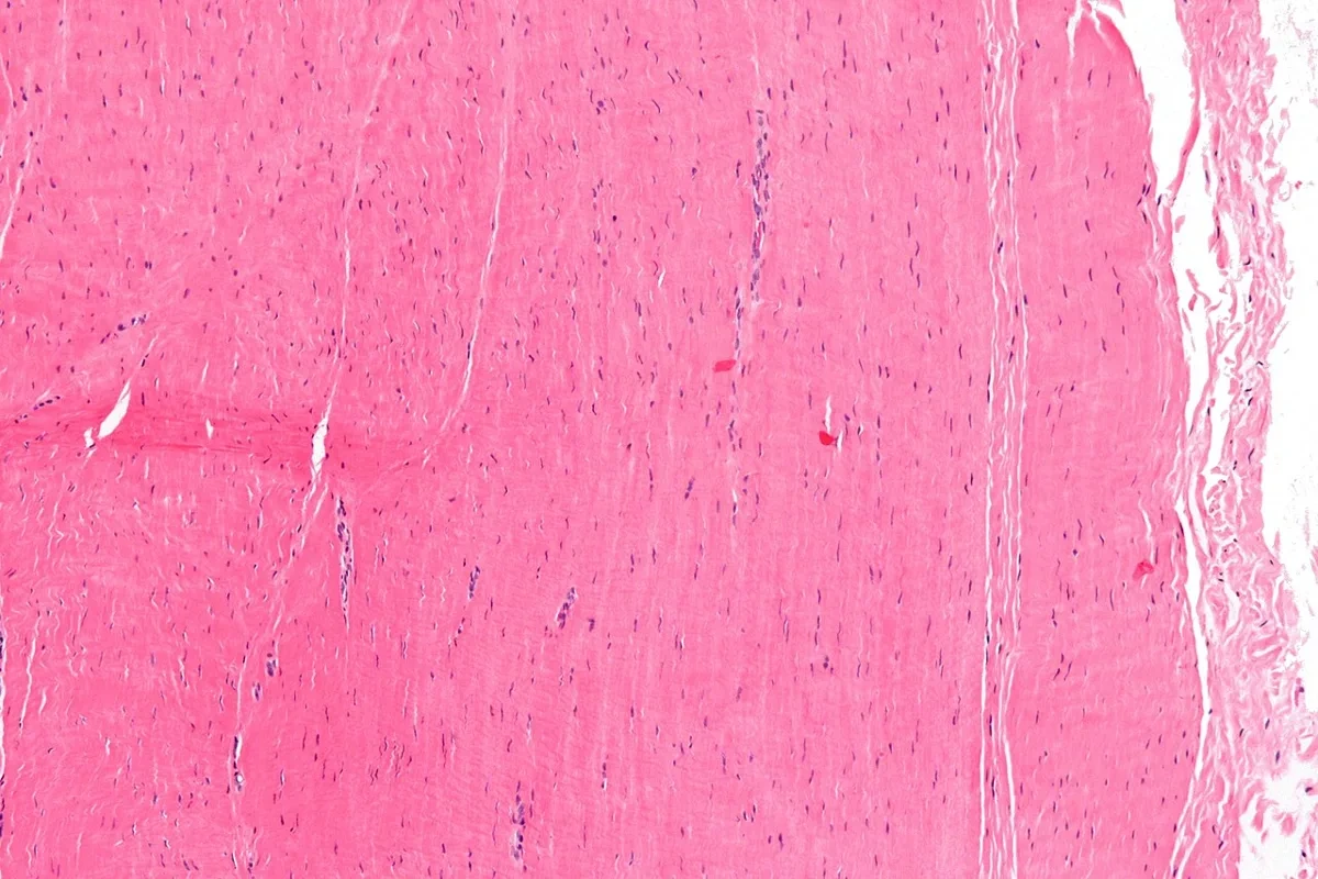
腱は密性規則性結合組織の典型である。縦断の標本では、
- 好酸性の膠原線維が平行に密に並び、ゆるく波打つ
- 線維の束のあいだに、細長く押しつぶされた核が1列に並ぶ——腱細胞(腱芽細胞・腱の線維芽細胞)である。横断では、細胞が薄い突起を線維のあいだへ伸ばし、翼のように見える(翼細胞)
束は段階的にまとまる。
| 段階 | 中身 | 包むもの |
|---|---|---|
| 一次腱束 | 膠原線維の束と、そのあいだの腱細胞 | — |
| 二次腱束 | 一次腱束がいくつか集まったもの | 腱内膜(endotenonium)——疎性結合組織。血管・神経・リンパ管が通る |
| (三次腱束) | 二次腱束の集まり | 腱内膜 |
| 腱全体 | — | 腱上膜(腱外膜)と、その外の腱傍組織または滑膜性の腱鞘 |
ヨーロッパの教科書では、腱の中の結合組織を腱内膜(内腱周膜)、腱全体を包むものを腱周膜(外腱周膜)と呼ぶこともある。名前は違っても、束の階層とそれを包む疎性結合組織という構造は同じである。
なぜ束が何段にも分かれているのか。ロープと同じで、1本の線維が切れても全体は切れにくい。束どうしは腱内膜を介して少しずつずれ合えるので、腱は曲がりながら力を伝えられる。線維のゆるい波(クリンプ)は、引っ張られた最初に伸びて衝撃を和らげる。
血管は腱内膜の中を走るだけで、束の中には入らない。腱細胞はほとんど分裂しない。だから腱は代謝がゆっくりで、治りにくい。修復には、腱自身の細胞(腱内膜・腱上膜の細胞)による内因性の治癒と、周りの組織から入ってくる細胞による外因性の治癒がある。外因性の治癒が強いと、腱が周りと癒着する。
手の屈筋腱が滑膜性の腱鞘の中で切れると、縫合したあと腱鞘との癒着で指が動かなくなりやすい。縫ったあと早くから計画的に動かすのは、腱を滑らせて癒着を防ぎながら内因性の治癒を促すためである。
使いすぎによる腱障害(アキレス腱・膝蓋腱・肘など)の組織は、炎症細胞が多い「炎症」ではなく、線維の配列が乱れ、基質が増え、細い血管が入り込み、腱細胞の核が丸くなるという変性の像である。ステロイドの注射が一時的にしか効かず、かえって腱を弱めうるのはこのためである。
English version
Core
Tendon is the typical dense regular connective tissue. In a longitudinal section:
- eosinophilic collagen fibres run parallel and tightly packed, in gentle waves;
- between the bundles, long flattened nuclei stand in rows — the tendon cells (tenoblasts, tendon fibroblasts). In cross-section the cells send thin processes between the fibres and look like wings (wing cells).
The bundles are grouped in steps.
| Level | Content | Wrapping |
|---|---|---|
| Primary bundle | collagen fibres with tendon cells between them | — |
| Secondary bundle | several primary bundles together | endotenon (endotenonium) — loose connective tissue carrying vessels, nerves and lymphatics |
| (Tertiary bundle) | groups of secondary bundles | endotenon |
| Whole tendon | — | epitenon, then paratenon or a synovial tendon sheath |
European textbooks may call the connective tissue inside the tendon the endotenon (internal peritenonium) and the wrapping of the whole tendon the peritenonium (external). The names differ, but the structure — a hierarchy of bundles wrapped in loose connective tissue — is the same.
Going further
Why so many levels of bundles? Like a rope: if one fibre breaks, the whole does not. Bundles can slide slightly against each other through the endotenon, so the tendon can bend while carrying force. The gentle waves of the fibres (crimp) straighten first when pulled and soften the shock.
Vessels run only in the endotenon and do not enter the bundles, and tendon cells rarely divide. So tendon has slow metabolism and heals poorly. Repair comes from the tendon’s own cells (endotenon and epitenon) — intrinsic healing — and from cells entering from surrounding tissue — extrinsic healing. Strong extrinsic healing makes the tendon stick to its surroundings.
In the clinic
When a flexor tendon of the hand is cut inside its synovial sheath, adhesions to the sheath after repair often stop the finger moving. Early, planned movement after repair lets the tendon glide, preventing adhesions while encouraging intrinsic healing.
In overuse tendinopathy (Achilles, patellar tendon, elbow), the tissue shows not "inflammation" with many inflammatory cells but degeneration: disordered fibres, more ground substance, ingrowth of small vessels and rounded tendon-cell nuclei. This is why steroid injections help only briefly and may even weaken the tendon.
脂肪組織Textus adiposus
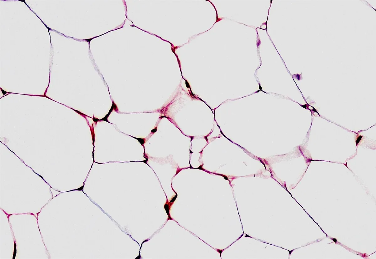
脂肪細胞(adipocytus)が主体の特殊な結合組織。貯蔵・断熱・緩衝に加え、内分泌器官でもある。
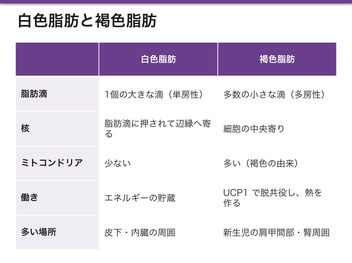
- 白色脂肪——単房性。1個の大きな脂肪滴に押されて核が辺縁へ寄る。エネルギー貯蔵
- 褐色脂肪——多房性。ミトコンドリアが多く(褐色の由来)、UCP1 で酸化的リン酸化を脱共役し熱を作る。新生児の肩甲間部・腎周囲に多い
HE では脂質が処理で溶け出すため、細胞質が空洞に見える。
脂肪組織はアディポサイトカインを分泌する内分泌器官である。レプチン(満腹シグナル)、アディポネクチン(インスリン感受性を上げる、肥満で減る唯一の主要なもの)、TNF-α・レジスチン(インスリン抵抗性を上げる)。
内臓脂肪と皮下脂肪は別物として扱う必要がある。内臓脂肪は門脈へ直接遊離脂肪酸とサイトカインを送るため、肝のインスリン抵抗性・脂質異常症・炎症を強く引き起こす。腹囲がメタボリックシンドロームの必須項目である理由がここにある。
新生児が褐色脂肪に頼るのは、ふるえ熱産生(骨格筋)がまだ使えないためである。低体温に弱い所以で、分娩室の保温が重要になる。
脂肪組織は内分泌器官である。レプチン(食欲抑制)・アディポネクチン(インスリン感受性を上げる)・TNF-α と IL-6(炎症)を出す。
内臓脂肪が増えると、アディポネクチンが減り炎症性サイトカインが増える——これがインスリン抵抗性の本体で、メタボリック症候群で腹囲を測る理由になっている。皮下脂肪ではなく内臓脂肪が問題なのは、門脈へ直接遊離脂肪酸を送り込むからである。
褐色脂肪は熱を作る。UCP-1(サーモゲニン)が電子伝達系の共役を外し、ATP ではなく熱にする。新生児の体温維持に不可欠で、肩甲間・腎周囲に多い。新生児は震えて熱を作れないので、これに頼っている。
脂肪壊死 — 急性膵炎ではリパーゼが脂肪を分解し、脂肪酸がカルシウムと結合して石鹸化する。低カルシウム血症が重症度の指標になるのはこのためである。
English version
Core
A specialised connective tissue composed chiefly of adipocytes. Besides storage, insulation and cushioning, it is an endocrine organ.
- White adipose tissue — unilocular; one large droplet pushes the nucleus to the rim. Energy storage.
- Brown adipose tissue — multilocular; abundant mitochondria (the source of the colour) and UCP1, which uncouples oxidative phosphorylation to produce heat. Abundant in the interscapular and perirenal regions of the newborn.
On H&E the lipid dissolves during processing, so the cytoplasm appears as an empty space.
Going further
Adipose tissue is an endocrine organ secreting adipocytokines: leptin (the satiety signal), adiponectin (which raises insulin sensitivity and is the one major adipokine that falls in obesity), and TNF-α and resistin (which raise insulin resistance).
Visceral and subcutaneous fat must be treated as different things. Visceral fat sends free fatty acids and cytokines directly into the portal vein, so it drives hepatic insulin resistance, dyslipidaemia and inflammation far more strongly. This is why waist circumference is the obligatory criterion of the metabolic syndrome.
The newborn depends on brown fat because shivering thermogenesis in skeletal muscle is not yet available. That is the reason for vulnerability to hypothermia and for keeping the delivery room warm.
In the clinic
Adipose tissue is an endocrine organ, secreting leptin (appetite suppression), adiponectin (insulin sensitisation) and TNF-α and IL-6 (inflammation). As visceral fat increases, adiponectin falls and inflammatory cytokines rise — this is the substance of insulin resistance, and the reason waist circumference is measured in the metabolic syndrome. Visceral rather than subcutaneous fat is the problem because it delivers free fatty acids straight into the portal vein.
Brown fat makes heat. UCP-1 (thermogenin) uncouples the electron transport chain so that the energy becomes heat rather than ATP. It is essential to the newborn’s temperature, is abundant interscapularly and perirenally, and is depended upon because the newborn cannot shiver.
Fat necrosis — in acute pancreatitis lipase digests fat and the fatty acids bind calcium to form soaps. This is why hypocalcaemia is an index of severity.
MMP — 細胞外基質を作り替える酵素
MMP(マトリックスメタロプロテアーゼ)は、活性中心に亜鉛を持つ一群のタンパク分解酵素。コラーゲン・エラスチン・プロテオグリカンなど、細胞外基質のほぼ全てを分解できる。
作られ方に3段の安全装置がある——①不活性な前駆体(プロ酵素)として分泌される ②細胞外でプロペプチドが切られて初めて働く ③TIMP(組織メタロプロテアーゼ阻害因子)が結合して止める。MMP と TIMP の均衡が基質の量を決める。
正常な役割——創傷治癒、骨のリモデリング、血管新生、月経周期の子宮内膜の崩壊、分娩時の子宮頸管の熟化、白血球が血管外へ出るときの通路づくり。
破綻するとき——癌の浸潤と転移(基底膜を破る)、関節リウマチと変形性関節症(軟骨の分解)、動脈硬化プラークの破綻、肺気腫。
基質は「作られたら終わり」ではなく、絶えず壊されて作り直されている——この動的な平衡が MMP を学ぶ理由である。MMP 阻害薬は癌治療で試みられたが、正常なリモデリングまで止めるため成功していない。
English version
Core
The matrix metalloproteinases are a family of proteases with zinc at the active site, able to degrade almost all of the extracellular matrix — collagen, elastin, proteoglycan.
Three safeguards govern how they are made:
- They are secreted as inactive proenzymes.
- They work only after the propeptide is cleaved outside the cell.
- TIMPs (tissue inhibitors of metalloproteinases) bind and stop them.
The balance between MMP and TIMP determines the amount of matrix.
Normal roles — wound healing, bone remodelling, angiogenesis, breakdown of the endometrium in the menstrual cycle, cervical ripening at labour, and making the route for leukocytes leaving a vessel.
When it fails — tumour invasion and metastasis (breaching the basement membrane), rheumatoid and osteoarthritis (cartilage degradation), rupture of the atherosclerotic plaque, emphysema.
Going further
The matrix is not finished once it is made: it is continuously broken down and rebuilt, and this dynamic equilibrium is the reason for studying the MMPs.
MMP inhibitors were tried as cancer treatment but have not succeeded, because they also stop normal remodelling.
まとめ
| 結合組織 | 細胞がまばらで、細胞外基質が体積の大半を占める組織。 |
|---|---|
| 間葉 — すべての結合組織の出発点 | 間葉は胎生期の結合組織で、のちの結合組織すべての出発点になる。 |
| 膠原線維(コラーゲン線維) | 体内で最も多いタンパク質。3本のポリペプチドが右巻きの三重らせんを組む。 |
| 弾性線維 | エラスチンの芯をフィブリリンの微細線維が取り巻く。 |
| 細網線維 | III型コラーゲンの細い線維が網目を作る。 |
| 基質 | 線維のあいだを埋める、無定形のゲル。主成分はグリコサミノグリカン(GAG)とそれが結合したプロテオグリカン、そして接着を担う糖タンパク(フィブロネクチン・ラミニン)。 |
| 線維芽細胞 | 結合組織の常在細胞の主役。線維も基質も、これが作って外に出している。 |
| 大食細胞(マクロファージ) | 血中の単球が組織に入って分化したもの。貪食・抗原提示・サイトカイン産生を担う。 |
| 肥満細胞 | 細胞質を好塩基性の顆粒が埋め尽くす細胞。 |
| 形質細胞 | B細胞が最終分化して抗体を作り続ける工場になったもの。 |
| 疎性結合組織 | 線維がまばらで、細胞と基質に富む。体の隙間を埋める汎用の充填材で、血管・神経の通り道でもある。 |
| 密性結合組織 | 膠原線維がぎっしり詰まり、細胞は少ない。 |
| 腱の組織 — 一次腱束・二次腱束と、腱内膜 | 腱は密性規則性結合組織の典型である。縦断の標本では、好酸性の膠原線維が平行に密に並び、ゆるく波打つ線維の束のあいだに、細長く押しつぶされた核が1列に並ぶ——腱細胞(腱芽細胞・腱の線… |
| 脂肪組織 | 脂肪細胞(adipocytus)が主体の特殊な結合組織。 |
| MMP — 細胞外基質を作り替える酵素 | MMP(マトリックスメタロプロテアーゼ)は、活性中心に亜鉛を持つ一群のタンパク分解酵素。 |
このノートは医学生が授業の内容を自分の言葉でまとめたもので、診療の判断に使うものではありません。誤りに気づいたらお知らせください。