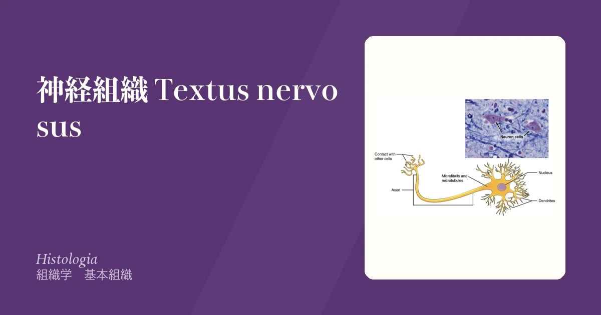
神経組織は2つの細胞集団でできている。信号を運ぶニューロンと、それを支えるグリア(神経膠細胞)。数の上ではグリアのほうが多く、単なる詰め物ではなく絶縁・栄養・免疫・環境の維持を担う。
この章の要点は中枢と末梢で担当する細胞が違うことに尽きる。髄鞘を作るのは中枢が稀突起膠細胞、末梢がSchwann 細胞。この一点が、なぜ末梢神経は再生するのに中枢神経は再生しないのか、なぜ多発性硬化症と Guillain-Barré 症候群は別の病気なのかを同時に説明する。
脱髄と軸索障害の区別も、組織の理解がそのまま神経伝導検査の読み方になる。脱髄なら伝導速度が落ち、軸索障害なら振幅が落ちる——絶縁体が壊れたのか、電線が切れたのか、という違いである。
- ニューロン(神経細胞)
- ニューロンの分類 — 突起の数・働き・軸索の長さ・形
- 髄鞘(ミエリン鞘)
- グリア(神経膠細胞)
- 末梢神経の3層 — 内膜・周膜・外膜
- シナプス
- 軸索輸送 — 1 m 先の末端まで、どうやって物を届けるか
- ニューロンの細胞質を読む — Nissl 小体と、溜まる色素
- 神経組織の発生
ニューロン(神経細胞)Neuronum
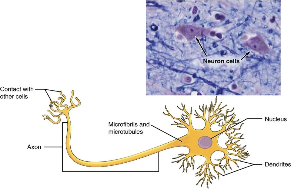
情報を受け取り、統合し、送る細胞。3つの部分からなる。
- 樹状突起——入力を受ける。数が多く、短く枝分かれする
- 細胞体(核周部)——核とNissl 小体(粗面小胞体の塊。好塩基性で青紫)。ここでタンパクを作る
- 軸索——出力。1本だけ。Nissl 小体は無い(軸索小丘から先には粗面小胞体が入らない)
成熟ニューロンは分裂しない(永久細胞)。
「軸索でタンパクを作れない」ことの帰結が大きい。必要な蛋白はすべて細胞体で作り、軸索輸送で運ぶ。
- 順行性(キネシン)——細胞体 → 終末。小胞・ミトコンドリア
- 逆行性(ダイニン)——終末 → 細胞体。老廃物・神経栄養因子。破傷風毒素・狂犬病ウイルス・単純ヘルペスもこの経路で中枢へ上がる
軸索は最長1m にもなるため、細胞体から遠いところほど維持が難しい。糖尿病性神経障害やビタミン欠乏で最も長い軸索(足趾)から症状が出る(長さ依存性、手袋靴下型)のはこのためである。
軸索が切れると細胞体に変化が起きる(中心性色素融解)。核が偏在し Nissl 小体が周辺へ散る——修復モードに入った証拠で、蛋白合成を再生へ振り向けている像である。
Nissl 小体が消える(中心性融解)のは軸索が傷ついたニューロンの反応である。核が辺縁へ寄り、細胞体が腫れ、Nissl 小体が周辺へ散る。「再生しようと蛋白を作り直している」形態であり、損傷の位置を推定する手がかりになる。
中枢のニューロンは再生しない。失われれば機能が戻らないので、脳梗塞の治療は「死なせない」ことに全力を注ぐ——ペナンブラを救う時間との勝負になる。
封入体が疾患の名前になる。Lewy 小体(αシヌクレイン)= Parkinson 病・Lewy 小体型認知症、神経原線維変化(リン酸化タウ)と老人斑(アミロイドβ)= Alzheimer 病、Negri 小体=狂犬病。
虚血に最も弱いのは海馬 CA1・小脳 Purkinje 細胞・大脳皮質第3・5層——心停止蘇生後に記憶障害が残るのはこの選択的脆弱性による。
English version
Core
A cell that receives, integrates and transmits information, in three parts.
- Dendrites — receive input; numerous, short and branching.
- Cell body (perikaryon) — nucleus and Nissl bodies (masses of rough ER, basophilic and blue-purple). Protein is made here.
- Axon — output; only one. It contains no Nissl bodies, because rough ER does not pass beyond the axon hillock.
A mature neuron does not divide — a permanent cell.
Going further
The consequence of "protein cannot be made in the axon" is large. Everything needed is made in the cell body and carried by axonal transport.
- Anterograde (kinesin) — cell body → terminal: vesicles, mitochondria.
- Retrograde (dynein) — terminal → cell body: worn material, neurotrophic factors. Tetanus toxin, rabies virus and herpes simplex ascend to the CNS by this route.
An axon may be up to a metre long, so the further from the cell body, the harder it is to maintain. This is why diabetic neuropathy and vitamin deficiency affect the longest axons (the toes) first — a length-dependent, glove-and-stocking distribution.
When an axon is cut, the cell body changes: central chromatolysis, the nucleus displaced to the periphery and the Nissl substance dispersed. It is the appearance of a cell that has switched into repair mode, redirecting protein synthesis to regeneration.
In the clinic
Loss of Nissl substance (central chromatolysis) is the reaction of a neuron whose axon has been injured: the nucleus moves to the rim, the cell body swells and the Nissl bodies scatter. It is the form of a cell rebuilding protein in order to regenerate, and it helps locate the injury.
Central neurons do not regenerate. Once lost, function does not return, so the treatment of stroke is devoted entirely to not letting them die — a race against time to save the penumbra.
Inclusion bodies become the names of diseases: Lewy bodies (α-synuclein) in Parkinson disease and dementia with Lewy bodies; neurofibrillary tangles (phosphorylated tau) and senile plaques (amyloid β) in Alzheimer disease; Negri bodies in rabies.
The most vulnerable to ischaemia are hippocampal CA1, cerebellar Purkinje cells and cortical layers 3 and 5 — this selective vulnerability is why memory impairment persists after resuscitation from cardiac arrest.
ニューロンの分類 — 突起の数・働き・軸索の長さ・形Neuronum multipolare
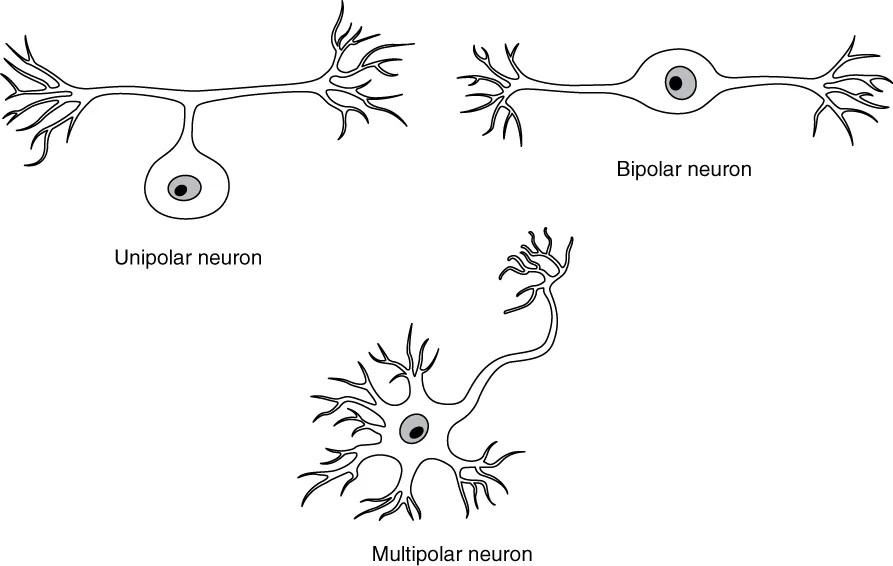
ニューロンはいくつもの基準で分けられる。最もよく使うのは突起の数である。
| 基準 | 分類 | 例 |
|---|---|---|
| 突起の数 | 多極——軸索1本と樹状突起多数。最も多い | 脊髄前角の運動ニューロン、錐体細胞、Purkinje 細胞、自律神経節の細胞 |
| 双極——細胞体の両端から1本ずつ | 網膜の双極細胞、嗅細胞、ラセン神経節・前庭神経節 | |
| 偽単極——1本出て、すぐ T 字に分かれる | 脊髄神経節・脳神経の感覚神経節 | |
| 単極——突起が1本だけ | 成体ではまれ(発生途中の神経芽細胞) | |
| 働き | 感覚(求心性)・運動(遠心性:体性と内臓性)・介在 | 介在ニューロンが大多数を占める |
| 軸索の長さ | Golgi I 型——長い軸索で遠くへ送る | 錐体細胞、Purkinje 細胞、前角細胞 |
| Golgi II 型——短く枝分かれし、近くで回路を作る | 大脳・小脳皮質の小型の細胞 | |
| 形 | 錐体形・星形・紡錘形・洋梨形・籠形・顆粒形 | Purkinje 細胞は洋梨形、小脳の籠細胞 |
| 大きさ | 小型(10〜20 µm)から、100 µm を超える巨大なものまで | Betz 細胞(運動野) |
| 伝達物質 | コリン作動性・アドレナリン作動性・グルタミン酸作動性・GABA 作動性など | — |
偽単極ニューロンは、発生のはじめは双極で、2本の突起が細胞体の近くで1本に合わさってできる。末梢へ向かう枝は働きの上では樹状突起だが、構造は軸索と同じで、髄鞘も持つ。皮膚からの信号は、細胞体を通らずに T 字の分岐をそのまま通り抜けて中枢へ行く。細胞体は、2本の長い枝を養う工場として神経節に置かれている。
Golgi I 型と II 型の区別は、回路の役割の区別である。I 型は領域の外へ出力し、II 型は領域の中で計算する。
偽単極ニューロンの細胞体そのものが壊れる病気がある(感覚性ニューロノパチー)。Sjögren 症候群、肺小細胞癌に伴う抗 Hu 抗体、シスプラチンなどが原因になる。細胞体が死ぬと中枢側と末梢側の両方の枝が失われるので、足先から始まる長さ依存性の神経障害とちがい、顔や体幹を含めて、長さに関係なく感覚が落ち、強い感覚性運動失調と腱反射の消失が出る。
帯状疱疹は、水痘のあと感覚神経節の偽単極ニューロンに潜んでいたウイルスが再び活動したもので、その神経節が受け持つ皮膚の帯に沿って出る。筋萎縮性側索硬化症は、多極の運動ニューロン(大脳の錐体細胞と脊髄前角細胞)を選んで侵す。
English version
Core
Neurons are classified by several criteria. The most common is the number of processes.
| Criterion | Class | Examples |
|---|---|---|
| Number of processes | Multipolar — one axon and many dendrites; the most common | spinal motor neurons, pyramidal cells, Purkinje cells, autonomic ganglion cells |
| Bipolar — one process from each end | retinal bipolar cells, olfactory cells, spiral and vestibular ganglia | |
| Pseudounipolar — one process that soon splits like a T | spinal and cranial sensory ganglia | |
| Unipolar — a single process | rare in adults (developing neuroblasts) | |
| Function | sensory (afferent), motor (efferent: somatic and visceral), interneuron | interneurons are the vast majority |
| Axon length | Golgi type I — long axon to distant targets | pyramidal cells, Purkinje cells, anterior horn cells |
| Golgi type II — short, branched axon for local circuits | small cells of cerebral and cerebellar cortex | |
| Shape | pyramidal, stellate, fusiform, pear-shaped, basket, granule | Purkinje cells are pear-shaped; cerebellar basket cells |
| Size | small (10–20 µm) to giant cells over 100 µm | Betz cells (motor cortex) |
| Transmitter | cholinergic, adrenergic, glutamatergic, GABAergic and others | — |
Going further
A pseudounipolar neuron starts as bipolar in development; its two processes fuse near the cell body. The peripheral branch works as a dendrite, but structurally it is an axon and can be myelinated. A signal from the skin passes straight through the T-junction to the centre without entering the cell body. The cell body sits in the ganglion as a factory feeding the two long branches.
Golgi type I versus II is a difference in circuit role: type I sends output out of the region; type II computes inside it.
In the clinic
Some diseases destroy the cell bodies of pseudounipolar neurons (sensory neuronopathy): Sjögren syndrome, anti-Hu antibodies with small-cell lung cancer, cisplatin and others. When the cell body dies, both the central and peripheral branches are lost. So, unlike length-dependent neuropathy that starts in the toes, sensation falls regardless of length, including the face and trunk, with severe sensory ataxia and loss of tendon reflexes.
Herpes zoster is the reactivation of virus that hid in pseudounipolar neurons of a sensory ganglion after chickenpox; it follows the skin band of that ganglion. Amyotrophic lateral sclerosis selectively attacks multipolar motor neurons (cortical pyramidal cells and anterior horn cells).
髄鞘(ミエリン鞘)Vagina myelini
軸索を何重にも巻いた細胞膜の層。脂質に富み絶縁体として働く。一定間隔で切れ目(Ranvier 絞輪)があり、そこにだけ Na⁺ チャネルが密集する。
活動電位が絞輪から絞輪へ飛ぶ跳躍伝導により、伝導速度が桁違いに速くなる。
作る細胞が中枢と末梢で違う——中枢は稀突起膠細胞(1個が複数の軸索を担当)、末梢はSchwann 細胞(1個が1本の軸索の1区間だけ)。
「1個が何本を担当するか」の違いが、再生能の差に直結する。
- 末梢——Schwann 細胞は軸索が切れると増殖し、基底膜の管(Büngner 帯)を残して軸索を誘導する。約1mm/日で再生する
- 中枢——稀突起膠細胞は増殖せず、むしろ再生を阻害する因子(Nogo など)を出す。さらに星状膠細胞がグリア瘢痕を作って伸長を物理的に阻む
「末梢は管が残るから治る、中枢は瘢痕が塞ぐから治らない」——脊髄損傷が回復しない組織学的理由である。
神経伝導検査の読み方も組織から出る。脱髄では伝導速度が低下し(絶縁が壊れ跳躍伝導ができない)、軸索障害では振幅(CMAP/SNAP)が低下する(伝わる線の本数が減る)。どちらが落ちたかで病態が決まる。
脱髄疾患はどちらの髄鞘かで分かれる。
- 多発性硬化症——中枢の脱髄。時間的・空間的多発が診断の核。視神経炎・核間性眼筋麻痺・Uhthoff 現象(体温上昇で悪化)。MRI で脳室周囲の白質病変
- Guillain-Barré 症候群——末梢の脱髄。先行感染(Campylobacter jejuni)の1〜3週後、上行性・左右対称の弛緩性麻痺と腱反射消失。髄液で蛋白細胞解離。多くは可逆(末梢だから再生できる)
予後の差が「再生できる組織かどうか」で説明できるのがこの対比の要点である。
English version
Core
Layers of cell membrane wrapped many times around the axon. Rich in lipid, it acts as insulation. At regular intervals there are gaps, the nodes of Ranvier, and sodium channels are concentrated there alone. The action potential jumps from node to node — saltatory conduction — making conduction faster by orders of magnitude.
The cell that makes it differs: centrally the oligodendrocyte (one cell serves several axons), peripherally the Schwann cell (one cell serves one internode of one axon).
Going further
The difference in how many axons one cell serves translates directly into the difference in regenerative capacity.
- Peripheral — when an axon is cut the Schwann cells proliferate, leave a tube of basal lamina (the band of Büngner) and guide the axon. Regeneration proceeds at about 1 mm a day.
- Central — oligodendrocytes do not proliferate and in fact release inhibitors of regeneration such as Nogo; astrocytes add a glial scar that physically blocks growth.
The periphery heals because the tube remains; the centre does not because the scar closes it — the histological reason spinal cord injury does not recover.
The reading of nerve conduction studies also comes out of the histology. In demyelination the conduction velocity falls (insulation is lost and saltatory conduction fails); in axonal damage the amplitude falls (fewer wires carry the signal). Which has fallen defines the pathology.
In the clinic
Demyelinating diseases divide by which myelin is affected.
- Multiple sclerosis — central demyelination. Dissemination in time and in space is the core of the diagnosis. Optic neuritis, internuclear ophthalmoplegia, Uhthoff phenomenon (worsening with a rise in body temperature); periventricular white matter lesions on MRI.
- Guillain–Barré syndrome — peripheral demyelination. One to three weeks after an antecedent infection (Campylobacter jejuni), an ascending, symmetrical flaccid paralysis with loss of tendon reflexes; albuminocytological dissociation in the CSF. Largely reversible — because it is peripheral and can regenerate.
The point of the contrast is that the difference in prognosis is explained by whether the tissue can regenerate.
グリア(神経膠細胞)Neuroglia
ニューロンを支える細胞群。中枢に4種、末梢に2種。
中枢
- 星状膠細胞——最多。血液脳関門の維持、K⁺と伝達物質の回収、グリア瘢痕
- 稀突起膠細胞——中枢の髄鞘
- 小膠細胞(ミクログリア)——中枢の免疫。卵黄嚢由来で他と系統が違う
- 上衣細胞——脳室の内面を覆う。脈絡叢で髄液を作る
末梢——Schwann 細胞(髄鞘)と外套細胞(衛星細胞)(神経節細胞を包む)。
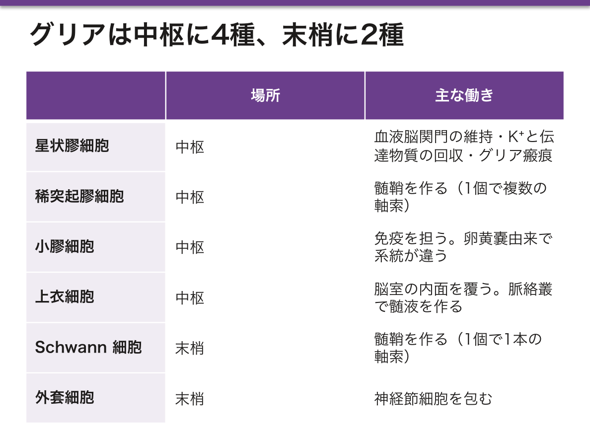
星状膠細胞は足突起(end-feet)で毛細血管を包み、内皮細胞に密着結合を作らせて血液脳関門を維持する。同時にシナプス間隙のグルタミン酸を回収しており、これが破綻すると興奮毒性でニューロンが死ぬ——虚血性脳障害の主要機序である。
グリアは脳腫瘍の主な発生母地でもある。ニューロンは分裂しないので腫瘍化しにくく、成人の原発性脳腫瘍の多くはグリオーマ(星状膠細胞腫・膠芽腫)である。「分裂する細胞が腫瘍になる」という原則が脳でも成り立っている。
上衣細胞と脈絡叢は髄液の産生・循環に関わる。この経路が塞がれると水頭症になり、閉塞部位(中脳水道・第四脳室出口)で非交通性、吸収障害(くも膜顆粒)で交通性と分かれる。
グリアは腫瘍になる。ニューロンはほとんどならない。成人の原発性脳腫瘍の大半がグリオーマなのは、分裂できるのがグリアだからである。膠芽腫は最も悪性で、偽柵状壊死と微小血管増殖が組織学的な決め手になる。
星状膠細胞は血液脳関門を維持し、傷ではグリア瘢痕を作る。中枢では線維芽細胞の瘢痕ではなくグリア瘢痕になり、これが軸索の再生を妨げる——中枢神経が再生しない理由の1つである。GFAP がマーカー。
稀突起膠細胞は1個で複数の軸索に髄鞘を作る(Schwann 細胞は1対1)。だから中枢の脱髄(多発性硬化症)は広い範囲に及ぶ。
ミクログリアは中枢のマクロファージで、中胚葉(卵黄嚢)由来——ほかのグリアと出自が違う。HIV 脳症では多核巨細胞を作る。
English version
Core
The supporting cells: four types centrally, two peripherally.
Central
- Astrocyte — the most numerous. Maintains the blood–brain barrier, takes up K⁺ and transmitters, forms the glial scar.
- Oligodendrocyte — central myelin.
- Microglia — central immunity. Derived from the yolk sac, a different lineage from the rest.
- Ependymal cell — lines the ventricles; produces CSF at the choroid plexus.
Peripheral — Schwann cells (myelin) and satellite cells (enveloping ganglion cells).
Going further
Astrocytes wrap capillaries with their end-feet and induce the endothelium to form tight junctions, maintaining the blood–brain barrier. At the same time they take up glutamate from the synaptic cleft, and when this fails, excitotoxicity kills neurons — the principal mechanism of ischaemic brain injury.
Glia are also the main source of brain tumours. Neurons do not divide and rarely become neoplastic, so most adult primary brain tumours are gliomas (astrocytoma, glioblastoma). The principle that dividing cells become tumours holds in the brain too.
Ependymal cells and the choroid plexus govern the production and circulation of CSF. Obstruction of this route causes hydrocephalus: non-communicating if the block is at the aqueduct or the outlets of the fourth ventricle, communicating if absorption at the arachnoid granulations fails.
In the clinic
Glia become tumours; neurons almost never do. Most adult primary brain tumours are gliomas because glia are the cells that can divide. Glioblastoma is the most malignant, with pseudopalisading necrosis and microvascular proliferation as the histological hallmarks.
Astrocytes maintain the blood–brain barrier and form the glial scar after injury. In the CNS the scar is glial rather than fibroblastic, and it obstructs axonal regeneration — one of the reasons the central nervous system does not recover. GFAP is the marker.
An oligodendrocyte myelinates several axons (a Schwann cell only one), so central demyelination such as multiple sclerosis affects a wide area.
Microglia are the macrophages of the CNS and derive from mesoderm (yolk sac) — a different origin from the other glia. In HIV encephalopathy they form multinucleate giant cells.
末梢神経の3層 — 内膜・周膜・外膜
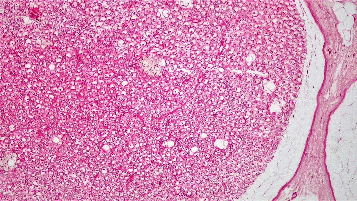
末梢神経は結合組織の3層に包まれる。骨格筋の3層と同じ入れ子構造。
- 神経内膜 endoneurium——軸索1本ずつを包む疎な結合組織
- 神経周膜 perineurium——神経束を包む。密着結合を持つ上皮様の細胞層で、血液神経関門を作る
- 神経外膜 epineurium——神経全体を包む密性結合組織。血管(vasa nervorum)が走る
この3層が、神経損傷の分類(Seddon / Sunderland)そのものを決めている。
- 一過性伝導障害(neurapraxia)——局所の脱髄のみ。軸索は連続。数週で完全回復。「土曜の夜麻痺」(橈骨神経圧迫)が典型
- 軸索断裂(axonotmesis)——軸索は切れるが内膜の管が残る。Wallerian 変性ののち1mm/日で再生。回復するが時間がかかる
- 神経断裂(neurotmesis)——管ごと断裂。自然回復は望めず縫合・移植が必要。誤った標的へ伸びれば神経腫を作る
「管が残っているか」で予後が決まる——軟骨・筋・神経に共通する原則がここでも効いている。整形外科・手外科で神経損傷を扱うとき、まずこの分類に落とす。
周膜が血液神経関門を作るため、局所麻酔薬は周膜を越えにくい。神経ブロックで効果発現に時間がかかり、神経束の外側の線維から先に効くのはこの障壁による。
3層のどこまで切れたかで、回復の見込みが決まる。Seddon/Sunderland 分類はこの層の話そのものである。
一過性伝導障害(neurapraxia) — 髄鞘だけの障害。軸索は連続しているので、数日〜数週で完全に回復する。正座のしびれ・土曜夜麻痺。
軸索断裂(axonotmesis) — 軸索は切れるが神経内膜の管が残る。Waller 変性の後、1日約1mm で再生し、元の標的に届く。
神経断裂(neurotmesis) — 管ごと切れる。自然回復は望めず、手術で縫合しないと迷走した軸索が神経腫(断端神経腫)を作って痛みの原因になる。
「管が残っているか」が予後を分ける——3層の解剖が、そのまま神経外傷の分類になっている。
English version
Core
A peripheral nerve is invested by three layers of connective tissue, the same nested arrangement as in skeletal muscle.
- Endoneurium — loose connective tissue around each individual axon.
- Perineurium — around a fascicle; an epithelium-like layer of cells joined by tight junctions, forming the blood–nerve barrier.
- Epineurium — dense connective tissue around the whole nerve, carrying the vasa nervorum.
Going further
These three layers are what define the classification of nerve injury (Seddon / Sunderland).
- Neurapraxia — local demyelination only; the axon is continuous. Complete recovery in weeks. Saturday night palsy (radial nerve compression) is typical.
- Axonotmesis — the axon is severed but the endoneurial tube remains. After Wallerian degeneration it regenerates at 1 mm a day. Recovery occurs but takes time.
- Neurotmesis — the tube is severed as well. Spontaneous recovery cannot be expected; suture or grafting is required, and axons growing to the wrong target form a neuroma.
Whether the tube remains decides the prognosis — the same principle that governs cartilage, muscle and nerve alike. In orthopaedic and hand surgery, a nerve injury is first placed into this classification.
Because the perineurium forms the blood–nerve barrier, local anaesthetic crosses it with difficulty: this is why a nerve block takes time to work and why the fibres at the outside of the fascicle are affected first.
In the clinic
How far through the three layers the injury goes determines the prospect of recovery; the Seddon and Sunderland classifications are precisely a statement about these layers.
- Neurapraxia — myelin alone is affected; the axon is continuous, so recovery is complete in days to weeks. The numbness of kneeling, Saturday night palsy.
- Axonotmesis — the axon is cut but the endoneurial tube remains. After Wallerian degeneration it regrows at about 1 mm a day and reaches its original target.
- Neurotmesis — the tube is cut too. Spontaneous recovery cannot be expected, and without surgical repair the misdirected axons form a stump neuroma and a source of pain.
Whether the tube remains divides the prognosis — the anatomy of the three layers is itself the classification of nerve trauma.
シナプスSynapsis
ニューロンが次の細胞へ情報を渡す接点。ほとんどが化学シナプス。
- 活動電位が終末に到達
- 電位依存性Ca²⁺チャネルが開く
- Ca²⁺ 流入 → シナプス小胞が膜と融合(SNARE 蛋白)
- 神経伝達物質が間隙へ放出
- 後シナプス膜の受容体に結合 → 脱分極または過分極
電気シナプス(ギャップ結合)は速いが数が少なく、心筋や一部の中枢で使われる。
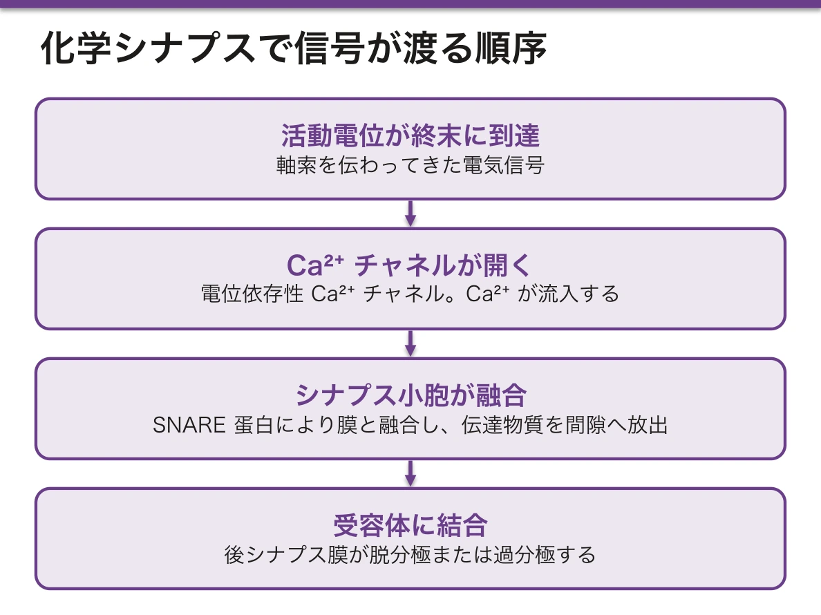
SNARE 蛋白は毒素の標的として臨床に直結する。
- ボツリヌス毒素——SNARE を切断してACh 放出を阻害。弛緩性麻痺。治療応用(痙縮・眼瞼痙攣・美容)もこの機序
- 破傷風毒素——逆行性軸索輸送で脊髄へ上がり、抑制性介在ニューロン(グリシン・GABA)の放出を止める。抑制が外れて強直性痙攣。「開口障害・後弓反張」
同じ SNARE を切っても、標的ニューロンが興奮性か抑制性かで麻痺と痙攣に分かれる——組織と回路の両方を見ないと説明できない好例である。
神経筋接合部では ACh がアセチルコリンエステラーゼで速やかに分解される。この酵素を阻害すると ACh が留まるため、重症筋無力症の治療(ピリドスチグミン)にも、有機リン中毒(コリン作動性クリーゼ)にもなる。同じ機序が治療にも中毒にもなる。
シナプスの各段階に、それぞれ薬と病気がある。
ボツリヌス毒素 — SNARE 蛋白を切断してアセチルコリンの放出を止める。弛緩性麻痺を起こすが、その性質を逆用して痙縮・眼瞼痙攣・多汗症の治療に使う。
破傷風毒素も同じく SNARE を切るが、標的が抑制性介在ニューロンなので抑制が外れて強直性痙攣(後弓反張・開口障害)になる。同じ機序でも、どのシナプスを止めるかで正反対の症状になる。
Lambert–Eaton 症候群 — シナプス前の P/Q 型 Ca チャネルに対する自己抗体。反復刺激で筋力が増す(waning ではなく waxing)ことが重症筋無力症との決定的な違いで、小細胞肺癌の傍腫瘍症候群として現れる。
再取り込みの阻害がSSRI・SNRI、分解酵素の阻害がコリンエステラーゼ阻害薬・MAO 阻害薬である。
English version
Core
The point at which a neuron passes information to the next cell. Almost all are chemical.
- The action potential reaches the terminal.
- Voltage-gated Ca²⁺ channels open.
- Ca²⁺ enters → synaptic vesicles fuse with the membrane (SNARE proteins).
- Transmitter is released into the cleft.
- It binds receptors on the postsynaptic membrane → depolarisation or hyperpolarisation.
Electrical synapses (gap junctions) are fast but few, used in cardiac muscle and parts of the CNS.
Going further
The SNARE proteins are a direct clinical target of toxins.
- Botulinum toxin — cleaves SNARE and blocks acetylcholine release, causing flaccid paralysis. Its therapeutic uses (spasticity, blepharospasm, cosmetic) work by the same mechanism.
- Tetanus toxin — ascends by retrograde axonal transport to the spinal cord and stops release from inhibitory interneurons (glycine, GABA). With inhibition removed, tonic spasm follows: trismus and opisthotonos.
The same SNARE is cleaved, yet the result is paralysis or spasm depending on whether the target neuron is excitatory or inhibitory — a fine example of something that cannot be explained without looking at both the tissue and the circuit.
At the neuromuscular junction acetylcholine is rapidly destroyed by acetylcholinesterase. Inhibiting that enzyme makes ACh persist, which is both the treatment of myasthenia gravis (pyridostigmine) and the mechanism of organophosphate poisoning (cholinergic crisis). One mechanism serves as both treatment and poison.
In the clinic
Each step of the synapse has its own drug and its own disease.
Botulinum toxin cleaves SNARE and stops acetylcholine release, causing flaccid paralysis — and that property is turned to use in spasticity, blepharospasm and hyperhidrosis. Tetanus toxin cleaves SNARE too, but its target is the inhibitory interneuron, so removing inhibition produces tonic spasm (opisthotonos, trismus). The same mechanism gives opposite symptoms depending on which synapse is stopped.
Lambert–Eaton syndrome — autoantibodies against presynaptic P/Q-type calcium channels. Strength increases with repetitive stimulation (waxing rather than waning), the decisive difference from myasthenia gravis, and it appears as a paraneoplastic syndrome of small cell lung cancer.
Blockade of reuptake gives the SSRIs and SNRIs; blockade of the degrading enzyme gives the cholinesterase and MAO inhibitors.
軸索輸送 — 1 m 先の末端まで、どうやって物を届けるかTransportus axonalis
蛋白合成は細胞体でしか行われない。軸索は最長 1 m を超える——だから運ぶ仕組みが要る。
| 向き | モーター蛋白 | 速さ | 運ぶもの |
|---|---|---|---|
| 順行性(細胞体 → 末端) | キネシン | 速い 200〜400 mm/日/遅い 0.2〜5 mm/日 | 速い:小胞・膜蛋白・ミトコンドリア/遅い:細胞骨格・可溶性蛋白 |
| 逆行性(末端 → 細胞体) | ダイニン | 100〜200 mm/日 | 古い小器官・神経栄養因子・取り込まれた物質 |
どちらも微小管の上を、ATP を使って歩く。微小管はプラス端が末端側を向く。
神経再生の速さ(約 1 mm/日)は、遅い順行性輸送の速さで決まっている。
「細胞体から遠いほど不利になる」という一点が、末梢神経障害の分布を決めている。最も長い軸索から先に壊れるので、症状が足先から始まり、手袋・靴下型に広がる(末梢神経障害の章)——「長さ依存性の障害という言葉が、輸送の話として理解できる」。
逆行性輸送は、病原体と毒素の通り道にもなる。破傷風毒素・狂犬病ウイルス・単純ヘルペスウイルス・ポリオウイルスが、これに乗って中枢へ達する(ウイルスの章)——「傷の場所から、症状が出る場所までの時間が、距離で決まる」。
ボツリヌス毒素が局所に留まるのは、逆に神経筋接合部で働くからになる(神経筋接合部の項)——「同じ細菌毒素でも、作用する場所が違えば全身症状の有無が変わる」。
微小管を壊す薬は、末梢神経障害を起こす(薬が臓器を壊す章)——「ビンクリスチン・パクリタキセルの用量規定毒性が、これになる」。
Waller 変性の後の再生が 1 日 1 mm なのも、この輸送の速さによる(末梢神経の変性と再生の項)——「回復の予測を、距離の割り算で出せる」。
軸索輸送は病原体と毒素の通り道でもある。
逆行性輸送(ダイニン)で中枢へ運ばれるもの — 狂犬病ウイルス・破傷風毒素・単純ヘルペスウイルス・ポリオウイルス。末梢から入って中枢で悪さをするのは、この経路を使うからである。ヘルペスが神経節に潜伏し、再活性化して同じデルマトームに出るのも軸索輸送による往復の結果である。
軸索は最も長いところで 1m を超えるので、輸送が滞れば末端から先に壊れる。糖尿病性・薬剤性のニューロパチーが手袋靴下型(遠位優位)になるのは、長い神経ほど不利だからである。「長さ依存性」という言い方はここから来る。
微小管を止める抗癌薬(ビンカアルカロイド・タキサン)が末梢神経障害を起こすのも、軸索輸送が微小管に依存しているためである。
English version
Core
Protein synthesis occurs only in the cell body, yet an axon may exceed a metre — so a transport system is required.
| Direction | Motor | Speed | Cargo |
|---|---|---|---|
| Anterograde (body → terminal) | Kinesin | Fast 200–400 mm/day; slow 0.2–5 mm/day | Fast: vesicles, membrane protein, mitochondria. Slow: cytoskeleton, soluble protein |
| Retrograde (terminal → body) | Dynein | 100–200 mm/day | Worn organelles, neurotrophic factors, internalised material |
Both walk along microtubules using ATP; the plus ends point towards the terminal.
The rate of nerve regeneration (about 1 mm a day) is set by the speed of slow anterograde transport.
Going further
The single fact that distance from the cell body is a disadvantage determines the distribution of peripheral neuropathy. The longest axons fail first, so symptoms begin in the toes and spread in a glove-and-stocking pattern — "length-dependent" becomes intelligible as a statement about transport.
Retrograde transport is also the route of pathogens and toxins. Tetanus toxin, rabies virus, herpes simplex and poliovirus all reach the CNS along it — and the time between the site of the wound and the appearance of symptoms is set by the distance. Botulinum toxin, by contrast, stays local because it acts at the neuromuscular junction — the same class of bacterial toxin differs in whether systemic symptoms occur, according to where it acts.
Drugs that disrupt microtubules cause peripheral neuropathy — the dose-limiting toxicity of vincristine and paclitaxel. And regeneration after Wallerian degeneration proceeds at 1 mm a day for the same reason — recovery can be predicted by dividing the distance.
In the clinic
Axonal transport is also the route of pathogens and toxins.
Carried centrally by retrograde transport (dynein): rabies virus, tetanus toxin, herpes simplex virus, poliovirus. They enter peripherally and cause disease centrally because they use this route. That herpes lies latent in a ganglion and reactivates in the same dermatome is the round trip along the same transport.
An axon may exceed a metre, so if transport falters the far end fails first. Diabetic and drug-induced neuropathies are distal-predominant (glove and stocking) because the longer the nerve the greater the disadvantage — the origin of the term "length-dependent".
Cytotoxic drugs that arrest microtubules (vinca alkaloids, taxanes) cause peripheral neuropathy for the same reason: axonal transport depends on microtubules.
ニューロンの細胞質を読む — Nissl 小体と、溜まる色素
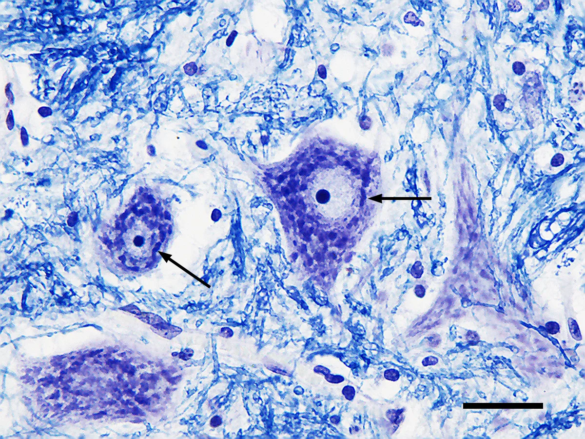
神経細胞の細胞質には、他の細胞に無い構造と色素がある。染色像でニューロンを見分け、その状態を推定する手がかりになる。
| Nissl 小体 | 粗面小胞体とリボソームの塊 | 塩基性色素で濃く染まる。蛋白合成が盛んなことを示す。樹状突起には入るが、軸索には入らない |
| 神経原線維 | 中間径フィラメントと微小管 | 鍍銀染色で見える。形を保ち、物を運ぶ |
| リポフスチン | 黄褐色の顆粒 | リソソームで処理しきれなかった脂質の残骸。加齢とともに増えるので「消耗色素」と呼ばれる |
| 神経メラニン | 黒褐色 | 黒質と青斑核にある。ドパミン・ノルアドレナリンの代謝産物由来 |
軸索に Nissl 小体が無いことが、軸索の性質を決めている——軸索は自分で蛋白を作れないので、細胞体から運んでもらうしかない。
色素の増減が、そのまま疾患の所見になる。
- 黒質の脱色——Parkinson 病では、肉眼で黒質が薄くなって見える。神経メラニンを持つドパミン神経が脱落するためで、切っただけで分かる数少ない神経変性疾患の所見である
- 青斑核の脱色——同じく Parkinson 病や Alzheimer 病で起こる
- リポフスチンの著明な蓄積——加齢のほか、神経セロイドリポフスチン症ではリソソームの異常で大量に溜まる
神経メラニンには二面性がある。鉄や金属を結合して無害化する保護的な働きがある一方、細胞が死ぬと放出されてミクログリアを活性化し、炎症を招くとも考えられている。ドパミンを作る細胞が選択的に脆いことの説明として議論が続いている。
Nissl 小体は染色法の名前としても残っている。Nissl 染色(クレシルバイオレット)は神経細胞体だけを染めるので、細胞の分布と皮質の層構造を見るのに使う。大脳皮質を6層に分ける記載も、この染色に基づいている。
細胞質の像が変わる代表が軸索損傷後の中心性クロマチン溶解で、Nissl 小体が分散し、核が辺縁へ寄る。これは死につつある像ではなく、修復のために合成を切り替えた像である(詳しくは末梢神経の変性と再生の項)。
Parkinson 病の病理を問われたら、黒質の脱色と Lewy 小体で答える。肉眼所見と顕微鏡所見が対応している。
English version
Core
The cytoplasm of a neuron contains structures and pigments found in no other cell — clues for identifying a neuron and for inferring its state.
| What it is | Meaning | |
|---|---|---|
| Nissl substance | Masses of rough ER and ribosomes | Stains densely with basic dyes; indicates active protein synthesis. Enters dendrites but not the axon |
| Neurofibrils | Intermediate filaments and microtubules | Seen with silver stains; maintain shape and carry cargo |
| Lipofuscin | Yellow-brown granules | Lipid residue the lysosome could not digest; increases with age — the wear-and-tear pigment |
| Neuromelanin | Black-brown | In the substantia nigra and locus coeruleus; derived from dopamine and noradrenaline metabolism |
That the axon contains no Nissl substance determines its nature — it cannot make its own protein and must have it delivered from the cell body.
Going further
The gain or loss of pigment is itself the finding in disease.
- Depigmentation of the substantia nigra — in Parkinson disease the substantia nigra is visibly pale to the naked eye, because the neuromelanin-containing dopaminergic neurons are lost. One of the few neurodegenerative diseases recognisable simply on cutting the brain.
- Depigmentation of the locus coeruleus — likewise in Parkinson and Alzheimer disease.
- Marked lipofuscin accumulation — with age, and massively in the neuronal ceroid lipofuscinoses, from a lysosomal defect.
Neuromelanin has two faces: it binds iron and metals and is protective, yet when the cell dies it is released, activates microglia and provokes inflammation. It remains under discussion as an explanation for the selective vulnerability of dopaminergic cells.
Nissl substance survives as the name of a stain: Nissl (cresyl violet) stains only neuronal cell bodies, and is used to show their distribution and the lamination of the cortex — the description of the cortex in six layers rests on this stain.
In the clinic
Asked about the pathology of Parkinson disease, answer with depigmentation of the substantia nigra and Lewy bodies. The macroscopic and the microscopic findings correspond.
神経組織の発生Histogenesis textus nervosi
出発点は外胚葉。脊索が上を通る外胚葉を誘導して神経板を作り、それが神経溝 → 神経管と閉じる(神経管形成、3〜4週)。閉じ損なうと二分脊椎・無脳症。
神経管の壁は神経上皮という多列上皮で、ここから①神経芽細胞(ニューロン)②膠芽細胞(星状膠細胞・希突起膠細胞)③上衣細胞が生じる。
小膠細胞だけは由来が違う——卵黄嚢の中胚葉(単球系)から入り込む。この例外が必ず問われる。
神経堤 crista neuralis——神経管が閉じるときに縁から遊離する細胞群。脊髄神経節・自律神経節・Schwann 細胞・副腎髄質・メラノサイト・鰓弓の間葉になる。末梢の髄鞘は Schwann 細胞=神経堤由来、中枢の髄鞘は希突起膠細胞=神経管由来——この対比が要点。
神経管の壁は脳室帯・外套層・辺縁層の3層に分かれ、それぞれが上衣・灰白質・白質になる。脊髄で翼板(背側=感覚)と基板(腹側=運動)に分かれ、境が境界溝——成人の菱形窩でも同じ配置が残っている。
English version
Core
The starting point is ectoderm. The notochord induces the overlying ectoderm to form the neural plate, which closes into the neural groove and then the neural tube (neurulation, weeks 3–4). Failure of closure gives spina bifida and anencephaly.
The wall of the tube is a pseudostratified neuroepithelium, from which arise ① neuroblasts (neurons), ② glioblasts (astrocytes, oligodendrocytes) and ③ ependymal cells.
Microglia alone have a different origin — they enter from the yolk sac mesoderm (monocyte lineage). This exception is always asked.
Neural crest — cells released from the margins as the tube closes; they give the spinal and autonomic ganglia, Schwann cells, adrenal medulla, melanocytes and the mesenchyme of the pharyngeal arches.
Peripheral myelin is made by Schwann cells, of neural crest origin; central myelin by oligodendrocytes, of neural tube origin — this contrast is the point.
Going further
The wall of the neural tube divides into three zones — ventricular, mantle and marginal — which become the ependyma, grey matter and white matter respectively.
In the spinal cord it separates into the alar plate (dorsal, sensory) and the basal plate (ventral, motor), with the sulcus limitans between them — and the same arrangement persists in the adult rhomboid fossa.
まとめ
| ニューロン(神経細胞) | 情報を受け取り、統合し、送る細胞。3つの部分からなる。 |
|---|---|
| ニューロンの分類 — 突起の数・働き・軸索の長さ・形 | ニューロンはいくつもの基準で分けられる。 |
| 髄鞘(ミエリン鞘) | 軸索を何重にも巻いた細胞膜の層。脂質に富み絶縁体として働く。 |
| グリア(神経膠細胞) | ニューロンを支える細胞群。中枢に4種、末梢に2種。 |
| 末梢神経の3層 — 内膜・周膜・外膜 | 末梢神経は結合組織の3層に包まれる。骨格筋の3層と同じ入れ子構造。 |
| シナプス | ニューロンが次の細胞へ情報を渡す接点。ほとんどが化学シナプス。 |
| 軸索輸送 — 1 m 先の末端まで、どうやって物を届けるか | 蛋白合成は細胞体でしか行われない。軸索は最長 1 m を超える——だから運ぶ仕組みが要る。 |
| ニューロンの細胞質を読む — Nissl 小体と、溜まる色素 | 神経細胞の細胞質には、他の細胞に無い構造と色素がある。 |
| 神経組織の発生 | 出発点は外胚葉。脊索が上を通る外胚葉を誘導して神経板を作り、それが神経溝 → 神経管と閉じる(神経管形成、3〜4週)。 |
このノートは医学生が授業の内容を自分の言葉でまとめたもので、診療の判断に使うものではありません。誤りに気づいたらお知らせください。