中枢神経の組織学は、末梢神経の章(神経組織)で扱った細胞がどう並べられているかの話である。読みどころは3つ。
- 層になっている——大脳皮質は6層、小脳皮質は3層。層ごとに入力と出力の役割が違う
- 関門がある——血液脳関門が、脳を血液の変動から切り離している
- 専用の液に浸かっている——脳脊髄液を脈絡叢が作り、くも膜顆粒が吸う
末梢との違いも押さえておく。髄鞘を作るのは Schwann 細胞ではなく稀突起膠細胞で、1個が複数の軸索を担当する。そして中枢では軸索が再生しない——傷は瘢痕ではなくグリア瘢痕で埋まる。
臨床で特に重要なのは脊髄である。灰白質と白質の配置がそのまま神経症状の地図になり、損傷高位の判定に直結する。
- 大脳皮質 — 6層と、その意味
- 小脳皮質 — 3層と1つの出口
- 小脳核 — 小脳の本当の出口
- 血液脳関門とグリア — 脳を隔てるもの
- 脊髄 — 灰白質の形が症状の地図になる
- 髄膜と脈絡叢 — 脳を浮かせる水を、どこで作るか
- 海馬 — 3層の古い皮質が、記憶の入口になっている
- 上衣細胞と脳室 — 水と脳のあいだの、もう1枚の面
- 傷んだニューロンは、決まった順序で決まった場所から消える
大脳皮質 — 6層と、その意味Cortex cerebri
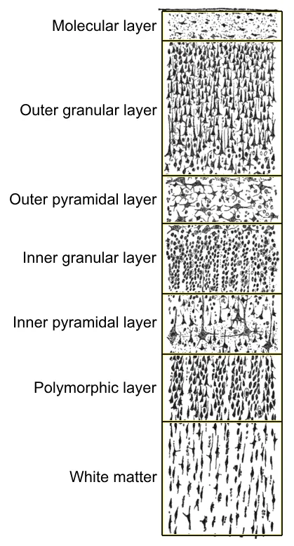
新皮質は表面から6層。要点は「4層が入口、5層が出口」である。
- I 分子層——細胞が少なく、樹状突起と軸索の層
- II 外顆粒層/III 外錐体細胞層——皮質どうしを結ぶ(連合・交連線維)
- IV 内顆粒層——視床からの入力を受ける
- V 内錐体細胞層——皮質から出る大型の錐体細胞。運動野の Betz 細胞
- VI 多形細胞層——視床へ戻す
層の厚さが機能で変わることが、組織像から領野を言い当てる根拠になる。
- 一次運動野(4野)——出力が主なのでV層が厚く、IV層がほとんど無い(無顆粒皮質)。Betz 細胞が並ぶ
- 一次感覚野・視覚野——入力が主なのでIV層が厚い(顆粒皮質)。視覚野では IV層がさらに細分され、肉眼でもGennari 線条として見える
「入力の多い場所は入口の層が厚く、出力の多い場所は出口の層が厚い」——構造が仕事量を映している。
もう一つの原理がコラム構造である。皮質は層と直交する方向に柱としてまとまり、同じ柱の細胞は同じ刺激に反応する(同じ指、同じ傾きの線)。水平方向が層、垂直方向が柱という二重の秩序で、皮質は情報を整理している。
この配置から病態も出る。低酸素・虚血に最も弱いのは第3・5・6層で、境界に沿って壊死すると層状壊死という特徴的な像になる。海馬の CA1(Sommer 領域)や小脳の Purkinje 細胞とともに、選択的脆弱性を示す代表である。
層ごとに違う病気が起こる。
Alzheimer 病 — 老人斑(アミロイドβ・細胞外)と神経原線維変化(リン酸化タウ・細胞内)。海馬と側頭葉内側から始まり、頭頂・側頭へ広がる。近時記憶の障害で発症するのは、始まる場所が海馬だからである。
層状壊死(laminar necrosis) — 心停止・低血糖などの全般性虚血では第3・5層が選択的に壊死する。代謝要求が高い層が先に落ちる。
皮質形成異常は難治性てんかんの原因になる。神経細胞の移動障害で層構造が乱れ、滑脳症・多小脳回になる。MRI で層構造の異常を探すのがてんかん外科の出発点である。
第5層の Betz 細胞が錐体路の起始である。筋萎縮性側索硬化症ではここが失われ、上位と下位の運動ニューロン徴候が同時に出る。
English version
Core
The neocortex has six layers from the surface. The point is that layer IV is the entrance and layer V the exit.
- I Molecular layer — few cells; a layer of dendrites and axons.
- II External granular / III External pyramidal — connect cortex to cortex (association and commissural fibres).
- IV Internal granular — receives input from the thalamus.
- V Internal pyramidal — large pyramidal cells projecting out of the cortex; the Betz cells of the motor cortex.
- VI Multiform layer — projects back to the thalamus.
Going further
That the thickness of the layers varies with function is what allows an area to be named from its histology.
- Primary motor cortex (area 4) — mainly output, so layer V is thick and layer IV almost absent (agranular cortex). Betz cells are lined up here.
- Primary sensory and visual cortex — mainly input, so layer IV is thick (granular cortex). In the visual cortex layer IV is further subdivided and is visible to the naked eye as the line of Gennari.
Where input predominates the entrance layer is thick; where output predominates the exit layer is thick — the structure mirrors the work done.
A second principle is the columnar organisation: the cortex is grouped into columns perpendicular to the layers, and cells in one column respond to the same stimulus (the same digit, the same line orientation). Layers horizontally and columns vertically — a double order by which the cortex organises information.
The pathology follows from the arrangement. Layers 3, 5 and 6 are the most vulnerable to hypoxia and ischaemia, and when they necrose along their plane the result is the characteristic laminar necrosis — one of the classic examples of selective vulnerability, along with hippocampal CA1 (Sommer sector) and the cerebellar Purkinje cells.
In the clinic
Different diseases occur in different layers.
Alzheimer disease — senile plaques (amyloid β, extracellular) and neurofibrillary tangles (phosphorylated tau, intracellular). It begins in the hippocampus and medial temporal lobe and spreads parietally and temporally. It presents with impairment of recent memory because of where it begins.
Laminar necrosis — in global ischaemia from cardiac arrest or hypoglycaemia, layers 3 and 5 necrose selectively. The layers with the highest metabolic demand fail first.
Cortical dysplasia causes intractable epilepsy: a disorder of neuronal migration disrupts the lamination, giving lissencephaly or polymicrogyria. Searching the MRI for disordered lamination is the starting point of epilepsy surgery.
The Betz cells of layer V are the origin of the pyramidal tract; in amyotrophic lateral sclerosis they are lost, so upper and lower motor neuron signs appear together.
小脳皮質 — 3層と1つの出口Cortex cerebelli
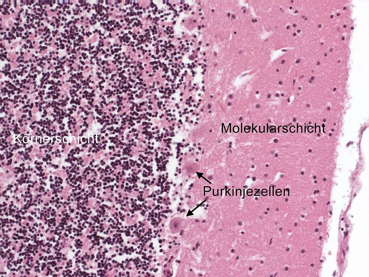
どこを切っても同じ3層で、全体が同じ回路の繰り返しになっている。
- 分子層——星状細胞・籠細胞と、平行線維
- Purkinje 細胞層——1列に並ぶ大型細胞。樹状突起を1つの平面に扇状に広げる
- 顆粒層——きわめて密な顆粒細胞。脳内で最も数の多い神経細胞
入力は2系統、出力は1つ。
- 苔状線維 → 顆粒細胞 → 平行線維 → Purkinje 細胞(多数が弱く)
- 登上線維(下オリーブ核から)→ Purkinje 細胞に1対1で強く巻きつく
- 出力は Purkinje 細胞だけ。しかも抑制性(GABA)
小脳皮質の唯一の出口が抑制性である——ここが小脳を理解する鍵になる。Purkinje 細胞は小脳核を抑える。小脳核は常に興奮しているので、小脳は「動きを止める側」から運動を調節している。だから小脳が壊れると動きが無くなるのではなく、止められなくなる(測定障害・企図振戦・反復拮抗運動不能)。
2種類の入力の違いにも意味がある。平行線維は多数が少しずつ、登上線維は1本が強烈にPurkinje 細胞へ入る。登上線維が発火すると複雑スパイクが生じ、同時に入っていた平行線維の結合が弱められる(長期抑圧)。
これが運動学習の実体である。登上線維が運ぶのは「今の動きは誤りだった」という誤差信号で、それが起きた瞬間の入力を弱める。失敗した組み合わせを消していくことで、動きが洗練される。
小脳の障害は同側に出る。小脳半球 → 上小脳脚で交叉 → 対側の視床・運動野 → 皮質脊髄路で再び交叉、と2回交叉するためである。脳幹梗塞で「顔は同側・体は対側」という交代性の所見が出るのと同じく、交叉の回数を数えることが局在診断になる。
Purkinje 細胞は小脳皮質の唯一の出口で、虚血・低酸素・アルコール・抗てんかん薬(フェニトイン)に非常に弱い。
アルコール性小脳変性症では小脳虫部(前上部)の Purkinje 細胞が優先的に脱落する。体幹失調と歩行障害が主体で、上肢は比較的保たれる——虫部が体幹を担うという局在がそのまま症状になる。
傍腫瘍性小脳変性症 — 抗 Yo 抗体(婦人科癌)・抗 Hu 抗体(小細胞肺癌)。亜急性に進行する小脳失調を見たら悪性腫瘍を探す。
髄芽腫は小脳虫部に発生する小児の悪性腫瘍で、第四脳室を塞いで水頭症を起こす。体幹失調+頭蓋内圧亢進という組み合わせで気づかれる。
English version
Core
The same three layers wherever it is cut — the whole organ is one circuit repeated.
- Molecular layer — stellate and basket cells, and the parallel fibres.
- Purkinje cell layer — a single row of large cells whose dendrites spread like a fan in one plane.
- Granular layer — extremely densely packed granule cells, the most numerous neurons in the brain.
Two inputs, one output.
- Mossy fibre → granule cell → parallel fibre → Purkinje cell (many, weakly).
- Climbing fibre (from the inferior olive) → wraps one to one around a Purkinje cell, powerfully.
- The output is the Purkinje cell alone — and it is inhibitory (GABA).
Going further
That the sole exit of the cerebellar cortex is inhibitory is the key to understanding the cerebellum. Purkinje cells inhibit the cerebellar nuclei, which are tonically active, so the cerebellum regulates movement from the side that stops it. Hence damage does not abolish movement but removes the ability to stop it — dysmetria, intention tremor, dysdiadochokinesis.
The two inputs differ in meaning as well. Parallel fibres act in large numbers, each weakly; the climbing fibre acts alone and powerfully. When a climbing fibre fires it produces a complex spike and simultaneously weakens the parallel fibre synapses that were active (long-term depression). This is the substance of motor learning: the climbing fibre carries the error signal "that movement was wrong", and weakens the inputs active at that moment. Movement is refined by erasing the combinations that failed.
Cerebellar signs appear on the same side, because there are two decussations: the hemisphere crosses in the superior cerebellar peduncle to the contralateral thalamus and motor cortex, and the corticospinal tract crosses again. Counting the decussations is localisation — as with the crossed findings of a brainstem infarct.
In the clinic
The Purkinje cell is the sole output of the cerebellar cortex and is highly vulnerable to ischaemia, hypoxia, alcohol and phenytoin.
In alcoholic cerebellar degeneration the Purkinje cells of the anterior superior vermis are preferentially lost: truncal ataxia and gait disturbance predominate while the arms are relatively spared — the localisation of the vermis to the trunk appearing directly as the symptom.
Paraneoplastic cerebellar degeneration — anti-Yo (gynaecological cancer), anti-Hu (small cell lung cancer). Subacutely progressive cerebellar ataxia should prompt a search for malignancy.
Medulloblastoma arises in the cerebellar vermis in children and obstructs the fourth ventricle, causing hydrocephalus. It is recognised by the combination of truncal ataxia with raised intracranial pressure.
小脳核 — 小脳の本当の出口Nucleus dentatus
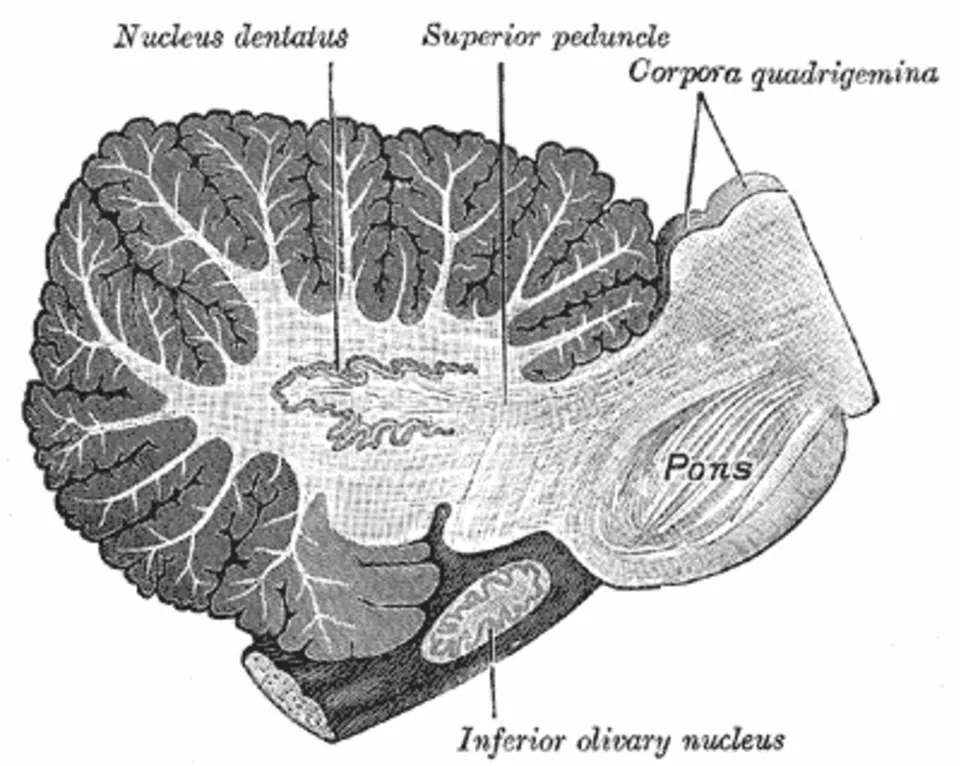
小脳の白質(髄体)の中、第四脳室の屋根の近くに、左右4対の小脳核が埋まっている。外側から内側へ、
| 核 | Purkinje 細胞の入力元 | 主な出力先 | 受け持つこと |
|---|---|---|---|
| 歯状核(最大。ひだのある袋の形) | 小脳半球の外側部 | 上小脳脚 → 反対側の視床 → 運動野・運動前野(一部は赤核) | 運動の計画とタイミング |
| 栓状核・球状核(あわせて中位核) | 半球の中間部 | 反対側の赤核、視床 | 進行中の手足の動きの修正 |
| 室頂核 | 虫部 | 前庭神経核・網様体 | 姿勢・平衡・眼球運動 |
組織像(鍍銀標本)では、層構造は無く、有髄線維の流れの中に、大型の多極ニューロンの細胞体が群れになって散らばる。3層に並ぶ小脳皮質との違いがはっきりしている。
小脳核には、Purkinje 細胞の軸索(抑制性、GABA)と、苔状線維・登上線維の側枝(興奮性)が入る。小脳核の出力ニューロンは興奮性で、小脳から外へ出る信号のほとんどすべてを担う(例外は片葉小節葉から前庭神経核への直接の線維)。
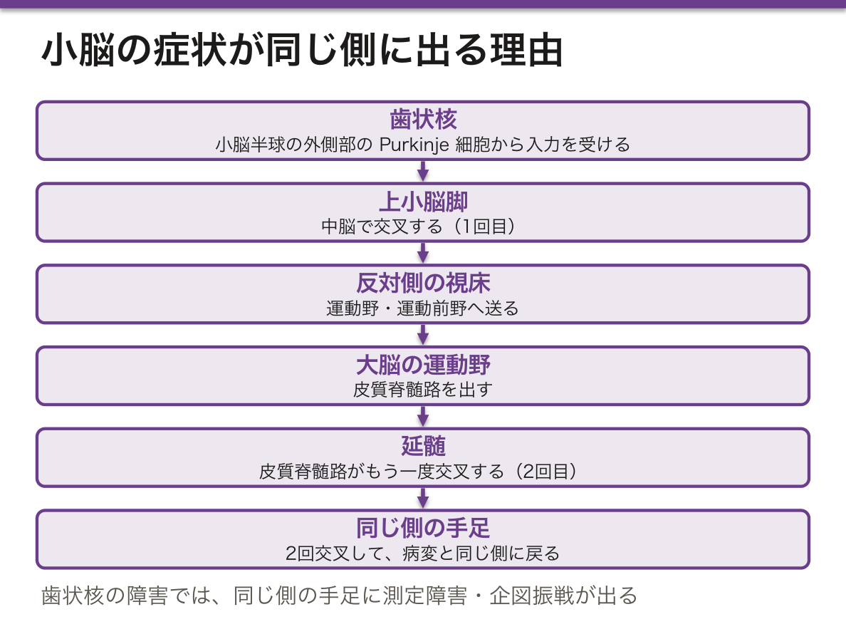
小脳核は、苔状線維・登上線維の側枝から絶えず興奮を受けて常に発火している。そこに Purkinje 細胞の抑制が加わったり外れたりして、出力の強さとタイミングが削り出される。皮質が「彫刻刀」、核が「素材」と考えるとよい。
小脳の症状が病変と同じ側に出る理由も、ここから説明できる。歯状核の出力は中脳で交叉して反対側の大脳へ行き、大脳からの皮質脊髄路は延髄でもう一度交叉する。2回交叉するので、結局は同じ側の手足に戻る。
どの核の系統が壊れたかで症状が変わる。小脳半球・歯状核の障害では、同じ側の手足の測定障害・企図振戦が出る。虫部・室頂核の障害では、手足の動きは比較的保たれるのに体幹が揺れて歩けない。アルコールによる小脳変性が主に前方の虫部を侵し、歩行の失調が目立つのはこのためである。
歯状核 → 反対側の赤核 → (赤核と同じ側の)下オリーブ核 → 反対側の歯状核という三角の回路(Guillain-Mollaret 三角)が途中で切れると、下オリーブ核が大きくなり、口蓋振戦が出る。
歯状核は画像でも話題になる。ガドリニウム造影剤の検査を何度も受けた患者で、造影していない T1 強調像で歯状核が白く見えることが報告され、ガドリニウムが脳に残ることが知られるようになった。日本に多い歯状核赤核淡蒼球ルイ体萎縮症(DRPLA)は、名前のとおり歯状核から赤核への系統を侵す。
English version
Core
Four pairs of deep cerebellar nuclei lie in the white matter (medullary body), near the roof of the fourth ventricle. From lateral to medial:
| Nucleus | Purkinje input from | Main output to | Role |
|---|---|---|---|
| Dentate (largest; a folded bag) | lateral hemisphere | superior peduncle → contralateral thalamus → motor and premotor cortex (some to red nucleus) | planning and timing of movement |
| Emboliform and globose (together: interposed) | intermediate hemisphere | contralateral red nucleus, thalamus | correcting ongoing limb movement |
| Fastigial | vermis | vestibular nuclei, reticular formation | posture, balance, eye movements |
On a slide (silver impregnation) there are no layers: groups of large multipolar cell bodies lie scattered among streams of myelinated fibres. The contrast with the three-layered cerebellar cortex is clear.
The nuclei receive Purkinje axons (inhibitory, GABA) and collaterals of mossy and climbing fibres (excitatory). Their output neurons are excitatory and carry almost every signal that leaves the cerebellum (the exception is direct fibres from the flocculonodular lobe to the vestibular nuclei).
Going further
The nuclei get constant excitation from mossy- and climbing-fibre collaterals, so they fire all the time. Purkinje inhibition, added or removed, carves out the strength and timing of the output. Think of the cortex as the chisel and the nuclei as the material.
This also explains why cerebellar signs appear on the same side as the lesion. Dentate output crosses in the midbrain to the opposite cerebrum, and the corticospinal tract from the cerebrum crosses again in the medulla. Two crossings bring it back to the limbs of the same side.
In the clinic
Symptoms depend on which nuclear system fails. Damage to the hemisphere and dentate causes dysmetria and intention tremor in the limbs on the same side. Damage to the vermis and fastigial nucleus leaves limb movement fairly intact but makes the trunk sway and walking unstable. Alcoholic cerebellar degeneration mainly hits the anterior vermis, so gait ataxia stands out.
If the triangle dentate → contralateral red nucleus → inferior olive (on the red nucleus side) → contralateral dentate (Guillain-Mollaret triangle) is cut, the inferior olive enlarges and palatal tremor appears.
The dentate nucleus also appears in imaging. In patients who had many gadolinium-enhanced scans, the dentate looked bright on unenhanced T1 images, which showed that gadolinium stays in the brain. Dentatorubral-pallidoluysian atrophy (DRPLA), relatively common in Japan, attacks, as its name says, the dentate-to-red-nucleus system.
血液脳関門とグリア — 脳を隔てるもの
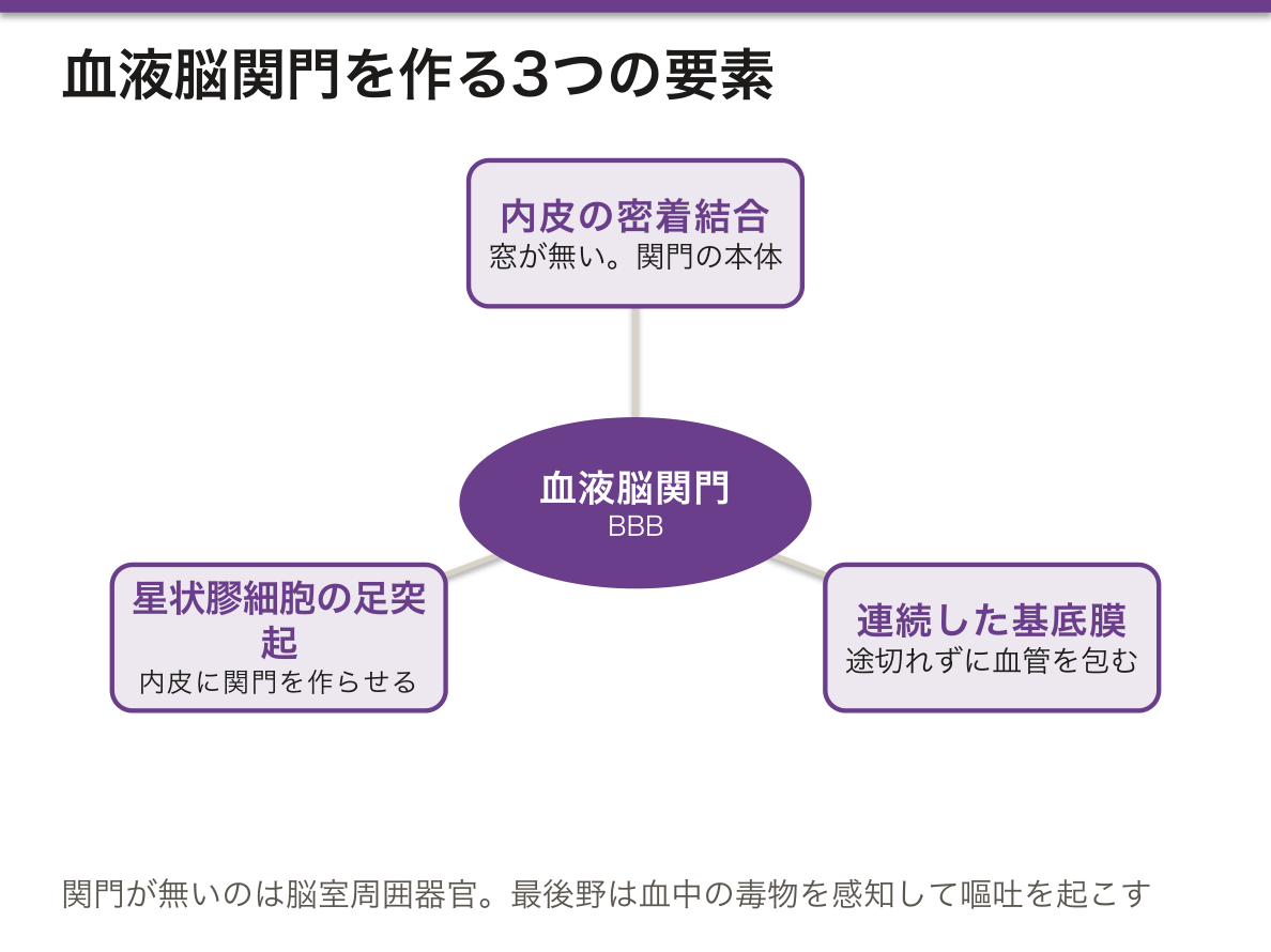
血液脳関門を作るのは3つの要素。
- 毛細血管内皮の密着結合——窓が無く、細胞のあいだを物が通れない。これが本体
- 連続した基底膜
- 星状膠細胞(アストロサイト)の足突起——血管を覆い、内皮に関門を作らせ、維持させる
グリアの分担。
- 星状膠細胞——関門の維持、カリウムの緩衝、グルタミン酸の回収、グリア瘢痕
- 稀突起膠細胞——髄鞘。1個が複数の軸索を担当する
- 小膠細胞(ミクログリア)——唯一の中胚葉/骨髄由来。脳の免疫担当
- 上衣細胞——脳室の内面。線毛を持つ
関門があることが、薬が効かない理由にも、効く理由にもなる。脂溶性が高く分子量の小さいものは通り、水溶性で大きいものは通らない。
- レボドパは通るがドパミンは通らない——だから Parkinson 病では前駆体を投与し、末梢での脱炭酸を阻害する薬(カルビドパ)を併用して脳へ届く量を増やす。末梢の阻害薬自身は関門を通らないことが前提になっている
- 第2世代の抗ヒスタミン薬で眠くならないのは、関門を通りにくく設計されているため
- 白血病の中枢神経再発には、静注では届かないので髄腔内投与を行う
関門が無い場所もある(脳室周囲器官)。最後野には関門が無く、血中の毒物を感知して嘔吐を起こす——化学受容器引金帯である。関門の穴が、防御の仕組みとして使われている。
関門は病気で壊れる。造影 CT・MRI で「造影される」ということは、そこで関門が破れているという意味である。腫瘍、膿瘍、脱髄の活動期——造影効果は病変の活動性そのものを見ている。高血圧性脳症や子癇での可逆性後頭葉白質脳症も、関門の破綻による浮腫である。
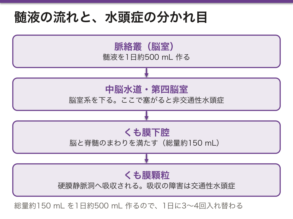
髄液は脈絡叢が作り、くも膜顆粒が吸う。1日約500mL 作られ、常に約150mL が存在する——1日3回ほど入れ替わる。
この流れのどこが詰まるかで水頭症の型が分かれる。
- 非交通性(閉塞性)——脳室系の途中(中脳水道など)で塞がる。閉塞より上流の脳室だけが拡大
- 交通性——くも膜下腔や吸収の障害。くも膜下出血・髄膜炎の後に多い
- 正常圧水頭症——歩行障害・認知機能低下・尿失禁の三徴。髄液を抜くと改善する=治療できる認知症として重要
髄膜炎で髄液の糖が下がるのは、細菌が消費するだけでなく関門での糖輸送そのものが障害されるためである。細菌性では糖が下がり、ウイルス性では保たれる——この一点が最初の鑑別になる。
English version
Core
Three elements form the blood–brain barrier.
- Tight junctions of the capillary endothelium — no fenestrations, nothing passes between the cells. This is the barrier itself.
- A continuous basal lamina.
- Astrocytic end-feet — covering the vessel, inducing and maintaining the barrier in the endothelium.
Division of labour among the glia
- Astrocyte — maintains the barrier, buffers potassium, takes up glutamate, forms the glial scar.
- Oligodendrocyte — myelin; one cell serves several axons.
- Microglia — the only one of mesodermal / marrow origin; immunity of the brain.
- Ependymal cell — lines the ventricles; ciliated.
Going further
The barrier is the reason a drug fails and the reason a drug works. Small, lipid-soluble molecules cross; large, water-soluble ones do not.
- Levodopa crosses, dopamine does not — hence in Parkinson disease the precursor is given, with a peripheral decarboxylase inhibitor (carbidopa) to increase the amount reaching the brain. It is a premise that the peripheral inhibitor itself does not cross.
- Second-generation antihistamines do not cause drowsiness because they are designed not to cross.
- Central nervous relapse of leukaemia is treated intrathecally, because intravenous drug does not reach.
Some places have no barrier (the circumventricular organs). The area postrema has none, so it detects circulating toxins and induces vomiting — the chemoreceptor trigger zone. A hole in the barrier is used as a mechanism of defence.
The barrier is broken by disease. That a lesion enhances on contrast CT or MRI means the barrier is broken there — in tumour, abscess and active demyelination, enhancement is a direct view of the activity of the lesion. The oedema of hypertensive encephalopathy and eclampsia (PRES) is likewise a failure of the barrier.
In the clinic
CSF is made by the choroid plexus and absorbed by the arachnoid granulations: about 500 mL a day against a standing volume of about 150 mL — replaced three times over.
Where this flow is obstructed determines the type of hydrocephalus.
- Non-communicating (obstructive) — a block within the ventricular system (the aqueduct, typically); only the ventricles upstream dilate.
- Communicating — impaired flow or absorption in the subarachnoid space; common after subarachnoid haemorrhage or meningitis.
- Normal pressure hydrocephalus — the triad of gait disturbance, cognitive decline and urinary incontinence, improving when CSF is removed. Important as a treatable dementia.
In meningitis the CSF glucose falls not only because bacteria consume it but because glucose transport across the barrier is itself impaired. It falls in bacterial and is preserved in viral meningitis — this single point is the first discriminator.
脊髄 — 灰白質の形が症状の地図になる
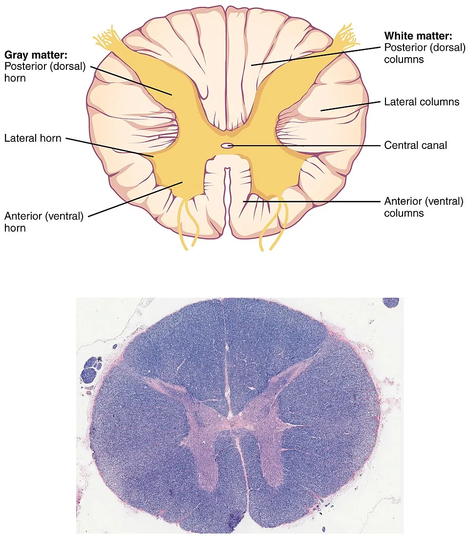
中心が灰白質(H字・蝶形)、外側が白質。大脳と逆である。
- 後角——感覚の入口
- 側角——胸髄〜上位腰髄のみ。交感神経の細胞体
- 前角——運動神経細胞(下位運動ニューロン)
白質の主な路。
- 後索——触覚・振動覚・位置覚。同側を上行し、延髄で交叉
- 外側脊髄視床路——痛覚・温度覚。入った高さで交叉して対側を上行
- 外側皮質脊髄路——運動。延髄の錐体で交叉済み
灰白質は頸膨大・腰膨大で大きい(四肢を支配するため)。
交叉する場所が違うという一点が、脊髄の症候学のすべてを決めている。
- 痛覚・温度覚は入った高さですぐ交叉する → 障害されると対側で失われる
- 触覚・位置覚は交叉せず上がる → 障害されると同側で失われる
だから半側を切るとBrown-Séquard 症候群——同側の運動麻痺と深部覚障害+対側の痛温覚障害という、一見ちぐはぐな所見になる。ちぐはぐさそのものが、交叉の位置の違いの証明である。
脊髄空洞症では中心管の周囲が広がるので、そこで交叉している痛温覚の線維だけが最初に切られる。結果、両側・その高さだけ・痛温覚だけが失われ、触覚は残る(宙吊り型の解離性感覚障害)。やけどに気づかないという主訴になる。
血流の分布も症候になる。前脊髄動脈は前3分の2を養うので、閉塞すると運動麻痺と痛温覚障害が出て、後索(触覚・位置覚)は残る。大動脈手術の合併症として現実的な問題である。
上位運動ニューロンと下位運動ニューロンの区別も脊髄で完結する。障害高位では前角が壊れて弛緩性麻痺・萎縮・線維束攣縮、その高位より下では錐体路が切れて痙性麻痺・腱反射亢進・Babinski 徴候。1か所の損傷で2種類の麻痺が同時に出るのはこのためで、弛緩性麻痺の高さが損傷高位を教える。
灰白質の形が、そのまま症状の地図になる。
筋萎縮性側索硬化症 — 前角細胞(下位)と側索の錐体路(上位)が同時に侵される。筋萎縮と線維束攣縮(下位)に、腱反射亢進と Babinski 徴候(上位)が同居するという矛盾した組み合わせが診断の決め手になる。感覚は保たれ、眼球運動と膀胱直腸は最後まで残る。
脊髄性筋萎縮症・ポリオ — 前角細胞のみ。純粋な下位運動ニューロン障害で、弛緩性麻痺・腱反射消失・感覚正常。
亜急性連合性脊髄変性症 — ビタミン B12 欠乏で後索と側索がやられる。深部感覚障害+痙性麻痺。
脊髄空洞症 — 中心管の周りで交叉する温痛覚だけが両側で落ちる宙吊り型の解離性感覚障害。灰白質の位置関係を知らないと説明できない所見である。
English version
Core
Grey matter centrally (H-shaped, like a butterfly), white matter outside — the reverse of the cerebrum.
- Dorsal horn — the entrance for sensation.
- Lateral horn — thoracic and upper lumbar cord only; the cell bodies of the sympathetic outflow.
- Ventral horn — motor neurons (lower motor neurons).
The principal tracts of the white matter
- Dorsal columns — touch, vibration, position. Ascend ipsilaterally and decussate in the medulla.
- Lateral spinothalamic tract — pain and temperature. Decussate at the level of entry and ascend contralaterally.
- Lateral corticospinal tract — motor; already decussated in the medullary pyramid.
The grey matter is enlarged at the cervical and lumbar enlargements, which supply the limbs.
Going further
The single fact that the tracts decussate in different places determines the whole symptomatology of the cord.
- Pain and temperature decussate immediately → damage causes loss on the opposite side.
- Touch and position ascend uncrossed → damage causes loss on the same side.
So hemisection gives Brown-Séquard syndrome — ipsilateral motor and deep sensory loss with contralateral loss of pain and temperature, an apparently incoherent set of findings. The incoherence is itself the proof that the decussations lie at different levels.
In syringomyelia the region around the central canal expands, so the pain and temperature fibres decussating there are cut first. The result is loss of pain and temperature, bilaterally, confined to those segments, with touch preserved — a suspended, dissociated sensory loss, presenting as failure to notice burns.
The blood supply is symptomatic too: the anterior spinal artery supplies the anterior two-thirds, so occlusion gives motor paralysis and loss of pain and temperature while the dorsal columns are spared — a real complication of aortic surgery.
The distinction between upper and lower motor neuron is completed within the cord: at the level of injury the ventral horn is destroyed giving flaccid paralysis, atrophy and fasciculation; below it the pyramidal tract is cut giving spastic paralysis, hyperreflexia and a Babinski sign. One lesion produces two kinds of paralysis, and the level of the flaccid paralysis gives the level of the injury.
In the clinic
The shape of the grey matter is directly a map of the symptoms.
Amyotrophic lateral sclerosis — the anterior horn cells (lower) and the corticospinal tract in the lateral column (upper) are affected together. The contradictory combination of wasting and fasciculation with hyperreflexia and a Babinski sign is the diagnostic key. Sensation is preserved, and eye movement and bladder function are spared until late.
Spinal muscular atrophy and poliomyelitis — anterior horn cells alone: pure lower motor neuron disease, with flaccid paralysis, absent reflexes and normal sensation.
Subacute combined degeneration — vitamin B12 deficiency affecting the dorsal and lateral columns: impaired deep sensation with spastic paralysis.
Syringomyelia — a suspended, dissociated loss of pain and temperature only, bilaterally. A finding that cannot be explained without knowing the topography of the grey matter.
髄膜と脈絡叢 — 脳を浮かせる水を、どこで作るかPlexus choroideus
脳と脊髄は3層の膜に包まれる。層の間に何があるかで、出血の名前が変わる。
| 層 | 組織 | その外側/内側の腔 |
|---|---|---|
| 硬膜 | 密性線維性結合組織。頭蓋では2葉(骨膜層と髄膜層)で、離れる所に硬膜静脈洞 | 硬膜外腔(頭蓋では潜在的。中硬膜動脈が走る) |
| くも膜 | 無血管の薄い膜。くも膜顆粒が硬膜静脈洞へ突出 | 硬膜下腔(架橋静脈が横切る) |
| 軟膜 | 脳表に密着し、血管とともに脳実質へ入る | くも膜下腔——ここに脳脊髄液と主要動脈 |
脈絡叢は軟膜の血管が脳室の上衣に押し込まれた構造で、有窓性毛細血管+立方の脈絡叢上皮からなる。脳脊髄液を1日約500 mL 産生(総量は約150 mL なので1日3〜4回入れ替わる)。
「どの層のどの血管が切れたか」で頭蓋内出血がすべて決まる。
| 出血 | 切れる血管 | CT所見 | 経過 |
|---|---|---|---|
| 硬膜外血腫 | 中硬膜動脈(側頭骨骨折を伴う) | 凸レンズ型(縫合線を越えない) | 動脈性で急速。意識清明期〈lucid interval〉の後に急変 |
| 急性硬膜下血腫 | 架橋静脈 | 三日月型(縫合線を越える) | 脳挫傷を伴い予後不良 |
| 慢性硬膜下血腫 | 架橋静脈(軽微な外傷) | 三日月型 | 高齢者・飲酒者。1〜2か月後に認知症様・片麻痺。穿頭ドレナージで劇的に治る「治せる認知症」 |
| くも膜下出血 | 脳動脈瘤(Willis 動脈輪) | 脳槽が高吸収(星型・五角形) | 突然の激しい頭痛。再破裂が最大の危険 |
血液脳関門〈BBB〉と血液脳脊髄液関門は別物——BBB は脳毛細血管内皮の密着結合が作り、後者は脈絡叢上皮の密着結合が作る。脈絡叢の毛細血管は有窓性で漏れるが、上皮で止めている。
BBB が無い場所〈脳室周囲器官〉——最後野(嘔吐中枢)・下垂体後葉・松果体・脳弓下器官・終板脈管器官。血中の情報を直接感知する必要がある場所だから開いている。最後野が化学受容器引金帯〈CTZ〉として血中の毒物を検知して嘔吐を起こすのはこのため。
水頭症は産生・循環・吸収のどこが壊れたかで分ける——非交通性(中脳水道・第四脳室の閉塞)、交通性(くも膜顆粒での吸収障害:くも膜下出血後・髄膜炎後)。正常圧水頭症は歩行障害・認知症・尿失禁の3徴で、シャント術で改善しうる。
層と腔の対応が、そのまま出血の名前になる。硬膜外=動脈・凸レンズ形、硬膜下=架橋静脈・三日月形、くも膜下=動脈瘤・脳槽に沿う(脳硬膜の章)。
髄膜炎の髄液所見は層の話ではなく中身の話だが、くも膜下腔に何が起きているかを映す。細菌性=多核球優位・糖低下・蛋白高値、ウイルス性=単核球優位・糖正常、結核性=単核球優位・糖低下・蛋白著増。糖が下がるかどうかが最初の分かれ道になる。
脈絡叢は血液髄液関門を作る。上衣細胞ではなく、脈絡叢上皮の密着結合がその実体である。
脈絡叢乳頭腫は髄液の産生過剰による水頭症を起こす数少ない腫瘍である。
English version
Core
Brain and cord are invested by three membranes, and what lies between the layers determines the name of a haemorrhage.
| Layer | Tissue | The space outside / inside it |
|---|---|---|
| Dura mater | Dense fibrous tissue; two laminae in the skull, separating to enclose the dural venous sinuses | Extradural space (potential in the skull; the middle meningeal artery runs here) |
| Arachnoid | Thin and avascular; arachnoid granulations project into the sinuses | Subdural space (crossed by the bridging veins) |
| Pia mater | Adherent to the surface, entering the parenchyma with the vessels | Subarachnoid space — CSF and the major arteries |
The choroid plexus is pial vessels pushed into the ependyma of a ventricle: fenestrated capillaries plus a cuboidal choroid epithelium. It produces about 500 mL of CSF a day against a volume of about 150 mL — replaced three or four times daily.
Going further
Which vessel in which layer has torn settles every intracranial haemorrhage.
| Haemorrhage | Vessel | CT appearance | Course |
|---|---|---|---|
| Extradural | Middle meningeal artery (with a temporal fracture) | Biconvex; does not cross sutures | Arterial and rapid; deterioration after a lucid interval |
| Acute subdural | Bridging veins | Crescentic; crosses sutures | Accompanied by contusion; poor prognosis |
| Chronic subdural | Bridging veins (trivial trauma) | Crescentic | Elderly, alcohol. Dementia-like or hemiparesis 1–2 months later. Dramatically cured by burr-hole drainage — a treatable dementia |
| Subarachnoid | Cerebral aneurysm (circle of Willis) | High density in the cisterns | Sudden severe headache; rebleeding is the greatest danger |
The blood–brain barrier and the blood–CSF barrier are different things: the former is made by the tight junctions of the cerebral capillary endothelium, the latter by those of the choroid epithelium. The choroidal capillaries are fenestrated and leak; the epithelium stops it.
Places without a barrier (circumventricular organs) — area postrema (vomiting), neurohypophysis, pineal gland, subfornical organ, organum vasculosum. They are open because they must sample the blood directly, which is why the area postrema serves as the chemoreceptor trigger zone.
In the clinic
The correspondence of layer and space is directly the name of the haemorrhage: extradural = artery, biconvex; subdural = bridging vein, crescentic; subarachnoid = aneurysm, along the cisterns.
The CSF findings in meningitis are a matter of content rather than of layer, but they mirror what is happening in the subarachnoid space. Bacterial — neutrophil predominance, low glucose, high protein. Viral — mononuclear predominance, normal glucose. Tuberculous — mononuclear predominance, low glucose, markedly raised protein. Whether the glucose falls is the first branch point.
The choroid plexus forms the blood–CSF barrier, and the substance of it is the tight junctions of the choroid epithelium, not of the ependyma. Choroid plexus papilloma is one of the few tumours that causes hydrocephalus by overproduction of CSF.
海馬 — 3層の古い皮質が、記憶の入口になっているHippocampus
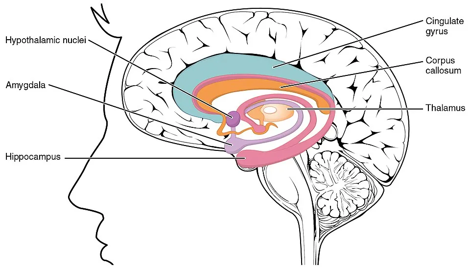
大脳皮質の6層とは別の、3層構造(古皮質)——分子層・錐体細胞層・多形層。
回路は一方向に流れる(三シナプス回路)——嗅内皮質 →(貫通線維)→ 歯状回 →(苔状線維)→ CA3 →(Schaffer 側枝)→ CA1 → 海馬支脚 → 嗅内皮質。
CA1(Sommer 扇形部)は、虚血に最も弱い。
歯状回の顆粒細胞層は、成人でも神経新生が起こる数少ない場所になる。
長期増強(LTP)が起こる場所として、記憶の細胞レベルの基盤とされる。
「一方向の回路」であることが、この領域の弱点と機能の両方を作っている。一箇所が落ちれば、その先へ情報が届かない(高次機能の章)——「並列でないから、脆い」。
CA1 が虚血に弱いことは、蘇生後の患者で必ず問題になる。心停止後に全身が回復しても、記憶だけが戻らないことがある(選択的脆弱性)(心拍再開後の章)——「同じ低酸素でも、壊れる場所には順番がある」。
Alzheimer 病でも、嗅内皮質と海馬から変性が始まる(認知症の章)——「だから最初の症状が、近時記憶の障害になる」。
両側の海馬が失われると、新しい記憶が作れなくなる(前向性健忘)。すでに固定された古い記憶と、手続き記憶は保たれる——「記憶は1つの機能ではなく、場所ごとに別の系になっている」。
てんかんとの関係も深い。内側側頭葉てんかんでは海馬硬化(CA1 の脱落とグリオーシス)が見られ、切除で発作が止まることがある(てんかんの章)——「薬で止まらない発作に、外科という選択肢がある」。
神経新生が起こる場所であることは、抗うつ薬の作用機序の仮説にもつながっている(向精神薬の章)——「効果が出るまで数週間かかる理由の説明として提案されている」。
海馬は最も虚血に弱く、最も早く Alzheimer 病に侵される。記憶が最初に失われるのは、この2つの理由による。
CA1(Sommer 区)は低酸素・低血糖に対して選択的に脆弱である。心停止蘇生後の記憶障害や一酸化炭素中毒後の遅発性障害はここが落ちた結果である。
海馬硬化症は側頭葉てんかんの最も多い原因で、CA1 の神経脱落とグリオーシスを示す。薬剤抵抗性なら側頭葉切除で発作が止まりうる——てんかん外科の主要な適応である。
抗 NMDA 受容体脳炎は若年女性の卵巣奇形腫に伴い、精神症状・けいれん・不随意運動で発症する。腫瘍の切除と免疫療法で回復しうるので、「若い人の急な精神症状」で必ず鑑別に挙げる。
English version
Core
A three-layered archicortex, distinct from the six-layered neocortex — molecular, pyramidal cell and polymorphic layers.
The circuit flows in one direction (the trisynaptic circuit): entorhinal cortex → (perforant path) → dentate gyrus → (mossy fibres) → CA3 → (Schaffer collaterals) → CA1 → subiculum → entorhinal cortex.
- CA1 (Sommer sector) is the most vulnerable to ischaemia.
- The granule cell layer of the dentate gyrus is one of the few sites where neurogenesis occurs in the adult.
- It is where long-term potentiation occurs, and is regarded as the cellular basis of memory.
Going further
Being a one-way circuit creates both the function and the fragility of this region. If one point fails, information cannot reach what lies beyond — it is fragile because it is not parallel.
That CA1 is vulnerable to ischaemia is always an issue in the resuscitated patient: the body may recover fully after cardiac arrest and memory alone fail to return — the same hypoxia damages structures in a fixed order.
Alzheimer disease likewise begins in the entorhinal cortex and hippocampus — which is why the first symptom is impairment of recent memory.
With loss of both hippocampi, new memories can no longer be formed (anterograde amnesia) while consolidated remote memory and procedural memory are preserved — memory is not one function but a set of separate systems in separate places.
The relation to epilepsy is close: mesial temporal lobe epilepsy shows hippocampal sclerosis (loss of CA1 with gliosis), and resection can stop the seizures — surgery is an option when drugs fail. That neurogenesis occurs here also feeds hypotheses about the action of antidepressants — proposed as an explanation for the several weeks before an effect appears.
In the clinic
The hippocampus is the most vulnerable to ischaemia and the earliest affected in Alzheimer disease. Memory is lost first for those two reasons.
CA1 (Sommer sector) is selectively vulnerable to hypoxia and hypoglycaemia; the memory impairment after resuscitation from cardiac arrest and the delayed injury after carbon monoxide poisoning are the result of its loss.
Hippocampal sclerosis is the commonest cause of temporal lobe epilepsy, showing neuronal loss in CA1 with gliosis. If drug-resistant, temporal lobectomy can stop the seizures — a principal indication for epilepsy surgery.
Anti-NMDA receptor encephalitis accompanies ovarian teratoma in young women and presents with psychiatric symptoms, seizures and involuntary movements. Because it can recover with removal of the tumour and immunotherapy, it must always be considered in a young person with sudden psychiatric symptoms.
上衣細胞と脳室 — 水と脳のあいだの、もう1枚の面Ependymocytus
脳室と脊髄中心管の内面を覆う、単層立方〜円柱の上皮様の細胞。グリアの一種になる。
- 線毛を持ち、髄液の流れを作る
- 密着結合を持たない——だから血液脳関門とは違い、髄液と脳の実質は物質をやり取りできる
- 基底膜が無い
脈絡叢では、上衣細胞が変化した脈絡叢上皮となり、密着結合を持って血液髄液関門を作る。
脳室下帯(SVZ)には神経幹細胞があり、上衣細胞がその環境を支えている。
タニサイト——第三脳室底の特殊な上衣細胞。突起を視床下部へ伸ばし、髄液と神経内分泌をつなぐ。
関門が「どこにあるか」を取り違えないことが、この項の要点になる。関門は脈絡叢の上皮にあって、脳室の内面(上衣)には無い(この章の血液脳関門の項)——「髄液に入った薬は、脳の実質へ届く」。
だから髄腔内投与という経路が成り立つ。メトトレキサートやシタラビンの髄注、バクロフェンの持続髄注(投与経路の章)——「血液脳関門を通れない薬を、内側から入れている」。
逆に、中枢神経白血病が起こる理由も同じ場所にある。全身の抗腫瘍薬が届きにくい髄腔が、腫瘍細胞の逃げ場(sanctuary site)になる(白血病の各論の章)——「だから予防的な髄注を行う」。
線毛が髄液の流れを作ることは、水頭症の一因になる。線毛の異常や、上衣の障害による中脳水道の閉塞(中枢神経の圧の章)——「流れが止まる場所より上流が拡大する」。
上衣腫は、この細胞から生じる腫瘍になる。小児では第四脳室に多く、脳室系に沿って播種する(神経の病理の章)——「発生した場所が、広がる道をそのまま持っている」。
上衣細胞には密着結合が無い。だから髄液と脳実質のあいだに関門は無く、物質は行き来する。髄液検査が脳の状態を映すのはこの性質のおかげであり、髄注(髄腔内投与)で薬を中枢へ届けられるのも同じ理由による。
急性リンパ性白血病では中枢神経が「聖域」になる。血液脳関門で全身化学療法が届きにくいため、髄注による中枢神経予防が標準治療に組み込まれている。
上衣腫は第四脳室に好発し、髄液の流れに乗って播種する(drop metastasis)。脊髄まで含めた造影 MRIが必要になる。
髄液細胞診は癌性髄膜炎の診断に使う。1回の陰性では否定できず、複数回・十分量を提出するのが実務である。
English version
Core
An epithelium-like layer of simple cuboidal to columnar cells lining the ventricles and the central canal — a kind of glia.
- Ciliated, generating the flow of CSF.
- It has no tight junctions — so, unlike the blood–brain barrier, substances can pass between the CSF and the brain parenchyma.
- It has no basal lamina.
In the choroid plexus the ependymal cells are modified into the choroid epithelium, which does have tight junctions and forms the blood–CSF barrier.
The subventricular zone contains neural stem cells, and the ependyma supports their environment. Tanycytes are specialised ependymal cells in the floor of the third ventricle that send processes into the hypothalamus, linking CSF with neuroendocrine function.
Going further
The point of this section is not to mistake where the barrier is. The barrier lies in the choroid epithelium, not in the ependymal lining of the ventricle — a drug placed in the CSF reaches the brain parenchyma.
This is what makes intrathecal administration possible: methotrexate and cytarabine by intrathecal injection, baclofen by continuous intrathecal infusion — drugs that cannot cross the blood–brain barrier are introduced from the inside.
Conversely, the same place explains central nervous relapse of leukaemia: the intrathecal space, which systemic drugs reach poorly, becomes a sanctuary site for tumour cells — which is why prophylactic intrathecal therapy is given.
That the cilia generate CSF flow makes them a cause of hydrocephalus — ciliary defects, or aqueductal obstruction from ependymal damage: whatever lies upstream of the blockage dilates.
Ependymoma arises from these cells, occurs in the fourth ventricle in children, and seeds along the ventricular system — the site of origin carries with it the route of spread.
In the clinic
Ependymal cells have no tight junctions, so there is no barrier between CSF and brain and substances move freely. This is why CSF examination reflects the state of the brain, and why intrathecal administration delivers drug centrally.
In acute lymphoblastic leukaemia the CNS becomes a sanctuary, because systemic chemotherapy is excluded by the blood–brain barrier; CNS prophylaxis by intrathecal therapy is therefore part of standard treatment.
Ependymoma favours the fourth ventricle and seeds along the flow of CSF (drop metastasis), so contrast MRI of the whole spine is required.
CSF cytology is used to diagnose carcinomatous meningitis. A single negative result does not exclude it; repeated samples of adequate volume are the practical rule.
傷んだニューロンは、決まった順序で決まった場所から消えるNucleus pyknoticus
脳が虚血に曝されたとき、すべての神経細胞が一様に死ぬわけではない。特に弱い場所が決まっている(選択的脆弱性)。
- 海馬 CA1(Sommer 域)——最も弱い。記憶障害が残る
- 小脳の Purkinje 細胞——失調が残る
- 大脳皮質の第 3・5・6 層——層状壊死になる
- 大脳基底核の一部
理由として、グルタミン酸受容体(特に NMDA 型)の密度が高いこと、代謝要求が大きいこと、抗酸化能が乏しいことが挙げられる。興奮性の伝達が盛んな細胞ほど、興奮毒性で死にやすいという関係にある。
組織像には時間の順序があり、経過時間の推定に使える。
| 12〜24 時間 | 赤色ニューロン | 細胞質が濃く好酸性になり、核が濃縮する。虚血の最初の確実な所見 |
| 1〜3 日 | 好中球の浸潤 | |
| 3〜7 日 | マクロファージ(泡沫細胞)が現れる | 壊死組織を貪食する。脳では液状壊死になる |
| 2 週以降 | 星状膠細胞が増える(グリオーシス) | 脳の「瘢痕」は線維芽細胞ではなくグリアが作る |
| 数か月 | 嚢胞になる | 周囲にグリア性の壁が残る |
他の臓器と違い、脳の壊死は凝固壊死ではなく液状壊死になる。脂質に富み、支持となる線維性結合組織が乏しいためで、だから梗塞巣は最終的に空洞として残る。
低酸素の原因によって、傷む場所が変わるという点も押さえる。血流が全体に落ちた場合(心停止・低血圧)は、動脈の支配領域の境目(分水嶺領域)が弱い——最も遠くまで血を運ぶ末端だからである。「上肢が下肢より強く麻痺する」型の症状が出る。
心停止から蘇生した患者の予後を考えるとき、どの領域が傷んだかで残る症状が変わる。記憶障害と失調は見落とされやすい。
English version
Core
When the brain is exposed to ischaemia, not all neurons die equally. The vulnerable places are fixed (selective vulnerability).
- Hippocampal CA1 (Sommer sector) — the most vulnerable; memory impairment remains.
- Cerebellar Purkinje cells — ataxia remains.
- Cortical layers 3, 5 and 6 — giving laminar necrosis.
- Parts of the basal ganglia.
The reasons offered are a high density of glutamate (especially NMDA) receptors, a large metabolic demand and poor antioxidant capacity. The more actively excitatory the cell, the more readily it dies of excitotoxicity.
Going further
The histology has a temporal order and can be used to estimate the time since the insult.
| Time | Finding | Meaning |
|---|---|---|
| 12–24 h | Red neurons | Cytoplasm densely eosinophilic, nucleus pyknotic. The first reliable sign of ischaemia |
| 1–3 days | Neutrophil infiltration | |
| 3–7 days | Macrophages (foam cells) | Phagocytosing the necrotic tissue; in the brain this is liquefactive necrosis |
| From 2 weeks | Astrocytosis (gliosis) | The brain’s scar is made by glia, not fibroblasts |
| Months | A cyst | With a surrounding glial wall |
Unlike other organs, necrosis in the brain is liquefactive rather than coagulative — it is rich in lipid and poor in supporting fibrous tissue, so an infarct ends as a cavity.
The cause of the hypoxia changes which areas are damaged. When flow falls globally (cardiac arrest, hypotension) the watershed territories between arterial supplies are vulnerable, because they lie at the far end of the delivery — producing the pattern in which the arm is weaker than the leg.
In the clinic
When considering the prognosis of a patient resuscitated from cardiac arrest, which regions were damaged determines what deficits remain. Memory impairment and ataxia are easily overlooked.
まとめ
| 大脳皮質 — 6層と、その意味 | 新皮質は表面から6層。要点は「4層が入口、5層が出口」である。 |
|---|---|
| 小脳皮質 — 3層と1つの出口 | どこを切っても同じ3層で、全体が同じ回路の繰り返しになっている。 |
| 小脳核 — 小脳の本当の出口 | 小脳の白質(髄体)の中、第四脳室の屋根の近くに、左右4対の小脳核が埋まっている。 |
| 血液脳関門とグリア — 脳を隔てるもの | 血液脳関門を作るのは3つの要素。毛細血管内皮の密着結合——窓が無く、細胞のあいだを物が通れない。 |
| 脊髄 — 灰白質の形が症状の地図になる | 中心が灰白質(H字・蝶形)、外側が白質。 |
| 髄膜と脈絡叢 — 脳を浮かせる水を、どこで作るか | 脳と脊髄は3層の膜に包まれる。層の間に何があるかで、出血の名前が変わる。 |
| 海馬 — 3層の古い皮質が、記憶の入口になっている | 大脳皮質の6層とは別の、3層構造(古皮質)——分子層・錐体細胞層・多形層。 |
| 上衣細胞と脳室 — 水と脳のあいだの、もう1枚の面 | 脳室と脊髄中心管の内面を覆う、単層立方〜円柱の上皮様の細胞。 |
| 傷んだニューロンは、決まった順序で決まった場所から消える | 脳が虚血に曝されたとき、すべての神経細胞が一様に死ぬわけではない。 |
このノートは医学生が授業の内容を自分の言葉でまとめたもので、診療の判断に使うものではありません。誤りに気づいたらお知らせください。