内耳は同じ有毛細胞という受容器を、2つの目的に使い回している。蝸牛では音(周波数)を、前庭では加速度(頭の動きと傾き)を測る。違うのは受容器そのものではなく、有毛細胞を何に載せ、何で覆うかである。
- コルチ器(らせん器)
- 前庭 — 同じ有毛細胞で加速度を測る
- 中耳と耳管 — 音を効率よく渡し、圧を合わせる
コルチ器(らせん器)Organum spirale
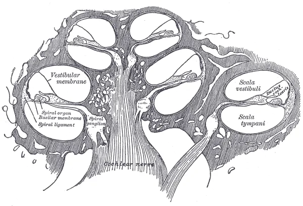
蝸牛管の基底板の上に乗る、聴覚の受容装置。
- 内有毛細胞——1列。求心性神経の約95%が接続する。実際に「聞いている」細胞
- 外有毛細胞——3列。遠心性支配を受け、自ら伸縮する
- 蓋膜——有毛細胞の上を覆う。基底板が振動するとずれが生じ、毛が傾く
- 支持細胞とコルチトンネル
蝸牛管の中は内リンパ(カリウムが高い)で、血管条が作る。前庭階・鼓室階は外リンパ。
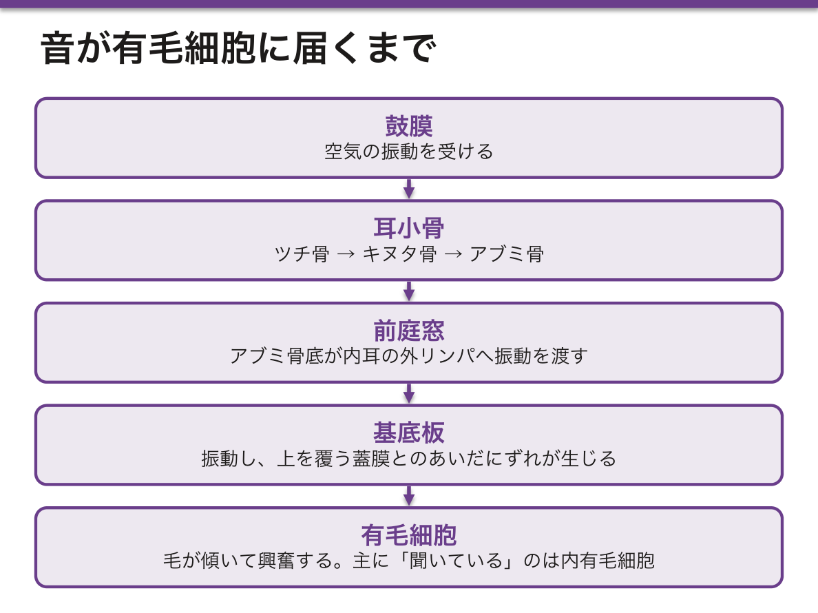
内リンパの高カリウムという異例の組成が、聴覚の感度を作っている。細胞の外がカリウムに富むため、毛が傾いてチャネルが開くとカリウムが細胞内へ流れ込んで脱分極する。細胞外液としては例外的な組成を、血管条が絶えずエネルギーを使って維持している。
この電位(蝸牛内電位、約+80mV)が崩れることが難聴になる。メニエール病は内リンパ水腫で、回転性めまい・変動する感音難聴・耳鳴・耳閉感が反復する。低音から落ちるのが特徴である。
外有毛細胞は聞くための細胞ではなく、増幅するための細胞である。プレスチンという蛋白で自ら伸縮し、基底板の振動を能動的に増幅する(蝸牛増幅器)。この動きが音として外へ漏れるため、外耳道にマイクを入れると測定できる——耳音響放射であり、新生児聴覚スクリーニングの原理である。返事のできない赤ん坊の聴力が測れるのは、耳が音を出しているからである。
基底板は基部で狭く硬く、頂部で広く柔らかいため、基部が高音、頂部が低音に共振する(場所説・トノトピー)。基部の有毛細胞はすべての音の振動が通過する場所にあり最も酷使されるので、騒音性難聴も老人性難聴も高音域(4kHz 付近)から落ちる。
有毛細胞は再生しない。だから感音難聴は基本的に不可逆で、予防が唯一の対策になる。
- 薬剤性——アミノグリコシド(ゲンタマイシンなど)・シスプラチン・ループ利尿薬が外有毛細胞を障害する。アミノグリコシドはミトコンドリア遺伝子 m.1555A>G を持つ人で少量でも高度難聴を起こす(母系遺伝)
- 騒音性——4kHz の谷(c⁵ dip)から始まり、自覚する頃には広がっている
- 突発性難聴——コルチ器の有毛細胞の変性が想定される。発症早期のステロイドが勝負になる
人工内耳が成立するのは、有毛細胞が失われてもらせん神経節と蝸牛神経が残っているためである。壊れた変換装置を電極で置き換え、トノトピーの順に電気刺激を配る——組織構造の理解がそのまま人工臓器の設計になっている。
English version
Core
The auditory receptor apparatus, sitting on the basilar membrane of the cochlear duct.
- Inner hair cells — a single row. About 95 per cent of the afferent fibres connect to them: these are the cells that actually hear.
- Outer hair cells — three rows. Efferently innervated, and they lengthen and shorten of their own accord.
- Tectorial membrane — covers the hair cells. When the basilar membrane vibrates a shear develops and the hairs are deflected.
- Supporting cells and the tunnel of Corti.
The cochlear duct contains endolymph (high in potassium), produced by the stria vascularis. The scala vestibuli and scala tympani contain perilymph.
Going further
The exceptional composition of endolymph — high potassium — creates the sensitivity of hearing. Because the outside of the cell is rich in potassium, deflection of the hairs opens the channels and potassium flows into the cell and depolarises it. A composition exceptional for an extracellular fluid, maintained by the stria vascularis at constant energetic cost. Collapse of this potential (the endocochlear potential, about +80 mV) is deafness. Ménière’s disease is endolymphatic hydrops, with recurrent rotatory vertigo, fluctuating sensorineural loss, tinnitus and aural fullness, characteristically losing the low frequencies first.
The outer hair cell is not a cell for hearing but a cell for amplifying. With the protein prestin it lengthens and shortens, actively amplifying the vibration of the basilar membrane (the cochlear amplifier). That movement leaks outwards as sound and can be recorded with a microphone in the ear canal — otoacoustic emissions, the principle of newborn hearing screening. The hearing of a baby who cannot answer can be measured because the ear emits sound.
The basilar membrane is narrow and stiff at the base and wide and slack at the apex, so the base resonates to high notes and the apex to low (the place principle, tonotopy). The basal hair cells lie where the vibration of every sound must pass and are the most heavily worked, so both noise-induced and age-related hearing loss begin in the high frequencies, around 4 kHz.
In the clinic
Hair cells do not regenerate. So sensorineural hearing loss is essentially irreversible and prevention is the only measure.
- Drug-induced — aminoglycosides (gentamicin and others), cisplatin and loop diuretics damage the outer hair cells. In a person carrying the mitochondrial variant m.1555A>G, even a small dose of an aminoglycoside causes profound deafness (maternal inheritance).
- Noise-induced — begins as a notch at 4 kHz (the c⁵ dip) and has already spread by the time it is noticed.
- Sudden sensorineural hearing loss — degeneration of the hair cells of the organ of Corti is presumed. Steroids given early are what count.
A cochlear implant works because the spiral ganglion and cochlear nerve survive the loss of the hair cells. The broken transducer is replaced by electrodes distributing stimulation in tonotopic order — understanding of the tissue architecture becomes directly the design of an artificial organ.
前庭 — 同じ有毛細胞で加速度を測る
平衡覚の受容器もコルチ器と同じ有毛細胞を使い、覆いをかぶせる物が違うだけである。
- 半規管(3本、互いに直交)——膨大部に膨大部稜。毛はクプラというゼラチン状の帽子に埋まる。回転(角)加速度を検出
- 耳石器(卵形嚢・球形嚢)——平衡斑。毛は耳石膜に埋まり、その上に炭酸カルシウムの結晶(耳石)が乗る。直線加速度と重力を検出
卵形嚢は水平、球形嚢は垂直の面を担当する。
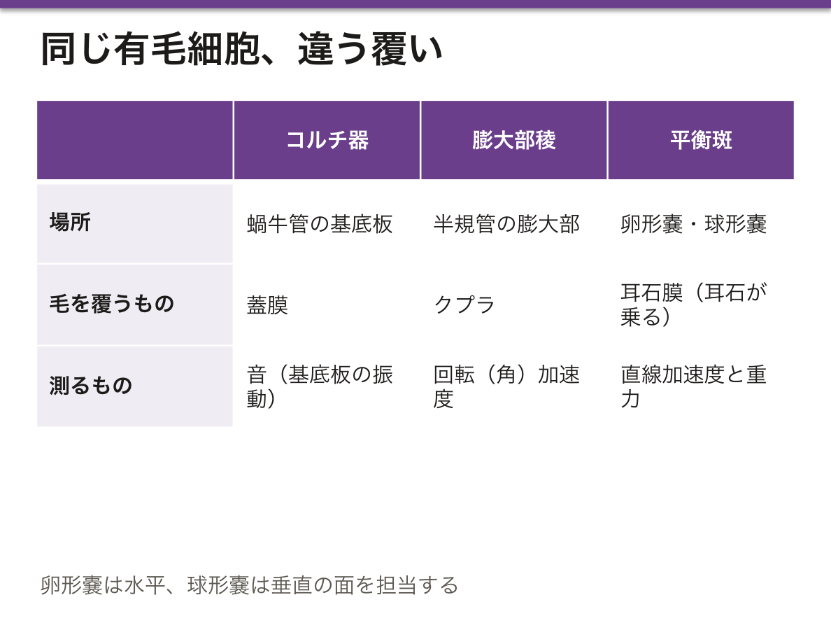
同じ細胞に何を載せるかで測る量が変わるという設計が要点である。
- クプラは内リンパと同じ比重なので重力では動かず、回転したときに内リンパが慣性で遅れることでのみ曲がる。だから半規管は角加速度だけを拾う
- 耳石膜には比重の大きい結晶が載っているので、頭が傾けば重力で、直線的に加速すれば慣性でずれる。だから耳石器は重力と直線加速度を拾う
載せる重りの比重を変えるだけで、測る物理量が変わっている。
また回転が一定速度で続くと内リンパも一緒に回り始め、クプラが元に戻って回転を感じなくなる。停止した瞬間には逆向きに曲がるので、止まったのに回っている感覚が生じる。回転椅子を止めたあとのめまいは、装置が加速度しか測れないことの当然の帰結である。
良性発作性頭位めまい症は剥がれた耳石が半規管(多くは後半規管)へ迷い込むことで起きる。本来なら動かないはずのクプラが、耳石の重みで頭位変換のたびに引っぱられるので、特定の頭の向きで数十秒の回転性めまいと眼振が出る。
だから治療は薬ではなく耳石を元の卵形嚢へ戻す頭位治療(Epley 法)になる。「異物が入り込んだ機械式センサー」という理解が、そのまま手技の根拠である。Dix-Hallpike 試験で誘発される眼振の向きと潜時(数秒)と疲労性が診断になる。
めまいの鑑別では、難聴を伴うかが最初の分岐になる——蝸牛と前庭は同じ内リンパの袋を共有しているので、内リンパ水腫(メニエール病)では両方が同時に障害され、前庭神経炎では前庭のみで聴力が保たれる。
English version
Core
The receptor for balance uses the same hair cell as the organ of Corti; only what is laid over it differs.
- Semicircular canals (three, mutually perpendicular) — the crista ampullaris in the ampulla. The hairs are embedded in a gelatinous cap, the cupula. Detects rotational (angular) acceleration.
- Otolith organs (utricle and saccule) — the macula. The hairs are embedded in the otolithic membrane, on which sit crystals of calcium carbonate (otoconia). Detects linear acceleration and gravity.
The utricle covers the horizontal plane, the saccule the vertical.
Going further
The point of the design is that what is laid on the same cell changes the quantity measured.
- The cupula has the same specific gravity as endolymph, so gravity does not move it; it bends only when rotation makes the endolymph lag by inertia. So the semicircular canals pick up angular acceleration alone.
- The otolithic membrane carries crystals of greater specific gravity, so it is displaced by gravity when the head tilts and by inertia on linear acceleration. So the otolith organs pick up gravity and linear acceleration.
Changing only the density of the weight laid on top changes the physical quantity measured.
And when rotation continues at constant speed the endolymph begins to turn with it, the cupula returns to rest, and the rotation is no longer felt. At the moment of stopping it bends the other way, giving the sensation of turning although one has stopped. The dizziness after a rotating chair is halted is the necessary consequence of an instrument that can measure only acceleration.
In the clinic
Benign paroxysmal positional vertigo arises when detached otoconia stray into a semicircular canal, usually the posterior. A cupula that ought not to move is pulled by their weight at every change of head position, so a particular orientation of the head produces some tens of seconds of rotatory vertigo with nystagmus.
So the treatment is not a drug but a positioning manoeuvre (Epley) returning the otoconia to the utricle. Understanding it as a mechanical sensor with a foreign body in it is itself the rationale for the technique. The direction, latency (a few seconds) and fatigability of the nystagmus provoked by the Dix–Hallpike test make the diagnosis.
In the differential diagnosis of vertigo, the first branch point is whether hearing loss accompanies it — cochlea and vestibule share the same endolymphatic sac, so in endolymphatic hydrops (Ménière’s disease) both are affected together, whereas in vestibular neuritis only the vestibule is affected and hearing is preserved.
中耳と耳管 — 音を効率よく渡し、圧を合わせるAuris media
空気で満たされた小部屋。鼓膜の振動を、内耳の液体へ渡す。
鼓膜は3層——外側は皮膚(重層扁平上皮)、中間は線維層、内側は粘膜(単層扁平〜立方)。上方の弛緩部には線維層が無い。
耳小骨は3つ——ツチ骨・キヌタ骨・アブミ骨。アブミ骨底が前庭窓(卵円窓)にはまる。
2つの筋が音を減衰させる——鼓膜張筋(三叉神経)とアブミ骨筋(顔面神経)。
耳管は中耳と上咽頭をつなぐ。粘膜は多列線毛円柱上皮。普段は閉じていて、嚥下・あくびで開く。
中耳がある理由は「気体と液体のあいだの段差」を埋めることにある。空気の振動をそのまま液体に当てると、ほとんど跳ね返って伝わらない(インピーダンス不整合)——「中耳は、この損失を取り戻すための増幅器になる」。
増幅の仕掛けは2つで、掛け算になっている。①鼓膜と前庭窓の面積比(約17倍) ②耳小骨のてこ比(約1.3倍)。合わせて約22倍、およそ 25〜30 dB を稼ぐ——「面積で圧を集め、てこで力を増やしている」。
だから鼓膜穿孔や耳小骨連鎖の離断では、この稼ぎぶんが失われて伝音難聴になる(中耳炎の章)——「難聴の大きさが、失われた増幅の量と対応する」。
耳管が普段閉じていることには、逆の理由がある。開きっぱなしなら自分の声と呼吸が響き(耳管開放症)、閉じたままなら圧が合わずに鼓膜が引き込まれる——「開くタイミングを制御することが仕事になる」。
小児の耳管は短く・太く・水平に近い。だから上咽頭の分泌物が中耳へ届きやすく、急性中耳炎が小児に多い(小児の気道の章)——「解剖の差が、そのまま疫学の差になっている」。
アブミ骨筋反射は、顔面神経麻痺の高さを決める道具にもなる(顔面神経麻痺の章)——「聴覚過敏を訴えるかどうかが、枝の分岐より上か下かを示す」。
耳管が短く・太く・水平なことが、小児の中耳炎を作っている。鼻咽腔の細菌が上がりやすく、排出されにくい。成長とともに減るのはこの形が変わるからである。
急性中耳炎 — 耳痛・発熱・鼓膜の発赤と膨隆。肺炎球菌・インフルエンザ桿菌。
滲出性中耳炎 — 痛みが無く、難聴だけ。小児では言語発達に影響するので見逃さない。成人の片側性なら上咽頭癌を除外する。
真珠腫性中耳炎 — 鼓膜の陥凹に扁平上皮が入り込んで角化物が溜まり、骨を破壊する。耳小骨・顔面神経管・内耳・頭蓋内へ及ぶ。良性だが破壊性で、手術が必要である。
耳小骨は音のインピーダンス整合器である。アブミ骨が固着するのが耳硬化症で、若年〜中年の伝音難聴を起こす。
English version
Core
An air-filled chamber. It hands the vibration of the tympanic membrane on to the fluid of the inner ear.
- The tympanic membrane has three layers — skin on the outside (stratified squamous), a fibrous layer between, and mucosa within (simple squamous to cuboidal). The pars flaccida above has no fibrous layer.
- Three ossicles — malleus, incus, stapes. The footplate of the stapes fits into the vestibular (oval) window.
- Two muscles damp the sound — tensor tympani (trigeminal) and stapedius (facial).
- The auditory tube joins middle ear to nasopharynx; the mucosa is pseudostratified ciliated columnar. Normally closed, it opens on swallowing and yawning.
Going further
The middle ear exists to bridge the step between gas and liquid. Air vibration striking fluid directly is almost entirely reflected (impedance mismatch) — the middle ear is the amplifier that recovers that loss.
The amplification has two parts, and they multiply. ① the area ratio of tympanic membrane to oval window (about 17×) ② the lever ratio of the ossicles (about 1.3×). Together about 22×, or some 25–30 dB — pressure concentrated by area, force increased by leverage.
So perforation of the drum or discontinuity of the ossicular chain loses that gain and gives conductive deafness — the size of the loss corresponds to the amount of amplification forfeited.
That the auditory tube is normally closed has the opposite reason. Held open, one’s own voice and breathing resound (patulous tube); held shut, the pressure cannot equalise and the drum is drawn in — its work is to control the timing of opening.
The child’s tube is short, wide and near-horizontal, so nasopharyngeal secretions reach the middle ear readily and acute otitis media is common in children — a difference in anatomy becomes directly a difference in epidemiology.
The stapedial reflex is also a tool for locating the level of a facial nerve palsy — whether hyperacusis is complained of shows whether the lesion is above or below the branch.
In the clinic
That the auditory tube is short, wide and horizontal creates otitis media in children: organisms ascend easily from the nasopharynx and drain poorly. It becomes less frequent with growth because that shape changes.
Acute otitis media — earache, fever, a red and bulging drum. Pneumococcus, Haemophilus influenzae.
Otitis media with effusion — no pain, hearing loss alone. In children it affects language development and must not be missed. Unilateral disease in an adult requires exclusion of nasopharyngeal carcinoma.
Cholesteatoma — squamous epithelium grows into a retraction of the drum, keratin accumulates and bone is destroyed, involving the ossicles, facial canal, inner ear and cranial cavity. Benign but destructive; surgery is required.
The ossicles are an impedance-matching device for sound. Fixation of the stapes is otosclerosis, causing conductive deafness in young and middle-aged adults.
まとめ
| コルチ器(らせん器) | 蝸牛管の基底板の上に乗る、聴覚の受容装置。 |
|---|---|
| 前庭 — 同じ有毛細胞で加速度を測る | 平衡覚の受容器もコルチ器と同じ有毛細胞を使い、覆いをかぶせる物が違うだけである。 |
| 中耳と耳管 — 音を効率よく渡し、圧を合わせる | 空気で満たされた小部屋。鼓膜の振動を、内耳の液体へ渡す。 |
このノートは医学生が授業の内容を自分の言葉でまとめたもので、診療の判断に使うものではありません。誤りに気づいたらお知らせください。