消化管は食道から直腸まで、どこを切っても同じ4層でできている。粘膜・粘膜下層・筋層・漿膜(または外膜)。部位ごとに違うのは粘膜の上皮の種類と、筋層の厚さだけである。
だから覚えるべきは4層の型ひとつと、「どこで上皮が切り替わるか」だけになる。食道は重層扁平、胃から大腸は単層円柱、肛門管下部でまた重層扁平——切り替わる場所が癌の好発部位であることは上皮の章で見たとおり。
そして病変がどの層まで及ぶかが、そのまま病期分類(T分類)と内視鏡治療の適応になる。潰瘍性大腸炎とCrohn病の違いも、深達度と分布で説明できる。層を数えることが臨床そのものである。
- 粘膜
- 粘膜下層
- 固有筋層
- 部位ごとの粘膜 — 食道・胃・小腸・大腸
- 炎症性腸疾患 — 深さと分布で分ける
- 胃底腺の細胞 — 酸を出す細胞と、酵素を出す細胞
- 小腸の吸収装置 — 表面積を600倍にする3段の仕掛け
- 腸管神経系 — 脳と独立に動く「第二の脳」
- 胃の粘膜防御 — 自分を消化しないための、幾重もの仕掛け
- 大腸の組織 — 吸収より、水を取り戻して溜めることが仕事になる
- 粘膜Tunica mucosa
- 粘膜下層Tela submucosa
- 固有筋層Tunica muscularis
- 部位ごとの粘膜 — 食道・胃・小腸・大腸
- 炎症性腸疾患 — 深さと分布で分ける
- 胃底腺の細胞 — 酸を出す細胞と、酵素を出す細胞Glandula gastrica propria
- 小腸の吸収装置 — 表面積を600倍にする3段の仕掛けVillus intestinalis
- 腸管神経系 — 脳と独立に動く「第二の脳」Plexus myentericus
- 胃の粘膜防御 — 自分を消化しないための、幾重もの仕掛けMucus gastricus
- 大腸の組織 — 吸収より、水を取り戻して溜めることが仕事になるIntestinum crassum
- まとめ
粘膜Tunica mucosa
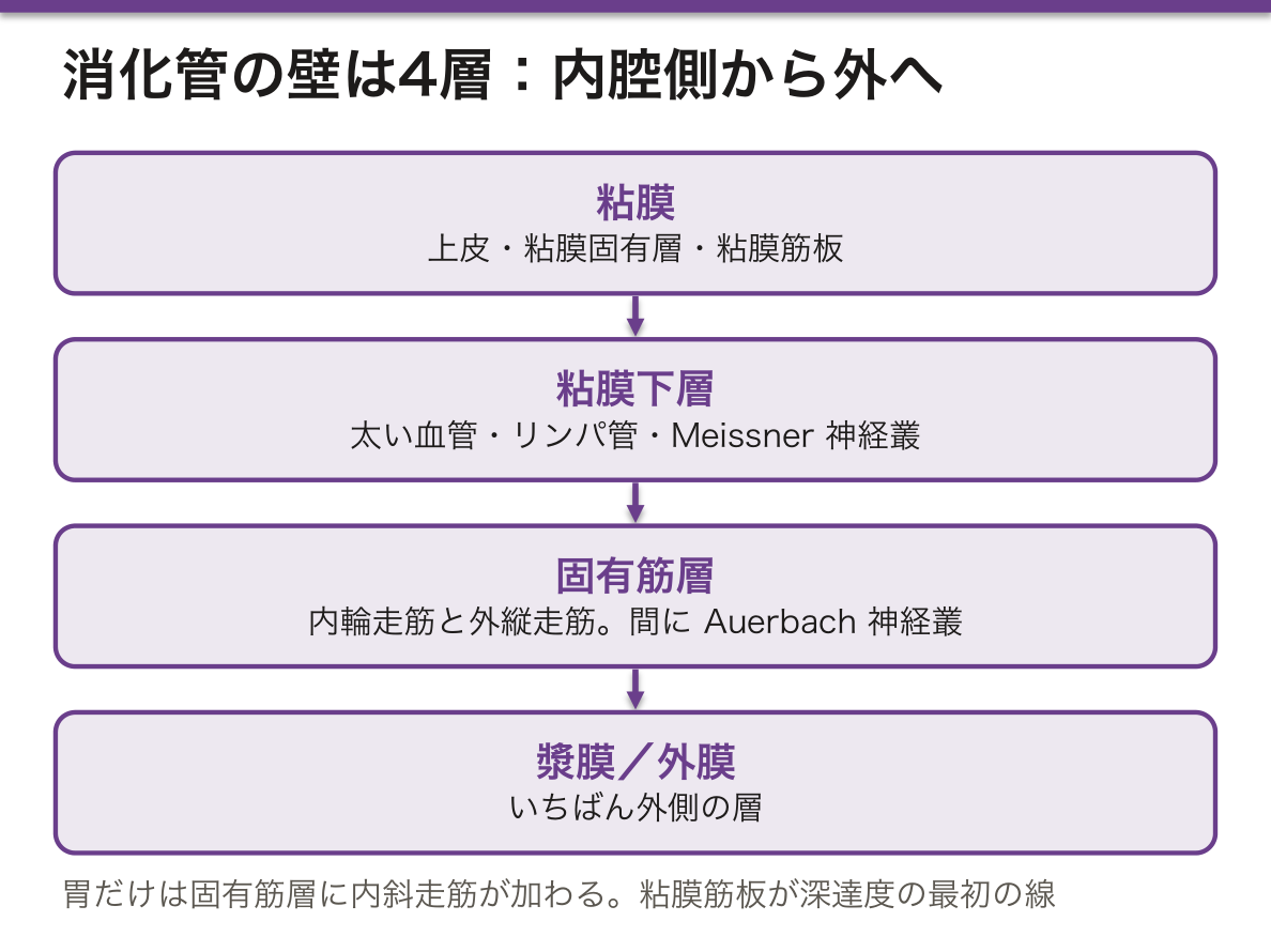
最内層。さらに3つに分かれる。
- 上皮——部位により変わる(食道=重層扁平、胃〜大腸=単層円柱)
- 粘膜固有層 lamina propria——疎性結合組織。毛細血管・リンパ管・免疫細胞(形質細胞・リンパ球)
- 粘膜筋板 lamina muscularis mucosae——薄い平滑筋。粘膜のひだを動かす
粘膜筋板が「深達度の最初の線」になる。
粘膜内には毛細リンパ管がほとんど無い(大腸では特に)。だから粘膜内にとどまる癌はリンパ節転移をほぼ起こさない。これが内視鏡的粘膜切除術(EMR)・内視鏡的粘膜下層剥離術(ESD)の適応の根拠である。
深達度の線を並べると治療が決まる。
- M(粘膜内)——リンパ節転移リスク極小 → 内視鏡治療で根治しうる
- SM(粘膜下層)——リンパ管が豊富 → 浸潤の深さ(大腸で1000μm など)で外科手術の適応が分かれる
- MP(固有筋層)以深——外科手術
「なぜ粘膜内なら内視鏡で済むのか」の答えは、リンパ管の分布という組織学的事実にある。大腸の粘膜内癌が「癌」でありながら転移しない扱いになるのはこのためで、欧米で「high-grade dysplasia」と呼ばれる所以でもある。
粘膜筋板を越えたかどうかが、早期癌の定義である。
胃癌・大腸癌の「早期」は「深達度が粘膜下層まで」と定義される。粘膜内にとどまれば、リンパ管が乏しいのでリンパ節転移はほぼ無い——だから内視鏡的粘膜下層剥離術(ESD)で治せる。
粘膜下層に達するとリンパ管に届き、転移のリスクが生じるので外科切除とリンパ節郭清を考える。1枚の薄い筋板が、治療法を分けている。
食道だけは粘膜固有層にもリンパ管があるため、より浅い段階から転移しうる——臓器ごとの組織の違いが、そのまま治療方針の違いになる。
びらんと潰瘍の違いも粘膜筋板で決まる。粘膜筋板を越えないのがびらん(瘢痕を残さない)、越えたのが潰瘍(瘢痕になり、変形を残す)。
English version
Core
The innermost layer, itself divided into three.
- Epithelium — varies with the region (oesophagus stratified squamous; stomach to colon simple columnar).
- Lamina propria — loose connective tissue with capillaries, lymphatics and immune cells (plasma cells, lymphocytes).
- Lamina muscularis mucosae — a thin sheet of smooth muscle that moves the mucosal folds.
The muscularis mucosae is the first line of depth.
Going further
There are almost no lymphatic capillaries within the mucosa (particularly in the colon). So a cancer confined to the mucosa hardly ever metastasises to lymph nodes. This is the basis for the indications for endoscopic mucosal resection (EMR) and endoscopic submucosal dissection (ESD).
Line up the levels of depth and the treatment follows.
- M (intramucosal) — negligible risk of nodal metastasis → curable endoscopically.
- SM (submucosal) — rich in lymphatics → the depth of invasion (1000 µm in the colon, for example) decides whether surgery is indicated.
- MP (muscularis propria) and deeper — surgery.
The answer to "why is endoscopy enough if it is intramucosal?" lies in a histological fact: the distribution of lymphatics. This is why an intramucosal cancer of the colon is treated as non-metastasising although it is a cancer, and why in Western practice it is called high-grade dysplasia.
In the clinic
Whether the muscularis mucosae has been crossed is the definition of early cancer. "Early" gastric and colorectal cancer is defined as invasion no deeper than the submucosa.
Confined to the mucosa, where lymphatics are scarce, nodal metastasis is essentially absent — so it can be cured by ESD. Reaching the submucosa it meets lymphatics and a risk of metastasis arises, so surgical resection with nodal dissection is considered. A single thin sheet of muscle separates the two treatments.
The oesophagus alone has lymphatics in the lamina propria as well, so it can metastasise from a shallower stage — a difference in tissue between organs becoming directly a difference in management.
The difference between an erosion and an ulcer is also set by the muscularis mucosae: not crossing it is an erosion (healing without scar), crossing it is an ulcer (scarring, leaving deformity).
粘膜下層Tela submucosa
密性結合組織の層。太い血管・リンパ管とMeissner 神経叢(粘膜下神経叢)が走る。
ここに腺があるのは食道腺と十二指腸の Brunner 腺だけ——粘膜下層に腺があれば十二指腸か食道と判定できる。
Brunner 腺はアルカリ性の粘液を出し、胃酸を中和して十二指腸粘膜を守る。
粘膜下層は血管が太いので、ここまで達した潰瘍が大出血を起こす。胃・十二指腸潰瘍の出血は粘膜下層〜筋層の動脈が露出することによる。
食道静脈瘤も粘膜下層の静脈が拡張したもので、門脈圧亢進時に左胃静脈 → 食道粘膜下静脈叢 → 奇静脈という側副路が開くために生じる。壁が薄い粘膜下層にあるため破裂しやすい。
Meissner 神経叢は粘膜の分泌と局所の血流を制御する。Hirschsprung 病ではこの神経叢と Auerbach 神経叢の両方の神経節細胞が欠如する(神経堤細胞の移動障害)。肛門側から連続して欠如するのが特徴で、生検は直腸で行う。無神経節部分が収縮したままなので、その口側が拡張する——「狭いのは病気の側、太いのは正常の側」という逆転が理解の鍵になる。
粘膜下層に腺があるのは食道と十二指腸だけ——この2つは酸にさらされる場所を守るために特別な腺を持っている。
Brunner 腺はアルカリ性の粘液を出して胃から来た酸を中和する。十二指腸潰瘍が球部(Brunner 腺より口側寄り)に多いことと関係している。
粘膜下層は太い血管が走る層である。消化管出血で大量出血になるのは、この層の動脈が露出したときで、露出血管を伴う潰瘍(Forrest 分類)は再出血のリスクが高い。Dieulafoy 潰瘍は粘膜下の異常に太い動脈が小さなびらんから破綻するもので、浅い病変なのに致命的な出血になる。
粘膜下層は内視鏡治療の作業層でもある。ここに液を注入して病変を持ち上げ、剥離する(ESD)。持ち上がらない(non-lifting sign)=粘膜下層へ浸潤しているというその場で使える所見になる。
English version
Core
A layer of dense connective tissue carrying the large vessels and lymphatics and Meissner’s (submucosal) plexus.
The only glands here are the oesophageal glands and the duodenal glands of Brunner — glands in the submucosa mean duodenum or oesophagus.
Brunner’s glands secrete alkaline mucus, neutralising gastric acid and protecting the duodenal mucosa.
Going further
The submucosa carries large vessels, so an ulcer reaching it bleeds heavily. Bleeding from gastric and duodenal ulcers comes from exposure of an artery in the submucosa or muscle.
Oesophageal varices are dilated submucosal veins, arising when portal hypertension opens the collateral left gastric vein → submucosal oesophageal plexus → azygos vein. Lying in the thin-walled submucosa, they rupture readily.
Meissner’s plexus controls mucosal secretion and local blood flow. In Hirschsprung’s disease the ganglion cells of both this and Auerbach’s plexus are absent (a failure of neural crest cell migration). Characteristically the absence is continuous from the anal end, so the biopsy is taken from the rectum.
The aganglionic segment stays contracted, so the bowel oral to it dilates — the inversion that the narrow part is the diseased side and the wide part the normal one is the key to understanding it.
In the clinic
Only the oesophagus and duodenum have glands in the submucosa — both have special glands to protect a place exposed to acid. Brunner’s glands secrete alkaline mucus neutralising the acid arriving from the stomach. This relates to the predilection of duodenal ulcers for the bulb (oral to the main mass of Brunner’s glands).
The submucosa is the layer of the large vessels. Gastrointestinal bleeding becomes massive when an artery of this layer is exposed, and an ulcer with a visible vessel (the Forrest classification) carries a high risk of rebleeding. A Dieulafoy lesion is an abnormally large submucosal artery rupturing through a small erosion — a superficial lesion causing fatal haemorrhage.
The submucosa is also the working layer of endoscopic treatment: fluid is injected into it to lift the lesion, which is then dissected off (ESD). Failure to lift (the non-lifting sign) means submucosal invasion — a finding usable on the spot.
固有筋層Tunica muscularis
内輪走筋と外縦走筋の2層(胃だけは内斜走筋を加えた3層)。あいだにAuerbach 神経叢(筋層間神経叢)が挟まる。
この2層が組んで蠕動運動を作る。輪走筋が内容物の後ろで締め、縦走筋が前方を短縮させる。
食道上部1/3は骨格筋、下部1/3は平滑筋、中1/3は混在——嚥下の随意/不随意の切り替わりに対応する。
蠕動のペースメーカーはCajal 介在細胞で、自発的に徐波を発生させ、平滑筋のギャップ結合を通じて広げる。消化管間質腫瘍(GIST)はこの細胞由来で、KIT(CD117)陽性が診断の決め手になり、イマチニブが奏効する——起源細胞が分かれば分子標的薬が決まる典型例。
神経叢の障害で運動が破綻する。
- アカラシア——食道下部の抑制性ニューロン(NO・VIP)が失われ、下部食道括約筋が弛緩しない。食道は拡張し、造影で鳥のくちばし様
- Chagas 病——Trypanosoma cruzi が神経叢を破壊し、巨大食道・巨大結腸をきたす
「収縮できない」のではなく「弛緩できない」のがアカラシアの本質で、抑制性の神経が選択的に失われることによる。治療(バルーン拡張・筋層切開・ボツリヌス毒素)はいずれも括約筋を機械的または薬理的に緩めることを狙っている。
2層が別々に働くから、蠕動が起こる。輪走筋が内容の口側で収縮し、縦走筋が肛門側で短縮する。この協調を作るのが筋層間神経叢である。
Hirschsprung 病では神経節が無い部分が弛緩できずに縮んだままになり、その口側が拡張する。「細いほうが病気」という逆転を必ず押さえる。
アカラシア——食道の筋層間神経叢の変性で下部食道括約筋が弛緩できない。食道造影で鳥のくちばし像、内圧検査で診断。Chagas 病でも同じ像になる。
胃だけ3層(内斜走筋がある)なのは、粥状にすりつぶすために回転する動きが要るからである。
固有筋層に達した癌は漿膜へ届きうる——腹膜播種の起点になる。
English version
Core
Two layers, inner circular and outer longitudinal (the stomach alone has three, with an inner oblique layer), with Auerbach’s (myenteric) plexus between them.
The two layers together produce peristalsis: the circular layer constricts behind the contents while the longitudinal layer shortens the segment ahead.
The upper third of the oesophagus is skeletal muscle, the lower third smooth, the middle third mixed — corresponding to the change from voluntary to involuntary swallowing.
Going further
The pacemaker of peristalsis is the interstitial cell of Cajal, which generates slow waves spontaneously and spreads them through the gap junctions of the smooth muscle. Gastrointestinal stromal tumour (GIST) arises from this cell, positivity for KIT (CD117) is decisive for the diagnosis, and imatinib is effective — a model example of the cell of origin determining the targeted drug.
Damage to the plexus breaks the movement.
- Achalasia — the inhibitory neurones (NO, VIP) of the lower oesophagus are lost and the lower oesophageal sphincter does not relax. The oesophagus dilates, with a bird’s beak on contrast studies.
- Chagas disease — Trypanosoma cruzi destroys the plexus, giving megaoesophagus and megacolon.
The essence of achalasia is not that it cannot contract but that it cannot relax, from selective loss of the inhibitory nerves. Every treatment (balloon dilatation, myotomy, botulinum toxin) aims to loosen the sphincter mechanically or pharmacologically.
In the clinic
Peristalsis occurs because the two layers act separately: the circular layer contracts oral to the contents while the longitudinal layer shortens the segment anal to it. The myenteric plexus creates that coordination.
In Hirschsprung’s disease the aganglionic segment cannot relax and stays contracted, and the bowel oral to it dilates. Always fix the inversion that the narrow part is the diseased one.
Achalasia — degeneration of the myenteric plexus of the oesophagus so that the lower sphincter cannot relax. A bird’s beak on barium swallow; the diagnosis is made by manometry. Chagas disease gives the same picture.
The stomach alone has three layers (with an inner oblique) because it needs a rotating movement to grind the contents to a paste.
A cancer reaching the muscularis propria can reach the serosa — the starting point of peritoneal dissemination.
部位ごとの粘膜 — 食道・胃・小腸・大腸
- 食道——重層扁平上皮(非角化)。粘液腺が粘膜下層に
- 胃——単層円柱上皮。胃小窩から胃腺が伸びる。絨毛は無い。表層粘液細胞・壁細胞(HCl と内因子)・主細胞(ペプシノゲン)・内分泌細胞(G細胞など)
- 小腸——単層円柱+杯細胞。輪状ヒダ・絨毛・微絨毛で表面積を600倍に。陰窩底にPaneth 細胞(抗菌ペプチド)
- 大腸——単層円柱。絨毛が無く、杯細胞が非常に多い。陰窩は真っ直ぐ
絨毛の有無が小腸と大腸を分ける最大の目印。
胃腺の細胞分布も部位で違う。胃底腺(体部)に壁細胞・主細胞が集中し、幽門腺(前庭部)にはG細胞(ガストリン)が多い。この分布が病態を決める。
- A型胃炎(自己免疫性)——体部の壁細胞が破壊される。酸が出ないのでフィードバックでガストリンが上昇し、内因子も失われて悪性貧血。カルチノイドのリスク
- B型胃炎(H. pylori)——前庭部優位に始まり、萎縮が体部へ広がる。腸上皮化生 → 胃癌
「どこの胃炎か」で貧血が出るか癌になるかが変わる——分布が診断そのものになる。
Paneth 細胞は好酸性の顆粒(リゾチーム・ディフェンシン)を持ち、小腸の陰窩底にのみある。大腸にPaneth細胞が現れれば化生であり、慢性炎症(潰瘍性大腸炎)の証拠になる。「本来無い細胞がある」ことが病気を語る。
上皮の切り替わる場所に病気が集まる。
食道胃接合部 — 重層扁平上皮と円柱上皮の境(SCJ)。逆流で円柱上皮に置き換わるのが Barrett 食道で、食道腺癌のリスクになる。扁平上皮癌は中部食道に多く、飲酒と喫煙が危険因子——同じ食道でも組織型で発生部位も原因も違う。
壁細胞が塩酸と内因子を出す。自己免疫性胃炎では壁細胞が破壊され、無酸症+内因子の欠乏=悪性貧血になり、胃酸が無いためガストリンが上がって神経内分泌腫瘍を生じうる。1種類の細胞が失われるだけで、貧血と腫瘍の両方が説明できる。
Paneth 細胞は小腸にあり、大腸には通常無い。大腸に Paneth 細胞が現れる(Paneth 細胞化生)=慢性の炎症のサインで、炎症性腸疾患の所見になる。
English version
Core
- Oesophagus — stratified squamous (non-keratinised). Mucous glands in the submucosa.
- Stomach — simple columnar. Gastric glands extend down from the gastric pits. No villi. Surface mucous cells, parietal cells (HCl and intrinsic factor), chief cells (pepsinogen), endocrine cells (G cells and others).
- Small intestine — simple columnar with goblet cells. Circular folds, villi and microvilli multiply the surface area 600-fold. Paneth cells (antimicrobial peptides) at the base of the crypts.
- Large intestine — simple columnar. No villi, very many goblet cells, straight crypts.
The presence or absence of villi is the chief marker separating small from large intestine.
Going further
The distribution of the cells of the gastric glands also differs by region. Parietal and chief cells are concentrated in the fundic glands (body), while the pyloric glands (antrum) are rich in G cells (gastrin). That distribution decides the pathology.
- Type A gastritis (autoimmune) — the parietal cells of the body are destroyed. No acid is produced, so gastrin rises by feedback; intrinsic factor is lost as well, giving pernicious anaemia. A risk of carcinoid.
- Type B gastritis (H. pylori) — begins in the antrum, and the atrophy spreads to the body. Intestinal metaplasia → gastric cancer.
Which gastritis it is changes whether the patient becomes anaemic or develops cancer — the distribution is the diagnosis itself.
Paneth cells have eosinophilic granules (lysozyme, defensins) and are found only at the base of the crypts of the small intestine. Paneth cells appearing in the colon are metaplasia, and evidence of chronic inflammation (ulcerative colitis). The presence of a cell that ought not to be there speaks of disease.
In the clinic
Disease gathers where the epithelium changes over.
Oesophagogastric junction — the boundary of stratified squamous and columnar epithelium (SCJ). Replacement by columnar epithelium from reflux is Barrett’s oesophagus, a risk for oesophageal adenocarcinoma. Squamous carcinoma is commoner in the middle oesophagus, with alcohol and smoking as risk factors — in the same oesophagus, the histological type changes both the site and the cause.
Parietal cells secrete hydrochloric acid and intrinsic factor. In autoimmune gastritis they are destroyed, giving achlorhydria plus intrinsic factor deficiency = pernicious anaemia, and the absence of acid raises gastrin so that a neuroendocrine tumour may arise. The loss of a single cell type explains both the anaemia and the tumour.
Paneth cells belong to the small intestine and are normally absent from the colon. Their appearance there (Paneth cell metaplasia) is a sign of chronic inflammation and a finding in inflammatory bowel disease.
炎症性腸疾患 — 深さと分布で分ける
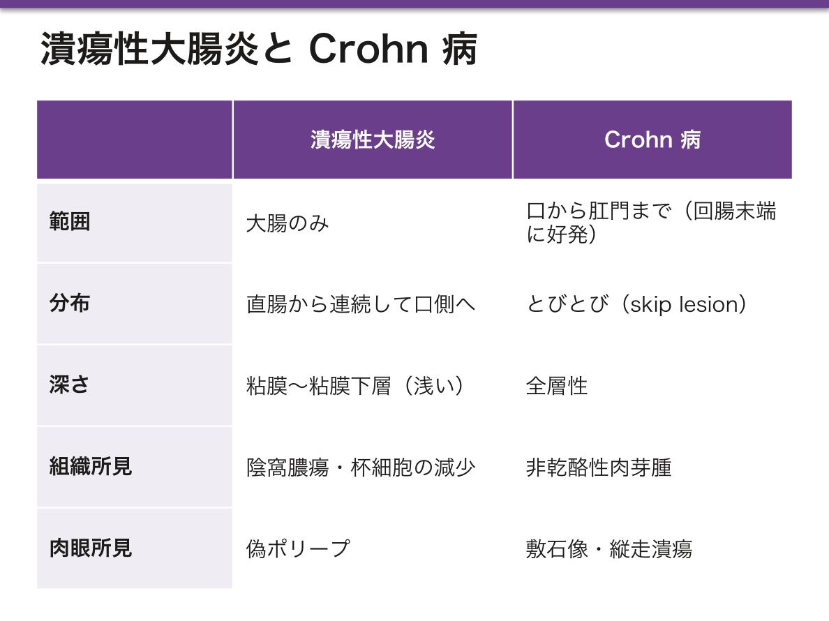
2つの疾患は「どこまで深く」「どう分布するか」で分かれる。
- 潰瘍性大腸炎——大腸のみ。直腸から連続して口側へ。炎症は粘膜〜粘膜下層まで(浅い)。陰窩膿瘍・杯細胞の減少。偽ポリープ
- Crohn 病——口から肛門までどこでも(回腸末端に好発)。とびとび(skip lesion)。炎症は全層性。非乾酪性肉芽腫・敷石像・縦走潰瘍
深さの違いが合併症の違いを作る——ここが理解の要点である。
- Crohn 病は全層性だから、壁を貫いて瘻孔・膿瘍・狭窄を作る。腸管どうし、腸と膀胱、腸と皮膚がつながる
- 潰瘍性大腸炎は浅いので瘻孔は作らないが、粘膜が広く連続して侵されるため大量の血性下痢と、重症時の中毒性巨大結腸症を起こす
癌化のリスクも分布から導ける。潰瘍性大腸炎では罹病期間と罹患範囲に比例して大腸癌が増えるため、発症8年ほどからサーベイランス内視鏡を行う。
回腸末端が侵される Crohn 病では、そこが吸収部位であるビタミンB12と胆汁酸が吸収されない。前者は巨赤芽球性貧血、後者は脂肪吸収障害とシュウ酸カルシウム結石(吸収されなかった脂肪酸がCaと結合し、遊離したシュウ酸が吸収される)を起こす。解剖学的な部位が、そのまま栄養と結石の問題になる。
深さと分布で分けるという原則が、そのまま合併症を予測させる。
潰瘍性大腸炎 — 粘膜〜粘膜下層の浅い炎症が、直腸から連続。陰窩膿瘍・杯細胞の減少。浅いので瘻孔・狭窄は少ないが、中毒性巨大結腸症と長期経過での大腸癌が問題になる。サーベイランス内視鏡が要る。
Crohn 病 — 全層性の炎症が、口から肛門まで飛び石状(skip lesion)。非乾酪性肉芽腫・縦走潰瘍・敷石像。全層だから瘻孔・狭窄・膿瘍を作る。回腸末端に好発するのでビタミン B12 欠乏と胆汁酸吸収障害を伴う。痔瘻が初発症状のことがある。
「浅くて連続=潰瘍性大腸炎、深くて飛び石=Crohn」と押さえれば、合併症は自分で導ける。
English version
Core
The two diseases separate by how deep and how distributed.
- Ulcerative colitis — colon only. Continuous from the rectum orally. The inflammation reaches mucosa to submucosa (shallow). Crypt abscesses, loss of goblet cells, pseudopolyps.
- Crohn’s disease — anywhere from mouth to anus (with a predilection for the terminal ileum). Skip lesions. The inflammation is transmural. Non-caseating granulomas, cobblestoning, longitudinal ulcers.
Going further
The difference in depth creates the difference in complications — this is the point to grasp.
- Crohn’s disease is transmural, so it penetrates the wall and forms fistulae, abscesses and strictures — between loops of bowel, bowel and bladder, bowel and skin.
- Ulcerative colitis is shallow and forms no fistulae, but the mucosa is involved widely and continuously, giving copious bloody diarrhoea and, when severe, toxic megacolon.
The risk of malignancy also follows from the distribution. In ulcerative colitis colorectal cancer increases in proportion to the duration and the extent of disease, so surveillance colonoscopy begins at about 8 years.
In Crohn’s disease affecting the terminal ileum, the very site of absorption of vitamin B12 and bile acids is involved. The former gives megaloblastic anaemia, the latter fat malabsorption and calcium oxalate stones (unabsorbed fatty acids bind calcium, and the freed oxalate is absorbed). An anatomical site becoming directly a problem of nutrition and of stones.
In the clinic
The principle of dividing by depth and distribution lets the complications be predicted.
Ulcerative colitis — shallow inflammation of mucosa and submucosa, continuous from the rectum. Crypt abscesses, loss of goblet cells. Being shallow, fistulae and strictures are uncommon, but toxic megacolon and, in the long term, colorectal cancer are the problems, requiring surveillance colonoscopy.
Crohn’s disease — transmural inflammation, in skip lesions from mouth to anus. Non-caseating granulomas, longitudinal ulcers, cobblestoning. Being transmural it makes fistulae, strictures and abscesses. With its predilection for the terminal ileum it is accompanied by B12 deficiency and bile acid malabsorption. An anal fistula may be the presenting symptom.
Fix "shallow and continuous = ulcerative colitis, deep and skipping = Crohn’s", and the complications can be derived for oneself.
胃底腺の細胞 — 酸を出す細胞と、酵素を出す細胞Glandula gastrica propria
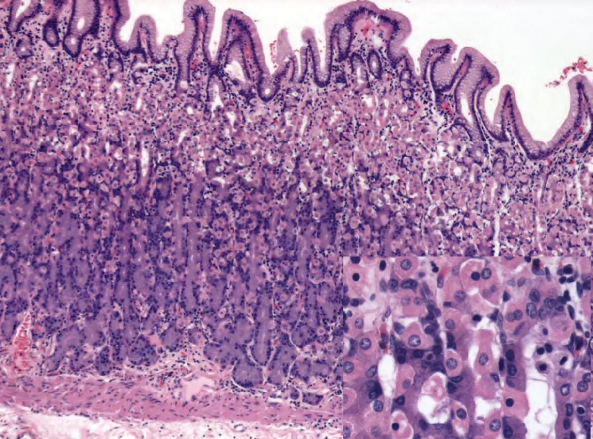
胃底腺は胃底部・胃体部にある管状腺。1本の腺を上から下へ辿ると細胞が入れ替わるのが観察の要点。
| 細胞 | 位置 | 染色性 | 分泌するもの |
|---|---|---|---|
| 副細胞(頸粘液細胞) | 頸部 | 淡明 | 可溶性粘液 |
| 壁細胞(傍細胞) | 頸部〜体部 | 好酸性(ピンク)——ミトコンドリアが多い | 塩酸と内因子 |
| 主細胞 | 底部 | 好塩基性(青紫)——粗面小胞体が多い | ペプシノゲン |
| 内分泌細胞 | 散在 | — | ヒスタミン(ECL細胞)、ソマトスタチン(D細胞) |
| 幹細胞 | 峡部 | — | 上下へ分化して腺を作り替える |
幽門腺(幽門部)には壁細胞がほとんど無く、代わりに G細胞がガストリンを出す。
染色性の違いは、細胞小器官の違いをそのまま見ている。 主細胞は蛋白(ペプシノゲン)を作るので粗面小胞体が発達し、リボソームのRNAがヘマトキシリンに染まって青い。壁細胞は酸を汲み出すためにATPを大量に要し、ミトコンドリアが詰まっていてエオジンに染まりピンクになる。「何を作っているか」が「どう染まるか」を決める。
壁細胞の分泌機構——静止時は細胞内小管とチューブロベシクルに H⁺/K⁺-ATPase が待機し、刺激されると管腔膜へ移動する。アクアポリン2 と同じ「小胞から膜へ」という仕組み。
壁細胞を刺激するのは3つの経路——①ガストリン(G細胞)②アセチルコリン(迷走神経、M3)③ヒスタミン(ECL細胞、H2)。ヒスタミンが最終共通経路に近いのでH2受容体拮抗薬が効き、PPI は H⁺/K⁺-ATPase そのもの(最終段階)を止めるのでさらに強力。
A型胃炎(自己免疫性胃炎)——抗壁細胞抗体・抗内因子抗体が壁細胞を壊す。胃体部優位の萎縮で、①内因子が出ずビタミンB12欠乏 → 悪性貧血、②胃酸が出ずフィードバックで高ガストリン血症 → ECL細胞が増えてカルチノイドを生じうる。B型(H. pylori)は前庭部優位で対照的。
PPI 長期投与も同じ経路をたどる——酸を止める → ガストリン上昇 → 胃底腺ポリープ・ECL細胞過形成。薬で作った低酸状態が、病気と同じ組織変化を起こすという例。
English version
Core
The fundic glands are tubular glands of the fundus and body. The point of the observation is that the cells change as a single gland is followed from top to bottom.
| Cell | Position | Staining | Secretes |
|---|---|---|---|
| Mucous neck cell | Neck | Pale | Soluble mucus |
| Parietal cell | Neck to body | Eosinophilic (pink) — many mitochondria | Hydrochloric acid and intrinsic factor |
| Chief cell | Base | Basophilic (blue-purple) — much rough ER | Pepsinogen |
| Endocrine cells | Scattered | — | Histamine (ECL cell), somatostatin (D cell) |
| Stem cells | Isthmus | — | Differentiate up and down, renewing the gland |
The pyloric glands (antrum) have almost no parietal cells; instead G cells secrete gastrin.
Going further
The difference in staining is a direct view of the difference in organelles. The chief cell makes a protein (pepsinogen), so its rough ER is developed and the ribosomal RNA takes up haematoxylin and stains blue. The parietal cell needs a great deal of ATP to pump acid, is packed with mitochondria, and takes up eosin and stains pink. What the cell makes decides how it stains.
The secretory mechanism of the parietal cell — at rest the H⁺/K⁺-ATPase waits in the intracellular canaliculi and tubulovesicles, and on stimulation it moves to the luminal membrane: the same "vesicle to membrane" arrangement as aquaporin 2.
Three pathways stimulate the parietal cell — ① gastrin (G cell) ② acetylcholine (vagus, M3) ③ histamine (ECL cell, H2). Histamine is nearest to the final common path, so H2 antagonists work; and a PPI blocks the H⁺/K⁺-ATPase itself — the final step — and is stronger still.
In the clinic
Type A gastritis (autoimmune) — anti-parietal cell and anti-intrinsic factor antibodies destroy the parietal cells. The atrophy predominates in the body, and ① no intrinsic factor is secreted, giving B12 deficiency → pernicious anaemia; ② no acid is secreted, so feedback gives hypergastrinaemia → ECL cell hyperplasia and possibly carcinoid. Type B (H. pylori) predominates in the antrum, by contrast.
Long-term PPI treatment follows the same pathway — stop the acid → gastrin rises → fundic gland polyps and ECL cell hyperplasia. An example of a low-acid state produced by a drug causing the same tissue change as a disease.
小腸の吸収装置 — 表面積を600倍にする3段の仕掛けVillus intestinalis
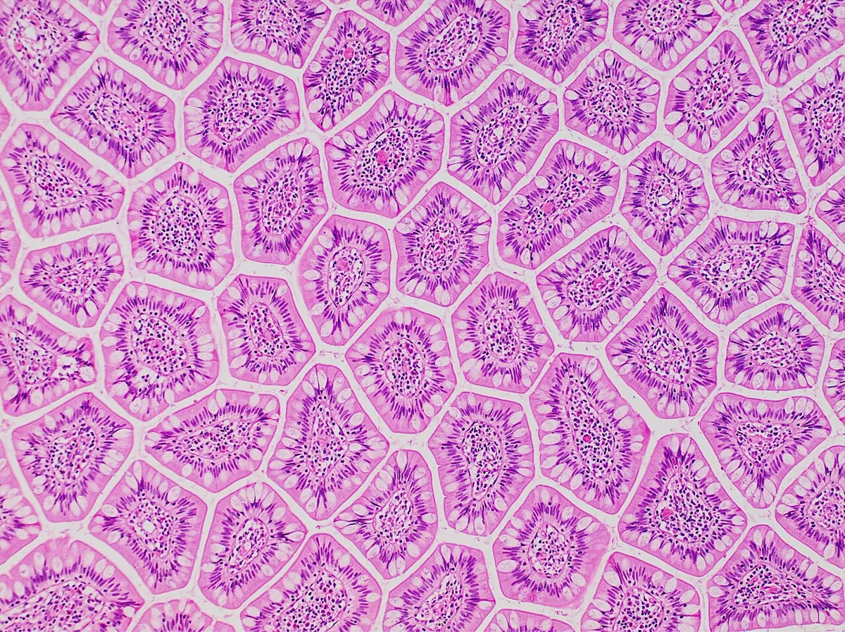
小腸は3段階で表面積を稼ぐ。
| 段 | 構造 | 倍率 |
|---|---|---|
| ① | 輪状ヒダ〈Kerckring ヒダ〉——粘膜と粘膜下層が作る大きなヒダ | ×3 |
| ② | 絨毛——粘膜が指状に突出(粘膜だけ。粘膜下層は入らない) | ×10 |
| ③ | 微絨毛〈刷子縁〉——吸収上皮細胞の表面 | ×20 |
合わせて約600倍、テニスコート1面分に達する。
絨毛の中身——中心に中心乳糜管〈リンパ管〉、その周りに毛細血管網と平滑筋。脂肪はリンパへ、糖とアミノ酸は門脈へという吸収経路の分岐がここで起きる。
陰窩〈Lieberkühn腺〉の底には幹細胞とPaneth 細胞(好酸性顆粒、リゾチーム・ディフェンシンを出す自然免疫)がある。
刷子縁は「膜そのものが消化酵素である」という点で特異。マルターゼ・スクラーゼ・ラクターゼ・ペプチダーゼが微絨毛の膜に埋め込まれ、分解と吸収が同じ場所で連続して起こる(膜消化)。
ラクターゼが最も先端側にあり、最も弱い——これが臨床的に効いてくる。ウイルス性腸炎で絨毛の先端が脱落すると、まずラクターゼが失われるので、感染後に一過性の乳糖不耐となり下痢が長引く。
上皮の入れ替わりは3〜5日と体内で最も速い。陰窩の幹細胞が分裂し、絨毛の先端へ押し上げられながら成熟し、先端から脱落する——ベルトコンベアのような構造。だから抗癌薬や放射線で分裂を止められると、真っ先に消化管粘膜が壊れる(口内炎・下痢)。
部位ごとの違い——十二指腸は粘膜下層に Brunner 腺(アルカリ性粘液で胃酸を中和)、回腸には Peyer 板(粘膜下層まで及ぶリンパ小節の集塊)。絨毛は空腸で最も長く、回腸へ向かって短くなる。
セリアック病——グルテンに対する免疫反応で絨毛が萎縮し、陰窩が過形成する(絨毛の平坦化+上皮内リンパ球の増加)。表面積を失うので吸収不良・下痢・鉄と葉酸の欠乏。HLA-DQ2/DQ8と関連し、疱疹状皮膚炎を合併する。
Whipple 病では PAS陽性のマクロファージが粘膜固有層を埋め、Tropheryma whipplei が原因。吸収不良+関節痛+神経症状という組み合わせで疑う。
English version
Core
The small intestine gains surface area in three stages.
| Stage | Structure | Factor |
|---|---|---|
| ① | Circular folds (of Kerckring) — large folds made by mucosa and submucosa | ×3 |
| ② | Villi — finger-like projections of mucosa (mucosa only; the submucosa does not enter) | ×10 |
| ③ | Microvilli (brush border) — on the surface of the absorptive cell | ×20 |
Together about 600-fold, reaching the area of a tennis court.
Inside a villus — a central lacteal (lymphatic) with a capillary network and smooth muscle around it. The divergence of the absorptive routes — fat to lymph, sugars and amino acids to the portal vein — happens here.
At the base of the crypts (of Lieberkühn) lie stem cells and Paneth cells (eosinophilic granules; lysozyme and defensins, innate immunity).
Going further
The brush border is peculiar in that the membrane itself is the digestive enzyme. Maltase, sucrase, lactase and peptidases are embedded in the membrane of the microvilli, so breakdown and absorption occur continuously in the same place (membrane digestion).
Lactase lies furthest towards the tip and is the weakest — and this tells clinically. When the tips of the villi are shed in viral enteritis, lactase is the first to be lost, so a transient lactose intolerance follows the infection and the diarrhoea is prolonged.
The epithelium turns over in 3–5 days, the fastest in the body. Stem cells in the crypt divide, mature as they are pushed up towards the tip of the villus, and are shed from the tip — a structure like a conveyor belt. So when division is halted by cytotoxic drugs or radiation, the gut mucosa is the first to break down (mucositis, diarrhoea).
Regional differences — the duodenum has Brunner’s glands in the submucosa (alkaline mucus neutralising gastric acid); the ileum has Peyer’s patches (aggregates of lymphoid follicles extending into the submucosa). The villi are longest in the jejunum and shorten towards the ileum.
In the clinic
Coeliac disease — an immune response to gluten atrophies the villi and hyperplases the crypts (villous flattening plus increased intraepithelial lymphocytes). Surface area is lost, giving malabsorption, diarrhoea and deficiency of iron and folate. Associated with HLA-DQ2/DQ8, and with dermatitis herpetiformis.
In Whipple’s disease, PAS-positive macrophages fill the lamina propria; the cause is Tropheryma whipplei. Suspect it from the combination of malabsorption, arthralgia and neurological symptoms.
腸管神経系 — 脳と独立に動く「第二の脳」Plexus myentericus
消化管の壁には2つの神経叢が層をなして埋め込まれている。層の位置が、そのまま役割を決める。
| 神経叢 | 位置 | 働き |
|---|---|---|
| Auerbach 神経叢(筋層間神経叢) | 固有筋層の内輪走筋と外縦走筋の間 | 蠕動運動の制御 |
| Meissner 神経叢(粘膜下神経叢) | 粘膜下層 | 分泌と局所の血流の制御 |
神経細胞の総数は1億個を超え、脊髄に匹敵する。迷走神経を切っても消化管は動き続ける——中枢から独立して蠕動反射を完結できるため「腸管神経系〈第二の脳〉」と呼ばれる。
これらの神経細胞は神経堤細胞に由来し、発生の途中で口側から肛門側へ向かって遊走してくる。
神経堤細胞が「口側から肛門側へ順に」遊走するという事実が、そのまま病気の形を決める。
Hirschsprung 病——遊走が途中で止まると、止まった場所から肛門側までが無神経節腸管になる。だから必ず直腸から連続して口側へ広がり、飛び石にならない。直腸S状部型が約80%と最多なのは、遊走の最終地点が最も届きにくいから。
| Hirschsprung病の要点 |
|---|
| 症状:胎便排泄の遅れ(生後24時間以内に出ない)・腹部膨満・嘔吐。腸炎を起こすと致死的 |
| 注腸造影:狭い無神経節部と拡張した口側の間に caliber change(移行帯) |
| 直腸粘膜生検:神経節細胞の欠如+アセチルコリンエステラーゼ陽性の神経線維が増生——これが確定診断 |
| 直腸肛門反射の消失(内圧検査) |
| Down症候群に合併しやすい。RET遺伝子が関与 |
Auerbach 神経叢だけが壊れる病気もある。食道アカラシアは下部食道の Auerbach 神経叢の変性で、抑制性ニューロン(NO・VIP)が失われ、下部食道括約筋が弛緩できなくなる。Chagas 病(Trypanosoma cruzi)は神経叢を直接破壊し、巨大食道・巨大結腸を起こす。
腸管神経系は約1億個のニューロンを持ち、脳と独立に動く。だから迷走神経を切っても蠕動は残る。
神経叢の位置が、そのまま病気の症状を決める。筋層間神経叢=運動なのでここが無ければ動かない(Hirschsprung 病・アカラシア)。粘膜下神経叢=分泌と血流。
セロトニンの大半(約90%)は腸のクロム親和性細胞にある。だからSSRI の副作用に消化器症状(悪心・下痢)が多く、5-HT3 拮抗薬(オンダンセトロン)が制吐薬として効く。「腸は最大のセロトニン器官」という事実が薬理に直結している。
過敏性腸症候群は腸管神経系と中枢の相互作用(脳腸相関)の異常として理解されている。器質的な異常が無いことが定義に含まれる。
English version
Core
Two plexuses are embedded in layers in the wall of the gut, and the position of the layer decides the role.
| Plexus | Position | Function |
|---|---|---|
| Auerbach’s (myenteric) | Between the inner circular and outer longitudinal muscle | Control of peristalsis |
| Meissner’s (submucosal) | In the submucosa | Control of secretion and local blood flow |
The total number of neurones exceeds 100 million, comparable to the spinal cord. The gut continues to move after the vagus is cut — able to complete the peristaltic reflex independently of the central nervous system, hence "the enteric nervous system, the second brain".
These neurones derive from neural crest cells and migrate during development from the oral end towards the anal end.
Going further
That the neural crest cells migrate in order from oral to anal decides the very form of the disease.
Hirschsprung’s disease — if the migration stops part way, everything from that point to the anus is aganglionic. So it always extends continuously from the rectum orally and never skips. The rectosigmoid type is the commonest at about 80 per cent because the end point of the migration is the hardest to reach.
Key points of Hirschsprung’s disease
- Symptoms: delayed passage of meconium (not passed within 24 hours), abdominal distension, vomiting. Enterocolitis is fatal.
- Contrast enema: a caliber change (transition zone) between the narrow aganglionic segment and the dilated bowel above.
- Rectal mucosal biopsy: absence of ganglion cells plus proliferation of acetylcholinesterase-positive nerve fibres — this is the confirmatory diagnosis.
- Loss of the rectoanal inhibitory reflex (manometry).
- Associated with Down syndrome. The RET gene is involved.
There are also diseases in which only Auerbach’s plexus is destroyed. In achalasia, degeneration of the myenteric plexus of the lower oesophagus loses the inhibitory neurones (NO, VIP) so the lower sphincter cannot relax. Chagas disease (Trypanosoma cruzi) destroys the plexus directly, causing megaoesophagus and megacolon.
In the clinic
The enteric nervous system has about 100 million neurones and works independently of the brain, so peristalsis remains after the vagus is cut.
The position of the plexus decides the symptoms of the disease. Myenteric = movement, so without it there is no movement (Hirschsprung’s disease, achalasia). Submucosal = secretion and blood flow.
Most of the body’s serotonin (about 90 per cent) is in the enterochromaffin cells of the gut. This is why gastrointestinal symptoms (nausea, diarrhoea) are common adverse effects of SSRIs, and why 5-HT3 antagonists (ondansetron) work as antiemetics. The fact that the gut is the largest serotonin organ bears directly on pharmacology.
Irritable bowel syndrome is understood as a disturbance of the interaction between the enteric nervous system and the centre (the brain–gut axis). The absence of an organic abnormality is part of its definition.
胃の粘膜防御 — 自分を消化しないための、幾重もの仕掛けMucus gastricus
胃は pH 1〜2 の酸とペプシンを出しながら、自分は溶けない。守りは層になっている。
| 層 | 中身 |
|---|---|
| ①粘液 | 表層粘液細胞が出す不溶性のゲル(約0.2 mm)。ペプシンを通さない |
| ②重炭酸 | 粘液の中に分泌され、粘液層の内側で pH を約7に保つ |
| ③上皮の疎水性と密着結合 | 細胞の表面が水と酸を弾く |
| ④粘膜の血流 | 逆拡散した H⁺ を洗い流し、重炭酸を供給する |
| ⑤速い再生 | 表層上皮は3〜5日で入れ替わる |
これらを支えているのがプロスタグランジンE₂(粘液と重炭酸を増やし、血流を保つ)。
幹細胞は胃小窩の峡部にあり、上へも下へも分化して移動する。
「守りの中心はプロスタグランジンである」という一点で、薬による潰瘍が説明できる。非ステロイド性抗炎症薬はシクロオキシゲナーゼ1 を止めるので、①②④が同時に落ちる(アラキドン酸カスケードの章)——「酸を増やしているのではなく、守りを外している」。
だから坐薬にしても注射にしても、潰瘍は起こる(抗炎症薬の章)——「局所刺激ではなく、全身の作用として起きるからになる」。
ミソプロストール(プロスタグランジン製剤)が予防に使われるのも、この機序による——「ただし子宮を収縮させるので、妊婦には使えない」。
Helicobacter pylori は、この守りを内側から崩している。ウレアーゼで尿素をアンモニアに変えて自分の周りを中和し、粘液層の中で生き延びる(らせん菌の章)——「尿素呼気試験は、この酵素を測っている検査になる」。
感染の場所が、その後の病気を分ける。前庭部が主体ならガストリンが増えて酸が過剰になり十二指腸潰瘍、体部に広がると萎縮性胃炎から胃癌へ向かう(胃の各論の章)——「同じ菌で、酸が増える人と減る人がいる」。
攻撃因子と防御因子の均衡という古典的な図式は、この層構造そのものである。
NSAIDはシクロオキシゲナーゼ1を阻害してプロスタグランジンを減らす。粘液・重炭酸・粘膜血流のすべてが同時に落ちるので潰瘍ができる。「防御を3つまとめて外す」から強力なのである。プロスタグランジン製剤(ミソプロストール)が予防薬になるのはこの機序の裏返しになる。
Helicobacter pylori — ウレアーゼでアンモニアを作り、酸を中和して粘液層に住みつく。慢性胃炎 → 萎縮 → 腸上皮化生 → 胃癌という道筋と、MALT リンパ腫を起こす。除菌で MALT リンパ腫が退縮する——抗菌薬で治る悪性腫瘍という稀な例である。
PPI と H2 遮断薬は攻撃因子を減らす側から働く。
English version
Core
The stomach secretes acid at pH 1–2 with pepsin, and does not dissolve itself. The defence is layered.
| Layer | What it is |
|---|---|
| ① Mucus | An insoluble gel from the surface mucous cells (about 0.2 mm). Pepsin cannot pass it |
| ② Bicarbonate | Secreted into the mucus, holding the pH near 7 on the inner side of the layer |
| ③ Hydrophobicity of the epithelium and tight junctions | The cell surface repels water and acid |
| ④ Mucosal blood flow | Washes away back-diffused H⁺ and supplies bicarbonate |
| ⑤ Rapid regeneration | The surface epithelium is replaced in 3–5 days |
All of this is supported by prostaglandin E₂ (which increases mucus and bicarbonate and preserves blood flow). The stem cells lie at the isthmus of the gastric pit and differentiate and move both up and down.
Going further
The single point that prostaglandin is the centre of the defence explains drug-induced ulceration. NSAIDs block cyclo-oxygenase 1, so ① ② and ④ fall together — they are not increasing the acid but removing the defence.
So ulcers occur whether the drug is given as a suppository or by injection — because it happens as a systemic action, not as local irritation. Misoprostol (a prostaglandin) is used in prevention for the same reason — though it contracts the uterus and cannot be used in pregnancy.
Helicobacter pylori breaks that defence from within. With urease it converts urea to ammonia, neutralises its surroundings and survives inside the mucus layer — the urea breath test is an assay of that enzyme.
Where the infection sits divides the subsequent disease. Predominantly in the antrum, gastrin rises, the acid is excessive and a duodenal ulcer follows; spreading to the body, it leads through atrophic gastritis to gastric cancer — with the same organism, some people make more acid and some less.
In the clinic
The classical scheme of a balance between aggressive and defensive factors is this layered structure itself.
NSAIDs inhibit cyclo-oxygenase 1 and reduce prostaglandin. Mucus, bicarbonate and mucosal blood flow all fall at once, and an ulcer forms. They are powerful because they remove three defences together. That a prostaglandin (misoprostol) prevents them is the converse of the same mechanism.
Helicobacter pylori — makes ammonia with urease, neutralises the acid and settles in the mucus layer. It gives the path chronic gastritis → atrophy → intestinal metaplasia → gastric cancer, and causes MALT lymphoma. MALT lymphoma regresses on eradication — a rare instance of a malignant tumour cured by an antibiotic.
PPIs and H2 blockers work from the side of reducing the aggressive factor.
大腸の組織 — 吸収より、水を取り戻して溜めることが仕事になるIntestinum crassum
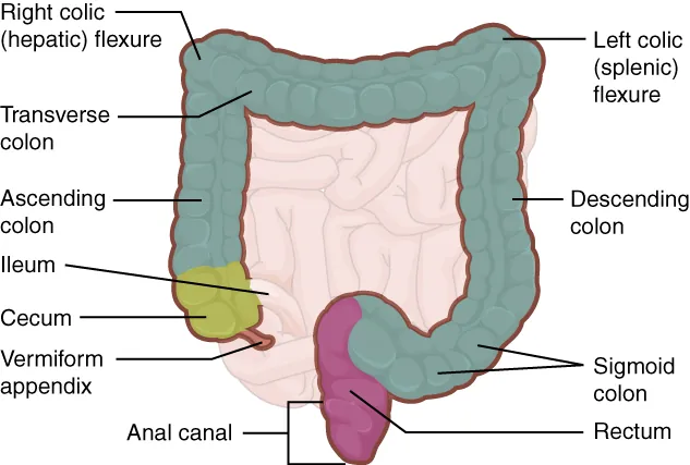
小腸との違いは、引き算で覚えると速い。
| 小腸 | 大腸 | |
|---|---|---|
| 輪状ひだ・絨毛 | ある | 無い |
| 腸腺(陰窩) | ある | ある(深く、まっすぐ) |
| 杯細胞 | あり | 非常に多い |
| Paneth 細胞 | ある | 通常は無い |
| 外縦走筋 | 全周 | 3本の結腸ヒモに集まる |
虫垂——粘膜固有層と粘膜下組織に、輪状のリンパ小節が密に並ぶ。内腔は狭く不規則。
直腸下部から肛門管へ——歯状線を境に、上は単層円柱上皮(内臓神経支配)、下は重層扁平上皮(体性神経支配)。
絨毛が無いことは、大腸が「吸収する器官ではない」ことを示している。表面積を稼ぐ必要がないほど、吸収するものが限られている(主に水と電解質)(短鎖脂肪酸の項)——「例外が短鎖脂肪酸で、これは細菌が作って上皮が直接使う」。
杯細胞が多いことは、糞便を滑らせるためになる——「粘液が減ると、便が硬くなるだけでなく粘膜も傷つく」。
歯状線の意味が、そのまま痔の症状を分ける。内痔核(上)は痛くなく出血する、外痔核(下)は激しく痛む(肛門の3つの病気の章)——「神経支配が変わる線が、症状の境界になっている」。
静脈還流も分かれる——上は門脈系、下は体循環系(門脈域の章)——「門脈圧亢進症で、直腸静脈瘤ができる理由がここにある」。
結腸ヒモに筋が集まっていることが、憩室の好発部位を決めている(腸の各論の章)——「血管が壁を貫く場所が弱点になり、そこから粘膜が押し出される」。
大腸に絨毛は無い。だから生検で「絨毛がある」なら小腸である——標本で最初に確かめる点になる。
杯細胞が減るのが潰瘍性大腸炎の活動性の指標である。陰窩膿瘍(陰窩の中に好中球が溜まる)と併せて評価する。
大腸癌の大半は腺腫から発生する(adenoma–carcinoma sequence)。APC → KRAS → TP53 と変異が積み重なる。腺腫を切除すれば癌を防げる——大腸内視鏡によるポリペクトミーが予防になる根拠である。
偽膜性腸炎(Clostridioides difficile) — 抗菌薬使用後の下痢。内視鏡で黄白色の偽膜、組織では火山の噴火のような(volcano lesion)フィブリンと好中球の噴出。
虚血性大腸炎は分水嶺(脾彎曲・S状結腸)に起こる。
English version
Core
The differences from the small intestine are learnt fastest by subtraction.
| Small intestine | Large intestine | |
|---|---|---|
| Circular folds, villi | Present | Absent |
| Crypts | Present | Present (deep and straight) |
| Goblet cells | Present | Very numerous |
| Paneth cells | Present | Normally absent |
| Outer longitudinal muscle | Around the whole circumference | Gathered into three taeniae coli |
Appendix — lymphoid follicles arranged densely in a ring in the lamina propria and submucosa. The lumen is narrow and irregular.
Lower rectum to anal canal — at the dentate line, above is simple columnar (visceral innervation), below is stratified squamous (somatic innervation).
Going further
The absence of villi shows that the colon is not an organ of absorption. What it absorbs is so limited (chiefly water and electrolytes) that it has no need to gain surface area — the exception being short-chain fatty acids, made by bacteria and used directly by the epithelium.
The abundance of goblet cells is to let the faeces slide — with less mucus the stool not only hardens but the mucosa is injured.
The meaning of the dentate line divides the symptoms of haemorrhoids directly. Internal piles (above) bleed painlessly; external piles (below) hurt severely — the line where the innervation changes is the boundary of the symptoms.
The venous drainage divides there too — above, portal; below, systemic — here is why rectal varices form in portal hypertension.
That the muscle is gathered into the taeniae decides where diverticula occur — the points where vessels pierce the wall are the weak spots, and the mucosa is pushed out through them.
In the clinic
The colon has no villi. So if a biopsy shows villi, it is small intestine — the first thing to confirm on a slide.
Loss of goblet cells is an index of activity in ulcerative colitis, assessed together with crypt abscesses (neutrophils accumulating within a crypt).
Most colorectal cancer arises from an adenoma (the adenoma–carcinoma sequence), with mutations accumulating APC → KRAS → TP53. Removing the adenoma prevents the cancer — the rationale for polypectomy at colonoscopy as prevention.
Pseudomembranous colitis (Clostridioides difficile) — diarrhoea after antibiotics. Yellow-white pseudomembranes at endoscopy; histologically an eruption of fibrin and neutrophils like a volcano (the volcano lesion).
Ischaemic colitis occurs at the watersheds (splenic flexure, sigmoid).
まとめ
| 粘膜 | 最内層。さらに3つに分かれる。 |
|---|---|
| 粘膜下層 | 密性結合組織の層。太い血管・リンパ管とMeissner 神経叢(粘膜下神経叢)が走る。 |
| 固有筋層 | 内輪走筋と外縦走筋の2層(胃だけは内斜走筋を加えた3層)。 |
| 部位ごとの粘膜 — 食道・胃・小腸・大腸 | 食道——重層扁平上皮(非角化)。粘液腺が粘膜下層に胃——単層円柱上皮。 |
| 炎症性腸疾患 — 深さと分布で分ける | 2つの疾患は「どこまで深く」「どう分布するか」で分かれる。 |
| 胃底腺の細胞 — 酸を出す細胞と、酵素を出す細胞 | 胃底腺は胃底部・胃体部にある管状腺。1本の腺を上から下へ辿ると細胞が入れ替わるのが観察の要点。 |
| 小腸の吸収装置 — 表面積を600倍にする3段の仕掛け | 小腸は3段階で表面積を稼ぐ。段構造倍率①輪状ヒダ〈Kerckring ヒダ〉——粘膜と粘膜下層が作る大きなヒダ×3②絨毛——粘膜が指状に突出(粘膜だけ。 |
| 腸管神経系 — 脳と独立に動く「第二の脳」 | 消化管の壁には2つの神経叢が層をなして埋め込まれている。 |
| 胃の粘膜防御 — 自分を消化しないための、幾重もの仕掛け | 胃は pH 1〜2 の酸とペプシンを出しながら、自分は溶けない。 |
| 大腸の組織 — 吸収より、水を取り戻して溜めることが仕事になる | 小腸との違いは、引き算で覚えると速い。小腸大腸輪状ひだ・絨毛ある無い腸腺(陰窩)あるある(深く、まっすぐ)杯細胞あり非常に多いPaneth 細胞ある通常は無い外縦走筋全周3本の結腸… |
このノートは医学生が授業の内容を自分の言葉でまとめたもので、診療の判断に使うものではありません。誤りに気づいたらお知らせください。