腎で作られた尿は、尿路では成分が変わらない。だから尿路の組織は「漏らさない」「伸び縮みする」「刺激に耐える」という3つの要求だけで説明できる。
その答えが尿路上皮(移行上皮)になる——伸びると平らになり、縮むと丸くなる。表層の傘細胞が、頂端の膜と密着結合で尿を遮る。
そしてこの上皮は、腎盂から尿道の一部まで一続きに広がっている——「同じ発癌因子が、同じ上皮のどこにでも当たる」。これが尿路上皮癌の多発性と再発性の理由になる。
男性の尿道だけが、途中で上皮の種類を変える。前立腺部・膜様部・海綿体部で作りが違う。
- 腎盂と尿管 — 3層の管
- 膀胱と尿道 — 伸びる袋と、変わる管
- 尿路上皮 — 伸び縮みし、尿を通さない特殊な上皮
- 膀胱三角 — 尿が逆流しない仕組みは、走り方にある
- 男性尿道 — 3つの部分で、上皮も走行も変わる
- 尿細胞診 — 剥がれた細胞だけで、癌を見つけられるか
- 尿膜管の遺残 — 膀胱の頂に、胎児の管の名残がある
- カテーテルが入った尿路に、何が起きるか
腎盂と尿管 — 3層の管Ureter
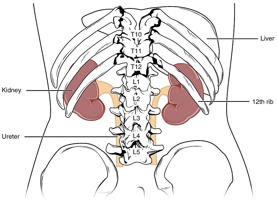
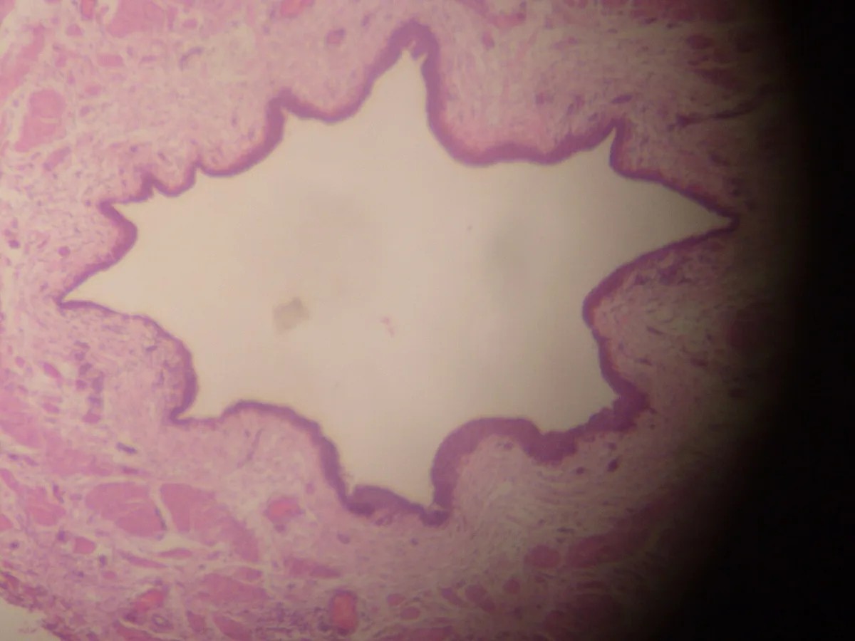
粘膜・筋層・外膜の3層。粘膜下組織は無い。
- 粘膜——尿路上皮(4〜5層)+固有層。縦のひだを作るので、内腔は星形に見える
- 筋層——上2/3が内縦・外輪の2層、下1/3で外縦が加わり3層になる
- 外膜——疎性結合組織。後腹膜に接する
蠕動——腎盂の平滑筋(ペースメーカー)から始まる。神経が無くても動く。
生理的狭窄部が3つ——腎盂尿管移行部・総腸骨動脈との交叉部・膀胱壁内部。結石はここで止まる。
膀胱壁内を斜めに貫くことが、逆流を防ぐ弁になっている。
内腔が星形に見えることが、尿管の標本を言い当てる決め手になる——「丸い管ではなく、ひだで畳まれた管として覚える」。
3つの狭窄部が、疝痛の部位と放散痛を決めている。上部なら側腹部、下部なら鼠径部と陰部へ放散する(尿路結石の章)——「痛みの場所が、石の高さを教える」。
蠕動が自律的であることが、移植腎でも尿が流れる理由になる。神経がつながっていなくても動く(腎移植の章)——「支配神経を失っても働き続ける平滑筋がある」。
斜めに貫く走行が失われると、膀胱尿管逆流になる。小児の反復性尿路感染と腎瘢痕の原因になる(膀胱尿管逆流の章)——「弁は構造でできているので、構造が崩れれば逆流する」。
だから小児の発熱性尿路感染では、画像で形態を調べる(小児の尿路感染の章)——「繰り返す感染の背後に、構造の問題を探す」。
尿管は後腹膜を走るので、婦人科と大腸の手術で損傷されやすい。子宮動脈との交叉が最も危険になる(手術の合併症の章)——「水の下を橋が通る、という位置関係で覚える」。
粘膜下組織が無いので、尿路上皮癌は早く筋層に達する。尿管と腎盂は壁が薄いぶん、同じ深達度でも予後が悪い。
尿路上皮癌は多発する(field defect)。尿路全体が同じ発癌物質にさらされているためで、上部尿路の癌を治療しても膀胱を監視し続ける必要がある。喫煙・芳香族アミン・シクロホスファミドが危険因子。
尿管の蠕動は自律的で、腎盂のペースメーカー細胞が起こす。結石が詰まると蠕動が痛みになる(疝痛)——間欠的で、体位で軽快しない。
尿管は後腹膜を走るので、後腹膜線維症で巻き込まれると両側水腎症になる。
English version
Core
Three layers — mucosa, muscle, adventitia. There is no submucosa.
- Mucosa — urothelium (4–5 layers) + lamina propria. It forms longitudinal folds, so the lumen looks star-shaped.
- Muscle — two layers (inner longitudinal, outer circular) in the upper two thirds; an outer longitudinal layer is added in the lower third, making three.
- Adventitia — loose connective tissue, against the retroperitoneum.
Peristalsis begins in the smooth muscle of the pelvis (the pacemaker). It moves without nerves.
Three physiological narrowings — the pelviureteric junction, the crossing of the common iliac artery, and the intramural part in the bladder wall. Stones lodge at these points.
That the ureter pierces the bladder wall obliquely makes a valve preventing reflux.
Going further
The star-shaped lumen is the decisive clue for identifying a ureter on a slide — learn it not as a round tube but as a tube folded by ridges.
The three narrowings decide the site of the colic and where it radiates. High up, the flank; low down, radiating to the groin and genitals — the site of the pain tells you the level of the stone.
That peristalsis is autonomous is why urine flows in a transplanted kidney: it moves although no nerve is connected — a smooth muscle that goes on working when it has lost its innervation.
When the oblique intramural course is lost, reflux follows. Vesicoureteric reflux is the cause of recurrent urinary infection and renal scarring in children — the valve is made of structure, so if the structure fails, urine refluxes. So in a febrile urinary infection in a child the anatomy is investigated by imaging — look for a structural problem behind a recurring infection.
The ureter runs in the retroperitoneum and is readily injured in gynaecological and colorectal surgery, the crossing with the uterine artery being the most dangerous — remember it as water passing under a bridge.
In the clinic
With no submucosa, urothelial carcinoma reaches the muscle early. The walls of ureter and pelvis being thin, the prognosis is worse at the same depth of invasion.
Urothelial carcinoma is multiple (a field defect), the whole tract having been exposed to the same carcinogen, so after treating an upper tract tumour the bladder must be kept under surveillance. Smoking, aromatic amines and cyclophosphamide are risk factors.
Ureteric peristalsis is autonomous, generated by pacemaker cells in the pelvis. When a stone obstructs, the peristalsis becomes the pain (colic) — intermittent, and not relieved by change of position.
The ureter runs in the retroperitoneum, so if it is caught up in retroperitoneal fibrosis, bilateral hydronephrosis results.
膀胱と尿道 — 伸びる袋と、変わる管Vesica urinaria
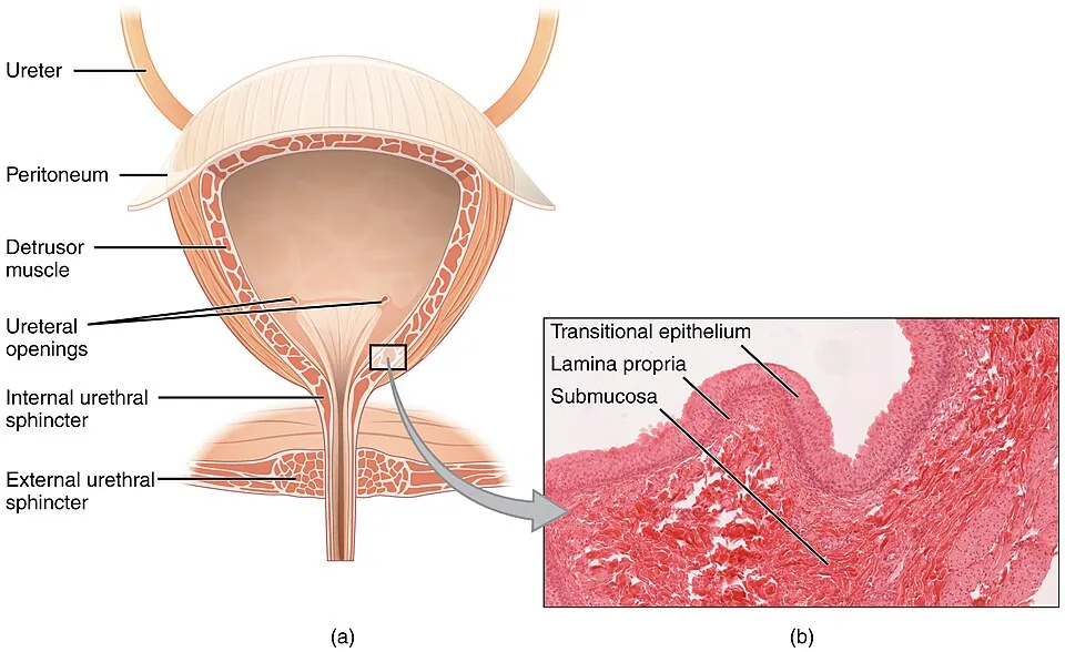
膀胱の尿路上皮は、空虚時6〜8層で表層に傘細胞、充満時は2〜3層に伸びて平らになる。
- 傘細胞——大きく、二核のこともある。頂端の膜(プラーク)と密着結合が尿を遮る
- 筋層——排尿筋。内縦・中輪・外縦の3層だが、実際は網目状に絡む
- 三角部——ひだが無く平滑。中腎管由来で、発生学的に他と異なる
男性尿道の3部。
| 部位 | 上皮 | 備考 |
|---|---|---|
| 前立腺部 | 尿路上皮 | 射精管が開口 |
| 膜様部 | 多列/重層円柱 | 外尿道括約筋(横紋筋) |
| 海綿体部 | 重層円柱→尿道口で重層扁平 | 尿道球腺・尿道腺が開く |
女性尿道は約4 cm と短く、重層扁平上皮が主体になる。
傘細胞と密着結合が「尿-血液関門」を作っている。膀胱は濃縮された尿を何時間も溜めるので、漏れれば組織が傷む——「腎が作った濃さを、膀胱が受け止めている」。
この関門が壊れることが、間質性膀胱炎の仮説の1つになっている(膀胱の機能異常の章)——「炎症の原因が見つからない痛みに、バリアの側から説明を与える」。
女性の尿道が短いことが、単純性膀胱炎が女性に多い理由になる(尿路感染症の章)——「解剖の差が、そのまま疫学の差になっている」。
膜様部の外尿道括約筋が横紋筋であることが、随意にとめられる理由になる(排尿の生理の章)——「内括約筋は自律神経、外括約筋は体性神経になる」。
だから前立腺手術では、この高さを傷つけないことが尿失禁の予防になる(前立腺肥大症の手術の章)——「機能の温存が、数ミリの位置で決まる」。
三角部が発生学的に別であることが、腺癌がここに出やすい理由の説明に使われる(膀胱癌の章)——「膀胱癌の大半は尿路上皮癌だが、例外の場所が決まっている」。
傘細胞と密着結合が「尿を漏らさない」バリアである。高浸透圧で毒性のある尿から、自分の組織を守っている。
このバリアが破れるのが間質性膀胱炎(膀胱痛症候群)と考えられている。尿が溜まると痛み、排尿で軽快する。細菌尿は無い——「膀胱炎症状があるのに培養が陰性」のときに思い出す。
膀胱癌は無痛性肉眼的血尿で発症することが多い。痛くない血尿は精査するのが原則で、膀胱鏡と尿細胞診を行う。
上皮内癌(CIS)は平坦で見えにくいため尿細胞診が有用である。
BCG 膀胱内注入が筋層非浸潤癌の再発予防に使われる——局所免疫を利用した治療である。
English version
Core
The bladder urothelium is 6–8 layers deep when empty, with umbrella cells at the surface, and stretches to 2–3 flattened layers when full.
- Umbrella cell — large, sometimes binucleate. The apical membrane (plaques) and the tight junctions shut out the urine.
- Muscle — the detrusor. Nominally three layers (inner longitudinal, middle circular, outer longitudinal) but in fact interwoven as a mesh.
- Trigone — no folds, always smooth. Derived from the mesonephric duct, developmentally different from the rest.
Three parts of the male urethra
| Part | Epithelium | Notes |
|---|---|---|
| Prostatic | Urothelium | The ejaculatory ducts open here |
| Membranous | Pseudostratified / stratified columnar | The external urethral sphincter (striated) |
| Spongy | Stratified columnar → stratified squamous at the meatus | The bulbourethral and urethral glands open here |
The female urethra is short, about 4 cm, and chiefly stratified squamous.
Going further
The umbrella cells with their tight junctions form a urine–blood barrier. The bladder holds concentrated urine for hours, and leakage would damage the tissue — the bladder receives the concentration the kidney made.
Breakdown of this barrier is one hypothesis for interstitial cystitis — an explanation from the side of the barrier for a pain in which no cause of inflammation can be found.
The shortness of the female urethra is why uncomplicated cystitis is commoner in women — a difference in anatomy becoming directly a difference in epidemiology.
That the external sphincter in the membranous part is striated muscle is why urination can be held voluntarily — the internal sphincter is autonomic, the external somatic. So in prostatic surgery, not damaging this level is the prevention of incontinence — preservation of function decided by a few millimetres.
That the trigone is developmentally separate is used to explain why adenocarcinoma tends to arise there — most bladder cancer is urothelial, but the exceptions have a fixed location.
In the clinic
The umbrella cells and tight junctions are the barrier that keeps urine out, protecting the tissue from urine that is hyperosmolar and toxic.
Rupture of that barrier is thought to be interstitial cystitis (bladder pain syndrome): pain as the bladder fills, relieved by voiding, with no bacteriuria. Remember it when there are symptoms of cystitis but the culture is negative.
Bladder cancer often presents with painless macroscopic haematuria. Painless haematuria must be investigated as a rule, with cystoscopy and urine cytology. Carcinoma in situ is flat and hard to see, so urine cytology is useful.
Intravesical BCG is used to prevent recurrence of non-muscle-invasive cancer — a treatment that exploits local immunity.
尿路上皮 — 伸び縮みし、尿を通さない特殊な上皮Urothelium
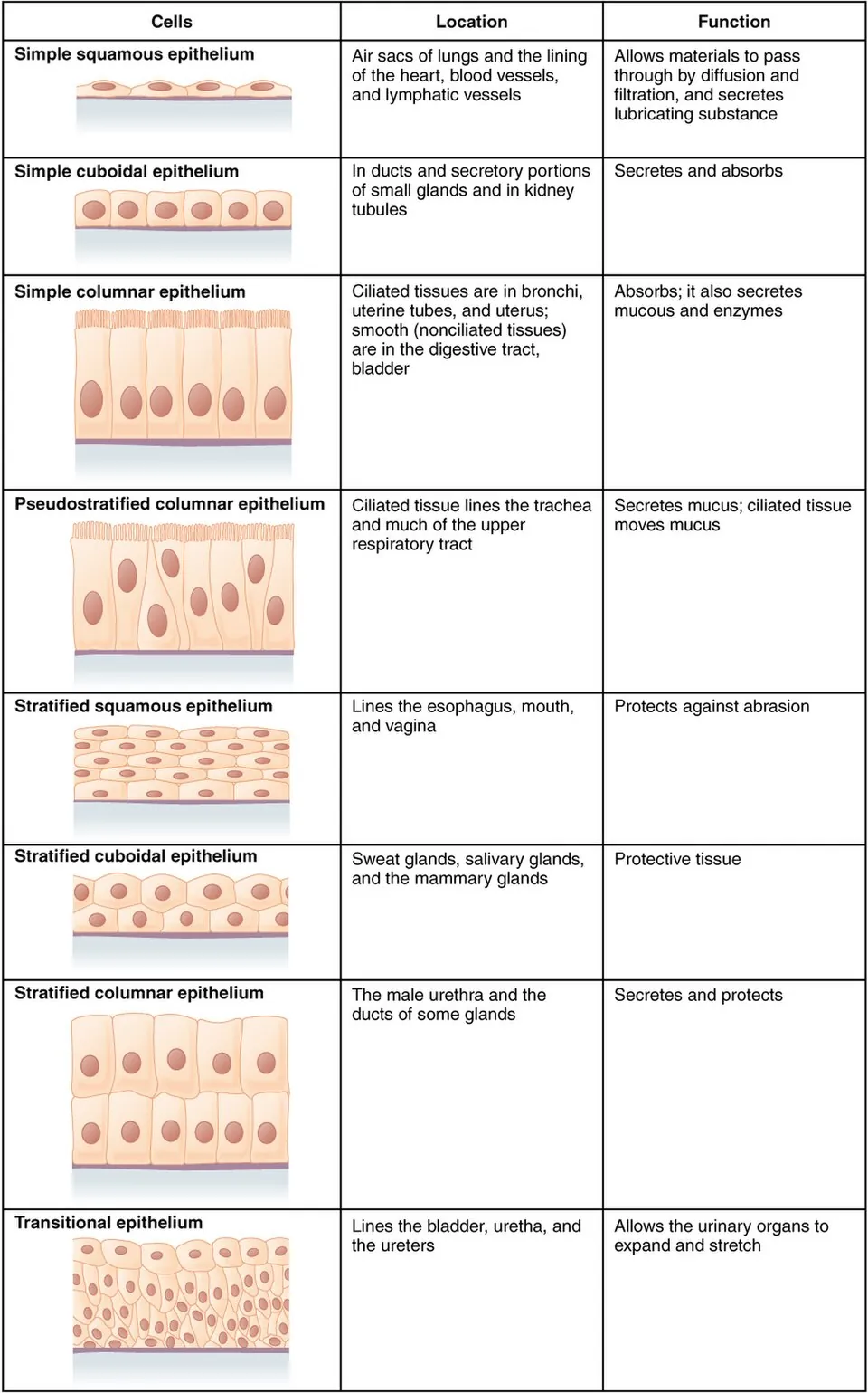
腎杯から尿道の一部までを覆う移行上皮〈尿路上皮〉。2つの相反する条件を同時に満たすために特殊化している。
| 条件 | 仕組み |
|---|---|
| 大きく伸び縮みする | 弛緩時は5〜6層で丸い細胞、伸展時は2〜3層で扁平に見える。層の数が変わるのではなく、重なり方が変わる |
| 高張・強酸性の尿を通さない | 表層の被蓋細胞〈umbrella cell〉——大型で、しばしば二核。管腔側の膜がウロプラキンという蛋白の板〈plaque〉で覆われ、密着結合で封じられる |
膜の余りは紡錘形の小胞〈fusiform vesicle〉として細胞内に畳まれ、伸展時に膜へ加わる——アコーディオンのように膜そのものを出し入れする。
「尿という毒から自分を守る上皮」が、逆に尿の中の発癌物質に最も長く曝される上皮でもある——これが尿路上皮癌の成り立ちになる。
| 危険因子 | 機序 |
|---|---|
| 喫煙(最大) | 発癌物質が尿中へ排泄される |
| 芳香族アミン(染料・ゴム・皮革工業) | 職業性膀胱癌。ベンジジン・2-ナフチルアミン。潜伏期が長い |
| シクロホスファミド | 代謝物アクロレインが膀胱粘膜を傷害。出血性膀胱炎と癌。メスナで予防 |
| フェナセチン、慢性刺激(結石・カテーテル) | — |
| ビルハルツ住血吸虫 | 虫卵の慢性刺激 → 扁平上皮化生 → 扁平上皮癌(尿路上皮癌ではない) |
尿路上皮は腎盂から膀胱・尿道まで連続している——だから癌も多発性・多中心性に発生する(field cancerization)。膀胱癌の患者では上部尿路も、上部尿路癌の患者では膀胱も検索する必要があり、術後も長期にわたり膀胱鏡でのサーベイランスが要る。
症状は「無症候性肉眼的血尿」——痛くない血尿ほど危ない。膀胱炎と違って排尿時痛を伴わないため軽視されやすい。40歳以上の肉眼的血尿は、証明されるまで悪性を疑う。
治療で特徴的なのがBCG 膀胱内注入療法——上皮内癌と表在性癌に対して、生きた弱毒結核菌を膀胱内に入れて局所の免疫反応を起こさせる。癌免疫療法として最も古く、いまなお有効な治療のひとつ。
伸び縮みするから、膀胱鏡の所見は充満の程度で変わる。空虚時は厚く見え、充満時は薄く伸びるので、生検の深達度評価では条件を揃える必要がある。
尿路上皮は腎杯から尿道の一部まで連続している。だから尿路上皮癌はどこにでも、何度でもできる。膀胱が最も多いが、腎盂・尿管にも起こる。
血尿の由来を分ける——変形赤血球と赤血球円柱があれば糸球体、均一な赤血球なら尿路。尿路由来なら画像と膀胱鏡へ進む。
尿路上皮が扁平上皮化生を起こすことがある。慢性刺激(結石・長期カテーテル・住血吸虫症)で起こり、扁平上皮癌のリスクになる。長期留置カテーテルの患者では膀胱癌に注意する。
English version
Core
The transitional epithelium (urothelium) covering everything from the renal calyces to part of the urethra. It is specialised to satisfy two contradictory requirements at once.
| Requirement | Mechanism |
|---|---|
| Stretch and recoil greatly | Relaxed, 5–6 layers of rounded cells; stretched, it looks like 2–3 flattened layers. The number of layers does not change; the way they overlap does |
| Do not let hypertonic, strongly acid urine through | The surface umbrella cell — large and often binucleate. Its luminal membrane is covered by plaques of the protein uroplakin and sealed by tight junctions |
The surplus membrane is folded away inside the cell as fusiform vesicles and added to the membrane on stretching — the membrane itself is taken in and out like an accordion.
Going further
The epithelium that protects itself from the poison of urine is also the epithelium exposed longest to the carcinogens in it — and this is how urothelial carcinoma comes about.
| Risk factor | Mechanism |
|---|---|
| Smoking (the greatest) | Carcinogens are excreted in the urine |
| Aromatic amines (dye, rubber, leather industries) | Occupational bladder cancer. Benzidine, 2-naphthylamine. Long latency |
| Cyclophosphamide | The metabolite acrolein injures the mucosa. Haemorrhagic cystitis and cancer. Prevented with mesna |
| Phenacetin, chronic irritation (stone, catheter) | — |
| Schistosoma haematobium | Chronic irritation by the eggs → squamous metaplasia → squamous carcinoma (not urothelial) |
The urothelium is continuous from renal pelvis to bladder and urethra — so the cancer arises multiply and multicentrically (field cancerisation). In a patient with bladder cancer the upper tract must be examined and vice versa, and long-term cystoscopic surveillance is required after treatment.
The symptom is painless macroscopic haematuria — the more painless the blood, the more dangerous. Unlike cystitis there is no pain on voiding, so it is easily dismissed. Macroscopic haematuria over the age of 40 is malignant until proved otherwise.
Characteristic in treatment is intravesical BCG — live attenuated tubercle bacilli instilled into the bladder for carcinoma in situ and superficial cancer to provoke a local immune response. The oldest form of cancer immunotherapy, and still one of the effective ones.
In the clinic
Because it stretches, the cystoscopic appearance changes with the degree of filling. It looks thick when empty and thin when full, so in assessing depth of invasion on biopsy the conditions must be matched.
The urothelium is continuous from calyx to part of the urethra. So urothelial carcinoma can arise anywhere, any number of times. The bladder is commonest, but it occurs in the pelvis and ureter too.
Separate the source of haematuria — dysmorphic red cells with red cell casts means glomerular; uniform red cells means the urinary tract. If it is the tract, proceed to imaging and cystoscopy.
The urothelium may undergo squamous metaplasia from chronic irritation (stone, long-term catheter, schistosomiasis), which is a risk for squamous carcinoma. In a patient with a long-term indwelling catheter, watch for bladder cancer.
膀胱三角 — 尿が逆流しない仕組みは、走り方にあるTrigonum vesicae
膀胱の後壁で、2つの尿管口と内尿道口を結ぶ三角形の領域。
- 粘膜がひだを作らず、常に平滑——他の部分は空虚時にひだが多い
- 粘膜が筋層に固く付着している
- 発生学的な由来が違う——三角部は中腎管(Wolff 管)由来、残りの膀胱は尿生殖洞由来
尿管は膀胱壁を斜めに、1〜2 cm にわたって貫く(壁内走行)。
弁は無い。膀胱内圧が上がると、この斜めの区間が押しつぶされて閉じる。
「弁が無いのに逆流しない」という点が、この章の核心になる。膨らむ袋の壁を斜めに貫くという幾何だけで、逆止弁として働いている——「構造物ではなく、配置が機能を作っている」。
だから壁内走行が短いと、逆流する。膀胱尿管逆流症は、この距離と膀胱壁の厚さの比が足りない状態になる(排尿の障害の章)——「先天的に短い場合と、膀胱が厚くなって相対的に短くなる場合がある」。
小児の腎瘢痕は、この逆流から生まれる。逆流した感染尿が腎盂へ届き、腎盂腎炎を繰り返して瘢痕を残す(小児に固有の病態の章)——「熱だけの乳児の尿路感染を追いかける理由が、将来の腎機能にある」。
三角部の粘膜が平滑であることは、膀胱鏡の目印になる。まず三角部と尿管口を見つけ、そこを基準に病変の位置を言う(泌尿器の悪性腫瘍の章)——「ひだの無い平らな面が、地図の原点になる」。
由来が違うことも、病気の分布に現れる。三角部には腺性膀胱炎や扁平上皮化生が生じやすい(泌尿器の発生の章)——「同じ袋の中で、一部だけ振る舞いが違う理由が発生にある」。
尿管が膀胱壁を斜めに貫くことが、逆流を防ぐ弁になっている。膀胱が充満すると壁内部が押しつぶされて閉じる——受動的な弁である。
膀胱尿管逆流症 — 壁内部が短いと弁が働かない。小児の反復性尿路感染で見つかり、腎盂腎炎を繰り返すと腎瘢痕を残し、将来の高血圧と慢性腎臓病につながる。排尿時膀胱尿道造影(VCUG)で診断する。
だから小児の発熱性尿路感染は「膀胱炎」で済ませない——背景の形態異常を探すのが原則になる。
三角部の粘膜は平滑で筋層に固く付着している。膀胱鏡ではここを基準に尿管口を探す。発生学的に中腎管由来で、周囲の膀胱(尿生殖洞由来)と出自が違う。
English version
Core
The triangular area on the posterior wall of the bladder joining the two ureteric orifices and the internal urethral orifice.
- The mucosa forms no folds and is always smooth — elsewhere the mucosa is much folded when empty.
- The mucosa is firmly attached to the muscle.
- Its developmental origin differs — the trigone from the mesonephric (Wolffian) duct, the rest of the bladder from the urogenital sinus.
The ureter pierces the bladder wall obliquely over 1–2 cm (the intramural course). There is no valve. When the intravesical pressure rises, that oblique segment is crushed shut.
Going further
The core of this chapter is that there is no valve and yet no reflux. The geometry alone — piercing the wall of a distending bag obliquely — acts as a non-return valve: function created by an arrangement, not by a structure.
So if the intramural course is short, urine refluxes. Vesicoureteric reflux is a state in which the ratio of that length to the thickness of the wall is insufficient — either congenitally short, or relatively short because the bladder wall has thickened.
Renal scarring in children is born of that reflux: infected urine refluxes to the pelvis and repeated pyelonephritis leaves scars — the reason a urinary infection in an infant with fever alone is pursued lies in the future renal function.
That the trigone mucosa is smooth makes it the landmark at cystoscopy: find the trigone and the ureteric orifices first, and describe the position of a lesion from there — the flat, fold-free surface as the origin of the map.
The difference in origin also shows in the distribution of disease: cystitis glandularis and squamous metaplasia arise readily in the trigone — the reason one part of the same bag behaves differently lies in its development.
In the clinic
That the ureter pierces the wall obliquely is the valve preventing reflux. When the bladder fills, the intramural segment is crushed shut — a passive valve.
Vesicoureteric reflux — if the intramural segment is short the valve does not work. Found in recurrent urinary infection in children; repeated pyelonephritis leaves renal scars and leads to hypertension and chronic kidney disease later. Diagnosed by micturating cystourethrography (VCUG).
So a febrile urinary infection in a child is never dismissed as "cystitis" — the rule is to look for an underlying structural abnormality.
The trigone mucosa is smooth and firmly attached to the muscle. At cystoscopy the ureteric orifices are found from it. Developmentally it derives from the mesonephric duct, unlike the surrounding bladder (urogenital sinus).
男性尿道 — 3つの部分で、上皮も走行も変わるUrethra masculina
長さ約 16〜20 cm。3部に分ける。
| 部 | 長さ | 上皮 | 特徴 |
|---|---|---|---|
| 前立腺部 | 約 3 cm | 尿路上皮 | 精丘に射精管と前立腺管が開く |
| 膜性部 | 約 1〜2 cm(最短・最狭) | 多列/単層円柱 | 外尿道括約筋(横紋筋)に囲まれる。最も固定されている |
| 海綿体部 | 約 15 cm | 多列円柱 →(舟状窩で)重層扁平 | 尿道球腺の導管が開く。可動性 |
女性尿道は約 4 cm と短く、外尿道口の近くだけが重層扁平上皮になる。
内尿道括約筋は平滑筋(交感神経)、外尿道括約筋は横紋筋(陰部神経)。
膜性部が最も短く、最も固定されていることが、外傷の型を決めている。骨盤骨折では、動く海綿体部と動かない膜性部の境で引きちぎられる(骨盤外傷の章)——「固定されている場所と、動く場所の継ぎ目が壊れる」。
だから骨盤骨折で外尿道口に出血があれば、導尿する前に尿道造影を行う(尿道損傷の章)——「入れてしまうと、部分断裂を完全断裂にしうる」。
2つの括約筋の支配が違うことは、術後の失禁を説明する。前立腺全摘では内尿道括約筋が失われ、外尿道括約筋だけで支えることになる(前立腺の章)——「残った一方を鍛えるのが、骨盤底筋訓練になる」。
女性尿道が短いことが、尿路感染の性差をそのまま作っている(尿路感染の章)——「距離が短ければ、菌は速く着く」。
上皮が場所ごとに違うことは、腫瘍の型にも現れる。近位は尿路上皮癌、遠位は扁平上皮癌が多い(この章の尿路上皮の項)——「その場所を覆っている上皮が、出る癌の型を決めている」。
3部の違いが、カテーテルと外傷の要点になる。
膜性部が最短で最も細く、外尿道括約筋に囲まれる。骨盤骨折で断裂しやすいのはここである。外尿道口からの出血・会陰部血腫・前立腺の浮動があれば尿道損傷を疑い、カテーテルを入れずに逆行性尿道造影を行う。
カテーテル挿入では陰茎を上前方へ引いて弯曲を伸ばす。抵抗があれば無理に押し込まない——仮道(false passage)を作ると出血と狭窄を招く。
前立腺部に射精管と前立腺管が開くので、前立腺の炎症は排尿症状と射精時痛の両方を起こす。
尿道狭窄は外傷・炎症・カテーテル留置後に起こり、尿勢低下と尿線の分裂で気づく。
女性尿道は短く直線的なのでカテーテル挿入は容易だが、感染しやすい。
English version
Core
About 16–20 cm long, in three parts.
| Part | Length | Epithelium | Features |
|---|---|---|---|
| Prostatic | About 3 cm | Urothelium | The ejaculatory and prostatic ducts open on the seminal colliculus |
| Membranous | About 1–2 cm (the shortest and narrowest) | Pseudostratified / simple columnar | Surrounded by the external urethral sphincter (striated). The most firmly fixed |
| Spongy | About 15 cm | Pseudostratified columnar → stratified squamous in the navicular fossa | The bulbourethral ducts open here. Mobile |
The female urethra is short, about 4 cm, stratified squamous only near the meatus.
The internal sphincter is smooth muscle (sympathetic); the external sphincter is striated (pudendal nerve).
Going further
That the membranous part is the shortest and the most firmly fixed decides the pattern of injury. In a pelvic fracture it is torn at the junction between the mobile spongy part and the immobile membranous part — what breaks is the seam between what is fixed and what moves.
So if there is blood at the meatus after a pelvic fracture, do a urethrogram before catheterising — passing a catheter can convert a partial tear into a complete one.
The difference in innervation of the two sphincters explains postoperative incontinence. In radical prostatectomy the internal sphincter is lost and continence rests on the external alone — training the one that remains is pelvic floor exercise.
The shortness of the female urethra creates the sex difference in urinary infection directly — a shorter distance means the organisms arrive sooner.
That the epithelium differs by site also shows in the type of tumour — urothelial carcinoma proximally, squamous carcinoma distally — the epithelium covering a place decides the type of cancer that arises there.
In the clinic
The differences between the three parts are the essentials for catheterisation and for trauma.
The membranous part is the shortest and narrowest and is surrounded by the external sphincter. It is where rupture occurs in a pelvic fracture. With blood at the meatus, a perineal haematoma or a high-riding prostate, suspect urethral injury and perform retrograde urethrography instead of passing a catheter.
In catheterisation, draw the penis upwards and forwards to straighten the curve. If there is resistance, do not force it — creating a false passage causes bleeding and stricture.
The ejaculatory and prostatic ducts open in the prostatic part, so prostatic inflammation causes both voiding symptoms and pain on ejaculation.
Urethral stricture follows trauma, inflammation or an indwelling catheter, and is noticed as a poor stream and a split stream.
The female urethra is short and straight, so catheterisation is easy but infection is common.
尿細胞診 — 剥がれた細胞だけで、癌を見つけられるかSedimentum urinae
尿は尿路の粘膜が剥がした細胞を、勝手に集めてくれている検体である。採るのに侵襲がまったく要らない。まず何が出ているかを形で分ける。
| 扁平上皮細胞 | 大きく平たい、核が小さい | 外尿道口・腟からの混入。多ければ採り直しの合図 |
| 尿路上皮細胞 | 洋梨形・核は中央 | 腎盂〜膀胱。炎症・結石・カテーテルでも増える |
| 異型のある尿路上皮細胞 | 核が大きく不整、N/C 比が上がる、クロマチンが粗い | 尿路上皮癌を疑う |
剥がれてくる細胞なので、腎盂か尿管か膀胱かは区別できない。陽性なら、どこから来たかを画像と内視鏡で追うことになる。
尿細胞診には、はっきりした得手不得手がある。ここを知らないと結果を読み違える。
| 高異型度癌・上皮内癌 | 感度が高い(80〜90%) | 細胞どうしの結合が弱く、ばらばらに剥がれてくる |
| 低異型度の乳頭状腫瘍 | 感度が低い(20〜40%) | 核の異型が乏しく、正常な尿路上皮と見分けがつかない |
この偏りが、かえって臨床的に都合がよい。膀胱鏡は乳頭状に盛り上がった腫瘍をよく見つけるが、平坦に広がる上皮内癌は「ただの発赤」に見えて見落としやすい。細胞診はまさにそこが得意なので、2つは互いの弱点を埋め合う。両方やる理由がここにある。
陰性でも癌を否定できないことは、そのまま覚えておく。細胞診陰性は「高異型度癌はたぶん無い」までしか言っていない。
偽陽性を作るものも決まっている——結石・カテーテルの機械的刺激・BCG 膀胱内注入後・放射線治療後・化学療法後。いずれも尿路上皮に反応性の異型を起こす。いつ何をされた患者かを添えずに細胞診へ出してはいけない。
尿路上皮癌の経過観察は、膀胱鏡と細胞診の組合せで行う。「細胞診が陰性だから膀胱鏡はやらない」という省き方はできない。
English version
Core
Urine is a specimen in which the mucosa of the tract has collected its own shed cells for us. It costs nothing invasive to obtain.
First separate what is present by its shape.
| Cell | Appearance | Meaning |
|---|---|---|
| Squamous cell | Large and flat, small nucleus | Contamination from the meatus or vagina. Many of them is a signal to repeat the sample |
| Urothelial cell | Pear-shaped, central nucleus | Pelvis to bladder. Also increased by inflammation, stones and catheters |
| Atypical urothelial cell | Large irregular nucleus, raised N/C ratio, coarse chromatin | Suspicious of urothelial carcinoma |
Being shed cells, they cannot tell pelvis from ureter from bladder. A positive result must be followed up with imaging and endoscopy to find where it came from.
Going further
Urine cytology has clear strengths and weaknesses, and misreading the result follows from not knowing them.
| Sensitivity | Why | |
|---|---|---|
| High-grade carcinoma, carcinoma in situ | High (80–90%) | Cell-to-cell adhesion is weak and they shed singly |
| Low-grade papillary tumour | Low (20–40%) | Nuclear atypia is slight and they cannot be told from normal urothelium |
That bias is clinically convenient. Cystoscopy finds papillary tumours that project well, but flat carcinoma in situ looks like mere redness and is easily missed. Cytology is strong exactly there, so the two make up for each other’s weaknesses. This is why both are done.
Remember that a negative result does not exclude cancer. A negative cytology says no more than "there is probably no high-grade carcinoma".
The causes of a false positive are also fixed — stones, mechanical irritation from a catheter, after intravesical BCG, after radiotherapy, after chemotherapy. All cause reactive atypia of the urothelium. Never send cytology without saying what was done to the patient and when.
In the clinic
Follow-up of urothelial carcinoma combines cystoscopy and cytology. "The cytology is negative, so we will not do cystoscopy" is not a permissible shortcut.
尿膜管の遺残 — 膀胱の頂に、胎児の管の名残があるUrachus
胎生期、膀胱の頂部と臍は尿膜管という管で繋がっている。通常は閉じて線維索(正中臍索)になり、腹膜のひだ(正中臍ヒダ)として残る。
閉じ方が不完全だと、どこが開いたままかで4つの形になる。
| 尿膜管開存 | 全長が開いている | 新生児の臍から尿が漏れる |
| 尿膜管洞 | 臍側だけ開いている | 臍からの分泌・臍炎を繰り返す |
| 尿膜管憩室 | 膀胱側だけ開いている | 無症状。感染・結石の巣になる |
| 尿膜管嚢胞 | 中央だけ残って袋になる | 感染すると臍下部の腫瘤と発熱 |
組織学的にここが重要なのは、遺残上皮が腸型だからである。尿膜管は総排泄腔由来で、円柱上皮(しばしば腸型)が残る。膀胱の他の場所を覆う尿路上皮とは別の上皮が、頂部にだけ紛れ込んでいることになる。
その帰結が尿膜管癌で、膀胱癌のうち 1% 未満と稀だが、膀胱癌なのに腺癌という例外的な組合せをとる。発生部位も決まっていて膀胱頂部の正中である。
だから膀胱頂部の腫瘍を見たら、通常の尿路上皮癌とは扱いが変わる——膀胱部分切除に加えて、尿膜管と臍まで一括して切除する。組織の由来が、手術の切除範囲をそのまま決めている例である。
腺癌はほかに、慢性刺激で腸上皮化生を起こした膀胱(膀胱外反症・住血吸虫症)からも出る。膀胱の腺癌を見たら、尿膜管か化生かを考える。
新生児の臍がいつまでも湿っている/尿の匂いがするときは、臍炎で片付けず尿膜管開存を疑って超音波を当てる。
English version
Core
In fetal life the apex of the bladder and the umbilicus are joined by the urachus. Normally it closes to a fibrous cord (the median umbilical ligament), remaining as a peritoneal fold (the median umbilical fold).
If closure is incomplete, four forms arise according to which part stays open.
| Form | What is open | Presentation |
|---|---|---|
| Patent urachus | The whole length | Urine leaks from the umbilicus of a newborn |
| Urachal sinus | The umbilical end only | Recurrent umbilical discharge and omphalitis |
| Urachal diverticulum | The bladder end only | Asymptomatic. A nidus for infection and stones |
| Urachal cyst | The middle remains as a sac | Infected, an infraumbilical mass with fever |
Going further
What matters histologically is that the residual epithelium is of intestinal type. The urachus derives from the cloaca and retains a columnar (often intestinal) epithelium. An epithelium different from the urothelium covering the rest of the bladder is thus mixed in at the apex alone.
The consequence is urachal carcinoma, less than 1 per cent of bladder cancers but taking the exceptional combination of an adenocarcinoma that is nonetheless a bladder cancer. Its site is fixed too — the midline at the apex of the bladder.
So on seeing a tumour at the bladder apex the management changes from ordinary urothelial carcinoma — partial cystectomy together with en-bloc excision of the urachus as far as the umbilicus. An example of the origin of a tissue deciding the extent of an operation directly.
Adenocarcinoma also arises from a bladder that has undergone intestinal metaplasia from chronic irritation (bladder exstrophy, schistosomiasis). On seeing an adenocarcinoma of the bladder, think urachus or metaplasia.
In the clinic
When a newborn’s umbilicus stays persistently moist or smells of urine, do not settle for omphalitis: suspect a patent urachus and put an ultrasound probe on it.
カテーテルが入った尿路に、何が起きるか
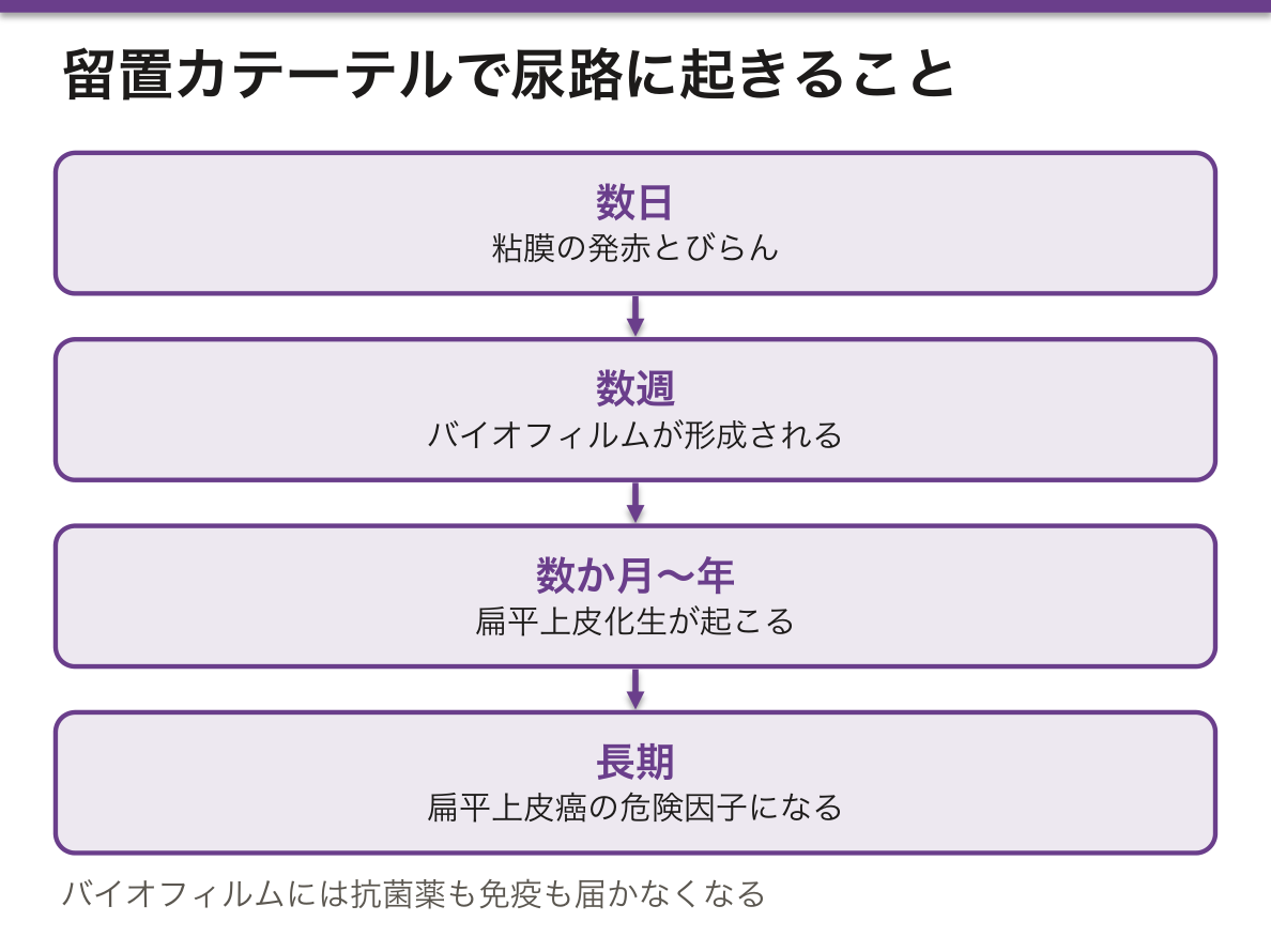
尿路上皮は異物が触れ続けることを想定していない。カテーテルを長く留置すると、組織が段階的に変わっていく。
- 数日——粘膜の発赤とびらん。機械的な刺激による反応性の変化
- 数週——バイオフィルムが形成される。細菌が多糖の膜を作って表面に定着し、抗菌薬も免疫も届かなくなる
- 数か月〜年——扁平上皮化生が起こる。慢性の刺激に対して、より丈夫な上皮へ置き換わる
⚠️ 長期の留置は、扁平上皮癌の危険因子になる。脊髄損傷で長年カテーテルを留置している患者で報告があり、膀胱癌の中では稀な扁平上皮癌が起こる——化生した上皮を出発点にしているためである。
カテーテル関連尿路感染は、避けられない部分と避けられる部分がある。
避けられない——留置している限り細菌尿は必ず起こる。1 日あたり 3〜10% の割合で増え、1 か月でほぼ全例が細菌尿になる。これは感染症ではない。
| 無症候性細菌尿 | 菌はいるが症状が無い | 治療しない。抗菌薬を出すと耐性菌を選ぶだけ。例外は妊婦と、泌尿器の手術前 |
| カテーテル関連尿路感染 | 発熱・側腹部痛・せん妄など | 治療する。可能ならカテーテルを入れ替えてから培養を採る |
最も効果のある対策は「入れないこと・早く抜くこと」である。毎日「まだ必要か」を確認するという単純な運用で、感染が大きく減ることが示されている。尿量測定や失禁の管理だけを理由に留置しない。
結石も問題になる。ウレアーゼ産生菌(Proteus など)が尿素を分解してアンモニアを出し、尿がアルカリ性になってリン酸マグネシウムアンモニウム結石ができる。カテーテルの内腔を塞ぎ、感染の巣になる——結石と感染が互いを維持するので、結石を取り除かないと感染は治らない。
入院患者の発熱で「尿培養が陽性だから尿路感染」と決めない。カテーテルが入っていれば、菌がいるのは当たり前である。症状で判断する。
English version
Core
The urothelium is not designed for a foreign body to remain against it. With a long-term indwelling catheter the tissue changes in stages.
- Days — redness and erosion of the mucosa: a reactive change to mechanical irritation.
- Weeks — a biofilm forms: bacteria settle on the surface within a polysaccharide film, beyond the reach of antibiotics and of the immune system.
- Months to years — squamous metaplasia: replacement by a tougher epithelium in response to chronic irritation.
⚠️ Long-term catheterisation is a risk factor for squamous carcinoma. It is reported in patients with spinal cord injury catheterised for many years, giving the squamous carcinoma that is rare among bladder cancers — because it starts from the metaplastic epithelium.
Going further
Catheter-associated urinary infection has an unavoidable part and an avoidable part.
Unavoidable — while the catheter is in, bacteriuria is inevitable. It accrues at 3–10 per cent per day, and by a month virtually every patient has bacteriuria. This is not an infection.
| What it is | What to do | |
|---|---|---|
| Asymptomatic bacteriuria | Bacteria present, no symptoms | Do not treat. An antibiotic only selects resistance. The exceptions are pregnancy and before urological surgery |
| Catheter-associated urinary tract infection | Fever, flank pain, delirium | Treat. Where possible change the catheter before taking the culture |
The most effective measure is not to insert one, and to remove it early. The simple practice of asking every day whether it is still needed has been shown to reduce infection greatly. Do not catheterise merely to measure urine output or to manage incontinence.
Stones are also a problem. Urease-producing organisms (Proteus and others) split urea to ammonia, the urine becomes alkaline and struvite stones form. They block the lumen of the catheter and become a nidus of infection — stone and infection maintain each other, so the infection will not clear until the stone is removed.
In the clinic
In an inpatient with fever, do not conclude "the urine culture is positive, therefore urinary infection". With a catheter in place, bacteria are to be expected. Judge by the symptoms.
まとめ
| 腎盂と尿管 — 3層の管 | 粘膜・筋層・外膜の3層。粘膜下組織は無い。 |
|---|---|
| 膀胱と尿道 — 伸びる袋と、変わる管 | 膀胱の尿路上皮は、空虚時6〜8層で表層に傘細胞、充満時は2〜3層に伸びて平らになる。 |
| 尿路上皮 — 伸び縮みし、尿を通さない特殊な上皮 | 腎杯から尿道の一部までを覆う移行上皮〈尿路上皮〉。 |
| 膀胱三角 — 尿が逆流しない仕組みは、走り方にある | 膀胱の後壁で、2つの尿管口と内尿道口を結ぶ三角形の領域。 |
| 男性尿道 — 3つの部分で、上皮も走行も変わる | 長さ約 16〜20 cm。3部に分ける。 |
| 尿細胞診 — 剥がれた細胞だけで、癌を見つけられるか | 尿は尿路の粘膜が剥がした細胞を、勝手に集めてくれている検体である。 |
| 尿膜管の遺残 — 膀胱の頂に、胎児の管の名残がある | 胎生期、膀胱の頂部と臍は尿膜管という管で繋がっている。 |
| カテーテルが入った尿路に、何が起きるか | 尿路上皮は異物が触れ続けることを想定していない。 |
このノートは医学生が授業の内容を自分の言葉でまとめたもので、診療の判断に使うものではありません。誤りに気づいたらお知らせください。