口腔は消化管でありながら、皮膚に近い作りをしている。重層扁平上皮で覆われ、こすれる場所は角化する——歯肉と硬口蓋が角化し、頬粘膜と口腔底は角化しない。
この「角化するかどうか」が、病変の見え方を決める——「白く見える病変は、角化が起きている場所の話になる」。
舌は、乳頭の形と味蕾の有無で4種類に分かれる。この見分けは標本でできるようにしておく。
歯は体の中で最も硬い組織を含み、しかも一部は再生しない。エナメル質を作る細胞は、萌出とともに失われる——「一度削れたエナメル質は、生涯戻らない」。
- 口腔粘膜と舌乳頭
- 味蕾 — 化学を電気に変える樽
- 口唇と口蓋 — 皮膚と粘膜が入れ替わる場所
- 舌の筋 — 内舌筋が形を変え、外舌筋が位置を動かす
- 舌の表面は、全身の状態を映している
口腔粘膜と舌乳頭Lingua
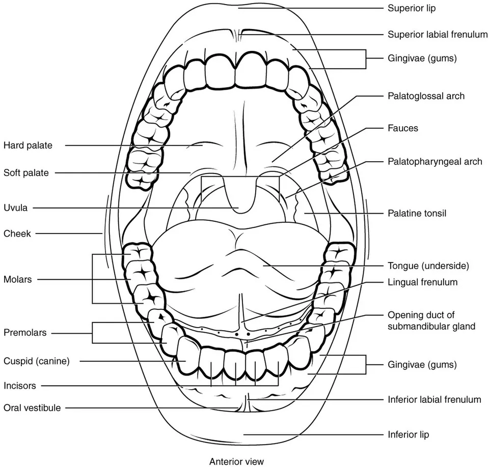
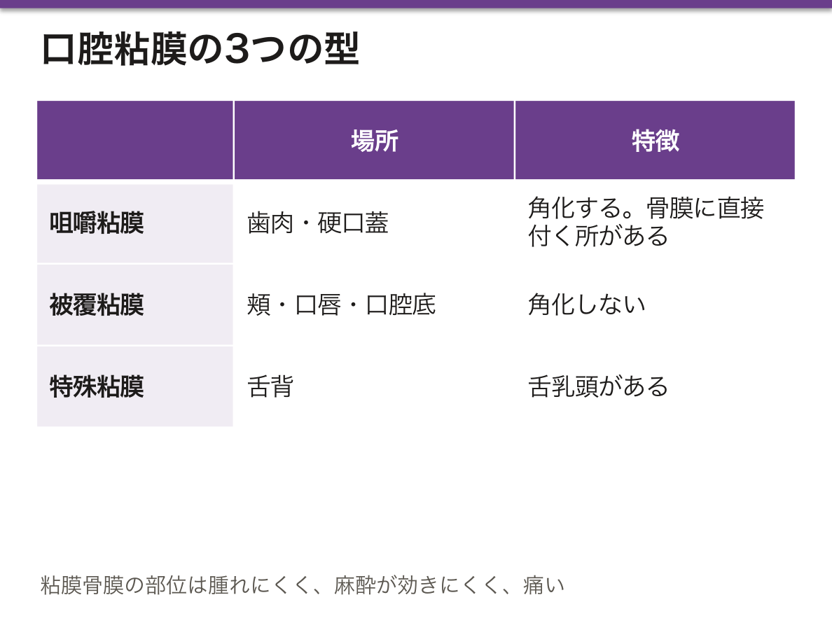
口腔粘膜の3型——咀嚼粘膜(歯肉・硬口蓋、角化する)・被覆粘膜(頬・口唇・口腔底、角化しない)・特殊粘膜(舌背)。
粘膜下組織を欠く場所がある——歯肉と硬口蓋の一部は骨膜に直接付く(粘膜骨膜)。だから腫れにくく、麻酔が効きにくく、痛い。
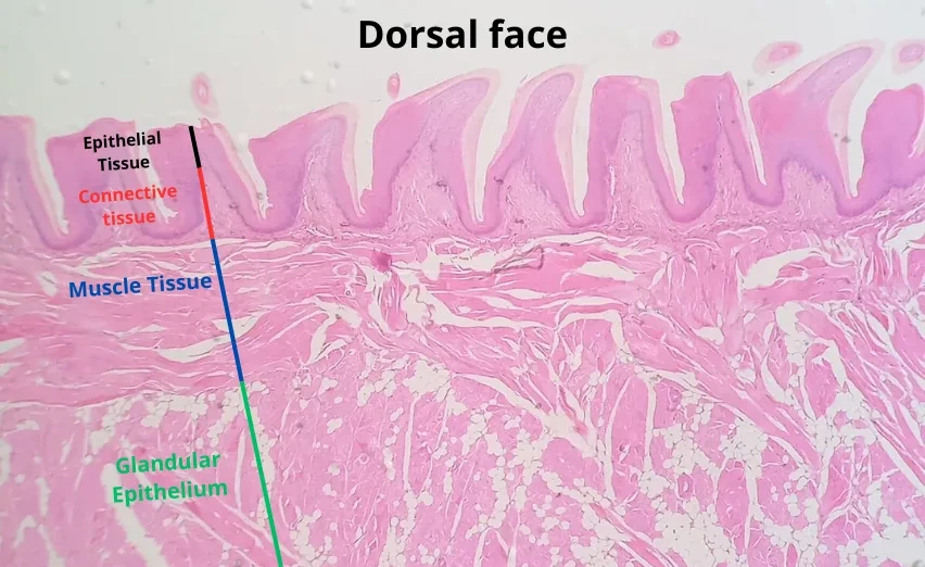
舌乳頭の4種。
| 乳頭 | 形 | 味蕾 | 分布 |
|---|---|---|---|
| 糸状乳頭 | 細く尖る・角化する | 無い | 舌背全体(最多) |
| 茸状乳頭 | キノコ状・赤い | あり | 舌尖と側縁 |
| 有郭乳頭 | 溝に囲まれる・大きい | 多い | 分界溝の前(8〜12個) |
| 葉状乳頭 | ひだ状 | あり | 舌の後外側 |
Ebner 腺——有郭乳頭の溝に開く純漿液腺。溝を洗って味を更新する。
糸状乳頭に味蕾が無いことが、この表の要点になる。最も数が多い乳頭が、味には関わっていない——「数と役割は一致しない」。
糸状乳頭が角化して伸びると、舌が白く見える(舌苔)。逆に萎縮すると赤く平らになる(悪性貧血の章)——「Hunter 舌炎の赤い舌は、乳頭が失われた像になる」。
だから鉄欠乏とビタミンB12欠乏で、同じように舌が滑らかになる(鉄欠乏性貧血の章)——「舌を見ることが、血算の前の手がかりになる」。
Ebner 腺が溝を洗う仕組みは、味覚が持続しない理由になっている。味物質が留まり続ければ、次の味が分からない(味覚の章)——「感覚は、消してから次を受け取る」。
口腔底と頬粘膜が角化しないことが、薬の吸収経路になる。舌下錠が速く効く(投与経路の章)——「角化していない粘膜が、初回通過効果を避ける入口になる」。
白板症は、こすっても取れない白色病変で、前癌病変になる。取れる白色は、多くはカンジダ症(口腔粘膜疾患の章)——「拭って取れるかどうかが、最初の鑑別になる」。
角化するかどうかで、病変の見え方が変わる。
白板症 — 擦っても取れない白色病変。過角化による。前癌病変であり、異形成の程度で経過観察か切除かを決める。紅板症のほうが悪性化率が高い。
口腔カンジダ症は擦ると取れる——白板症との決定的な違い。吸入ステロイド後のうがい不足・義歯・免疫低下・抗菌薬が誘因。成人で誘因なく出たら HIV や糖尿病を疑う。
粘膜下組織を欠く歯肉と硬口蓋は骨膜に直接付いている(粘膜骨膜)ので腫れても逃げ場が無く、強く痛む。局所麻酔の注入も痛い。
Koplik 斑(麻疹)は頬粘膜に出る。発疹より先に現れるので早期診断の手がかりになる。
English version
Core
Three types of oral mucosa — masticatory (gingiva, hard palate; keratinised), lining (cheek, lip, floor of mouth; non-keratinised) and specialised (dorsum of tongue).
In places the submucosa is absent — parts of the gingiva and hard palate attach directly to periosteum (mucoperiosteum). So they swell poorly, take anaesthetic poorly, and hurt.
Four kinds of lingual papilla.
| Papilla | Shape | Taste buds | Distribution |
|---|---|---|---|
| Filiform | Slender, pointed, keratinised | None | The whole dorsum (most numerous) |
| Fungiform | Mushroom-shaped, red | Present | Tip and lateral border |
| Circumvallate | Surrounded by a trench, large | Many | In front of the sulcus terminalis (8–12) |
| Foliate | Folded | Present | Posterolateral tongue |
Von Ebner’s glands — purely serous glands opening into the trench of the circumvallate papillae. They rinse the trench and refresh the taste.
Going further
That the filiform papillae have no taste buds is the point of the table — the most numerous papilla plays no part in taste: number and role do not coincide.
When the filiform papillae keratinise and lengthen, the tongue looks white (coating). When they atrophy it becomes red and flat — the red tongue of Hunter’s glossitis is a picture of papillae that have been lost. So iron deficiency and B12 deficiency both smooth the tongue in the same way — looking at the tongue is a clue that comes before the blood count.
The rinsing of the trench by von Ebner’s glands is why a taste does not persist. If the tastant stayed, the next taste could not be told — a sense erases before it receives the next.
That the floor of the mouth and buccal mucosa do not keratinise makes them a route of drug absorption. A sublingual tablet acts quickly — non-keratinised mucosa as the entrance that avoids first-pass metabolism.
Leukoplakia is a white lesion that cannot be rubbed off, and is premalignant. White that can be wiped away is usually candidiasis — whether it wipes off is the first differential.
In the clinic
Whether it keratinises changes how a lesion looks.
Leukoplakia — a white lesion that cannot be rubbed off, due to hyperkeratosis. It is premalignant, and the degree of dysplasia decides between observation and excision. Erythroplakia has the higher rate of malignant change.
Oral candidiasis rubs off — the decisive difference from leukoplakia. Precipitants are failure to rinse after inhaled steroids, dentures, immunosuppression and antibiotics. Appearing in an adult without a precipitant, suspect HIV or diabetes.
The gingiva and hard palate, lacking submucosa, are attached directly to periosteum (mucoperiosteum), so swelling has nowhere to go and hurts severely. Injection of local anaesthetic there is also painful.
Koplik’s spots (measles) appear on the buccal mucosa. They precede the rash and are a clue to early diagnosis.
味蕾 — 化学を電気に変える樽Gemma gustatoria
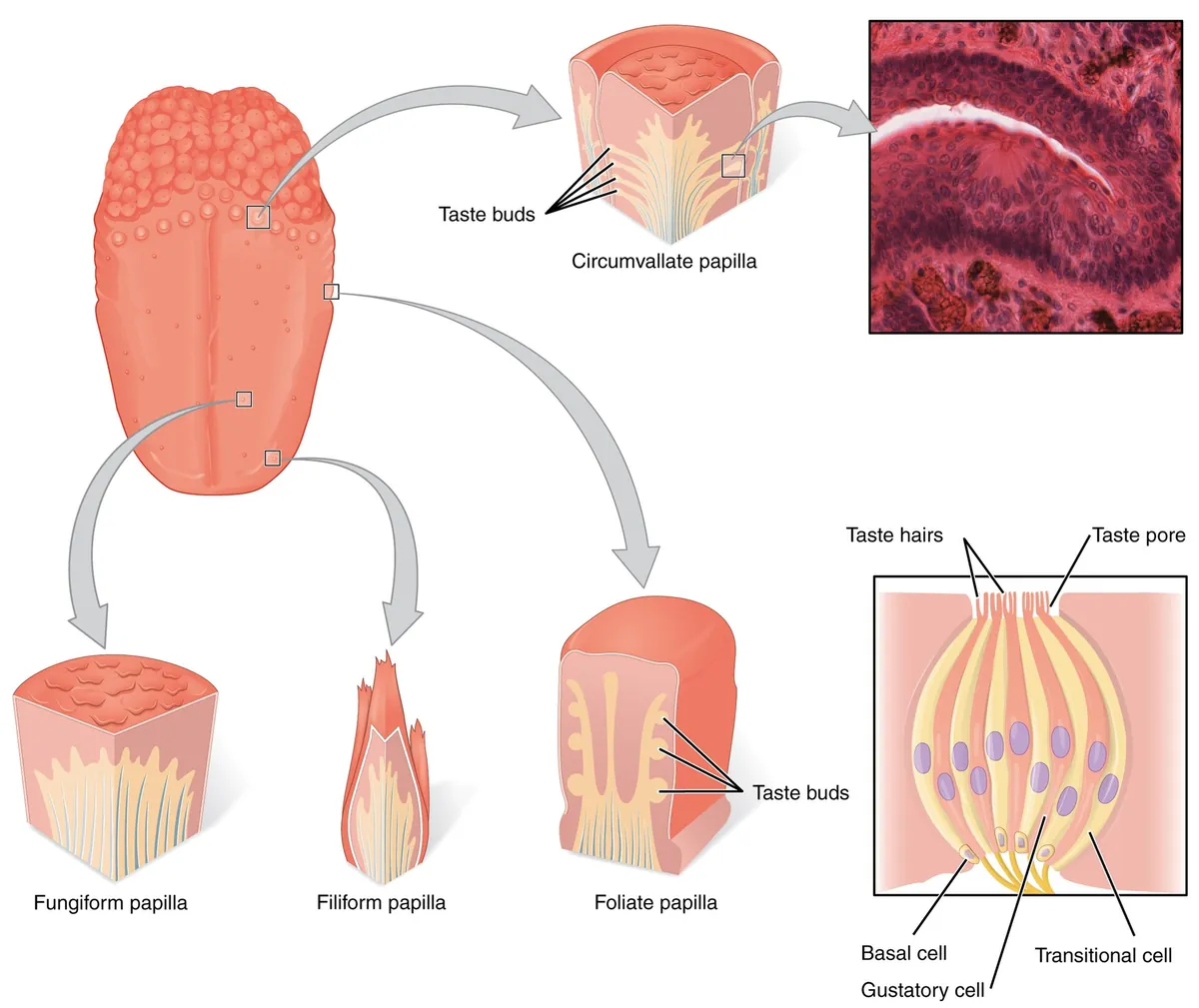
上皮内に埋まった樽状の細胞集団。頂端の味孔が口腔に開く。
- 味細胞(I〜III型)——寿命は約10日で入れ替わる
- 基底細胞——供給源になる幹細胞
基本味は5つ——甘味・塩味・酸味・苦味・うま味。辛味は味覚ではなく三叉神経の痛覚になる。
受容の仕方が2群に分かれる。
- 甘味・苦味・うま味——Gタンパク質共役型受容体
- 塩味・酸味——イオンチャネル
神経支配——舌前2/3は顔面神経(鼓索神経)、後1/3は舌咽神経、喉頭蓋は迷走神経。
味の地図(舌の場所で味が決まる)は誤りとされている。
味覚の神経支配が3つに分かれることが、障害の部位診断に直結する。前2/3の味覚脱失+同側の顔面神経麻痺=鼓索神経を含む高さの病変(顔面神経麻痺の章)——「味が分かるかどうかを聞くだけで、麻痺の高さが絞れる」。
だから Bell 麻痺の診察では、必ず味覚と聴覚過敏と涙を確認する。鼓索神経・アブミ骨筋神経・大錐体神経の順に分岐する——「枝が出る順番が、そのまま診察項目の順番になる」。
味細胞が10日で入れ替わることが、味覚障害の回復と悪化の速さを説明する。亜鉛欠乏で新生が滞る(亜鉛の章)——「入れ替わる細胞の病気は、材料の不足に弱い」。
薬剤性味覚障害も多い。降圧薬・抗菌薬・抗腫瘍薬・利尿薬(薬の副作用の章)——「訴えが漠然としているので、薬歴を先に見る」。
辛味が痛覚であることは、臨床でそのまま使われる。カプサイシンが TRPV1 を介して痛覚線維に作用し、繰り返すと感受性が落ちる(局所鎮痛の章)——「辛い成分が、鎮痛の貼付薬になっている」。
そして高齢者では、苦味の閾値が最も上がりにくい。甘味と塩味が先に鈍るので、味付けが濃くなる(高齢者の栄養の章)——「減塩指導が難しい理由の1つが、感覚の側にある」。
味細胞は約10日で入れ替わるので、味覚障害は回復しうる——ただし供給源と栄養が保たれていればである。
亜鉛欠乏が味覚障害の代表的な原因である。味細胞の turnover に亜鉛が要る。高齢者・偏食・薬剤性(利尿薬・降圧薬・抗癌薬)で起こる。薬剤性が最も多いので服薬歴を必ず確認する。
味覚の神経は3本に分かれる——前2/3が鼓索神経(VII)、後1/3が舌咽神経(IX)、喉頭蓋周辺が迷走神経(X)。障害部位で「どこの味が分からないか」が変わる。Bell 麻痺で味覚が落ちるのは鼓索神経を巻き込むからである。
放射線治療(頭頸部)では味蕾と唾液腺の両方が障害されるので味覚障害と口腔乾燥が重なり、栄養摂取が難しくなる。
English version
Core
A barrel-shaped cluster of cells embedded in the epithelium, opening to the mouth by an apical taste pore.
- Taste cells (types I–III) — replaced about every 10 days.
- Basal cells — the stem cells that supply them.
Five basic tastes — sweet, salt, sour, bitter, umami. Pungency is not a taste but trigeminal pain.
Reception falls into two groups.
- Sweet, bitter, umami — G protein-coupled receptors
- Salt, sour — ion channels
Innervation — anterior two thirds by the facial nerve (chorda tympani), posterior third by the glossopharyngeal, epiglottis by the vagus. The taste map (a taste assigned to a region of the tongue) is regarded as wrong.
Going further
That the innervation of taste divides into three leads straight to localisation of a lesion. Loss of taste over the anterior two thirds with an ipsilateral facial palsy places the lesion at a level that includes the chorda tympani — simply asking whether taste is intact narrows the height of the palsy.
So in examining Bell’s palsy, always check taste, hyperacusis and tearing. The branches leave in the order chorda tympani, nerve to stapedius, greater petrosal — the order the branches leave becomes the order of the examination.
That taste cells turn over in 10 days explains how quickly taste disturbance both develops and recovers. Zinc deficiency stalls their renewal — a disease of a cell that turns over is vulnerable to a shortage of materials.
Drug-induced taste disturbance is also common — antihypertensives, antibiotics, cytotoxics, diuretics — the complaint is vague, so look at the drug history first.
That pungency is pain is used directly in the clinic: capsaicin acts on pain fibres through TRPV1 and, repeated, reduces their sensitivity — a pungent substance has become an analgesic patch.
And in the elderly the threshold for bitter rises least. Sweet and salt dull first, so the seasoning grows stronger — one reason advice to reduce salt is difficult lies on the side of the senses.
In the clinic
Taste cells are replaced about every 10 days, so taste disturbance can recover — provided the source and the nutrition are intact.
Zinc deficiency is the representative cause, zinc being required for the turnover of the taste cells. It occurs in the elderly, in restricted diets, and from drugs (diuretics, antihypertensives, cytotoxics). Drugs are the commonest cause, so always check the medication history.
The nerves of taste divide into three — anterior two thirds chorda tympani (VII), posterior third glossopharyngeal (IX), around the epiglottis vagus (X). The site of the lesion changes which taste is lost. Taste falls in Bell’s palsy because the chorda tympani is involved.
Head and neck radiotherapy damages both taste buds and salivary glands, so taste disturbance and dry mouth are superimposed and nutrition becomes difficult.
口唇と口蓋 — 皮膚と粘膜が入れ替わる場所Labium oris
口唇は3つの区域が連続している。
| 区域 | 上皮 | 付属器 |
|---|---|---|
| 皮膚部 | 角化重層扁平 | 毛・脂腺・汗腺がある |
| 赤唇(移行部) | 薄く角化する重層扁平 | どれも無い——だから乾く |
| 粘膜部 | 非角化重層扁平 | 小唾液腺(口唇腺) |
赤唇が赤く見えるのは、上皮が薄く、真皮乳頭の毛細血管が透けるため。
口蓋は前後で作りが違う——硬口蓋は骨に密着した咀嚼粘膜(角化する)、軟口蓋は可動性で、口腔側が重層扁平・鼻腔側が多列線毛円柱上皮。
赤唇に付属器が無いことが、そのまま臨床の問題になる。皮脂も汗も出ないので、水分を保てず荒れる——「唇だけが乾く理由は、腺が無いという構造にある」。
そして紫外線を遮る角層も薄い。下口唇は日光角化症と扁平上皮癌の好発部位になる(皮膚の腫瘍の章)——「上を向いている面が、最も光を浴びる」。
軟口蓋で上皮が2種類になることが、呼吸と嚥下の切り替えを支えている。口腔側はこすれるので重層扁平、鼻腔側は空気を運ぶので線毛上皮になる(上皮組織の章)——「1枚の膜の裏表で、上皮が違う」。
口蓋裂は、この境界の癒合が起きなかった結果になる。一次口蓋(切歯孔より前)と二次口蓋で由来が違うので、唇裂と口蓋裂は別々にも一緒にも起こる(咽頭弓の章)——「切歯孔が、2つの発生の継ぎ目になる」。
Fordyce 斑——頬粘膜や赤唇に見える黄白色の粒。毛が無いのに存在する異所性の脂腺で、正常所見になる——「病気に見える正常を1つ知っておくと、無用な生検を減らせる」。
赤唇には付属器が無い。汗腺も脂腺も無いので乾く——口唇が荒れやすいのはこの構造による。舐めると悪化する(舐性口唇炎)。
日光に当たりやすく、角層が薄いので下口唇は日光角化症と扁平上皮癌の好発部位である。上口唇より下口唇に多いのは日光の当たり方の違いによる。
チアノーゼと貧血を見る場所でもある。角層が薄く血管が透けて見えるからで、口唇・眼瞼結膜・爪床を診るのはこの理由である。
口唇ヘルペスは三叉神経節に潜伏したウイルスの再活性化でいつも同じ場所(皮膚と赤唇の境)に出る。
口唇口蓋裂は哺乳・言語・中耳炎に影響する。
English version
Core
The lip is three continuous zones.
| Zone | Epithelium | Appendages |
|---|---|---|
| Skin | Keratinised stratified squamous | Hair, sebaceous and sweat glands |
| Vermilion (transitional) | Thin, slightly keratinised stratified squamous | None at all — hence it dries |
| Mucosa | Non-keratinised stratified squamous | Minor salivary (labial) glands |
The vermilion looks red because the epithelium is thin and the capillaries of the dermal papillae show through.
The palate is built differently front and back — the hard palate is masticatory mucosa bound to bone (keratinised); the soft palate is mobile, stratified squamous on the oral side and pseudostratified ciliated columnar on the nasal side.
Going further
That the vermilion has no appendages becomes directly a clinical problem. Producing neither sebum nor sweat, it cannot hold water and becomes chapped — the reason the lips alone dry out is the structural absence of glands.
And the horny layer that screens ultraviolet is thin, so the lower lip is a site of predilection for actinic keratosis and squamous cell carcinoma — the surface that faces upward receives the most light.
That the soft palate carries two epithelia supports the switch between breathing and swallowing: the oral side is rubbed, hence stratified squamous; the nasal side carries air, hence ciliated — one membrane with a different epithelium front and back.
Cleft palate is the failure of that boundary to fuse. The primary palate (in front of the incisive foramen) and the secondary palate differ in origin, so cleft lip and cleft palate occur separately or together — the incisive foramen is the seam between two developments.
Fordyce spots — yellowish-white granules on the buccal mucosa and vermilion, ectopic sebaceous glands present without hair, and a normal finding — knowing one normality that looks like a disease saves needless biopsies.
In the clinic
The vermilion has no appendages. With neither sweat nor sebaceous glands it dries — this structure is why the lips chap so readily. Licking makes it worse (lick eczema).
Exposed to sunlight and with a thin horny layer, the lower lip is a site of predilection for actinic keratosis and squamous cell carcinoma. It is commoner on the lower than the upper lip because of the way the light falls.
It is also a place to look for cyanosis and anaemia, the horny layer being thin and the vessels visible — the same reason the lips, palpebral conjunctiva and nail beds are examined.
Herpes labialis is reactivation of virus latent in the trigeminal ganglion, appearing always at the same place — the border of skin and vermilion.
Cleft lip and palate affect feeding, speech and otitis media.
舌の筋 — 内舌筋が形を変え、外舌筋が位置を動かすMusculus genioglossus
| 内舌筋 | 外舌筋 | |
|---|---|---|
| 付着 | 舌の中で完結する | 骨から起こって舌へ入る |
| 働き | 舌の形を変える | 舌の位置を動かす |
| 例 | 上縦筋・下縦筋・横筋・垂直筋 | オトガイ舌筋(前へ出す)・舌骨舌筋(下げる)・茎突舌筋(後上方へ引く)・口蓋舌筋 |
神経支配——口蓋舌筋(迷走神経=咽頭神経叢)を除き、すべて舌下神経。
組織学的には骨格筋だが、線維が3方向に交差して走る——この立体的な配置が、舌の自在な変形を可能にしている。
粘膜と筋のあいだに粘膜下組織が乏しく、粘膜が筋に固く付いている。
オトガイ舌筋が「舌を前へ出す」筋であることが、いくつもの臨床に直結する。片側が麻痺すると、健側に押されて舌が患側へ偏る(脳神経の章)——「舌を出させる診察で、偏る側が障害側になる」。
意識が落ちると、この筋の緊張が失われて舌根が沈下する(気道確保の章)——「頭部後屈・下顎挙上は、オトガイ舌筋の付着部ごと前へ引き出す手技になる」。
閉塞性睡眠時無呼吸も、同じ筋の緊張低下が関わる(眠るときの気道の章)——「舌下神経刺激療法という治療が、この機序から生まれた」。
粘膜下組織が乏しいことは、舌癌の性質に現れる(頭頸部の腫瘍の章)——「早くから筋層へ浸潤し、リンパ節転移が早い」。
舌の運動が言語と嚥下の両方を担うので、切除の範囲が生活を決める(声と言葉のリハビリテーションの章)——「再建では、動くかどうかが形より重要になる」。
舌の運動は舌下神経(XII)が支配する——口蓋舌筋だけが迷走神経。
舌下神経麻痺では舌を出すと患側へ偏る。健側のオトガイ舌筋だけが前へ押し出すためで、「舌は悪いほうを向く」。核性・末梢性では患側の萎縮と線維束攣縮を伴う。
意識障害での舌根沈下はオトガイ舌筋の緊張が失われるために起こる。下顎挙上(jaw thrust)で気道が開くのは下顎に付く舌の筋を前へ引くからで、気道確保の手技は舌の筋の解剖そのものである。
閉塞性睡眠時無呼吸でも上気道の筋緊張の低下が関わる。
巨舌 — 先端巨大症・甲状腺機能低下症・アミロイドーシス・Down 症候群・Beckwith–Wiedemann 症候群。
English version
Core
| Intrinsic muscles | Extrinsic muscles | |
|---|---|---|
| Attachment | Contained within the tongue | Arise from bone and enter the tongue |
| Action | Change the shape of the tongue | Move the position of the tongue |
| Examples | Superior and inferior longitudinal, transverse, vertical | Genioglossus (protrudes), hyoglossus (depresses), styloglossus (retracts and elevates), palatoglossus |
Innervation — all by the hypoglossal nerve except palatoglossus (vagus, via the pharyngeal plexus).
Histologically skeletal muscle, but the fibres run crossing in three directions — this three-dimensional arrangement is what allows the tongue to deform at will. There is little submucosa, and the mucosa is bound firmly to the muscle.
Going further
That genioglossus is the muscle that protrudes the tongue leads to several clinical points at once. If one side is paralysed the tongue is pushed by the sound side and deviates towards the lesion — on asking the patient to put out the tongue, the side it deviates to is the damaged side.
When consciousness falls, the tone of this muscle is lost and the tongue base drops back — head tilt and jaw thrust are manoeuvres that pull the attachment of genioglossus, and with it the tongue, forward.
Obstructive sleep apnoea involves loss of tone in the same muscle — the treatment by hypoglossal nerve stimulation was born from this mechanism.
The scarcity of submucosa shows in the behaviour of tongue cancer — it invades the muscle early and metastasises early to lymph nodes.
Because the movement of the tongue serves both speech and swallowing, the extent of resection decides the patient’s life — in reconstruction, whether it moves matters more than its shape.
In the clinic
The movement of the tongue is supplied by the hypoglossal nerve (XII) — palatoglossus alone by the vagus.
In hypoglossal palsy the protruded tongue deviates to the affected side, because only the sound genioglossus pushes forward: the tongue points to the bad side. Nuclear and peripheral lesions add atrophy and fasciculation on that side.
Falling back of the tongue in impaired consciousness is loss of genioglossus tone. Jaw thrust opens the airway because it pulls forward the tongue muscles attached to the mandible — the technique of securing an airway is the anatomy of the lingual muscles itself. Loss of upper airway muscle tone also underlies obstructive sleep apnoea.
Macroglossia — acromegaly, hypothyroidism, amyloidosis, Down syndrome, Beckwith–Wiedemann syndrome.
舌の表面は、全身の状態を映している
舌背は糸状乳頭が密に生えていて白っぽく見える。この乳頭は角化して伸び、こすれて摩耗するという入れ替わりを続けている。入れ替わりが速い上皮なので、全身の変化が早く現れる。
| 舌乳頭の萎縮(平滑舌) | 赤くつるつるになり、痛む | 鉄欠乏・ビタミン B12 欠乏・葉酸欠乏。B12 欠乏のものを Hunter 舌炎と呼ぶ |
| イチゴ舌 | 糸状乳頭が脱落し、茸状乳頭が赤く目立つ | 猩紅熱・川崎病 |
| 地図状舌 | 脱落した領域が地図のように移動する | 良性。乾癬との関連が指摘される |
| 黒毛舌 | 糸状乳頭が伸びて着色する | 抗菌薬・喫煙・口腔衛生。無害だが見た目で驚かれる |
| 白い苔で、こすると剥がれる | 下は赤くただれる | 口腔カンジダ症。吸入ステロイド・免疫抑制・糖尿病を考える |
| 白斑で、こすっても取れない | 境界が明瞭 | 白板症。前癌病変として扱う |
「こすって取れるかどうか」が、最初の分岐になる。取れれば感染(カンジダ)、取れなければ上皮そのものの変化(白板症)。白板症は生検が必要で、異形成の程度によって扱いが変わる。
⚠️ 舌癌は、見える場所にできるのに進行して見つかることがある。
- 好発部位は舌の側縁——舌背ではない。舌を出させて見るだけでは側縁が見えないので、ガーゼで舌をつまんで左右に引き出す
- 治らない潰瘍は 2 週間で疑う——口内炎は通常 1〜2 週で治る。治らない、硬い、痛みが乏しいものは癌を考える
- 危険因子は喫煙・飲酒。合併すると相乗的に上がる。慢性の機械的刺激(不適合な義歯・尖った歯)も関わる
舌の所見は費用がかからず、器具も要らない。貧血・ビタミン欠乏・感染・免疫抑制・悪性腫瘍が、同じ 10 秒の診察で拾える——身体診察の効率という点で、極めて割のよい部位である。
舌を診るときは、必ず側縁と裏側まで見る。「あーん」と言わせて舌背を見ただけでは、癌の好発部位を見ていない。
English version
Core
The dorsum of the tongue looks whitish from the dense filiform papillae. These keratinise and lengthen, and are worn away by friction — a continuing turnover. Being a rapidly renewing epithelium, systemic change appears there early.
| Finding | Appearance | Meaning |
|---|---|---|
| Atrophic (smooth) tongue | Red, glossy, painful | Iron, vitamin B12 or folate deficiency. The B12 form is Hunter’s glossitis |
| Strawberry tongue | Filiform papillae shed, fungiform papillae red and prominent | Scarlet fever, Kawasaki disease |
| Geographic tongue | Denuded areas migrating like a map | Benign; an association with psoriasis is reported |
| Black hairy tongue | Filiform papillae elongated and pigmented | Antibiotics, smoking, oral hygiene. Harmless but alarming to look at |
| White coating that rubs off | Raw and red underneath | Oral candidiasis. Consider inhaled steroids, immunosuppression, diabetes |
| White patch that will not rub off | Sharply defined | Leukoplakia — treat as premalignant |
Going further
Whether it rubs off is the first branch point. If it comes off, infection (candida); if it does not, a change in the epithelium itself (leukoplakia). Leukoplakia requires biopsy, and the degree of dysplasia changes the management.
⚠️ Tongue cancer can be found late despite arising where it can be seen.
- The site of predilection is the lateral border of the tongue — not the dorsum. Asking the patient to put out the tongue does not show the border: grasp it with gauze and draw it to each side.
- Suspect an ulcer that has not healed in two weeks — aphthous ulcers heal in one to two. One that does not heal, is indurated, and hurts little, suggests cancer.
- The risk factors are smoking and alcohol, and together they multiply. Chronic mechanical irritation (an ill-fitting denture, a sharp tooth) also contributes.
Examining the tongue costs nothing and needs no instrument. Anaemia, vitamin deficiency, infection, immunosuppression and malignancy can all be picked up in the same ten seconds — an extraordinarily good return for the effort of a physical examination.
In the clinic
When you look at the tongue, always see the lateral border and the underside. Saying "open wide" and looking at the dorsum does not show the site where cancer occurs.
まとめ
| 口腔粘膜と舌乳頭 | 口腔粘膜の3型——咀嚼粘膜(歯肉・硬口蓋、角化する)・被覆粘膜(頬・口唇・口腔底、角化しない)・特殊粘膜(舌背)。 |
|---|---|
| 味蕾 — 化学を電気に変える樽 | 上皮内に埋まった樽状の細胞集団。頂端の味孔が口腔に開く。 |
| 口唇と口蓋 — 皮膚と粘膜が入れ替わる場所 | 口唇は3つの区域が連続している。区域上皮付属器皮膚部角化重層扁平毛・脂腺・汗腺がある赤唇(移行部)薄く角化する重層扁平どれも無い——だから乾く粘膜部非角化重層扁平小唾液腺(口唇腺)… |
| 舌の筋 — 内舌筋が形を変え、外舌筋が位置を動かす | 内舌筋外舌筋付着舌の中で完結する骨から起こって舌へ入る働き舌の形を変える舌の位置を動かす例上縦筋・下縦筋・横筋・垂直筋オトガイ舌筋(前へ出す)・舌骨舌筋(下げる)・茎突舌筋(後上方… |
| 舌の表面は、全身の状態を映している | 舌背は糸状乳頭が密に生えていて白っぽく見える。 |
このノートは医学生が授業の内容を自分の言葉でまとめたもので、診療の判断に使うものではありません。誤りに気づいたらお知らせください。