腺はすべて上皮から出芽してできる。途中で導管を残せば外分泌腺、切り離せば内分泌腺——それだけの違いになる。
だから内分泌腺の周りには毛細血管が密に走り、外分泌腺には管が付く——「顕微鏡で管が見えるかどうかが、最初の分岐になる」。
外分泌腺の記述は、3つの軸の掛け算で決まる。
- 終末部の形——管状・胞状(腺房)・管状胞状
- 分泌物の性質——漿液性(薄い・酵素)/粘液性(粘る・ムチン)/混合性
- 分泌の様式——メロクリン/アポクリン/ホロクリン
唾液腺は、この3軸を全部使って見分ける——耳下腺は純漿液性、舌下腺は粘液優位、顎下腺は混合性。
- 外分泌腺の分類と、漿液・粘液の見分け
- 3つの大唾液腺 — スライドで言い当てる
- 内分泌腺の共通の型 — 3つの並び方
- 分泌の様式 — 分泌物と一緒に、細胞の何を捨てるか
- 分泌周期 — 取り込み・合成・蓄積・放出・回復
- 腺の形 — 導管が枝分かれするか、終末部が丸いか
- 唾液 — 何が入っていて、誰が命令するか
- 筋上皮細胞 — 腺を外から絞る細胞
外分泌腺の分類と、漿液・粘液の見分けGlandula exocrina
漿液細胞——円錐形。核は丸く中央よりやや基底側。細胞質は好塩基性(粗面小胞体が多い)。頂端に好酸性の分泌顆粒。
粘液細胞——細胞質が明るく泡状に抜ける(ムチンが溶け出す)。核は扁平で基底部に押し付けられる。PAS 陽性。
漿粘液腺(混合腺)——粘液腺房の周りに漿液細胞が三日月状に付く。漿液半月(Gianuzzi 半月)。
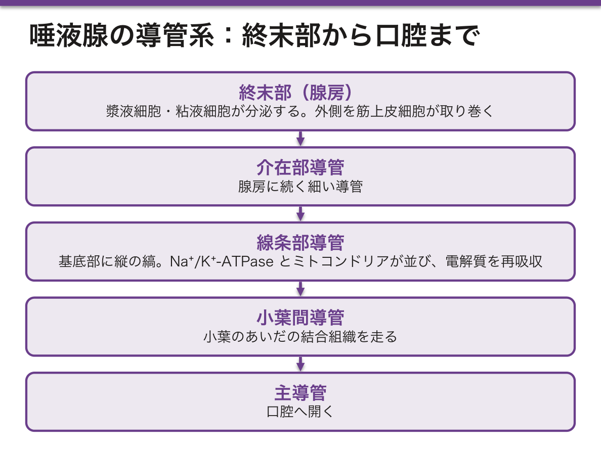
導管系——介在部導管 → 線条部導管 → 小葉間導管 → 主導管。
- 線条部導管——基底部に縦の縞が見える。Na⁺/K⁺-ATPase とミトコンドリアが並ぶ。電解質を再吸収する
筋上皮細胞——腺房と導管の外側、基底膜の内側にある。収縮して分泌物を押し出す。
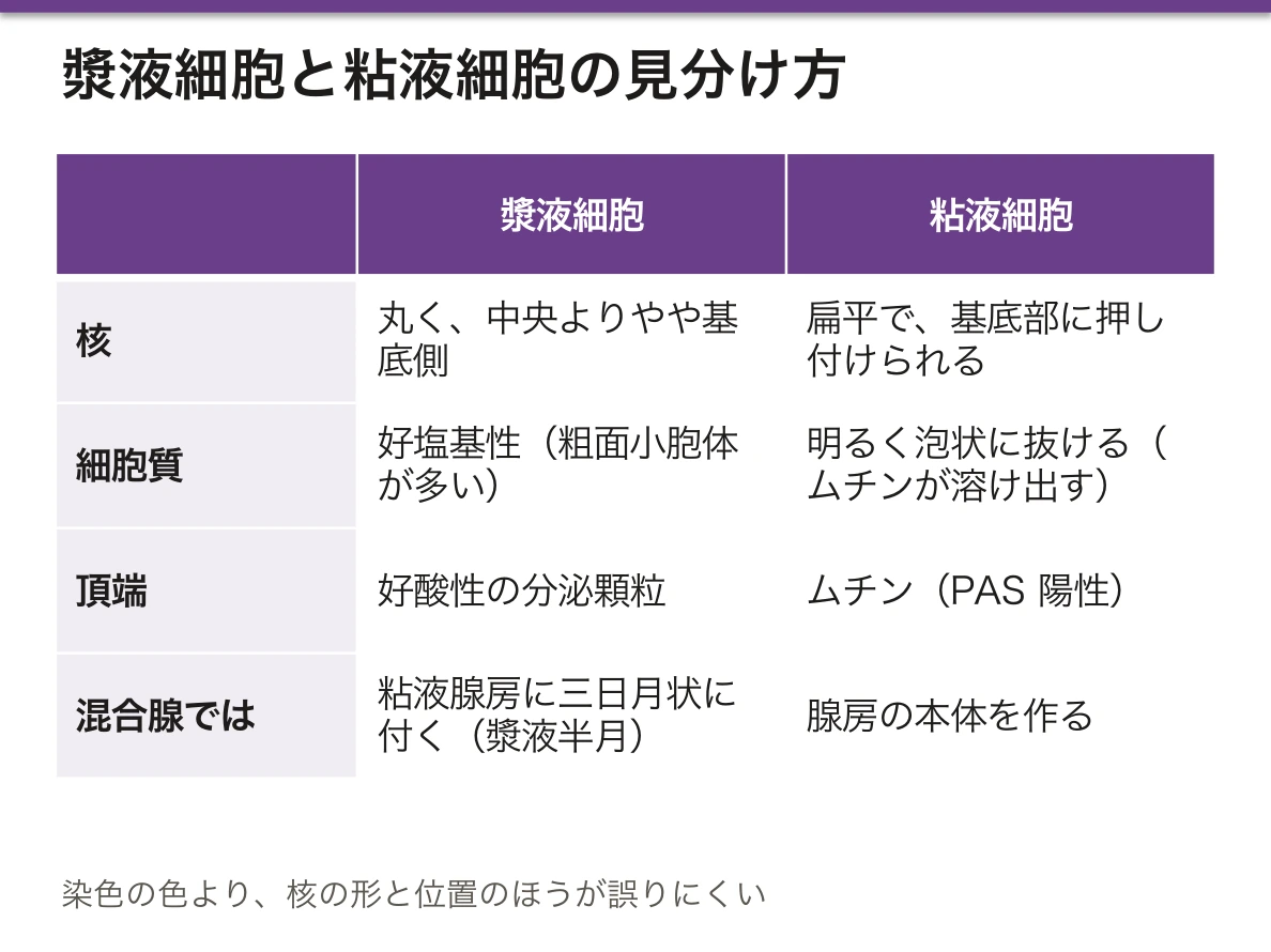
核の位置で漿液か粘液かを言うのが、標本で最も速い判別になる。丸い核が中央寄り=漿液、扁平な核が基底に押し付けられている=粘液——「染色の色より、核の形と位置のほうが誤りにくい」。
粘液細胞が明るく抜けて見えるのは、処理でムチンが失われるからになる(標本の作り方の章)——「空白が、あったものの跡になっている」。
線条部導管が電解質を再吸収するので、唾液は血漿より低張になる。流量が速いと再吸収が追いつかず、Na⁺ 濃度が上がる(唾液分泌の章)——「速く出た唾液は、しょっぱくなる」。
筋上皮細胞は、乳腺と汗腺と唾液腺に共通してある。だからこの3つに、筋上皮由来の腫瘍が共通して出る(唾液腺腫瘍の章)——「多形腺腫が上皮と間葉の両方の顔を持つ理由が、この細胞にある」。
そして筋上皮細胞の有無が、良性と悪性の判定に使われる。乳腺では、筋上皮層が保たれていれば浸潤ではない(乳腺病理の章)——「1層の細胞があるかどうかで、上皮内か浸潤かが決まる」。
嚢胞性線維症は、外分泌腺全体の病気になる。CFTR の異常で分泌物が濃くなり、膵管と気道と汗腺が同時に障害される(嚢胞性線維症の章)——「腺という括りが、そのまま病気の広がりになっている」。
漿液性か粘液性かで、腫れ方も腫瘍も変わる。
唾液の性状が変われば、齲蝕と口腔カンジダ症が増える。Sjögren 症候群では腺房が破壊されてリンパ球が浸潤する(口唇小唾液腺生検で診断)。乾燥性角結膜炎と口腔乾燥に加え、悪性リンパ腫(MALT)のリスクが上がる。
粘液が濃くなると導管が詰まる。嚢胞性線維症では膵管が詰まって膵は線維化・嚢胞化し、外分泌不全(脂肪便・脂溶性ビタミン欠乏)になる。気道では粘液が運べず反復感染——1つのチャネルの異常が、粘液を出すすべての腺を巻き込む。
粘液細胞の細胞質が明るく抜けて見えるのはムチンが処理中に溶け出すためで、証明したければ PAS かアルシアン青を追加する。
English version
Core
- Serous cell — conical; the nucleus is round and slightly basal. The cytoplasm is basophilic (abundant rough ER) with acidophilic secretory granules apically.
- Mucous cell — the cytoplasm is pale and foamy because the mucin dissolves out; the nucleus is flattened and pressed against the base. PAS positive.
- Seromucous (mixed) gland — serous cells cap a mucous acinus as a crescent: the serous demilune of Gianuzzi.
Duct system — intercalated duct → striated duct → interlobular duct → main duct. The striated duct shows vertical striations at the base, where Na⁺/K⁺-ATPase and mitochondria are arranged; it reabsorbs electrolytes.
Myoepithelial cells lie outside the acini and ducts but inside the basal lamina, and contract to expel the secretion.
Going further
Being able to call serous or mucous from the position of the nucleus is the fastest discrimination on a slide. A round nucleus towards the middle means serous; a flattened nucleus pressed against the base means mucous — the shape and position of the nucleus are less liable to error than the colour. The mucous cell looks pale because the mucin is lost during processing: the blank space is the trace of what was there.
Because the striated duct reabsorbs electrolytes, saliva is hypotonic to plasma. At high flow rates there is not enough time for reabsorption and the sodium concentration rises — saliva produced quickly tastes salty.
Myoepithelial cells are common to the mammary, sweat and salivary glands, and tumours derived from them therefore occur in all three — this cell is why the pleomorphic adenoma shows both epithelial and mesenchymal faces. Their presence or absence is used to judge benign from malignant: in the breast, if the myoepithelial layer is preserved the lesion is not invasive — whether one layer of cells is present decides in situ from invasive.
Cystic fibrosis is a disease of the exocrine glands as a class: the CFTR defect thickens the secretions and damages the pancreatic ducts, airways and sweat glands together — the category "gland" is itself the extent of the disease.
In the clinic
Whether a gland is serous or mucous changes both how it swells and what tumours it produces.
If the character of the saliva changes, caries and oral candidiasis increase. In Sjögren syndrome the acini are destroyed and infiltrated by lymphocytes — diagnosed by labial minor salivary gland biopsy. In addition to keratoconjunctivitis sicca and xerostomia, there is an increased risk of MALT lymphoma.
When mucus becomes thick, the ducts obstruct. In cystic fibrosis the pancreatic ducts block and the gland becomes fibrotic and cystic, giving exocrine insufficiency (steatorrhoea, deficiency of fat-soluble vitamins); in the airway mucus cannot be cleared and infection recurs. One channel defect involves every gland that secretes mucus.
The pale cytoplasm of the mucous cell is the mucin dissolved during processing; to demonstrate it, add PAS or Alcian blue.
3つの大唾液腺 — スライドで言い当てるGlandula salivaria
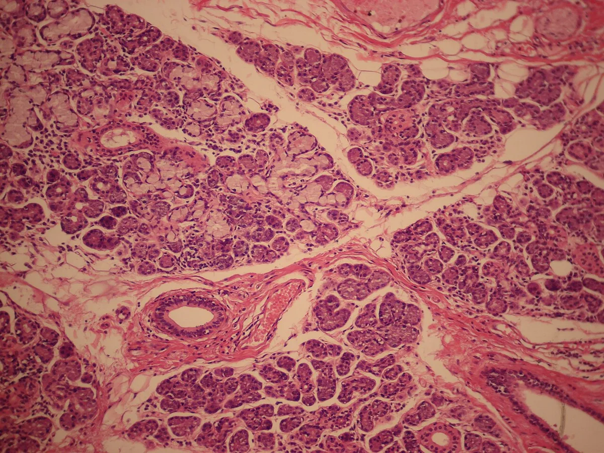
| 耳下腺 | 顎下腺 | 舌下腺 | |
|---|---|---|---|
| 分泌 | 純漿液性 | 混合(漿液優位) | 混合(粘液優位) |
| 漿液半月 | — | あり | あり |
| 線条部導管 | よく発達 | 最もよく発達 | 乏しい |
| 脂肪組織 | 多い(加齢で増える) | 少ない | 少ない |
| 導管 | Stensen 管 | Wharton 管 | Bartholin/Rivinus 管 |
| 唾石 | 少ない | 最も多い | 少ない |
見分けの順——①粘液細胞(明るい抜け)があるか ②漿液半月があるか ③線条部導管の発達 ④脂肪の量。
唾液の量は顎下腺が最多(安静時の約6〜7割)、刺激時は耳下腺が主役になる。
顎下腺に唾石が多い理由は、3つ重なっている。粘液を含んで粘り、導管が長く、重力に逆らって上向きに走る(唾石症の章)——「食事のたびに腫れて痛むという訴えが、この解剖から出てくる」。
耳下腺は顔面神経が貫くので、腫瘍の手術で最も気を遣う場所になる(耳下腺腫瘍の章)——「良性でも、神経を残せるかが術式を決める」。
流行性耳下腺炎(ムンプス)は耳下腺を侵し、思春期以降では精巣炎と難聴を起こす(ムンプスの章)——「唾液腺の病気が、聴力と生殖能に関わる」。
Sjögren 症候群では、リンパ球が腺房を置き換えていく。小唾液腺(口唇)の生検で診断する(Sjögren症候群の章)——「大きな腺を切らずに、小さな腺で証明する」。
そして乾燥は、齲蝕と口腔カンジダ症を増やす。唾液が持つ洗浄と抗菌の働きが失われるからになる(口腔の章)——「症状は乾きだが、問題は感染になる」。
頭頸部への放射線照射でも、同じ乾燥が起きる。漿液細胞が特に放射線に弱い(放射線の組織影響の章)——「照射野に唾液腺が入るかどうかが、生活の質を大きく変える」。
3つの唾液腺は、腫瘍の頻度と性質が違う。「大きい腺ほど腫瘍は多いが、良性の割合が高い」という逆相関で覚える。
耳下腺 — 腫瘍の約8割がここ、うち約8割が良性。最多は多形腺腫。顔面神経が腺の中を走るので、顔面神経麻痺を伴えば悪性を強く疑う。
顎下腺・舌下腺・小唾液腺は、腫瘍そのものは少ないが悪性の割合が高い。舌下腺と小唾液腺では悪性が多数を占める。
唾石は顎下腺に最も多い——導管が長く上向きで、分泌が粘稠だから。食事のたびに腫れて痛む。
線条部導管はイオンを再吸収するので、唾液は低張になる。アルドステロンが働く点で汗腺と共通している。
English version
Core
| Parotid | Submandibular | Sublingual | |
|---|---|---|---|
| Secretion | Purely serous | Mixed, serous predominant | Mixed, mucous predominant |
| Serous demilunes | — | Present | Present |
| Striated ducts | Well developed | Best developed | Sparse |
| Adipose tissue | Abundant (increases with age) | Little | Little |
| Duct | Stensen | Wharton | Bartholin / Rivinus |
| Calculi | Uncommon | Commonest | Uncommon |
Order of identification — ① are there mucous cells (pale gaps)? ② are there serous demilunes? ③ how developed are the striated ducts? ④ how much fat?
The submandibular gland produces most saliva (about 60–70 per cent at rest); on stimulation the parotid takes over.
Going further
Three reasons combine to make calculi commonest in the submandibular gland: the secretion is viscid because it contains mucus, the duct is long, and it runs upward against gravity — the complaint of swelling and pain with every meal follows from this anatomy.
The facial nerve traverses the parotid, making it the gland requiring most care at operation — even for a benign tumour, whether the nerve can be preserved decides the procedure.
Mumps attacks the parotid and, after puberty, causes orchitis and deafness — a disease of a salivary gland that involves hearing and fertility.
In Sjögren syndrome lymphocytes progressively replace the acini, and the diagnosis is made by biopsy of the labial minor glands — proving it in a small gland rather than cutting a large one. The dryness increases caries and oral candidiasis, because the cleansing and antibacterial actions of saliva are lost — the symptom is dryness but the problem is infection.
Radiotherapy to the head and neck produces the same dryness, because serous cells are especially radiosensitive — whether the salivary glands lie in the field greatly affects quality of life.
In the clinic
The three glands differ in the frequency and nature of their tumours. Remember the inverse relation: the larger the gland the more tumours, but the higher the proportion that are benign.
Parotid — about 80 per cent of salivary tumours, of which about 80 per cent are benign; the commonest is the pleomorphic adenoma. Because the facial nerve runs through the gland, facial palsy strongly suggests malignancy.
Submandibular, sublingual and minor glands have fewer tumours but a higher proportion of malignancy; in the sublingual and minor glands most are malignant.
Calculi are commonest in the submandibular gland — a long duct running upward with viscid secretion. It swells and hurts with every meal.
Because the striated duct reabsorbs ions, saliva is hypotonic; aldosterone acts here as it does in the sweat gland.
内分泌腺の共通の型 — 3つの並び方
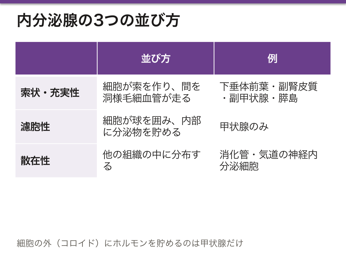
内分泌腺には導管が無く、細胞が毛細血管(有窓性)に接して並ぶ。並び方は3つに集約される。
- 索状・充実性——細胞が索を作り、間を洞様毛細血管が走る。下垂体前葉・副腎皮質・副甲状腺・膵島
- 濾胞性——細胞が球を囲み、内部に分泌物を貯める。甲状腺のみ
- 散在性——他の組織の中に分布する。消化管・気道の神経内分泌細胞(びまん性神経内分泌系)
甲状腺だけが、ホルモンを細胞の外(濾胞腔のコロイド)に貯める。
ホルモンの種類で細胞の姿が変わる。
- ペプチド——粗面小胞体と分泌顆粒が発達。好塩基性
- ステロイド——滑面小胞体・管状クリスタ・脂肪滴。淡く泡状
細胞の姿からホルモンの種類を言えることが、この章の実用点になる。泡状で淡い=ステロイド、顆粒が詰まって濃い=ペプチド(ミトコンドリアの章)——「像を見て、作っているものを当てる」。
甲状腺がコロイドに貯めることで、数か月分の備蓄ができている。だからヨウ素が不足しても、症状が出るまでに時間がかかる(甲状腺の章)——「唯一の細胞外貯蔵が、唯一の長い緩衝になっている」。
逆に、破壊されると貯めていたホルモンが一気に漏れる。亜急性甲状腺炎と無痛性甲状腺炎の一過性中毒症(破壊性甲状腺炎の章)——「作りすぎではなく、漏れているだけなので、抗甲状腺薬は効かない」。
散在性の神経内分泌細胞が、消化管と肺の神経内分泌腫瘍の起源になる(神経内分泌腫瘍の章)——「臓器はばらばらでも、細胞の系統は同じになる」。
だから染色も共通になる。クロモグラニンA・シナプトフィジン(免疫組織化学の章)——「どこにできても、同じ抗体で拾える」。
有窓性毛細血管であることが、ホルモンを速く血中へ送る条件になる(毛細血管の章)——「内分泌腺・腎糸球体・消化管粘膜という、物を通す場所に共通する構造になる」。
内分泌腺は毛細血管に接して並ぶ。有窓性毛細血管と洞様毛細血管がホルモンをすぐ血中へ渡すためで、血流が豊富=出血しやすいことでもある。
下垂体卒中 — 腺腫への出血で突然の頭痛・視力障害・下垂体機能低下。Sheehan 症候群は分娩時の大量出血による下垂体前葉の梗塞で、産後に乳汁が出ないことが最初の症状になる。妊娠で下垂体が肥大しているぶん虚血に弱い。
濾胞性(甲状腺)はホルモンを細胞の外に貯蔵する唯一の型である。だから破壊性甲状腺炎では、蓄えが一気に漏れて一過性の中毒症になり、その後に機能低下へ移る。摂取率が低いことが Basedow 病との鑑別点になる。
導管が無いので内分泌腫瘍は閉塞症状を出さず、ホルモン過剰で見つかる。
English version
Core
An endocrine gland has no duct; its cells lie against fenestrated capillaries. The arrangements reduce to three.
- Cords / solid — cells form cords with sinusoidal capillaries between them. Anterior pituitary, adrenal cortex, parathyroid, pancreatic islet.
- Follicular — cells enclose a sphere and store the product inside it. The thyroid alone.
- Diffuse — scattered within other tissues. The neuroendocrine cells of the gut and airway (the diffuse neuroendocrine system).
Only the thyroid stores its hormone outside the cell, in the follicular colloid.
The appearance of the cell changes with the type of hormone. Peptide — well-developed rough ER and secretory granules; basophilic. Steroid — smooth ER, tubular cristae, lipid droplets; pale and foamy.
Going further
The practical point of this chapter is being able to name the class of hormone from the appearance of the cell: pale and foamy means steroid, densely granular means peptide — look at the image and say what is being made.
Because the thyroid stores hormone in colloid, it holds several months’ supply; so iodine deficiency takes time to become symptomatic — the only extracellular store is also the only long buffer. Conversely, when it is destroyed the stored hormone leaks out at once: the transient thyrotoxicosis of subacute and painless thyroiditis — it is leakage, not overproduction, so antithyroid drugs do not work.
The diffuse neuroendocrine cells are the origin of the neuroendocrine tumours of gut and lung — the organs differ but the cell lineage is the same, which is why the same stains apply everywhere: chromogranin A and synaptophysin.
Fenestrated capillaries are the condition for delivering hormone rapidly into the blood — a structure shared by the endocrine glands, the renal glomerulus and the intestinal mucosa, all places where material must pass.
In the clinic
Endocrine cells lie against capillaries so that hormone passes immediately into the blood, which also means a rich blood supply and a tendency to haemorrhage.
Pituitary apoplexy — haemorrhage into an adenoma causing sudden headache, visual disturbance and hypopituitarism. Sheehan syndrome is infarction of the anterior pituitary after massive obstetric haemorrhage, and failure of lactation is the first symptom; the gland is enlarged in pregnancy and therefore vulnerable to ischaemia.
The follicular pattern (thyroid) is the only one that stores hormone outside the cell. Hence in destructive thyroiditis the store leaks out, causing transient thyrotoxicosis followed by hypothyroidism; a low uptake distinguishes it from Graves disease.
Because there is no duct, endocrine tumours do not obstruct and are found through hormone excess.
分泌の様式 — 分泌物と一緒に、細胞の何を捨てるかSecretio merocrina
3つに分ける。分かれ目は「細胞のどこまでを一緒に放出するか」になる。
| 様式 | 捨てるもの | 細胞 | 例 |
|---|---|---|---|
| メロクリン(開口分泌) | 分泌顆粒の中身だけ | 無傷で残る | 唾液腺・膵外分泌部・エクリン汗腺(大多数) |
| アポクリン(離出分泌) | 頂端の細胞質ごと | 頂部を失う | 乳腺の脂肪分・アポクリン汗腺 |
| ホロクリン(全分泌) | 細胞そのもの | 死ぬ | 脂腺・瞼板腺(Meibom 腺) |
乳腺は1つの腺で2つの様式を使う——蛋白質と乳糖はメロクリン、脂肪はアポクリン。
ホロクリンの腺には、必ず細胞を供給し続ける基底細胞がある。
ホロクリンが「死ぬことで分泌する」ことが、脂腺の病気の形を決めている。細胞が絶えず作られては死ぬので、増殖の速さと導管の開き具合の均衡が崩れると詰まる(痤瘡の章)——「面皰は、供給が排出を上回った状態になる」。
だから痤瘡の薬は、この2つのどちらかを動かしている。レチノイドは角化と排出の側、アンドロゲンを抑える薬は産生の側に効く——「詰まりは、出口を広げるか、量を減らすかしかない」。
アポクリン汗腺が頂部を捨てることは、においの発生源になっている。捨てられた細胞質を皮膚常在菌が分解する。汗そのものは無臭である(皮膚科の処置と生活の章)——「においを作っているのは、人ではなく菌になる」。
メロクリンが細胞を保つことが、分泌腺の疲れにくさを説明する。膜だけを融合させて中身を出し、膜はすぐ回収される(開口分泌と膜の再利用)(Golgi装置と分泌の経路の章)——「使い捨てにしない仕組みが、1日1.5 L の唾液を可能にしている」。
乳腺はアポクリンとメロクリンが同居する。脂肪は離出分泌、蛋白は開口分泌で出るため、母乳には細胞膜に包まれた脂肪球が含まれる。
全分泌(ホロクリン)は脂腺だけである。細胞が丸ごと壊れて分泌物になるので、導管が詰まると内容が溜まって炎症を起こす——痤瘡・粉瘤(表皮嚢腫)・霰粒腫(マイボーム腺)はいずれも同じ形である。
マイボーム腺は瞼板の中の脂腺で、詰まれば霰粒腫(無痛性の硬結)、感染すれば麦粒腫(痛い)——痛みの有無で区別する。
汗腺の分泌様式の違いが体臭の分布を決めることは上皮の章のとおりである。
English version
Core
Three modes, distinguished by how much of the cell is released with the product.
| Mode | What is lost | The cell | Examples |
|---|---|---|---|
| Merocrine (exocytosis) | Only the contents of the granule | Remains intact | Salivary glands, exocrine pancreas, eccrine sweat glands (the great majority) |
| Apocrine | Together with the apical cytoplasm | Loses its apex | The lipid of the mammary gland, apocrine sweat glands |
| Holocrine | The cell itself | Dies | Sebaceous glands, Meibomian glands |
The breast uses two modes in one gland — protein and lactose merocrine, fat apocrine. A holocrine gland always has basal cells that keep supplying replacements.
Going further
That holocrine secretion means "secreting by dying" determines the form of sebaceous disease. Cells are continuously made and destroyed, so if the balance between the rate of production and the patency of the duct is lost, it blocks — a comedone is the state in which supply has outstripped drainage.
So the drugs for acne move one or other of these two: retinoids act on keratinisation and drainage, antiandrogens act on production. A blockage can only be treated by widening the outlet or reducing the amount.
That the apocrine gland discards its apex is the source of body odour: skin commensals break down the discarded cytoplasm. The sweat itself is odourless — it is the bacteria, not the person, that make the smell.
That merocrine secretion preserves the cell explains why a secretory gland does not tire: only the membrane fuses, the contents are released, and the membrane is retrieved immediately — the refusal to be disposable is what makes 1.5 litres of saliva a day possible.
In the clinic
The breast combines apocrine and merocrine secretion. Fat leaves by apocrine and protein by merocrine secretion, so breast milk contains fat globules enclosed in cell membrane.
Holocrine secretion belongs to the sebaceous gland alone. The whole cell disintegrates to form the secretion, so if the duct obstructs the contents accumulate and inflame — acne, epidermoid cyst and chalazion are all the same form.
The Meibomian gland is a sebaceous gland within the tarsal plate: obstruction gives a chalazion (a painless nodule), infection gives a hordeolum (painful) — distinguished by whether it hurts.
分泌周期 — 取り込み・合成・蓄積・放出・回復Cyclus secretorius
腺細胞は、分泌を決まった順序の繰り返し(分泌周期)として行う。相の数え方は本によって3〜5つと違うが、中身は同じである。
| 相 | 何が起きるか | 場所 |
|---|---|---|
| 1 取り込み | 血液から材料(アミノ酸・糖・脂肪酸・水・イオン)を取り込む | 基底側の細胞膜 |
| 2 合成 | 蛋白は粗面小胞体で、脂質やステロイドは滑面小胞体で作る | 核の周りと基底側 |
| 3 加工と包装 | Golgi 装置で糖鎖を付け、濃縮して分泌顆粒に詰める | 核の上 |
| 4 蓄積 | 顆粒が細胞の頂上にたまり、合図を待つ | 頂上側 |
| 5 放出 | 開口分泌(メロクリン)・離出分泌(アポクリン)・全分泌(ホロクリン) | 頂上の膜 |
| 6 回復 | 使った膜を回収し、次の周期に備える。全分泌では新しい細胞が入れ替わる | — |
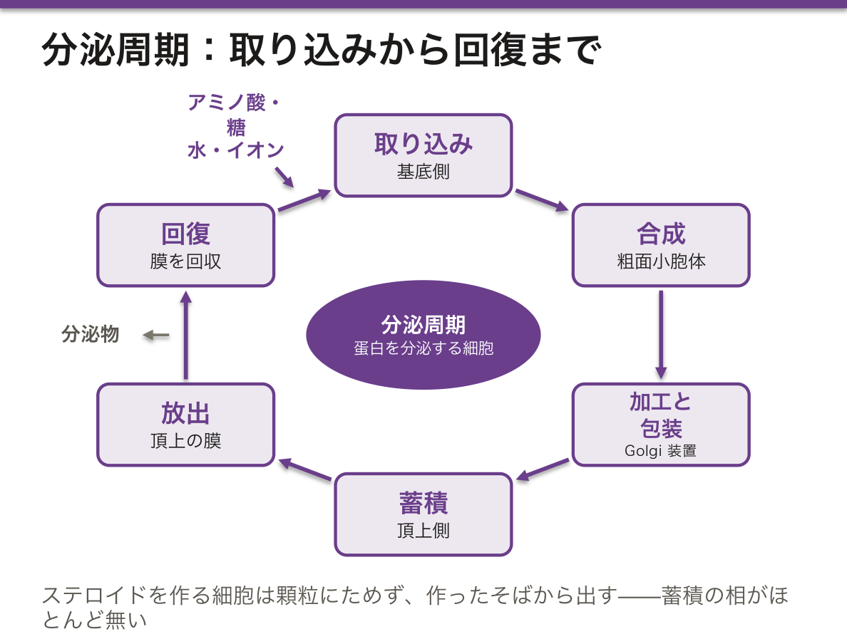
この順序がそのまま腺細胞の微細構造の並びになっている。蛋白を分泌する細胞(膵の腺房細胞が典型)を下から見ると、基底側に粗面小胞体と核、核の上に Golgi 装置、頂上に分泌顆粒が並ぶ。だから HE では基底側が青く(リボソームの RNA)、頂上がピンク(酵素原顆粒)に染め分けられる。色の並びを見れば、蛋白を作って出す細胞だと読める。
ステロイドを作る細胞(副腎皮質・Leydig 細胞)は違う。滑面小胞体と、管状のクリスタを持つミトコンドリアと、脂肪滴が多く、細胞質は泡のように明るい。ステロイドは膜を通り抜けるので顆粒にためず、作ったそばから出す——蓄積の相がほとんど無い。
分泌には、合図を待つ調節性分泌(顆粒をためる)と、作ったら絶えず出す構成性分泌(基底膜の成分や抗体など)がある。
膵の消化酵素は不活性な前駆体(酵素原)として顆粒に詰められ、十二指腸で初めて活性化される。蓄積の相で酵素が働いてしまえば、細胞が自分を消化するからである。この安全装置が破れて細胞の中でトリプシンが活性化されるのが急性膵炎の始まりで、トリプシノーゲン遺伝子(PRSS1)の変異は遺伝性膵炎を起こす。
嚢胞性線維症では分泌物そのものは出るが、CFTR(塩化物イオンのチャネル)の異常で水が伴わず、粘い分泌物が導管を詰まらせる。放出の相の異常である。
English version
Core
A gland cell secretes as a repeated sequence (the secretory cycle). Books count three to five phases, but the content is the same.
| Phase | What happens | Where |
|---|---|---|
| 1 Uptake | takes materials from blood (amino acids, sugars, fatty acids, water, ions) | basal membrane |
| 2 Synthesis | proteins on rough ER; lipids and steroids on smooth ER | around the nucleus and basally |
| 3 Processing and packaging | the Golgi adds sugars, concentrates the product and packs it into secretory granules | above the nucleus |
| 4 Storage | granules gather at the apex and wait for a signal | apical |
| 5 Release | merocrine (exocytosis), apocrine or holocrine | apical membrane |
| 6 Recovery | membrane is taken back for the next cycle; in holocrine glands new cells replace the old | — |
Going further
The order is also the layout of the gland cell. In a protein-secreting cell (the pancreatic acinar cell is typical), from base to apex there are rough ER and nucleus at the base, Golgi above the nucleus, and secretory granules at the apex. So in H&E the base is blue (ribosomal RNA) and the apex is pink (zymogen granules). This colour pattern tells you the cell makes and exports protein.
Steroid-making cells (adrenal cortex, Leydig cells) differ. They are rich in smooth ER, mitochondria with tubular cristae, and lipid droplets, and their cytoplasm looks pale and foamy. Steroids pass through membranes, so they are not stored in granules but released as soon as they are made — there is almost no storage phase.
Secretion is either regulated (granules are stored and wait for a signal) or constitutive (released continuously as made, e.g., basement-membrane components and antibodies).
In the clinic
Pancreatic enzymes are packed in granules as inactive precursors (zymogens) and activated only in the duodenum. If they worked during storage, the cell would digest itself. Acute pancreatitis begins when this safeguard fails and trypsin is activated inside the cell; mutations of the trypsinogen gene (PRSS1) cause hereditary pancreatitis.
In cystic fibrosis secretion is released, but a faulty CFTR (a chloride channel) means water does not follow, and thick secretions block the ducts — a defect of the release phase.
腺の形 — 導管が枝分かれするか、終末部が丸いかGlandula tubuloacinosa composita
外分泌腺の形は、2つの軸の掛け合わせで名前が付く。
軸① 導管が枝分かれするか——単一腺(しない)/複合腺(する)。
軸② 終末部(分泌部)の形——管状/胞状(腺房状)/管状胞状。
| 形 | 例 |
|---|---|
| 単一管状腺 | 大腸の腸腺(Lieberkühn 腺) |
| 単一分枝管状腺 | 胃底腺・子宮腺 |
| 単一コイル管状腺 | エクリン汗腺 |
| 単一分枝胞状腺 | 脂腺 |
| 複合管状腺 | Brunner 腺・腎(尿細管系) |
| 複合胞状腺 | 耳下腺・外分泌膵 |
| 複合管状胞状腺 | 顎下腺・気管腺 |
腺は上皮が下の結合組織へ落ち込んで作られる——導管を残せば外分泌腺、導管を失えば内分泌腺になる。
「腺は上皮の陥入である」という一文が、腺腫の性質を説明する。腺は上皮なので基底膜を持ち、そこを越えるかどうかが上皮内癌と浸潤癌を分ける(腫瘍総論の章)——「腺癌の診断が、線の越え方の話になる理由がここにある」。
終末部の形は、そのまま分泌物の粘さと対応している。さらさらした漿液は細い腺房から、粘い粘液は太い管状の終末部から出る——「形は、流すものの性質に合わせて決まっている」。
導管を失うと内分泌腺になる、という見方は発生でそのまま起きる。下垂体前葉は Rathke 嚢という上皮の陥入から作られ、導管を失って内分泌腺になった(器官の発生の章)——「消え残れば頭蓋咽頭腫になる」。
甲状舌管も同じ筋書きになる。舌盲孔から下降した上皮が導管を閉じ、残ると正中頸嚢胞になる(咽頭弓・嚢・溝の章)——「正中にある頸部の腫瘤は、まず発生の遺残を疑う」。
腺の形が分かると、内視鏡と生検の所見が読める。
大腸の腸腺(単一管状腺)が枝分かれし、不規則に融合するのが腺腫の像である。管状腺腫・絨毛腺腫・管状絨毛腺腫という分類は、腺の形そのものの名前になっている。絨毛成分が多いほど癌化のリスクが高い。
胃の腺の形が変わることが萎縮性胃炎の指標になる。固有腺が減り、腸型の腺に置き換わる(腸上皮化生)。
複合胞状腺(膵・耳下腺)は導管系が発達するので、導管が詰まると腺房が萎縮して線維に置き換わる——慢性膵炎の成り立ちである。
組織型を言うときは「腺房が丸いか、管が伸びるか」から入ると標本で迷わない。
English version
Core
The name of an exocrine gland is the product of two axes.
- Axis ①: does the duct branch? — simple (no) / compound (yes).
- Axis ②: the shape of the secretory portion — tubular / acinar (alveolar) / tubuloacinar.
| Form | Example |
|---|---|
| Simple tubular | Intestinal glands of the colon (crypts of Lieberkühn) |
| Simple branched tubular | Gastric fundic glands, uterine glands |
| Simple coiled tubular | Eccrine sweat gland |
| Simple branched alveolar | Sebaceous gland |
| Compound tubular | Brunner glands |
| Compound acinar | Parotid, exocrine pancreas |
| Compound tubuloacinar | Submandibular gland, tracheal glands |
A gland is formed by epithelium sinking into the connective tissue beneath — keep the duct and it is exocrine, lose it and it is endocrine.
Going further
The single sentence "a gland is an invagination of epithelium" explains the nature of adenomas. Because a gland is epithelium it has a basement membrane, and whether that is crossed separates carcinoma in situ from invasive carcinoma — this is why the diagnosis of adenocarcinoma is a question about crossing a line.
The shape of the secretory portion corresponds to the viscosity of the product: thin serous fluid from narrow acini, viscid mucus from wide tubular endpieces — form is matched to what has to flow through it.
The idea that losing the duct produces an endocrine gland happens literally in development. The anterior pituitary is formed from an epithelial invagination, Rathke’s pouch, which loses its duct — and if it persists, a craniopharyngioma. The thyroglossal duct follows the same script: epithelium descending from the foramen caecum closes its duct, and if it remains, a midline cervical cyst — a midline neck mass should first raise a developmental remnant.
In the clinic
Knowing the form of a gland lets you read endoscopic and biopsy findings. The branching and irregular fusion of the simple tubular colonic glands is the appearance of an adenoma. The classification into tubular, villous and tubulovillous adenoma is a name for the shape of the gland itself, and the more villous the component, the higher the risk of malignant change.
A change in the form of the gastric glands is an index of atrophic gastritis: the proper glands diminish and are replaced by intestinal-type glands (intestinal metaplasia).
Compound acinar glands (pancreas, parotid) have a well-developed duct system, so duct obstruction causes acinar atrophy and replacement by fibrous tissue — the development of chronic pancreatitis.
When stating a histological type, begin with "is the endpiece round, or is it a tube?" and identification on the slide becomes straightforward.
唾液 — 何が入っていて、誰が命令するかSaliva
1日 1.0〜1.5 L。安静時は顎下腺が約7割、刺激時は耳下腺が主役になる。
2段階でできる——①腺房が血漿とほぼ等張の一次唾液を作る ②線条部導管が Na⁺ と Cl⁻ を再吸収し、K⁺ と HCO₃⁻ を出す。結果として低張になる。
流量が増えると導管を通る時間が短くなり、再吸収が追いつかず、等張に近づく。
主な成分。
- α-アミラーゼ(プチアリン)——デンプンを分解する。耳下腺に多い
- ムチン——潤滑。舌下腺に多い
- リゾチーム・ラクトフェリン・分泌型 IgA——防御
- HCO₃⁻——緩衝。酸を中和して歯を守る
- ハプトコリン(R蛋白)——ビタミンB12 を胃酸から守る
支配は自律神経の両方だが、性質が違う——副交感神経が量(漿液性で多量)、交感神経が質(粘稠で少量)。
唾液が低張であることは、導管が「作った後で組成を変えている」ことの証拠になる。腎の尿細管と同じ設計で、まず作ってから取り戻す(腎と体液の章)——「分泌する器官は、たいてい一度多めに作って回収する」。
だから流量で組成が変わる。脱水で流量が落ちると Na⁺ が下がり、逆に嚢胞性線維症では導管の Cl⁻ 輸送が壊れて唾液と汗の塩分が上がる(チャネル病の章)——「汗が塩辛い子どもの検査が、この生理から出てくる」。
唾液分泌が両方の自律神経に支配されることが、薬の副作用の頻度を説明する。抗コリン作用を持つ薬(三環系抗うつ薬・抗ヒスタミン薬・過活動膀胱の薬)は例外なく口渇を起こす(自律神経作用薬の章)——「口が渇くという訴えは、まず薬歴で説明がつく」。
そして唾液が減ることは、齲蝕と感染の増加として現れる。緩衝も洗浄も抗菌も止まる(口腔の公衆衛生の章)——「Sjögren 症候群や頭頸部への放射線治療の後に、虫歯が一気に増える」。
唾液腺が腫れる病態を、痛みと左右で分ける。
- 急に痛くて片側——唾石症(顎下腺が最多。Wharton 管が上向きで長く、分泌が粘い)・急性化膿性唾液腺炎
- 痛くて両側——流行性耳下腺炎(ムンプス)。精巣炎・髄膜炎・難聴を合併しうる
- 痛くなくて両側・持続——Sjögren 症候群・IgG4 関連疾患・サルコイドーシス(膠原病の章)
- 片側で硬く、顔面神経麻痺を伴う——悪性腫瘍を疑う。耳下腺腫瘍は大きい腺ほど良性が多く、小さい腺ほど悪性が多い
English version
Core
1.0–1.5 litres a day. At rest the submandibular gland provides about 70 per cent; on stimulation the parotid takes the leading role.
Formed in two stages — ① the acini make a primary saliva nearly isotonic with plasma; ② the striated duct reabsorbs Na⁺ and Cl⁻ and secretes K⁺ and HCO₃⁻, so the result is hypotonic. At higher flow there is less time in the duct, reabsorption cannot keep up, and the saliva approaches isotonicity.
Principal constituents — α-amylase (ptyalin), digesting starch, chiefly from the parotid; mucins for lubrication, chiefly sublingual; lysozyme, lactoferrin and secretory IgA for defence; bicarbonate for buffering, neutralising acid and protecting the teeth; and haptocorrin (R protein), protecting vitamin B12 from gastric acid.
Both limbs of the autonomic system supply it, but differently — parasympathetic governs quantity (copious and serous), sympathetic governs quality (scanty and viscid).
Going further
That saliva is hypotonic is the evidence that the duct alters the composition after it has been made — the same design as the renal tubule: make it first, then take back. A secreting organ usually makes a surplus and recovers it.
Hence the composition changes with flow. In dehydration flow falls and sodium falls with it; conversely in cystic fibrosis the ductal chloride transport is broken and the salt content of saliva and sweat rises — the test for a child whose sweat tastes salty comes out of this physiology.
Dual autonomic control explains the frequency of drug side effects. Any drug with anticholinergic action — tricyclic antidepressants, antihistamines, drugs for overactive bladder — causes dry mouth without exception. A complaint of dry mouth is usually explained by the drug history first.
And reduced saliva presents as increased caries and infection, because buffering, cleansing and antibacterial action all stop — which is why caries increases sharply after Sjögren syndrome or head and neck radiotherapy.
In the clinic
Separate the causes of salivary swelling by pain and by laterality.
- Sudden, painful, unilateral — sialolithiasis (commonest in the submandibular gland: Wharton’s duct is long, runs upward and carries viscid secretion) or acute suppurative sialadenitis.
- Painful and bilateral — mumps, which may be complicated by orchitis, meningitis and deafness.
- Painless, bilateral and persistent — Sjögren syndrome, IgG4-related disease, sarcoidosis.
- Unilateral, hard, with facial palsy — suspect malignancy. In salivary tumours, the larger the gland the more often benign; the smaller the gland the more often malignant.
筋上皮細胞 — 腺を外から絞る細胞
分泌された液は、放っておいても導管へは出ていかない。終末部と導管を、籠のように取り巻いて絞り出す細胞がいる——筋上皮細胞である。
- 上皮由来なのに、収縮する——アクチンとミオシンを持ち、平滑筋アクチンが陽性になる。上皮でもあるのでサイトケラチンも陽性
- 基底膜と分泌細胞のあいだにいる——星形の突起を伸ばして取り巻く
- いる場所は決まっている——唾液腺・乳腺・汗腺・涙腺・気管支腺
乳腺では、オキシトシンがこの細胞を収縮させて乳汁を押し出す(射乳反射)。分泌そのものと、押し出すことは別の細胞が担当している。
この細胞の有無が、病理診断の決め手になる。
正常な腺と良性の病変では、分泌細胞の外側に筋上皮細胞の層が残っている。ところが浸潤癌ではこの層が消える——癌細胞が基底膜を破って外へ出ているからである。
| 筋上皮の層がある | 正常・腺症・乳管内癌(非浸潤癌) | 基底膜の内側に留まっている |
| 筋上皮の層が消えている | 浸潤癌 | 治療方針が大きく変わる |
だから乳腺の病理では、筋上皮のマーカー(p63・カルポニン・平滑筋ミオシン重鎖)を染めて浸潤の有無を判定する。「細胞が悪性かどうか」ではなく「どこまで出ているか」を、別の細胞の有無で判定しているという点が要である。前立腺でも同じ理屈で基底細胞のマーカーが使われる。
筋上皮細胞そのものが腫瘍になることもある。多形腺腫(唾液腺で最も多い良性腫瘍)は筋上皮細胞が軟骨様・粘液様の基質を作るため、1 つの腫瘍の中に上皮と間葉のような像が混在する。「多形」という名は、由来が複数あるからではなく、1 種類の細胞が多彩な像を作るためである。
乳癌の病理報告で「浸潤性」か「非浸潤性」かは、手術と薬物療法の内容を変える。この区別が筋上皮の有無で付いている。
English version
Core
Secreted fluid does not leave for the duct of its own accord. A cell wraps the endpieces and ducts like a basket and squeezes it out — the myoepithelial cell.
- Epithelial in origin, yet contractile — it contains actin and myosin and is positive for smooth muscle actin; being epithelial, it is also cytokeratin positive.
- It lies between the basal lamina and the secretory cells, sending out stellate processes.
- Its distribution is fixed — salivary, mammary, sweat, lacrimal and bronchial glands.
In the breast, oxytocin contracts this cell to expel milk (the milk-ejection reflex). Secreting and expelling are done by different cells.
Going further
The presence or absence of this cell is decisive in pathological diagnosis. In normal glands and benign lesions a layer of myoepithelial cells remains outside the secretory cells; in invasive carcinoma that layer disappears, because the tumour has broken through the basement membrane.
| Meaning | |
|---|---|
| Myoepithelial layer present | Normal, adenosis, ductal carcinoma in situ — still inside the basement membrane |
| Myoepithelial layer absent | Invasive carcinoma — the treatment changes fundamentally |
So breast pathology stains for myoepithelial markers — p63, calponin, smooth muscle myosin heavy chain — to judge invasion. The essential point is that what is being judged is not whether the cells are malignant but how far they have got, and it is judged by the presence of a different cell. The same logic uses basal cell markers in the prostate.
The myoepithelial cell can itself become a tumour. In the pleomorphic adenoma, the commonest benign salivary tumour, myoepithelial cells produce a cartilaginous and myxoid matrix, so epithelial and mesenchymal-looking areas coexist in one tumour. "Pleomorphic" refers not to several origins but to the many appearances produced by one kind of cell.
In the clinic
In a breast pathology report, whether the tumour is invasive or non-invasive changes both the surgery and the systemic therapy — and that distinction rests on the presence of the myoepithelial cell.
まとめ
| 外分泌腺の分類と、漿液・粘液の見分け | 漿液細胞——円錐形。核は丸く中央よりやや基底側。 |
|---|---|
| 3つの大唾液腺 — スライドで言い当てる | 耳下腺顎下腺舌下腺分泌純漿液性混合(漿液優位)混合(粘液優位)漿液半月—ありあり線条部導管よく発達最もよく発達乏しい脂肪組織多い(加齢で増える)少ない少ない導管Stensen 管W… |
| 内分泌腺の共通の型 — 3つの並び方 | 内分泌腺には導管が無く、細胞が毛細血管(有窓性)に接して並ぶ。 |
| 分泌の様式 — 分泌物と一緒に、細胞の何を捨てるか | 3つに分ける。分かれ目は「細胞のどこまでを一緒に放出するか」になる。 |
| 分泌周期 — 取り込み・合成・蓄積・放出・回復 | 腺細胞は、分泌を決まった順序の繰り返し(分泌周期)として行う。 |
| 腺の形 — 導管が枝分かれするか、終末部が丸いか | 外分泌腺の形は、2つの軸の掛け合わせで名前が付く。 |
| 唾液 — 何が入っていて、誰が命令するか | 1日 1.0〜1.5 L。安静時は顎下腺が約7割、刺激時は耳下腺が主役になる。 |
| 筋上皮細胞 — 腺を外から絞る細胞 | 分泌された液は、放っておいても導管へは出ていかない。 |
このノートは医学生が授業の内容を自分の言葉でまとめたもので、診療の判断に使うものではありません。誤りに気づいたらお知らせください。