組織学は細胞学(Cytologia)から始まる。組織の像は、細胞小器官の分布がそのまま染まった結果だからである。
- 塩基好性(好塩基性)に青く染まる——粗面小胞体とリボソーム、つまり蛋白を作っている細胞
- 酸好性(好酸性)に赤く染まる——ミトコンドリアと滑面小胞体、つまりエネルギーを使う細胞・ステロイドを作る細胞
だから「この細胞は何をしているか」は、色から言える——「染色の濃淡は、細胞の職業を映している」。
そして電子顕微鏡の像は、光学顕微鏡の色を説明するために見る。2つは別の科目ではなく、同じものを倍率を変えて見ている。
- 細胞膜
- ミトコンドリア
- 小胞体 — 粗面と滑面
- Golgi装置と分泌の経路
- 鍍銀で見る Golgi 装置 — 黒い網として見つかった小器官
- リソソームとペルオキシソーム
- 細胞骨格 — 3つの太さ
- 線毛・鞭毛・中心小体 — 9+2 と 9+0
- 上皮細胞は、上と下を知っている — 細胞の極性
細胞膜Membrana cellularis
脂質二重層に蛋白が浮かぶ流動モザイク構造。厚さ約7.5〜10 nm。
- 電子顕微鏡では3層に見える——濃・淡・濃。単位膜(unit membrane)と呼ぶ
- 光学顕微鏡では見えない。細胞の輪郭として認識しているのは、細胞膜そのものではない
- 脂質——リン脂質・コレステロール・糖脂質。コレステロールが流動性を調節する
- 糖衣(グリコカリックス)——外側だけにある糖鎖。PAS染色で陽性
膜蛋白——内在性(貫通する)と表在性(表面に乗る)。輸送体・受容体・接着分子・酵素として働く。
脂質の分布は非対称——ホスファチジルセリンは内側にある。
好酸性・好塩基性の話に入る前に、細胞膜が光学顕微鏡で見えないことを確認しておく。見えているのは細胞膜の直下に集まった細胞質と、隣接する細胞との境界の重なり——「輪郭が見えることと、膜が見えることは違う」。
ホスファチジルセリンが内側にあることが、アポトーシスの目印になる。細胞が死ぬと外側に反転し、それをマクロファージが認識して食べる(アポトーシスの章)——「膜の非対称が崩れることが、片付けてくれという合図になる」。
臨床では、この反転が血液凝固でも使われる。活性化した血小板がホスファチジルセリンを表に出し、凝固因子の足場になる(止血機構の章)——「同じ仕掛けが、死と止血の両方の合図になっている」。
糖衣は、細胞の身分証明になる。ABO 血液型も、細胞表面の糖鎖の違いになる(糖鎖の章)——「輸血で問題になるものが、顕微鏡では PAS 陽性として見えている」。
そして糖衣は、消化管上皮の微絨毛の表面で特に厚い。二糖類分解酵素がそこに埋まっている(微絨毛の章)——「刷子縁は、吸収面であると同時に最後の消化装置になる」。
コレステロールが流動性を調節する仕組みは、温度によって向きが逆になる。高温では動きを抑え、低温では固まるのを防ぐ——「緩衝材として働くので、多いか少ないかでは答えられない」。
膜そのものが病気の舞台になる。
遺伝性球状赤血球症 — スペクトリン・アンキリンなど膜骨格の異常。赤血球が球形になり、変形能を失って脾で壊される。脾摘で改善するのは、壊す場所を除くからである。
膜蛋白の異常が病気の名前になっているものは多い。嚢胞性線維症(CFTR=塩素チャネル)、筋ジストロフィー(ジストロフィン=膜と細胞骨格をつなぐ)。
補体による膜傷害 — 発作性夜間ヘモグロビン尿症ではGPI アンカーが作れず、CD55・CD59 が膜に留まらないため補体で赤血球が壊れる。膜に「留める仕組み」が壊れただけで溶血が起こる。
膜は薬の標的でもある。アムホテリシン B は真菌のエルゴステロールに結合して膜に穴をあける。
English version
Core
A fluid mosaic of proteins floating in a lipid bilayer, 7.5–10 nm thick. On electron microscopy it appears as three layers — dark, light, dark — the unit membrane. It cannot be seen by light microscopy: what we recognise as the outline of a cell is not the membrane itself.
- Lipids — phospholipids, cholesterol, glycolipids. Cholesterol regulates fluidity.
- Glycocalyx — carbohydrate chains on the outer surface only. PAS positive.
- Membrane proteins — integral (spanning) and peripheral (resting on the surface); transporters, receptors, adhesion molecules, enzymes.
- The lipid distribution is asymmetrical — phosphatidylserine lies on the inner leaflet.
Going further
Before discussing acidophilia and basophilia, confirm that the plasma membrane is not visible by light microscopy. What is seen is the cytoplasm gathered just beneath it and the overlap of adjacent cell borders — seeing an outline is not the same as seeing a membrane.
That phosphatidylserine lies on the inner leaflet becomes the marker of apoptosis. When a cell dies it flips to the outside and macrophages recognise it and engulf the cell — the collapse of membrane asymmetry is the signal that says "clear me away".
Clinically the same flip is used in coagulation: an activated platelet exposes phosphatidylserine and provides the scaffold for clotting factors — one mechanism serving as the signal for both death and haemostasis.
The glycocalyx is the cell’s identity card. The ABO blood groups are differences in these surface carbohydrates — what matters in transfusion is what appears as PAS positivity down the microscope. It is especially thick over the microvilli of the intestinal epithelium, where the disaccharidases are embedded — the brush border is both an absorptive surface and the final digestive apparatus.
The way cholesterol regulates fluidity reverses with temperature: at high temperature it restrains movement, at low temperature it prevents solidification. It acts as a buffer, so "more or less fluid" is not an answerable question.
In the clinic
The membrane itself becomes the stage for disease.
- Hereditary spherocytosis — a defect of the membrane skeleton such as spectrin or ankyrin. The red cell becomes spherical, loses deformability and is destroyed in the spleen. Splenectomy helps because it removes the place of destruction.
- Many diseases are named after a membrane protein: cystic fibrosis (CFTR, a chloride channel), muscular dystrophy (dystrophin, linking membrane to cytoskeleton).
- Complement-mediated membrane injury — in paroxysmal nocturnal haemoglobinuria the GPI anchor cannot be made, so CD55 and CD59 do not stay on the membrane and complement lyses the red cells. Haemolysis follows from the failure of the anchoring mechanism alone.
- The membrane is also a drug target: amphotericin B binds fungal ergosterol and punches holes in the membrane.
ミトコンドリアMitochondrion
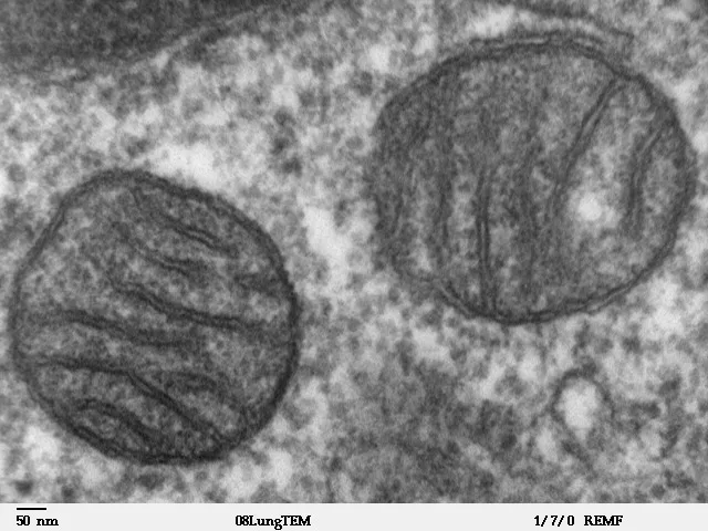
二重膜構造。外膜は平滑で、内膜が内側に折れ込んでクリスタを作る。
- クリスタの形が細胞で違う——板状クリスタが一般的。ステロイド産生細胞では管状クリスタ
- 基質(マトリックス)——TCA回路の酵素・独自の環状DNA・リボソーム
- 内膜——電子伝達系とATP合成酵素。心筋・骨格筋・肝ではクリスタが密
- 好酸性に染まる——ミトコンドリアが多い細胞ほど、細胞質が赤い
管状クリスタを持つ細胞——副腎皮質・精巣Leydig細胞・卵巣の莢膜細胞と黄体。
母性遺伝——受精時に精子のミトコンドリアは伝わらない。
クリスタの形からステロイド産生細胞を言い当てられる。管状クリスタ+豊富な滑面小胞体+脂肪滴——この3つが揃えばステロイドを作っている(副腎の章)——「電子顕微鏡の像が、そのままホルモンの種類を教えている」。
そして光学顕微鏡でも、副腎皮質は淡く泡状に見える。脂肪滴が処理の過程で溶け出すからになる(脱脂の章)——「空胞に見えるものが、あったことの証拠になる」。
ミトコンドリアが独自のDNAとリボソームを持つことは、細菌に由来する(細胞内共生説)ことを示している。リボソームは 70S で細菌型になる(翻訳の章)——「だから細菌のリボソームを狙う薬が、ヒトのミトコンドリアにも当たる」。
実際、リネゾリドとテトラサイクリン系とアミノグリコシド系の副作用の一部が、これで説明される(抗菌薬の副作用の章)——「選択毒性の穴が、細胞小器官の起源にある」。
母性遺伝は、家系図の形をはっきり変える。母親が発症すれば子は全員が受け継ぎ、父親が発症しても子には伝わらない(ミトコンドリア遺伝の章)——「一目で分かる遺伝形式なので、国試では家系図の形で問われる」。
そして症状はエネルギー要求の高い組織に出る。脳・骨格筋・心筋・網膜・内耳(ミトコンドリア病の章)——「臓器の組み合わせが不自然に見えるとき、共通項はエネルギーになる」。
ミトコンドリアは母性遺伝する。精子のミトコンドリアは受精後に分解されるため、ミトコンドリア病は母親からのみ伝わる——家系図の形で見当がつく。
ヘテロプラスミー(正常と変異のミトコンドリアが混在)のため同じ変異でも症状の重さが人によって違う。エネルギー需要の高い組織(神経・筋・心・内耳)から症状が出る——MELAS・MERRF・Leber 遺伝性視神経症。
筋生検の Gomori トリクローム変法で赤色ぼろ線維(ragged-red fiber)が見えれば、異常なミトコンドリアが筋線維の辺縁に集積している。
ステロイド産生細胞(副腎皮質・黄体・精巣 Leydig 細胞)は管状クリスタを持つ。クリスタの形を見れば、その細胞が何を作っているか分かる。
アポトーシスの内因性経路はミトコンドリアから始まる(チトクロム c の放出)。細胞の死のスイッチもここにある。
English version
Core
A double-membrane organelle. The outer membrane is smooth; the inner membrane folds inward to form cristae, and the shape of the cristae differs between cells — lamellar cristae are usual, but steroid-producing cells have tubular cristae.
- Matrix — the enzymes of the TCA cycle, its own circular DNA and ribosomes.
- Inner membrane — the electron transport chain and ATP synthase. Cristae are densely packed in cardiac and skeletal muscle and in liver.
- Stains acidophilic — the more mitochondria, the redder the cytoplasm.
- Cells with tubular cristae — adrenal cortex, testicular Leydig cells, ovarian theca and corpus luteum.
- Maternal inheritance — the sperm’s mitochondria are not transmitted at fertilisation.
Going further
A steroid-producing cell can be identified from the shape of its cristae. Tubular cristae, abundant smooth ER and lipid droplets — when these three coincide, the cell is making steroid: the electron micrograph is telling you which hormone. Even by light microscopy the adrenal cortex looks pale and foamy, because the lipid droplets dissolve out during processing — what appears as vacuoles is the evidence that they were there.
That mitochondria have their own DNA and ribosomes points to a bacterial origin (endosymbiosis); the ribosomes are 70S, the bacterial type. Hence drugs aimed at bacterial ribosomes also hit human mitochondria — part of the toxicity of linezolid, the tetracyclines and the aminoglycosides is explained this way. The gap in selective toxicity lies in the origin of the organelle.
Maternal inheritance changes the shape of the pedigree unmistakably: if the mother is affected all her children inherit it; if the father is affected none do. And symptoms appear in tissues with high energy demand — brain, skeletal muscle, heart, retina, inner ear. When a combination of organs looks unnatural, the common factor is energy.
In the clinic
Mitochondria are maternally inherited, because the sperm’s are degraded after fertilisation; the pedigree alone suggests the diagnosis. Heteroplasmy — a mixture of normal and mutant mitochondria — means the same mutation causes different severity in different people.
Symptoms appear in tissues with the highest energy demand — nerve, muscle, heart, inner ear: MELAS, MERRF, Leber hereditary optic neuropathy. On muscle biopsy, the modified Gomori trichrome shows ragged-red fibres, abnormal mitochondria accumulated at the periphery of the fibre.
Steroid-producing cells — adrenal cortex, corpus luteum, Leydig cells — have tubular cristae. The shape of the cristae tells you what the cell makes.
The intrinsic pathway of apoptosis begins at the mitochondrion, with the release of cytochrome c. The switch for cell death is here as well.
小胞体 — 粗面と滑面Reticulum endoplasmicum
粗面小胞体——リボソームが付く。分泌蛋白と膜蛋白を作る。
- 強い好塩基性——RNAのリン酸基がヘマトキシリンに染まる
- 発達している細胞——形質細胞・膵外分泌細胞・線維芽細胞・肝細胞
- 光学顕微鏡での呼び名——Nissl 小体(神経細胞)・エルゴプラズム
滑面小胞体——リボソームが付かない。脂質・ステロイド合成、解毒、カルシウム貯蔵。
- 好酸性。発達している細胞は副腎皮質・Leydig細胞・肝細胞・骨格筋(筋小胞体)
遊離リボソーム——細胞質に留まる蛋白を作る。
細胞質全体が青い細胞は、蛋白を分泌している。
形質細胞の見分け方が、この章で最も実用的になる。核が偏在して車軸状、細胞質が濃い青、核の近くに明るい部分(Golgi野)がある(形質細胞の章)——「抗体を作っている細胞は、作っている装置ごと染まって見える」。
だから慢性炎症の像に形質細胞が多ければ、抗体が関わる病態を考える。自己免疫性肝炎・IgG4関連疾患・Helicobacter pylori 胃炎(慢性炎症の章)——「細胞の種類が、炎症の性格を語る」。
神経細胞の Nissl 小体は、軸索が傷むと消える。核が偏在し、細胞体が腫れる——中心性色質融解になる(神経細胞の章)——「軸索の損傷が、細胞体の染色像に出る」。
滑面小胞体は、薬を飲むと増える。肝細胞のチトクロム P450 が誘導されるからになる(薬物代謝の章)——「フェノバルビタールを飲んだ肝細胞は、電子顕微鏡で見て分かる」。
これが薬物相互作用と耐性の組織学的な実体になる(酵素誘導の章)——「効かなくなったという臨床の訴えに、形の裏づけがある」。
小胞体は、折り畳みに失敗した蛋白が溜まる場所でもある。小胞体ストレス応答が起き、続けば細胞は死ぬ(蛋白の折り畳みの章)——「作る場所が、品質検査の場所も兼ねている」。
青い細胞質は「作っている細胞」の印である。
形質細胞 — 粗面小胞体が発達して細胞質が濃く青く、核は車軸状(cartwheel)で偏在し、核の近くに明るい部分(Golgi)がある。抗体を大量に作る細胞の形がそのまま像になっている。形質細胞が集まる=慢性炎症のサインである。
多発性骨髄腫では形質細胞が単クローン性に増える。M 蛋白・骨融解像(punched-out lesion)・高 Ca 血症・腎障害・貧血。
滑面小胞体は薬物代謝の場である。フェノバルビタールやアルコールの慢性投与で肝細胞の滑面小胞体が増える——薬物耐性と薬物相互作用の形態学的な裏づけになる。
小胞体ストレスと折り畳み異常はα1-アンチトリプシン欠乏症で見える。異常蛋白が肝細胞の小胞体に溜まり(PAS 陽性・ジアスターゼ抵抗性の封入体)、肝硬変を起こす一方、肺では酵素が足りず若年性肺気腫になる——1つの蛋白の異常が、溜まる臓器と足りない臓器の両方を壊す。
English version
Core
Rough ER — studded with ribosomes; makes secretory and membrane proteins. Strongly basophilic, because the phosphate groups of RNA bind haematoxylin.
- Well developed in plasma cells, pancreatic exocrine cells, fibroblasts, hepatocytes.
- Its light-microscopic names: Nissl bodies in neurons, ergastoplasm.
Smooth ER — no ribosomes; lipid and steroid synthesis, detoxification, calcium storage. Acidophilic. Well developed in the adrenal cortex, Leydig cells, hepatocytes and skeletal muscle (the sarcoplasmic reticulum).
Free ribosomes make protein that stays in the cell. A cell whose whole cytoplasm is blue is secreting protein.
Going further
The most immediately useful thing in this chapter is how to recognise a plasma cell: an eccentric cartwheel nucleus, deeply blue cytoplasm, and a pale area beside the nucleus, the Golgi zone. A cell making antibody is stained together with the machinery that makes it.
So when plasma cells are numerous in chronic inflammation, think of an antibody-driven process: autoimmune hepatitis, IgG4-related disease, Helicobacter pylori gastritis. The type of cell tells you the character of the inflammation.
The Nissl substance of a neuron disperses when the axon is injured; the nucleus moves to the periphery and the cell body swells — central chromatolysis. Damage to the axon shows itself in the staining of the cell body.
Smooth ER increases when drugs are taken, because hepatic cytochrome P450 is induced — a hepatocyte exposed to phenobarbital can be recognised on electron microscopy. This is the histological substance of drug interaction and of tolerance: the clinical complaint that a drug has stopped working has a morphological counterpart.
The ER is also where misfolded protein accumulates, provoking the unfolded protein response; if it continues the cell dies. The place of manufacture is also the place of quality control.
In the clinic
Blue cytoplasm is the mark of a cell that is making something.
Plasma cell — abundant rough ER gives deeply blue cytoplasm, the nucleus is eccentric and cartwheel, and there is a pale perinuclear Golgi zone. The shape of a cell producing large amounts of antibody is the image itself. Aggregated plasma cells signify chronic inflammation. In multiple myeloma they proliferate monoclonally: M protein, punched-out lytic lesions, hypercalcaemia, renal impairment and anaemia.
Smooth ER is the site of drug metabolism. Chronic phenobarbital or alcohol increases the smooth ER of hepatocytes — the morphological basis of tolerance and interaction.
ER stress and misfolding are visible in α1-antitrypsin deficiency: abnormal protein accumulates in the hepatocyte ER as PAS-positive, diastase-resistant inclusions and causes cirrhosis, while in the lung the enzyme is lacking and early emphysema follows. One abnormal protein damages both the organ where it accumulates and the organ where it is missing.
Golgi装置と分泌の経路Complexus Golgiensis
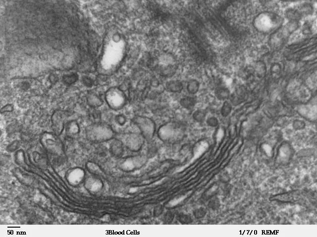
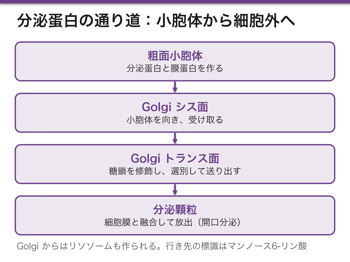
扁平な袋(槽)が重なった構造。極性がある。
- シス面——小胞体を向く。受け取る側
- トランス面——細胞膜を向く。送り出す側
働き——糖鎖の修飾・蛋白の選別・分泌顆粒とリソソームの形成。
光学顕微鏡では染まらず、核の近くの明るい抜けとして見える——Golgi野(negative Golgi image)。
分泌の様式は3つ。
- 開口分泌(メロクリン)——顆粒の膜が細胞膜と融合。ほとんどの腺
- 離出分泌(アポクリン)——細胞質の一部ごと放出。乳腺の脂肪・アポクリン汗腺
- 全分泌(ホロクリン)——細胞ごと壊れる。脂腺
Golgi 野が「明るい抜け」として見えることが、形質細胞の見分けに直結する。濃青の細胞質の中に、核に接した淡い領域がある——「染まらないことが所見になる」。
3つの分泌様式は、どこで問われるかが決まっている。脂腺だけがホロクリンで、これが座瘡の理解の土台になる(尋常性痤瘡の章)——「細胞が丸ごと皮脂になるので、出口が詰まれば溜まり続ける」。
そして乳腺は、成分によって様式が違う。蛋白(カゼイン)はメロクリン、脂肪はアポクリン(乳腺の章)——「1つの細胞が、2つの出し方を使い分けている」。
アポクリン汗腺という名前は、実は誤りだったことが分かっている。電子顕微鏡ではメロクリンに近い——「名前が歴史の名残になっている例なので、名称と機序を分けて覚える」。
Golgi での糖鎖修飾に異常があると、先天性グリコシル化異常症になる。発達遅滞・肝障害・凝固異常が組み合わさる(糖蛋白の章)——「修飾の異常が、全身のあちこちに出る」。
そしてリソソーム酵素の行き先も、Golgi で決まる。マンノース6-リン酸の標識が付かないと、酵素が細胞外へ漏れる——「宛先を書き損ねると、荷物が街に散らばる」。
Golgi は「宛先を書く」場所である。リソソーム酵素にマンノース6-リン酸の荷札を付けるのもここで、この標識が付かないと酵素が細胞外へ出てしまう。これが ムコリピドーシス II 型(I 細胞病)——酵素は作られているのに、届くべき場所に届かない。血中のリソソーム酵素が高値という逆説的な所見が診断の手がかりになる。
核の近くの明るい部分(Golgi 野・perinuclear hof)は形質細胞を見分ける目印になる。
分泌顆粒が見えるかで細胞の状態が読める。空腹時の膵外分泌細胞は顆粒に満ち、食後は減る。
コレラ毒素・百日咳毒素はG 蛋白を修飾して分泌の制御を壊す——止まらない分泌(コレラの水様下痢)という形で臨床に出る。
English version
Core
A stack of flattened cisternae, and it is polarised.
- Cis face — faces the ER; the receiving side.
- Trans face — faces the plasma membrane; the dispatching side.
- Functions — glycosylation, sorting of proteins, formation of secretory granules and lysosomes.
It does not stain by light microscopy and appears as a pale gap beside the nucleus — the negative Golgi image.
Three modes of secretion
- Merocrine (exocytosis) — the granule membrane fuses with the plasma membrane. Most glands.
- Apocrine — released together with part of the cytoplasm. The lipid of the mammary gland, apocrine sweat glands.
- Holocrine — the whole cell disintegrates. Sebaceous glands.
Going further
That the Golgi zone appears as a pale gap connects directly to recognising a plasma cell: within deeply blue cytoplasm there is a pale area against the nucleus — not staining is itself a finding.
Each of the three modes of secretion has its own examination context. Only the sebaceous gland is holocrine, and this is the foundation for understanding acne — the whole cell becomes sebum, so if the outlet is blocked it simply keeps accumulating.
The breast uses different modes for different components: protein (casein) merocrine, fat apocrine. One cell uses two ways of releasing its product.
The name "apocrine sweat gland" has turned out to be a misnomer — on electron microscopy the mechanism is closer to merocrine. The name is a historical relic, so keep the name and the mechanism apart.
Defective glycosylation in the Golgi gives the congenital disorders of glycosylation: developmental delay, liver disease and coagulopathy together. And the destination of lysosomal enzymes is also decided in the Golgi: without the mannose-6-phosphate tag the enzyme leaks out of the cell — write the address wrongly and the parcel is scattered through the city.
In the clinic
The Golgi is where the address is written. It attaches the mannose-6-phosphate label to lysosomal enzymes, and without it the enzymes leave the cell. This is mucolipidosis II (I-cell disease) — the enzymes are made but never arrive. The paradoxical finding of high lysosomal enzymes in the blood is the diagnostic clue.
The pale perinuclear hof is a mark by which the plasma cell is recognised.
The state of a cell can be read from its secretory granules: pancreatic acinar cells are packed with granules when fasting and depleted after a meal.
Cholera and pertussis toxins modify G proteins and destroy the control of secretion — presenting clinically as secretion that cannot be switched off, the watery diarrhoea of cholera.
鍍銀で見る Golgi 装置 — 黒い網として見つかった小器官Apparatus reticularis internus
HE 染色では Golgi 装置は染まらず、核の脇の明るい抜けとしてしか見えない。ところが組織を銀(硝酸銀)やオスミウムに浸すと、金属が Golgi 装置の膜に沈着し、黄色っぽい細胞質の中に黒〜黒褐色の糸や網として浮かび上がる。核は染まらず、明るく抜ける。
1898年に Golgi がこの方法で神経細胞の中に見つけ、「内網装置」と名付けた。Golgi 装置の古い名前である。
細胞の種類によって形と位置が違う。
| 型 | どう見えるか | 例 |
|---|---|---|
| 散在型(diffuse) | 核の周り全体に網が広がる | 脊髄神経節などの神経細胞 |
| 密集型(compact) | 核の片側、多くは核と細胞の頂上のあいだに1か所に集まる | 膵の腺房細胞・杯細胞などの分泌細胞、形質細胞 |
位置は細胞の極性を表す。分泌細胞では Golgi 装置が核より上(核上部)に集まり、分泌物はそこから細胞の頂上へ送り出される。だから密集型の Golgi 装置がどちらを向いているかで、どちらへ分泌する細胞かが分かる。
神経細胞の Golgi 装置が核を取り巻くほど大きいのは、長い突起の膜や伝達物質の小胞に使う蛋白を大量に仕上げて送り出すからである。細胞体だけで全部を作る神経細胞の事情(軸索では蛋白を作れない)と合う。
この黒い網は、見つかってから半世紀ほど「金属が沈着しただけの人工産物ではないか」と疑われた。電子顕微鏡で重なった扁平な袋が見えて、ようやく実在が確かめられた。1つの染色で見えたものは、別の方法で確かめる——組織学の基本的な態度を示す例である。
English version
Core
In H&E the Golgi apparatus does not stain and shows only as a pale area beside the nucleus. But if tissue is soaked in silver (silver nitrate) or osmium, the metal is deposited on the Golgi membranes, and it stands out as black to dark-brown threads or a net in a yellowish cytoplasm. The nucleus is not stained and looks pale.
Golgi found it in nerve cells with this method in 1898 and named it the "internal reticular apparatus", the old name of the Golgi apparatus.
Its shape and position differ by cell type.
| Type | Appearance | Examples |
|---|---|---|
| Diffuse | a net spread all around the nucleus | nerve cells such as those of the spinal ganglion |
| Compact | gathered in one place on one side of the nucleus, usually between nucleus and cell apex | secretory cells such as pancreatic acinar cells and goblet cells; plasma cells |
Going further
Position shows cell polarity. In secretory cells the Golgi gathers above the nucleus (supranuclear), and products are sent from there to the apex. So the direction a compact Golgi faces tells which way the cell secretes.
The neuronal Golgi is large enough to surround the nucleus because it finishes and sends out large amounts of protein for the membranes of long processes and for transmitter vesicles. This fits a neuron that must make everything in its cell body (the axon cannot make protein).
For about half a century after its discovery, this black net was suspected to be "just an artefact of metal deposits". Only the electron microscope, showing stacked flat sacs, proved it real. Confirm what one stain shows with another method — a basic attitude in histology.
リソソームとペルオキシソームLysosoma
リソソーム——加水分解酵素を含む小胞。内部は酸性(pH 約5)で、H⁺ポンプが酸性を保つ。
- 酵素は Golgi でマンノース6-リン酸の標識を受けて送られる
- 一次リソソーム(未使用)と二次リソソーム(食胞と融合した後)
- 残余小体——分解しきれなかったもの。リポフスチン=加齢色素
ペルオキシソーム——カタラーゼを含む。極長鎖脂肪酸のβ酸化・過酸化水素の分解・胆汁酸とプラスマローゲンの合成。
リポフスチン——心筋・肝・神経細胞に加齢とともに蓄積。褐色。褐色萎縮。
ライソゾーム病——酵素の欠損で基質が溜まる。細胞が泡沫状に膨らむ。
リポフスチンは、光学顕微鏡でそのまま褐色に見える。染めていないのに色がある——内因性色素になる(色素の章)——「染色の色ではない色を、区別して読む」。
だから高齢者の心筋と肝は、褐色を帯びて小さくなる。褐色萎縮と呼ぶ(萎縮の章)——「加齢そのものが、顕微鏡で見える」。
ライソゾーム病では、蓄積する物質によって溜まる臓器が決まる。Gaucher 病は脾と骨、Tay-Sachs 病は神経、Fabry 病は腎と血管と皮膚(ライソゾーム病の章)——「どの酵素が欠けたかが、症状の場所を決めている」。
そして酵素補充療法が成立するのは、マンノース6-リン酸受容体があるからになる。点滴した酵素を、細胞が自分で取り込んでリソソームへ運ぶ——「宛先の仕組みが、そのまま治療の入り口になった」。
ペルオキシソームの異常では、極長鎖脂肪酸が溜まる。副腎白質ジストロフィーと Zellweger 症候群(β酸化の章)——「ミトコンドリアが扱えない長さの脂肪酸を、こちらが切っている」。
そして I-細胞病(ムコリピドーシスII型)が、標識の仕組みの証明になっている。マンノース6-リン酸が付かず、酵素が血中に増えて細胞内に無い——「血中の酵素が高いのに、細胞は分解できないという逆説になる」。
リソソーム酵素が1つ欠けると、基質が溜まり続ける。これがリソソーム蓄積症で、何が溜まるかで、どの臓器が腫れるかが決まる。
Gaucher 病 — グルコセレブロシダーゼ欠損。肝脾腫・骨病変、しわくちゃの紙のような細胞質を持つ Gaucher 細胞。
Niemann–Pick 病 — スフィンゴミエリナーゼ欠損。泡沫細胞と桜紅斑(cherry-red spot)。
Tay–Sachs 病 — ヘキソサミニダーゼ A 欠損。桜紅斑はあるが、肝脾腫は無い——Niemann–Pick との鑑別点。
Pompe 病(酸性マルターゼ欠損)はグリコーゲンがリソソームに溜まり、心肥大を起こす。
ペルオキシソームの異常 — Zellweger 症候群(形成不全)と副腎白質ジストロフィー(極長鎖脂肪酸のβ酸化障害)。
English version
Core
Lysosome — a vesicle of hydrolytic enzymes. Its interior is acidic (about pH 5), maintained by a proton pump. The enzymes are delivered from the Golgi bearing the mannose-6-phosphate tag.
- Primary (unused) and secondary (after fusion with a phagosome) lysosomes.
- Residual bodies — what could not be digested. Lipofuscin is the pigment of ageing.
Peroxisome — contains catalase; β-oxidation of very-long-chain fatty acids, breakdown of hydrogen peroxide, synthesis of bile acids and plasmalogens.
Lipofuscin accumulates with age in cardiac muscle, liver and neurons; brown, giving brown atrophy. In lysosomal storage disease an enzyme is missing, substrate accumulates, and the cell swells into a foamy appearance.
Going further
Lipofuscin is brown to the naked eye down the microscope, without being stained — an endogenous pigment. Distinguish a colour that is not the colour of a dye. Hence the heart and liver of an elderly person are brown and shrunken: brown atrophy — ageing itself made visible.
In lysosomal storage disease, the substance that accumulates determines the organ that is affected: Gaucher disease the spleen and bone, Tay–Sachs the nervous system, Fabry the kidney, vessels and skin. Which enzyme is missing decides where the symptoms appear.
Enzyme replacement therapy works because the mannose-6-phosphate receptor exists: infused enzyme is taken up by the cell itself and carried to the lysosome. The addressing mechanism became the route of treatment.
Peroxisomal disease allows very-long-chain fatty acids to accumulate: adrenoleukodystrophy and Zellweger syndrome — the peroxisome cuts the fatty acids that are too long for the mitochondrion to handle.
And I-cell disease is the proof of the labelling mechanism: without mannose-6-phosphate the enzymes are high in the blood and absent from the cell — the paradox of high circulating enzyme with a cell that cannot digest.
In the clinic
Lose a single lysosomal enzyme and its substrate accumulates indefinitely. What accumulates decides which organ enlarges.
- Gaucher disease — glucocerebrosidase deficiency. Hepatosplenomegaly and bone lesions; Gaucher cells with crumpled-tissue-paper cytoplasm.
- Niemann–Pick disease — sphingomyelinase deficiency. Foam cells and a cherry-red spot.
- Tay–Sachs disease — hexosaminidase A deficiency. A cherry-red spot but no hepatosplenomegaly — the point of distinction from Niemann–Pick.
- Pompe disease — acid maltase deficiency; glycogen accumulates in lysosomes and causes cardiomegaly.
Peroxisomal disorders — Zellweger syndrome (failure of formation) and adrenoleukodystrophy (defective β-oxidation of very-long-chain fatty acids).
細胞骨格 — 3つの太さCytoskeleton
太さで3種類に分かれ、役割も薬の標的も違う。
| 種類 | 径 | 成分 | 働き |
|---|---|---|---|
| 微小管 | 25 nm | チューブリン | 細胞内輸送・紡錘糸・線毛 |
| 中間径フィラメント | 10 nm | 細胞種で異なる | 力学的な支え |
| 微細線維(アクチン) | 7 nm | アクチン | 収縮・微絨毛・細胞運動 |
中間径フィラメントは細胞の由来を示す——サイトケラチン=上皮、ビメンチン=間葉、デスミン=筋、GFAP=グリア、ニューロフィラメント=神経。
これが免疫組織化学で原発巣を推定する根拠になる。
中間径フィラメントの種類が細胞の由来を示すことが、病理診断の実務そのものになる。転移巣しか見つからない癌で、何由来かを決める(免疫組織化学の章)——「原発不明癌の検索は、この表から始まる」。
だから未分化な腫瘍でも、染め分けで系統が言える。サイトケラチン陽性なら癌腫、ビメンチンのみなら肉腫の可能性(腫瘍の分類の章)——「形が壊れても、骨格の蛋白は出自を覚えている」。
微小管は、薬の標的として最も分かりやすい。重合を止めるもの(ビンカアルカロイド・コルヒチン)と、脱重合を止めるもの(タキサン)がある(微小管阻害薬の章)——「同じ構造に、逆向きの薬が2種類ある」。
どちらも分裂を止めるが、副作用は末梢神経に出る。軸索輸送に微小管が要るからになる(薬剤性末梢神経障害の章)——「分裂しない細胞が、分裂を狙う薬で傷む理由がここにある」。
アクチンは、微絨毛の芯として光学顕微鏡の刷子縁を作っている(微絨毛の章)——「見えている縁取りは、束ねたアクチンの断面になる」。
ケラチンの遺伝子変異は、表皮を裂けやすくする。単純型表皮水疱症(遺伝性皮膚疾患の章)——「支えの蛋白が弱いと、擦れただけで水疱ができる」。
細胞骨格は抗癌薬の主要な標的である。
微小管 — ビンカアルカロイド(重合を妨げる)とタキサン(脱重合を妨げる)。作用は逆だが、どちらも紡錘体を機能させないので分裂が止まる。共通の副作用が末梢神経障害なのは、軸索輸送も微小管に依存しているからである。コルヒチンも微小管を止めて好中球の遊走を抑え、痛風に効く。
中間径フィラメントは細胞の種類ごとに違うので、そのまま免疫染色のマーカーになる(サイトケラチン=上皮、ビメンチン=間葉、デスミン=筋、GFAP=グリア、ニューロフィラメント=神経)。原発不明癌の由来を決めるのはこの性質である。
アクチンの異常はWiskott–Aldrich 症候群(湿疹・血小板減少・免疫不全)で見える。
English version
Core
Three kinds by diameter, with different roles and different drug targets.
| Type | Diameter | Component | Role |
|---|---|---|---|
| Microtubule | 25 nm | Tubulin | Intracellular transport, mitotic spindle, cilia |
| Intermediate filament | 10 nm | Differs with cell type | Mechanical support |
| Microfilament (actin) | 7 nm | Actin | Contraction, microvilli, cell motility |
The intermediate filament indicates the origin of the cell — cytokeratin for epithelium, vimentin for mesenchyme, desmin for muscle, GFAP for glia, neurofilament for neurons. This is the basis for inferring the primary site by immunohistochemistry.
Going further
That intermediate filaments identify the cell of origin is the practice of pathological diagnosis itself: deciding the origin of a carcinoma when only a metastasis has been found. The search for an occult primary begins with this table.
So even an undifferentiated tumour can be assigned a lineage: cytokeratin positive means carcinoma, vimentin alone raises the possibility of sarcoma. The form may be destroyed, but the skeletal protein still remembers where the cell came from.
Microtubules are the clearest example of a drug target. There are agents that prevent polymerisation (vinca alkaloids, colchicine) and agents that prevent depolymerisation (taxanes). Two classes of drug act in opposite directions on the same structure. Both arrest division, but the toxicity appears in peripheral nerve, because axonal transport also requires microtubules — this is why cells that do not divide are damaged by drugs aimed at dividing cells.
Actin forms the core of the microvillus and thereby creates the brush border seen by light microscopy — the visible border is a section through bundled actin. Mutations in keratin make the epidermis split easily: epidermolysis bullosa simplex — when the supporting protein is weak, friction alone raises a blister.
In the clinic
The cytoskeleton is a principal target of anticancer drugs.
Microtubules — vinca alkaloids prevent polymerisation, taxanes prevent depolymerisation. The actions are opposite but both prevent the spindle from working, so division stops. Their shared toxicity is peripheral neuropathy, because axonal transport also depends on microtubules. Colchicine also blocks microtubules, suppressing neutrophil migration, and works in gout.
Intermediate filaments differ by cell type and therefore serve directly as immunohistochemical markers — cytokeratin for epithelium, vimentin for mesenchyme, desmin for muscle, GFAP for glia, neurofilament for nerve. This property is what determines the origin of an occult primary.
A defect of actin is seen in Wiskott–Aldrich syndrome — eczema, thrombocytopenia and immunodeficiency.
線毛・鞭毛・中心小体 — 9+2 と 9+0Cilium
運動線毛——微小管が 9対+中央2本(9+2)。ダイニン腕が滑りを起こす。
- 基部に基底小体(9+0の三連微小管)
- ある場所——気道・卵管・脳室上衣・精子の鞭毛(同じ構造)
一次線毛(不動線毛)——9+0。1細胞に1本。感覚とシグナルの受信装置。
- 腎尿細管・軟骨細胞・網膜視細胞の外節
中心小体——三連微小管の 9+0 が直交する2本。紡錘体の極になる。
不動線毛症候群(Kartagener 症候群)——ダイニン腕の欠損。慢性副鼻腔炎・気管支拡張症・内臓逆位・男性不妊。
微絨毛は微小管ではなくアクチンで、動かない。
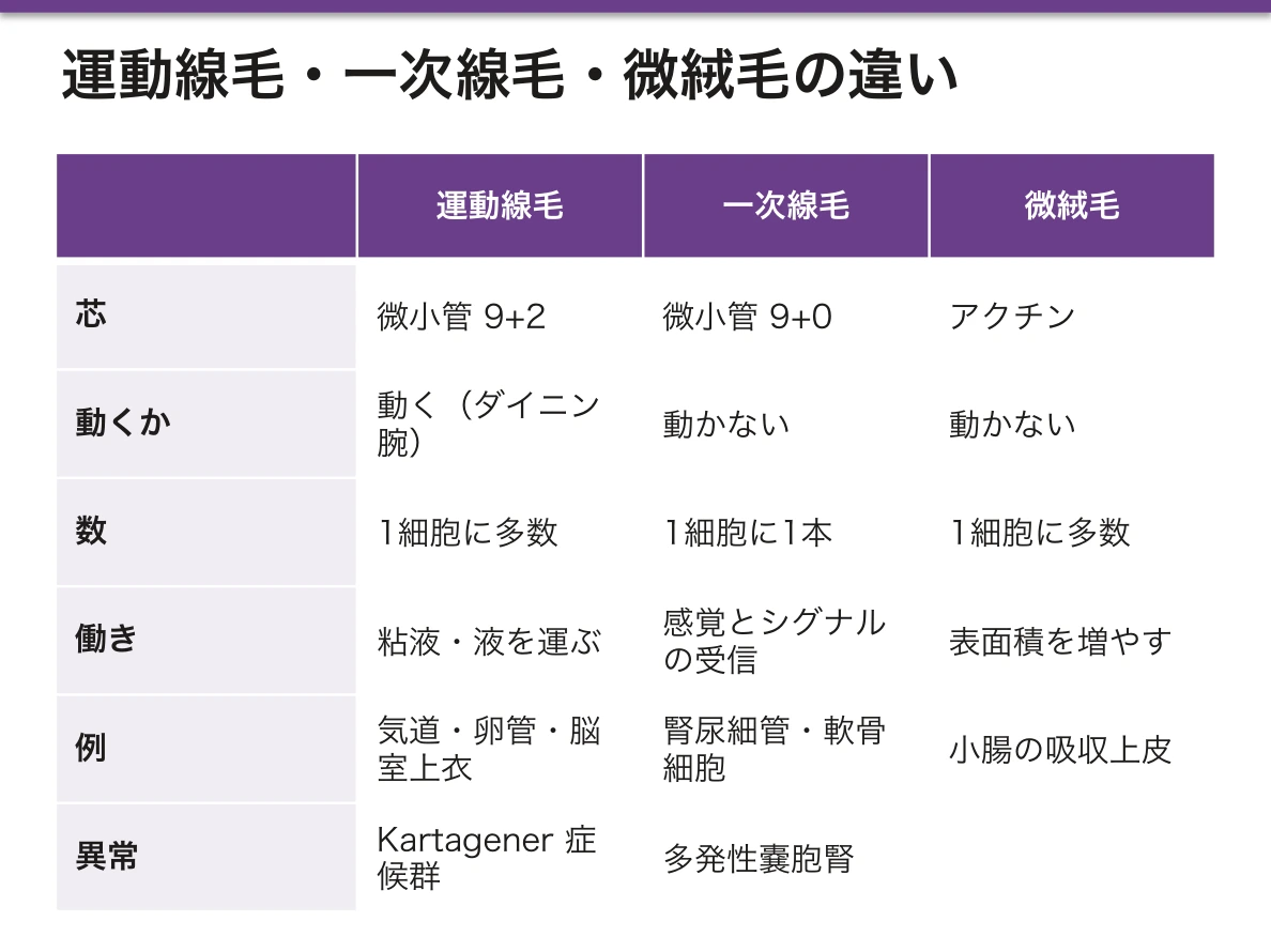
線毛と微絨毛の混同が、この分野で最も多い取り違えになる。線毛は微小管で動く、微絨毛はアクチンで動かない——「太さも成分も働きも別なので、同じ突起として括らない」。
Kartagener 症候群で内臓逆位が起きるのは、胎生期の結節(node)の線毛が体の左右を決めているからになる(左右軸の決定の章)——「呼吸器の病気に内臓逆位が付いてくる理由が、発生にある」。
そして逆位は約半数にしか起きない。線毛が動かないと左右がランダムに決まるからになる——「逆位が無いことは、この症候群を否定しない」。
精子の鞭毛が同じ構造なので、男性不妊を伴う。精子はあるが動かない(男性不妊の章)——「精液検査で数ではなく運動率が落ちる」。
一次線毛の異常は、多発性嚢胞腎を起こす。尿細管の細胞が管腔内の流れを感じ取れなくなる(多発性嚢胞腎の章)——「動かない1本の毛が、腎臓の形を保っていた」。
これらをまとめて繊毛病(ciliopathy)と呼ぶ。腎嚢胞・網膜変性・多指症・肥満・肝線維症が繰り返し組み合わさる——「臓器の組み合わせが奇妙に見えるとき、共通の細胞小器官を疑う」。
同じ 9+2 構造だから、繊毛が動かないと全身で困る。
原発性繊毛運動不全症(Kartagener 症候群) — ダイニン腕の欠損。慢性副鼻腔炎・気管支拡張症・内臓逆位に加え、男性は精子が泳げず不妊、女性は卵管輸送が悪く異所性妊娠が増える。1つの構造の異常で、呼吸器・左右軸・生殖のすべてが説明できる。
喫煙は気道の線毛を麻痺させ、やがて扁平上皮化生で線毛を失わせる。粘液は運べなくなり、咳でしか出せない——喫煙者の咳はこの結果である。
一次線毛(9+0)はアンテナである。多発性嚢胞腎(ポリシスチン)をはじめとする繊毛病(ciliopathy)——網膜色素変性・多指症・腎嚢胞が組み合わさる症候群群は、この不動の線毛の異常による。
English version
Core
Motile cilium — nine doublets plus a central pair (9+2). Dynein arms generate the sliding. At the base is the basal body, nine triplets (9+0).
- Found in the airways, uterine tube, ependyma, and as the flagellum of the spermatozoon (the same structure).
Primary (non-motile) cilium — 9+0, one per cell; a sensory and signalling antenna. Renal tubules, chondrocytes, the outer segment of the retinal photoreceptor.
Centriole — two mutually perpendicular cylinders of nine triplets (9+0); becomes the pole of the spindle.
Immotile cilia syndrome (Kartagener) — absence of the dynein arms: chronic sinusitis, bronchiectasis, situs inversus, male infertility.
Microvilli are actin, not microtubules, and do not move.
Going further
Confusing cilia with microvilli is the commonest error in this area. Cilia are microtubular and move; microvilli are actin and do not. Different diameter, different composition, different function — do not group them as "projections".
Situs inversus occurs in Kartagener syndrome because the cilia of the embryonic node determine left and right. The reason a respiratory disease comes with transposed viscera lies in development. And it occurs in only about half of cases, because when the cilia do not move laterality is decided at random — the absence of situs inversus does not exclude the syndrome.
Because the sperm flagellum has the same structure, male infertility accompanies it: sperm are present but do not move — semen analysis shows reduced motility rather than reduced count.
Defects of the primary cilium cause polycystic kidney disease: the tubular cell can no longer sense flow within the lumen — a single hair that does not move was maintaining the shape of the kidney. Collectively these are the ciliopathies, in which renal cysts, retinal degeneration, polydactyly, obesity and hepatic fibrosis recur together — when a combination of organs looks strange, suspect a shared organelle.
In the clinic
Because the 9+2 structure is the same everywhere, immotile cilia cause trouble throughout the body.
Primary ciliary dyskinesia (Kartagener syndrome) — absence of the dynein arms. Chronic sinusitis, bronchiectasis and situs inversus, and in addition male infertility because the sperm cannot swim and, in women, an increased rate of ectopic pregnancy from poor tubal transport. One structural defect explains the respiratory tract, the left–right axis and reproduction together.
Smoking paralyses the airway cilia and eventually removes them through squamous metaplasia. Mucus can no longer be transported and must be cleared by coughing — the smoker’s cough is the consequence.
The primary cilium (9+0) is an antenna. Polycystic kidney disease (polycystin) and the other ciliopathies — syndromes combining retinitis pigmentosa, polydactyly and renal cysts — arise from defects of this immotile cilium.
上皮細胞は、上と下を知っている — 細胞の極性Polaritas cellularis
上皮細胞はどちらが管腔側でどちらが基底側かを区別して、膜の構成を作り分けている。この非対称が方向を持った輸送を可能にしている。
- 頂端膜——微絨毛・線毛。吸収と分泌の面。SGLT1 のような取り込みの装置が並ぶ
- 側方膜——細胞どうしの接着。タイト結合・接着結合・デスモソーム
- 基底膜側——Na⁺/K⁺-ATPase と、汲み出しの輸送体。基底膜を介して結合組織に接する
タイト結合が「柵」として働き、頂端膜の蛋白が側方へ流れるのを防いでいる。つまり接着装置が、同時に膜の区画を作る仕切りにもなっている。
この配置があって初めて、「腸から血へ」という一方向の輸送が成立する。頂端側で取り込み、基底側で放出する——同じ物質を運ぶのに、両側で別の輸送体が要るのはこのためである。極性が崩れれば、物質は行ったり来たりするだけになる。
極性の喪失が、病気の像として見えてくる。
| 癌 | 極性が失われ、腺の構造が崩れる | 「腺が腺らしく並んでいるか」が分化度の判定そのものになる |
| 虚血の尿細管 | Na⁺/K⁺-ATPase が頂端側へ移動する | ナトリウムの再吸収ができなくなる。急性尿細管壊死の機序の一部 |
| 微絨毛封入体病 | 頂端膜の輸送がうまくいかない | 新生児からの重症の水様下痢 |
極性は上皮だけのものではない。神経細胞は樹状突起と軸索、遊走する細胞は前縁と後縁、骨細胞と破骨細胞は骨に面する側とそれ以外を区別している。「細胞が方向を持っていること」自体が、組織が仕事をするための条件である。
上皮が極性を失って動けるようになる過程を上皮間葉転換と呼び、発生・創傷治癒・線維化・癌の浸潤で共通して使われている。
病理報告の「高分化・低分化」は、もとの組織らしい構造をどれだけ保っているかの判定である。極性の保たれ方が、そのまま分化度になる。
English version
Core
An epithelial cell distinguishes which side faces the lumen and which faces the base, and builds a different membrane on each. This asymmetry is what makes directional transport possible.
- Apical membrane — microvilli and cilia; the surface for absorption and secretion. Uptake systems such as SGLT1 are arranged here.
- Lateral membrane — cell-to-cell adhesion: tight junctions, adherens junctions, desmosomes.
- Basal membrane — Na⁺/K⁺-ATPase and the export transporters; rests on the basal lamina against the connective tissue.
The tight junction acts as a fence, preventing apical proteins from drifting laterally. The adhesion apparatus is at the same time the partition that creates the membrane domains.
Going further
Only with this arrangement does one-way transport from gut to blood become possible: uptake at the apical side, release at the basal side. This is why moving one substance requires a different transporter on each face. If polarity is lost, material simply moves back and forth.
The loss of polarity becomes visible as disease.
| What happens | Consequence | |
|---|---|---|
| Carcinoma | Polarity is lost and the glandular architecture breaks down | Whether glands are still arranged like glands is the grading of differentiation itself |
| Ischaemic tubule | Na⁺/K⁺-ATPase migrates to the apical side | Sodium can no longer be reabsorbed; part of the mechanism of acute tubular necrosis |
| Microvillus inclusion disease | Apical transport fails | Severe watery diarrhoea from the neonatal period |
Polarity is not confined to epithelium. A neuron distinguishes dendrite from axon, a migrating cell its leading from its trailing edge, an osteocyte and an osteoclast the surface facing bone from the rest. That a cell has a direction is itself the condition for a tissue to do work.
The process by which an epithelium loses polarity and becomes able to move is called epithelial–mesenchymal transition, and it is used in common in development, wound healing, fibrosis and tumour invasion.
In the clinic
The terms well differentiated and poorly differentiated in a pathology report are a judgement of how far the tumour retains the architecture of the original tissue. How well polarity is preserved is the grade of differentiation.
まとめ
| 細胞膜 | 脂質二重層に蛋白が浮かぶ流動モザイク構造。 |
|---|---|
| ミトコンドリア | 二重膜構造。外膜は平滑で、内膜が内側に折れ込んでクリスタを作る。 |
| 小胞体 — 粗面と滑面 | 粗面小胞体——リボソームが付く。分泌蛋白と膜蛋白を作る。 |
| Golgi装置と分泌の経路 | 扁平な袋(槽)が重なった構造。極性がある。 |
| 鍍銀で見る Golgi 装置 — 黒い網として見つかった小器官 | HE 染色では Golgi 装置は染まらず、核の脇の明るい抜けとしてしか見えない。 |
| リソソームとペルオキシソーム | リソソーム——加水分解酵素を含む小胞。内部は酸性(pH 約5)で、H⁺ポンプが酸性を保つ。 |
| 細胞骨格 — 3つの太さ | 太さで3種類に分かれ、役割も薬の標的も違う。 |
| 線毛・鞭毛・中心小体 — 9+2 と 9+0 | 運動線毛——微小管が 9対+中央2本(9+2)。 |
| 上皮細胞は、上と下を知っている — 細胞の極性 | 上皮細胞はどちらが管腔側でどちらが基底側かを区別して、膜の構成を作り分けている。 |
このノートは医学生が授業の内容を自分の言葉でまとめたもので、診療の判断に使うものではありません。誤りに気づいたらお知らせください。