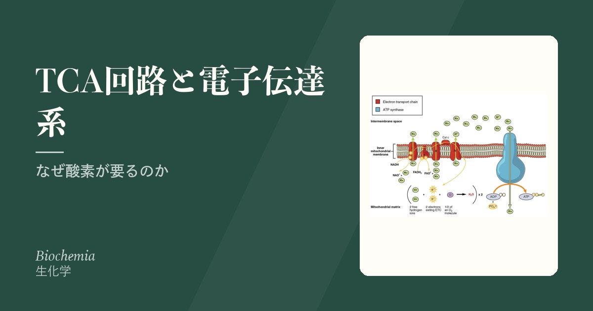
「呼吸で酸素を使う」と言うが、酸素そのものはエネルギーを持っていない。酸素の役割はただ一つ、電子伝達系の最後で電子を受け取ってどくことである。受け手がいなければ電子が詰まり、その手前がすべて止まる。
だから理解の順序はこうなる——栄養素を壊して電子を NADH・FADH₂ に載せる(TCA回路)→ 電子を段階的に渡してH⁺を汲み出す(電子伝達系)→ H⁺が戻る力でATPを作る(ATP合成酵素)。ATPは電子から直接できるのではなく、H⁺の勾配を経由して作られる——これが化学浸透説の核心である。
この章の臨床は「電子の流れがどこで止まるか」で全部説明できる。シアン中毒も、一酸化炭素中毒も、ミトコンドリア病も、褐色脂肪の熱産生も、止まる場所と漏れる場所が違うだけである。
- TCA回路 — 電子を集める回転木馬
- ピルビン酸脱水素酵素複合体 — 3つの酵素の順番と、リン酸化による開け閉め
- クエン酸回路の8段階 — 酵素・クラス・反応の型・調節因子
- スクシニル CoA 合成酵素 — クエン酸回路の中の基質レベルのリン酸化
- 電子伝達系 — 電子を渡してH⁺を汲む
- 電子の運び手 — フラビン・Fe-S クラスター・ユビキノン・シトクロムと Q サイクル
- ATP 合成酵素の回り方 — F₀ が回り、F₁ の3つの部屋が形を変える
- ATP合成と脱共役 — 勾配を熱に変える
- ADP が呼吸の速さを決める — 呼吸調節とアデニル酸キナーゼ
- 細胞質の NADH をミトコンドリアへ — 2つのシャトルと輸送体ファミリー
- ミトコンドリア病 — 母から伝わる病気
- 呼吸鎖を止めるもの — 作用点が違えば、症状も治療も違う
- TCA回路 — 電子を集める回転木馬
- ピルビン酸脱水素酵素複合体 — 3つの酵素の順番と、リン酸化による開け閉め
- クエン酸回路の8段階 — 酵素・クラス・反応の型・調節因子
- スクシニル CoA 合成酵素 — クエン酸回路の中の基質レベルのリン酸化
- 電子伝達系 — 電子を渡してH⁺を汲む
- 電子の運び手 — フラビン・Fe-S クラスター・ユビキノン・シトクロムと Q サイクル
- ATP 合成酵素の回り方 — F₀ が回り、F₁ の3つの部屋が形を変える
- ATP合成と脱共役 — 勾配を熱に変える
- ADP が呼吸の速さを決める — 呼吸調節とアデニル酸キナーゼ
- 細胞質の NADH をミトコンドリアへ — 2つのシャトルと輸送体ファミリー
- ミトコンドリア病 — 母から伝わる病気
- 呼吸鎖を止めるもの — 作用点が違えば、症状も治療も違う
- まとめ
TCA回路 — 電子を集める回転木馬
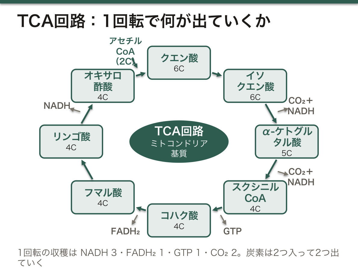
ミトコンドリア基質で回る。アセチルCoA(2炭素)がオキサロ酢酸(4炭素)と結合してクエン酸(6炭素)になり、CO₂を2つ捨てながら一周してオキサロ酢酸に戻る。
1回転あたりの収穫——NADH 3・FADH₂ 1・GTP(=ATP)1・CO₂ 2。
調節点は3か所で、いずれもATP・NADHで抑制、ADP・Ca²⁺で促進される。
- クエン酸シンターゼ
- イソクエン酸デヒドロゲナーゼ(律速)
- α-ケトグルタル酸デヒドロゲナーゼ
TCA回路はエネルギーを作る場所であると同時に、材料の集散地でもある。中間体が抜き取られて他の合成に使われる(cataplerosis)ので、補充(anaplerosis)が要る。
- クエン酸 → 細胞質へ出て脂肪酸合成の材料に
- α-ケトグルタル酸 ⇄ グルタミン酸(アミノ酸代謝の要)
- オキサロ酢酸 ⇄ アスパラギン酸、そして糖新生の入口
- スクシニルCoA → ヘム合成の材料
主な補充路はピルビン酸カルボキシラーゼ(ピルビン酸 → オキサロ酢酸、ビオチン要求)である。
回路が回るにはオキサロ酢酸が要る——これが飢餓でケトン体が出る理由になる。飢餓では糖新生にオキサロ酢酸が奪われ、TCA回路に入りきれないアセチルCoA がケトン体へ流れる。「脂肪はグルコースの炎で燃える」という古い言い回しは、この事情を指している。
α-ケトグルタル酸デヒドロゲナーゼは PDH と同じ5つの補酵素を使う。だからビタミンB1欠乏では TCA回路も同時に止まり、障害が二重にかかる。
English version
Core
Turns in the mitochondrial matrix. Acetyl-CoA (2 carbons) joins oxaloacetate (4 carbons) to make citrate (6 carbons), which goes round shedding two CO₂ and returns to oxaloacetate.
The yield per turn — 3 NADH, 1 FADH₂, 1 GTP (= ATP), 2 CO₂.
Three control points, all inhibited by ATP and NADH and activated by ADP and Ca²⁺.
- Citrate synthase
- Isocitrate dehydrogenase (rate-limiting)
- α-ketoglutarate dehydrogenase
Going further
The TCA cycle is both a place where energy is made and a depot of raw materials. Intermediates are drawn off for other syntheses (cataplerosis), so replenishment (anaplerosis) is required.
- Citrate → leaves for the cytosol as substrate for fatty acid synthesis.
- α-ketoglutarate ⇄ glutamate (the crux of amino acid metabolism).
- Oxaloacetate ⇄ aspartate, and the entrance to gluconeogenesis.
- Succinyl-CoA → substrate for haem synthesis.
The chief replenishing route is pyruvate carboxylase (pyruvate → oxaloacetate, requiring biotin).
The cycle needs oxaloacetate to turn — and this is why ketone bodies appear in starvation. In starvation the oxaloacetate is taken for gluconeogenesis, and the acetyl-CoA that cannot enter the cycle overflows into ketone bodies. The old saying that "fat burns in the flame of carbohydrate" points to exactly this.
α-ketoglutarate dehydrogenase uses the same five coenzymes as PDH. So in thiamine deficiency the TCA cycle stops as well, and the damage is doubled.
ピルビン酸脱水素酵素複合体 — 3つの酵素の順番と、リン酸化による開け閉め
ピルビン酸はミトコンドリア内膜のピルビン酸輸送体で基質に入り、酸化的脱炭酸を受けてアセチル CoA になる。
総反応:ピルビン酸+CoA+NAD⁺ → アセチル CoA+CO₂+NADH+H⁺
| 酵素 | 補酵素(ビタミン) | していること |
|---|---|---|
| E1 ピルビン酸脱水素酵素 | TPP(B1) | 脱炭酸して CO₂ を外し、残った2炭素をヒドロキシエチル基として TPP に載せる |
| E2 ジヒドロリポイルトランスアセチラーゼ | リポ酸・CoA(B5) | 2炭素を酸化してアセチル基にし、リポ酸を経て CoA に渡す → アセチル CoA |
| E3 ジヒドロリポイル脱水素酵素 | FAD(B2)・NAD⁺(B3) | 還元されたリポ酸を FAD で酸化して戻し、電子を NAD⁺ へ → NADH |
リポ酸はリシン残基に共有結合した長い「腕」で、E1→E2→E3 の活性部位を順に回る。中間体が複合体の外に出ずに次の酵素へ渡されるので(基質チャネリング)、反応が速く、副反応が起きにくい。
調節:①産物阻害——アセチル CoA が E2 を、NADH が E3 を阻害する。②リン酸化——PDH キナーゼ(PDK)が E1 をリン酸化すると不活性、PDH ホスファターゼ(PDP)が脱リン酸化すると活性になる。
PDK と PDP を動かすものは、「燃料が足りているか」の信号として読める。
| 活性化するもの | 抑えるもの | 結果 | |
|---|---|---|---|
| PDK(止める側) | アセチル CoA・NADH・ATP | ピルビン酸・ADP | エネルギーが十分なら PDH を止める |
| PDP(開ける側) | Ca²⁺(筋収縮)・Mg²⁺・インスリン(脂肪組織) | — | 収縮中の筋と、食後の脂肪合成で PDH を開ける |
飢餓では PDK(特に PDK4)が増えて PDH が閉じる。脂肪酸からアセチル CoA が来ている間は、ピルビン酸を燃やさずに乳酸・アラニンとして肝へ返し、糖新生の材料に回すためである。PDH は不可逆なので、ここを閉じることが「糖を節約する」ことになる。E3 は α-ケトグルタル酸脱水素酵素と分枝鎖ケト酸脱水素酵素と共通の部品なので、E3 の欠損では3つが同時に止まる。
PDH 欠損症(多くは X 連鎖の E1α 遺伝子の変異)では、ピルビン酸がアセチル CoA に進めず、乳酸とピルビン酸がそろって上がる(乳酸/ピルビン酸比は正常)。電子伝達系の異常では NADH が溜まるので比が上がり、これが鑑別の手がかりになる。治療は、PDH を通らずにアセチル CoA を作れるケトン食と、ビタミン B1 の大量投与(反応する型がある)。PDK を阻害するジクロロ酢酸は PDH を開いて乳酸を下げる薬として研究されてきた。
English version
Core
Pyruvate enters the matrix by the mitochondrial pyruvate carrier and undergoes oxidative decarboxylation to acetyl-CoA.
Overall: pyruvate + CoA + NAD⁺ → acetyl-CoA + CO₂ + NADH + H⁺
| Enzyme | Cofactor (vitamin) | What it does |
|---|---|---|
| E1 pyruvate dehydrogenase | TPP (B1) | Removes CO₂ and holds the remaining 2 carbons on TPP as a hydroxyethyl group |
| E2 dihydrolipoyl transacetylase | Lipoate, CoA (B5) | Oxidises the 2-carbon unit to acetyl and passes it via lipoate to CoA → acetyl-CoA |
| E3 dihydrolipoyl dehydrogenase | FAD (B2), NAD⁺ (B3) | Re-oxidises reduced lipoate with FAD and passes electrons to NAD⁺ → NADH |
Lipoate is a long arm attached to a lysine; it swings through the active sites of E1, E2 and E3. Intermediates never leave the complex (substrate channelling), so the reaction is fast with few side reactions.
Regulation: (1) product inhibition — acetyl-CoA inhibits E2, NADH inhibits E3; (2) phosphorylation — PDH kinase (PDK) phosphorylates E1 and inactivates it; PDH phosphatase (PDP) removes the phosphate and activates it.
Going further
Read the controls of PDK and PDP as ‘is there enough fuel?’.
| Activated by | Inhibited by | Result | |
|---|---|---|---|
| PDK (switches off) | Acetyl-CoA, NADH, ATP | Pyruvate, ADP | Stops PDH when energy is sufficient |
| PDP (switches on) | Ca²⁺ (muscle contraction), Mg²⁺, insulin (adipose tissue) | — | Opens PDH in contracting muscle and for fat synthesis after meals |
In starvation PDK (especially PDK4) rises and PDH closes. While fatty acids supply acetyl-CoA, pyruvate is not burned but returned to the liver as lactate and alanine for gluconeogenesis. Because PDH is irreversible, closing it saves glucose. E3 is shared with α-ketoglutarate dehydrogenase and branched-chain keto acid dehydrogenase, so E3 deficiency stops all three.
In the clinic
In PDH deficiency (often X-linked, E1α gene) pyruvate cannot become acetyl-CoA, so lactate and pyruvate rise together (normal lactate/pyruvate ratio). In respiratory chain defects NADH accumulates and the ratio rises — a useful clue. Treatment is a ketogenic diet, which makes acetyl-CoA without PDH, and high-dose thiamine (some forms respond). Dichloroacetate, which inhibits PDK and opens PDH, has been studied for lowering lactate.
クエン酸回路の8段階 — 酵素・クラス・反応の型・調節因子
| 段階 | 反応 | 酵素(クラス) | 反応の型 | 出るもの・調節 |
|---|---|---|---|---|
| 1 | アセチル CoA+オキサロ酢酸+H₂O → クエン酸+CoA | クエン酸合成酵素(転移酵素) | 縮合(C–C 結合をつくる唯一の段階) | 不可逆。オキサロ酢酸の量で決まる。クエン酸・スクシニル CoA・NADH・ATP で阻害、ADP で促進 |
| 2 | クエン酸 ⇄ イソクエン酸(シス-アコニット酸を経る) | アコニターゼ(脱離酵素) | 脱水 → 水和(異性化) | Fe-S クラスターを持つ |
| 3 | イソクエン酸+NAD⁺ → α-ケトグルタル酸+CO₂+NADH | イソクエン酸脱水素酵素(酸化還元酵素) | 酸化的脱炭酸 | NADH①・CO₂①。ADP・Ca²⁺ で促進、ATP・NADH で阻害 |
| 4 | α-KG+NAD⁺+CoA → スクシニル CoA+CO₂+NADH | α-ケトグルタル酸脱水素酵素複合体(酸化還元酵素) | 酸化的脱炭酸 | NADH②・CO₂②。スクシニル CoA・NADH で阻害、Ca²⁺ で促進。PDH と同じ5つの補酵素 |
| 5 | スクシニル CoA+GDP+Pᵢ ⇄ コハク酸+GTP+CoA | スクシニル CoA 合成酵素(合成酵素) | 基質レベルのリン酸化 | GTP(または ATP) |
| 6 | コハク酸+FAD ⇄ フマル酸+FADH₂ | コハク酸脱水素酵素(酸化還元酵素) | 脱水素(単結合 → 二重結合) | FADH₂。内膜にあり、電子伝達系の複合体 II を兼ねる。マロン酸が競合阻害 |
| 7 | フマル酸+H₂O ⇄ L-リンゴ酸 | フマラーゼ(脱離酵素) | 水和(二重結合への付加) | L 型だけができる |
| 8 | L-リンゴ酸+NAD⁺ ⇄ オキサロ酢酸+NADH | リンゴ酸脱水素酵素(酸化還元酵素) | 脱水素(アルコール → ケトン) | NADH③。平衡は大きく逆向きだが、オキサロ酢酸がすぐ1で使われるので進む |
1回転の総反応:アセチル CoA+3 NAD⁺+FAD+GDP+Pᵢ+2 H₂O → 2 CO₂+3 NADH+3 H⁺+FADH₂+GTP+CoA。
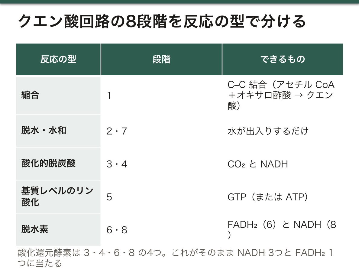
反応の型は、構造の変化を見れば決まる。炭素が CO₂ として抜けて NADH ができれば酸化的脱炭酸(3・4)、H が2つ抜けて二重結合やケトンができれば脱水素(6・8)、水が出入りするだけなら脱水・水和(2・7)、2つの分子がつながれば縮合(1)、高エネルギーのチオエステルを使って GTP を作れば基質レベルのリン酸化(5)。酸化還元酵素は 3・4・6・8 の4つで、これがそのまま NADH 3つと FADH₂ 1つに当たる。
律速はどこか。ヨーロッパの教科書(Lehninger 系)は、オキサロ酢酸の濃度に左右されるクエン酸合成酵素を回路の速さを決める段階として強調し、日本の教科書の多くはイソクエン酸脱水素酵素を律速酵素とする。どちらも不可逆で調節を受ける3段階(1・3・4)に含まれ、条件によってどこが律速にもなりうる。共通する原則は「基質があれば進み、産物(NADH・ATP・スクシニル CoA)が溜まれば止まり、ADP と Ca²⁺ が来れば加速する」である。Ca²⁺ は筋収縮の合図なので、ATP を使い始める前から回路を速めておける。
アセチル CoA の2炭素は、その回転では CO₂ にならない。3・4で外れる CO₂ の炭素は、もとのオキサロ酢酸に由来する。
殺鼠剤のフルオロ酢酸は、体内でフルオロクエン酸になってアコニターゼを止め、回路を断つ。フマラーゼや コハク酸脱水素酵素の遺伝的な欠損は、溜まったフマル酸・コハク酸が「がん代謝物」として働き、腎細胞癌・子宮筋腫(フマラーゼ)や褐色細胞腫・パラガングリオーマ(コハク酸脱水素酵素)の原因になる。
English version
Core
| Step | Reaction | Enzyme (class) | Reaction type | Output / regulation |
|---|---|---|---|---|
| 1 | Acetyl-CoA + oxaloacetate + H₂O → citrate + CoA | Citrate synthase (transferase) | Condensation (the only C–C bond formation) | Irreversible; set by oxaloacetate; inhibited by citrate, succinyl-CoA, NADH, ATP; activated by ADP |
| 2 | Citrate ⇄ isocitrate (via cis-aconitate) | Aconitase (lyase) | Dehydration → hydration (isomerisation) | Has an Fe-S cluster |
| 3 | Isocitrate + NAD⁺ → α-ketoglutarate + CO₂ + NADH | Isocitrate dehydrogenase (oxidoreductase) | Oxidative decarboxylation | NADH 1, CO₂ 1; activated by ADP, Ca²⁺; inhibited by ATP, NADH |
| 4 | α-KG + NAD⁺ + CoA → succinyl-CoA + CO₂ + NADH | α-Ketoglutarate dehydrogenase complex (oxidoreductase) | Oxidative decarboxylation | NADH 2, CO₂ 2; inhibited by succinyl-CoA, NADH; activated by Ca²⁺; same five cofactors as PDH |
| 5 | Succinyl-CoA + GDP + Pᵢ ⇄ succinate + GTP + CoA | Succinyl-CoA synthetase (ligase) | Substrate-level phosphorylation | GTP (or ATP) |
| 6 | Succinate + FAD ⇄ fumarate + FADH₂ | Succinate dehydrogenase (oxidoreductase) | Dehydrogenation (single → double bond) | FADH₂; in the inner membrane, also complex II; malonate is a competitive inhibitor |
| 7 | Fumarate + H₂O ⇄ L-malate | Fumarase (lyase) | Hydration (addition to a double bond) | Only L-malate |
| 8 | L-Malate + NAD⁺ ⇄ oxaloacetate + NADH | Malate dehydrogenase (oxidoreductase) | Dehydrogenation (alcohol → ketone) | NADH 3; equilibrium lies far back, but step 1 removes oxaloacetate at once |
One turn: acetyl-CoA + 3 NAD⁺ + FAD + GDP + Pᵢ + 2 H₂O → 2 CO₂ + 3 NADH + 3 H⁺ + FADH₂ + GTP + CoA.
Going further
The reaction type follows from the change in structure. Carbon leaves as CO₂ and NADH forms: oxidative decarboxylation (3, 4). Two H leave and a double bond or ketone forms: dehydrogenation (6, 8). Water only goes in or out: dehydration/hydration (2, 7). Two molecules join: condensation (1). A thioester’s energy makes GTP: substrate-level phosphorylation (5). The four oxidoreductases (3, 4, 6, 8) give exactly the 3 NADH and 1 FADH₂.
Which step is rate-limiting? European (Lehninger-style) teaching stresses citrate synthase, which depends on oxaloacetate; many Japanese textbooks name isocitrate dehydrogenase. Both are among the three irreversible, regulated steps (1, 3, 4), and each can limit the rate under some conditions. The shared rule: it runs when substrate is present, stops when products (NADH, ATP, succinyl-CoA) build up, and speeds up with ADP and Ca²⁺. Ca²⁺ signals contraction, so the cycle speeds up even before ATP is used.
The two carbons of acetyl-CoA are not lost as CO₂ in the same turn; the carbons released in steps 3 and 4 come from the original oxaloacetate.
In the clinic
The rat poison fluoroacetate becomes fluorocitrate in the body and blocks aconitase, breaking the cycle. Inherited defects of fumarase and succinate dehydrogenase let fumarate and succinate accumulate as ‘oncometabolites’, causing renal cell carcinoma and uterine leiomyomas (fumarase) or phaeochromocytoma and paraganglioma (SDH).
スクシニル CoA 合成酵素 — クエン酸回路の中の基質レベルのリン酸化
α-ケトグルタル酸脱水素酵素が作ったスクシニル CoA は、チオエステル結合(–CO–S–CoA)に大きなエネルギーを持っている(加水分解の ΔG°′ 約 −33 kJ/mol、ATP とほぼ同じ)。スクシニル CoA 合成酵素はこのエネルギーで、GDP(または ADP)にリン酸を付けて GTP(ATP)を作る。クエン酸回路で、膜も酸素も使わずに高エネルギーのリン酸ができる唯一の段階である。
反応は3段階で進む:①Pᵢ が CoA を追い出してスクシニルリン酸ができる、②そのリン酸が酵素のヒスチジン残基に移る(リン酸化酵素の中間体)、③ヒスチジンから GDP へリン酸が渡る。
名前は逆向き(GTP を使ってスクシニル CoA を「合成する」向き)で付けられているので、クラスは合成酵素(リガーゼ)になる。反応は可逆で、産物のコハク酸がすぐ次へ使われることで前へ進む。
GTP か ATP かは、組織によって違う。この酵素には GTP を作る型と ATP を作る型があり、肝や腎のように糖新生をする組織では GTP 型が多い。糖新生の酵素 PEP カルボキシキナーゼは GTP を使うので、理にかなっている。脳・骨格筋・心筋のように ATP をそのまま使う組織では ATP 型が多い。GTP はヌクレオシド二リン酸キナーゼで ATP とリン酸を交換できるので、エネルギーの量としてはどちらも同じ1つと数える。
スクシニル CoA は回路の外でも使われる。ヘム合成の材料(グリシンと縮合)になり、肝以外の組織ではケトン体を使うときに CoA を貸す(スクシニル CoA:3-ケト酸 CoA 転移酵素)。このとき CoA を渡したスクシニル CoA は GTP を作らずにコハク酸になるので、ケトン体を燃やすたびに GTP 1つぶんを払っていることになる。肝はこの転移酵素を持たないので、自分で作ったケトン体を使えない。
ATP 型のスクシニル CoA 合成酵素の遺伝子(SUCLA2)の変異では、ミトコンドリア DNA が減ってしまう病気(ミトコンドリア DNA 枯渇症候群)が起き、乳児期からの筋緊張低下・難聴・脳症と、軽いメチルマロン酸尿を示す。メチルマロン酸が出るのは、溜まったスクシニル CoA がメチルマロニル CoA の側へ反応を押し戻すためと考えられている。
English version
Core
Succinyl-CoA, made by α-ketoglutarate dehydrogenase, holds much energy in its thioester bond (ΔG°′ of hydrolysis about −33 kJ/mol, close to ATP). Succinyl-CoA synthetase uses it to add phosphate to GDP (or ADP), making GTP (ATP) — the only step in the cycle that makes a high-energy phosphate without a membrane or oxygen.
Three steps: (1) Pᵢ displaces CoA, giving succinyl phosphate; (2) the phosphate moves to a histidine of the enzyme (phospho-enzyme intermediate); (3) histidine passes the phosphate to GDP.
The name describes the reverse reaction (using GTP to ‘synthesise’ succinyl-CoA), so the class is ligase. The reaction is reversible and is pulled forward because succinate is used at once.
Going further
GTP or ATP depends on the tissue. One form makes GTP and one makes ATP. Gluconeogenic tissues such as liver and kidney have more of the GTP form, which suits PEP carboxykinase, a GTP-using enzyme of gluconeogenesis. Brain, skeletal muscle and heart, which use ATP directly, have more of the ATP form. Nucleoside diphosphate kinase exchanges phosphate between GTP and ATP, so both count as one high-energy phosphate.
Succinyl-CoA is also used outside the cycle: it condenses with glycine for haem synthesis, and in tissues other than the liver it lends CoA to activate ketone bodies (succinyl-CoA:3-ketoacid CoA transferase). That succinyl-CoA becomes succinate without making GTP, so burning ketone bodies costs one GTP each time. The liver lacks this transferase and cannot use the ketone bodies it makes.
In the clinic
Variants of the ATP-forming subunit gene (SUCLA2) cause a mitochondrial DNA depletion syndrome: hypotonia from infancy, deafness, encephalopathy and mild methylmalonic aciduria. Methylmalonic acid is thought to appear because accumulated succinyl-CoA pushes the reaction back towards methylmalonyl-CoA.
電子伝達系 — 電子を渡してH⁺を汲む
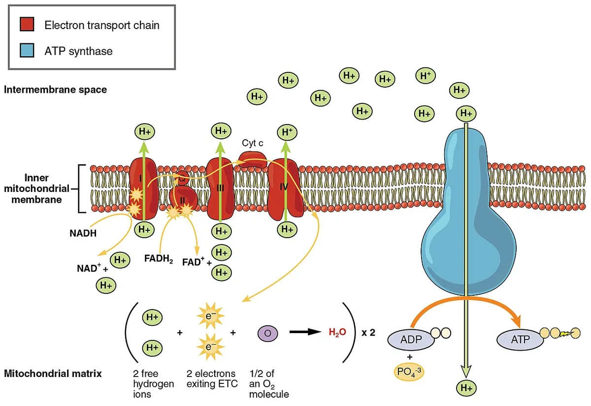
ミトコンドリア内膜に並ぶ4つの複合体。NADHとFADH₂が持ってきた電子を順に渡し、そのエネルギーでH⁺を基質側から膜間腔へ汲み出す。
- 複合体I(NADH→ユビキノン)——H⁺を汲む
- 複合体II(コハク酸→ユビキノン。TCA回路のコハク酸デヒドロゲナーゼそのもの)——H⁺を汲まない
- 複合体III(ユビキノン→シトクロムc)——汲む
- 複合体IV(シトクロムc→O₂)——汲む。ここで水ができる
FADH₂ が複合体II から入る=複合体I を飛ばすので、汲めるH⁺が少なく、得られるATPも少ない(NADH ≒2.5 ATP、FADH₂ ≒1.5 ATP)。
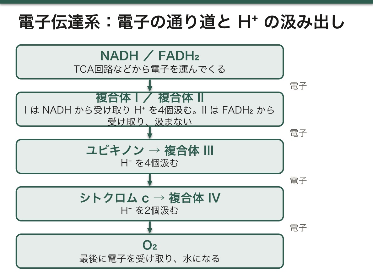
電子は酸化還元電位の低いほうから高いほうへしか流れない。最終受容体である酸素は電位が最も高く、だから電子を引き抜ける。酸素は「燃やす」のではなく「電子を受け取って引きずり出す」役である。
したがって複合体IV が止まると、上流の全部が止まる。これが毒物の作用点である。
- シアン・アジ化物・硫化水素——複合体IV を阻害。細胞は酸素があっても使えない。動脈血と静脈血の酸素差が縮まり静脈血が鮮紅色になる。乳酸アシドーシスが急激に進む
- 一酸化炭素——ヘモグロビンへの結合(酸素運搬の阻害)に加え、複合体IV も阻害する
- ロテノン・バルビツール酸——複合体I
- アンチマイシンA——複合体III
- オリゴマイシン——ATP合成酵素そのもの
シアン中毒の治療がこの理解の上に立つ。ヒドロキソコバラミンがシアンを直接捕まえるほか、亜硝酸でメトヘモグロビンを作り、シアンをそちらへ引き剥がすという方法が使われる(複合体IV より Fe³⁺ への親和性が高いことを利用する)。
English version
Core
Four complexes in the inner mitochondrial membrane. The electrons brought by NADH and FADH₂ are passed along in turn, and the energy pumps H⁺ from the matrix into the intermembrane space.
- Complex I (NADH → ubiquinone) — pumps H⁺.
- Complex II (succinate → ubiquinone; succinate dehydrogenase of the TCA cycle itself) — does not pump.
- Complex III (ubiquinone → cytochrome c) — pumps.
- Complex IV (cytochrome c → O₂) — pumps. Water is formed here.
FADH₂ enters at complex II and so skips complex I, pumping fewer H⁺ and yielding less ATP (NADH ≈ 2.5 ATP, FADH₂ ≈ 1.5 ATP).
Going further
Electrons flow only from lower to higher redox potential. Oxygen, the terminal acceptor, has the highest potential and can therefore pull the electrons through. Oxygen does not "burn" anything; it accepts the electrons and draws them out.
So when complex IV stops, everything upstream stops. This is the point of attack of the poisons.
- Cyanide, azide, hydrogen sulphide — inhibit complex IV. The cell cannot use oxygen although it is there. The arteriovenous oxygen difference narrows and the venous blood is bright red. Lactic acidosis advances rapidly.
- Carbon monoxide — besides binding haemoglobin (blocking oxygen carriage), it also inhibits complex IV.
- Rotenone, barbiturates — complex I. Antimycin A — complex III. Oligomycin — ATP synthase itself.
The treatment of cyanide poisoning rests on this understanding. Hydroxocobalamin captures cyanide directly; alternatively nitrites are used to make methaemoglobin and pull the cyanide away onto it (exploiting its greater affinity for Fe³⁺ than for complex IV).
電子の運び手 — フラビン・Fe-S クラスター・ユビキノン・シトクロムと Q サイクル
電子伝達系の部品は、電子を2個まとめて運ぶものと、1個ずつ運ぶものに分かれる。この違いが、部品の並び方を決めている。
| 運び手 | 運ぶ電子 | どこにあるか | 特徴 |
|---|---|---|---|
| NAD⁺/NADH | 2個(ヒドリドイオン H⁻ として) | 基質に溶けている | ニコチンアミド環が H⁻ を受け取る。複合体 I への入口 |
| FMN・FAD(フラビン) | 1個でも2個でも | 複合体 I(FMN)・II(FAD) | イソアロキサジン環。2電子を受け取り1電子ずつ渡す「じょうご」 |
| Fe-S クラスター | 1個 | 複合体 I・II・III | 鉄と硫黄が同数。Fe³⁺ ⇄ Fe²⁺ で1電子を運ぶ |
| ユビキノン(CoQ) | 1個でも2個でも | 内膜の脂質の中を動く | Q → セミキノン(Q·⁻)→ ユビキノール(QH₂)。電子と一緒に H⁺ も運ぶ |
| シトクロム(a・b・c) | 1個 | 複合体 III・IV、シトクロム c は膜間腔 | ヘムの鉄が Fe³⁺ ⇄ Fe²⁺。シトクロム c は水溶性で、III から IV へ1電子ずつ運ぶ |
| Cu(CuA・CuB) | 1個 | 複合体 IV | ヘム a₃ と組んで O₂ を受け取る |
移動できる運び手は2つ——膜の中を動くユビキノンと、膜の外(膜間腔)を動くシトクロム c。複合体どうしは直接つながらず、この2つが電子を渡し歩く。
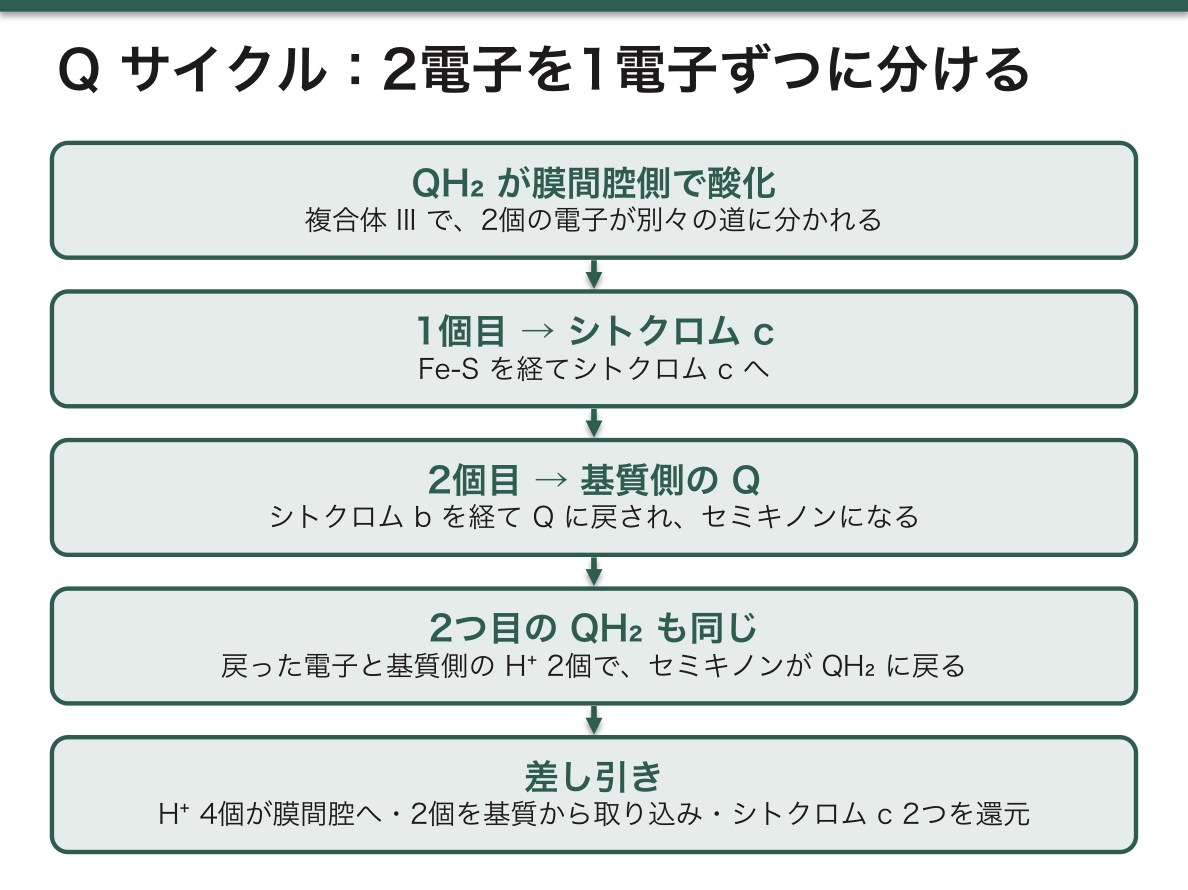
Q サイクルは、2電子の運び手(QH₂)から1電子の運び手(シトクロム c)へ電子を渡すための仕組みである。複合体 III で QH₂ が膜間腔側で酸化されると、2個の電子は別々の道に分かれる。1個は Fe-S を経てシトクロム c へ、もう1個はシトクロム b を経て基質側の Q に戻され、セミキノンになる。2つ目の QH₂ でも同じことが起き、戻された2個目の電子と基質側の H⁺ 2個でセミキノンが QH₂ に戻る。差し引くと、QH₂ が1つ酸化されるごとに H⁺ が4個膜間腔へ出て、2個が基質から取り込まれ、シトクロム c が2つ還元される。
各複合体が膜間腔へ出す H⁺ は、電子2個あたり複合体 I が4個、II が0個、III が4個、IV が2個。NADH から入れば 4+4+2=10個、FADH₂(複合体 II)から入れば 4+2=6個で、この差がそのまま ATP の収量の差(約2.5 と 1.5)になる。勾配は、ポンプで汲み出すこと(I・IV)と、基質側で H⁺ を化学的に使い(Q の還元・O₂ の還元)、膜間腔側で放す(QH₂ の酸化)ことの両方で作られる。
コエンザイム Q10 は体内で作られ、その合成はコレステロールと同じメバロン酸経路の途中から分かれる。だからスタチンは CoQ10 の合成も下げる。スタチンの筋症状との関係が議論されてきたが、補充の効果ははっきりしていない。原発性の CoQ10 欠乏症はまれだが、補充で改善しうるミトコンドリア病として重要である。
English version
Core
Parts of the respiratory chain carry either two electrons together or one at a time; this difference decides their order.
| Carrier | Electrons | Location | Notes |
|---|---|---|---|
| NAD⁺/NADH | 2 (as hydride, H⁻) | Dissolved in the matrix | Nicotinamide ring takes H⁻; entry to complex I |
| FMN, FAD (flavins) | 1 or 2 | Complex I (FMN), II (FAD) | Isoalloxazine ring; takes 2 electrons and passes them 1 at a time (‘funnel’) |
| Fe-S clusters | 1 | Complexes I, II, III | Equal numbers of Fe and S; Fe³⁺ ⇄ Fe²⁺ |
| Ubiquinone (CoQ) | 1 or 2 | Moves within the membrane lipid | Q → semiquinone (Q·⁻) → ubiquinol (QH₂); carries H⁺ with the electrons |
| Cytochromes (a, b, c) | 1 | Complexes III, IV; cytochrome c in the intermembrane space | Haem iron Fe³⁺ ⇄ Fe²⁺; water-soluble cytochrome c carries one electron from III to IV |
| Copper (CuA, CuB) | 1 | Complex IV | With haem a₃, accepts O₂ |
There are two mobile carriers: ubiquinone moving in the membrane and cytochrome c moving outside it (intermembrane space). The complexes do not touch; these two carry electrons between them.
Going further
The Q cycle passes electrons from a two-electron carrier (QH₂) to a one-electron carrier (cytochrome c). When QH₂ is oxidised on the intermembrane side of complex III, its two electrons split: one goes via Fe-S to cytochrome c, the other via cytochrome b back to a Q on the matrix side, making a semiquinone. A second QH₂ does the same; the second returned electron and two matrix H⁺ turn the semiquinone back into QH₂. Net: for each QH₂ oxidised, 4 H⁺ go out, 2 H⁺ are taken from the matrix, and 2 cytochrome c are reduced.
H⁺ moved out per two electrons: complex I 4, II 0, III 4, IV 2. From NADH: 4 + 4 + 2 = 10; from FADH₂ (complex II): 4 + 2 = 6. This difference is the difference in ATP yield (about 2.5 and 1.5). The gradient is built both by pumping (I, IV) and by using H⁺ chemically on the matrix side (reducing Q and O₂) and releasing it outside (oxidising QH₂).
In the clinic
Coenzyme Q10 is made in the body; its synthesis branches from the mevalonate pathway, as cholesterol does. Statins therefore also lower CoQ10. A link with statin muscle symptoms has been debated, but the benefit of supplements is unclear. Primary CoQ10 deficiency is rare but important as a mitochondrial disease that can improve with supplementation.
ATP 合成酵素の回り方 — F₀ が回り、F₁ の3つの部屋が形を変える
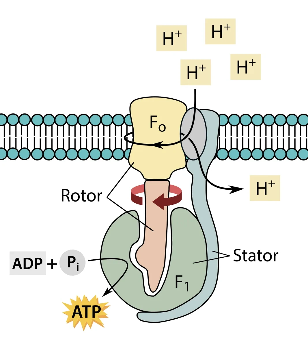
ATP 合成酵素(複合体 V)は2つの部分でできている。
| 部分 | 場所 | 部品 | 働き |
|---|---|---|---|
| F₀ | 内膜に埋まる | c サブユニットの輪(c リング)と a サブユニット | H⁺ が膜間腔から基質へ通る道。H⁺ が通るたびに c リングが回る。オリゴマイシンが結合する(名前の o の由来) |
| F₁ | 基質に突き出す | α₃β₃ の6量体と、中心の軸 γ | β サブユニットの3か所で ATP を作る |
結合変化機構:H⁺ の流れで c リングと γ 軸が回ると、偏った形の γ が αβ の3つの組を順に押し、それぞれの形を変える。3つの組は常に違う状態にある——O(開いた形・空)・L(ゆるい形・ADP と Pᵢ を抱える)・T(きつい形・ATP をつくって抱える)。γ が 120 度回るごとに T が O に変わって ATP が放出され、1回転で ATP が3つできる。
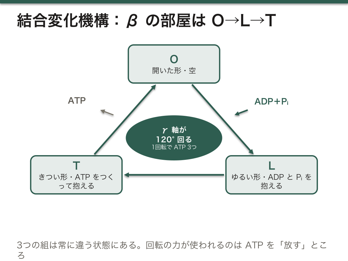
エネルギーが使われるのは、ATP を「つくる」ことではなく「放す」ことである。T の部屋の中では ADP+Pᵢ ⇄ ATP の平衡がほぼ1で、ATP は自然にできる。ATP がきつく結合しているので、それを部屋から外すのに回転の力が要る。これは、ATP のエネルギーの大部分が溶液中での「濃度と溶媒和」の差から来ることと対応している。
ATP 1つに H⁺ はいくつ要るか。ヒトの c リングは c サブユニット8個なので、1回転(ATP 3つ)に H⁺ 8個、ATP 1つあたり約2.7個。これに、ADP と Pᵢ を基質へ運び込み ATP を外へ出すための H⁺ 約1個(ATP/ADP 交換体とリン酸輸送体)を足して、約4個になる。NADH の10個 ÷ 4 ≒ 2.5、FADH₂ の6個 ÷ 4 ≒ 1.5 という P/O 比はここから出ている。
勾配が無くなると、この酵素は逆に回って ATP を分解し、H⁺ を汲み出してしまう。酸素が来ないときに ATP を無駄にしないため、阻害蛋白 IF1 が pH の低下(電子伝達が止まると基質が酸性に傾く)を合図に F₁ に結合して逆回転を止める。
心筋梗塞や脳梗塞の虚血では、電子伝達が止まり勾配が崩れる。ATP 合成酵素が逆回転すると残った ATP まで失われるので、IF1 の働きが細胞の生き残りに関わると考えられている。回転する分子モーターであることは、1997年のノーベル化学賞(Boyer・Walker)の対象になった。
English version
Core
ATP synthase (complex V) has two parts.
| Part | Location | Components | Function |
|---|---|---|---|
| F₀ | In the inner membrane | Ring of c subunits (c-ring) and subunit a | Path for H⁺ from the intermembrane space to the matrix; the c-ring turns as H⁺ passes; binds oligomycin (hence the ‘o’) |
| F₁ | Projects into the matrix | α₃β₃ hexamer and central γ shaft | Makes ATP at three sites on the β subunits |
Binding-change mechanism: H⁺ flow turns the c-ring and the γ shaft; the asymmetric γ pushes each αβ pair in turn and changes its shape. The three pairs are always in different states — O (open, empty), L (loose, holding ADP + Pᵢ), T (tight, making and holding ATP). Each 120° turn changes T to O and releases ATP; one full turn makes 3 ATP.
Going further
Energy is used to release ATP, not to make it. Inside the T site the equilibrium ADP + Pᵢ ⇄ ATP is close to 1, so ATP forms by itself. Because ATP is bound tightly, rotation is needed to free it.
How many H⁺ per ATP? The human c-ring has 8 c subunits: 8 H⁺ per turn (3 ATP), about 2.7 H⁺ per ATP. Add about 1 H⁺ to bring ADP and Pᵢ in and send ATP out (ATP/ADP translocase and phosphate carrier): about 4. NADH’s 10 H⁺ ÷ 4 ≈ 2.5 and FADH₂’s 6 ÷ 4 ≈ 1.5 — the P/O ratios.
If the gradient disappears, the enzyme runs backwards, splitting ATP to pump H⁺ out. To avoid wasting ATP without oxygen, the inhibitor protein IF1 binds F₁ when the matrix becomes acidic (as happens when electron transport stops) and blocks reverse rotation.
In the clinic
In ischaemia (myocardial or cerebral infarction) electron transport stops and the gradient collapses. Reverse rotation would waste the remaining ATP, so IF1 is thought to matter for cell survival. The rotary motor was the subject of the 1997 Nobel Prize in Chemistry (Boyer, Walker).
ATP合成と脱共役 — 勾配を熱に変える
電子伝達で作られたH⁺の勾配(プロトン駆動力)が、ATP合成酵素(複合体V)を回してATPを作る。化学浸透説。
だから電子伝達とATP合成は「勾配」を介してつながっているだけで、直結してはいない。両者を切り離すことを脱共役という。
脱共役すると——電子は流れ、酸素は消費されるが、ATPはできず熱になる。
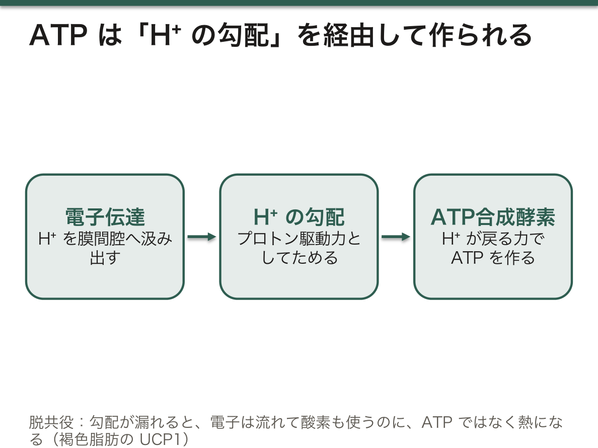
褐色脂肪組織はこれを生理的に利用している。UCP1(サーモゲニン)がH⁺を素通りさせ、勾配を熱に変える。新生児がふるえずに体温を保てるのはこの仕組みによる。
病的な脱共役は危険である。2,4-ジニトロフェノール(DNP)は膜を通れる弱酸としてH⁺を運び込み、勾配を潰す。かつて痩身薬として使われたが、致死的な高体温で禁止された。「痩せる」のはATPが作れず基質を燃やし続けるためで、原理としては正しいが制御が効かない。
アスピリンの大量服用でも軽度の脱共役が起きるため、高体温が中毒症状に含まれる。同時に呼吸中枢刺激による呼吸性アルカローシスと、代謝阻害による代謝性アシドーシスが併存する——混合性酸塩基平衡異常の代表例になる。
収支をまとめると、グルコース1分子から約30〜32 ATP(シャトルの種類で変わる)。嫌気的解糖の2 ATPと比べて15倍以上。酸素があることの意味はこの効率にある。
English version
Core
The H⁺ gradient made by electron transport (the proton-motive force) turns ATP synthase (complex V) and makes ATP — the chemiosmotic theory.
So electron transport and ATP synthesis are connected only through the gradient and are not directly coupled. Separating them is uncoupling.
When uncoupled, electrons flow and oxygen is consumed, but no ATP is made and the energy becomes heat.
Going further
Brown adipose tissue uses this physiologically. UCP1 (thermogenin) lets H⁺ pass straight through and turns the gradient into heat. This is how the newborn maintains its temperature without shivering.
Pathological uncoupling is dangerous. 2,4-dinitrophenol (DNP), a membrane-permeable weak acid, carries H⁺ in and destroys the gradient. Once sold as a slimming agent, it was banned after fatal hyperthermia. It causes weight loss because no ATP can be made and substrate is burnt continuously — the principle is correct but there is no control over it.
A large overdose of aspirin also causes mild uncoupling, so hyperthermia is among the features of poisoning. At the same time respiratory alkalosis from stimulation of the respiratory centre coexists with metabolic acidosis from the metabolic block — the classic mixed acid–base disturbance.
The balance: about 30–32 ATP from one molecule of glucose (varying with the shuttle used). More than fifteen times the 2 ATP of anaerobic glycolysis. The meaning of having oxygen lies in that efficiency.
ADP が呼吸の速さを決める — 呼吸調節とアデニル酸キナーゼ
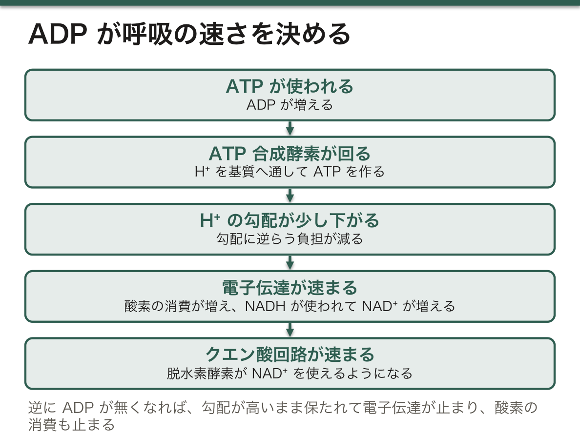
ミトコンドリアの酸素の消費速度は、主に ADP(と Pᵢ)がどれだけあるかで決まる。これを呼吸調節(受容体調節)という。
流れは一続きである。ATP が使われる → ADP が増える → ATP 合成酵素が H⁺ を基質へ通して ATP を作る → H⁺ の勾配が少し下がる → 勾配に逆らう負担が減って電子伝達が速まる → NADH が使われて NAD⁺ が増える → クエン酸回路の脱水素酵素が速まる。逆に ADP が無くなれば、勾配が高いまま保たれて電子伝達が止まり、酸素の消費も止まる。
アデニル酸キナーゼ(ミオキナーゼ)は、2 ADP ⇄ ATP+AMP を触媒する。ADP に残った2つ目のリン酸も ATP に回せるので、激しく ATP を使う筋で役に立つ。クレアチンキナーゼ(クレアチンリン酸+ADP ⇄ クレアチン+ATP)とともに、ATP の濃度を一定に保つ緩衝の働きをする。
アデニル酸キナーゼは、エネルギー不足の信号を増幅する。細胞の中では ATP が AMP よりはるかに多いので、ATP がほんの数%減っただけでも、この反応の平衡を通して AMP は何倍にも増える。だから代謝酵素の多くは ATP ではなく AMP を見張っている——PFK-1 と筋のグリコーゲンホスホリラーゼは AMP で活性化され、AMP 活性化プロテインキナーゼ(AMPK)は、糖の取り込みと脂肪酸の酸化を進め、合成系を止める。
呼吸調節が外れた状態が脱共役である。H⁺ が ATP 合成酵素を通らずに漏れると、ADP が無くても電子伝達が走り続け、酸素は消費されるのに ATP はできない(熱になる)。単離したミトコンドリアでは、ADP を加えたときと使い切ったときの酸素消費の比(呼吸調節比)が、膜がどれだけ健全かの指標になる。
ほかにも、Ca²⁺ が PDH ホスファターゼと TCA 回路の脱水素酵素を直接速め、ATP の需要が増える前から供給を準備する。
メトホルミンは複合体 I を軽く抑えて肝の ATP を減らし、AMP を増やすことで AMPK を活性化し、糖新生を抑えると考えられている。ミトコンドリアのアデニル酸キナーゼ2の欠損は、白血球が作られない重症複合免疫不全症(細網異形成症)と難聴を起こす。
English version
Core
The rate at which mitochondria use oxygen is set mainly by how much ADP (and Pᵢ) is available: respiratory (acceptor) control.
The chain: ATP is used → ADP rises → ATP synthase lets H⁺ into the matrix to make ATP → the gradient falls a little → electron transport has less to push against and speeds up → NADH is used, NAD⁺ rises → TCA dehydrogenases speed up. When ADP runs out, the gradient stays high, electron transport stops, and so does oxygen use.
Adenylate kinase (myokinase) catalyses 2 ADP ⇄ ATP + AMP. It lets the second phosphate of ADP be used too, which helps muscle that uses ATP fast. With creatine kinase (phosphocreatine + ADP ⇄ creatine + ATP) it buffers the ATP concentration.
Going further
Adenylate kinase amplifies the low-energy signal. Cells contain much more ATP than AMP, so a fall of only a few per cent in ATP raises AMP several-fold through this equilibrium. That is why many enzymes watch AMP, not ATP: PFK-1 and muscle glycogen phosphorylase are activated by AMP, and AMP-activated protein kinase (AMPK) increases glucose uptake and fatty acid oxidation and switches off synthesis.
Uncoupling is respiratory control lost. If H⁺ leaks past ATP synthase, electron transport runs without ADP; oxygen is used but no ATP is made (heat instead). In isolated mitochondria the ratio of oxygen use with ADP to that after ADP is used up (respiratory control ratio) shows how intact the membrane is.
Ca²⁺ also directly speeds PDH phosphatase and the TCA dehydrogenases, preparing supply before demand rises.
In the clinic
Metformin is thought to inhibit complex I slightly, lower hepatic ATP, raise AMP and activate AMPK, reducing gluconeogenesis. Deficiency of mitochondrial adenylate kinase 2 causes reticular dysgenesis, a severe combined immunodeficiency with no white cells, together with deafness.
細胞質の NADH をミトコンドリアへ — 2つのシャトルと輸送体ファミリー
ミトコンドリア内膜は NADH も NAD⁺ も通さない。解糖でできた細胞質の NADH は、電子だけを別の分子に載せ替えて運び込む。方法は2つある。
| リンゴ酸-アスパラギン酸シャトル | グリセロール3-リン酸シャトル | |
|---|---|---|
| 細胞質で | リンゴ酸脱水素酵素がオキサロ酢酸を NADH で還元 → リンゴ酸 | グリセロール3-リン酸脱水素酵素が DHAP を NADH で還元 → グリセロール3-リン酸 |
| 膜を越える | リンゴ酸が α-ケトグルタル酸と交換で入る | 膜を越えない。内膜の外側の面にある酵素が直接受け取る |
| 基質側で | リンゴ酸 → オキサロ酢酸で NADH に戻る → 複合体 I | ミトコンドリアの酵素が FAD に電子を渡す → ユビキノン(複合体 I を飛ばす) |
| 戻し方 | オキサロ酢酸は膜を通れないので、アミノ基転移(AST)でアスパラギン酸にして、グルタミン酸と交換で外へ出す | DHAP が細胞質に残るので戻す必要がない |
| NADH 1つあたり | 約2.5 ATP | 約1.5 ATP |
| 向き・主な組織 | 酵素の反応は可逆(Asp/Glu 交換体は一方向に働く)。肝・心筋・腎 | 不可逆。骨格筋(速筋)・脳・褐色脂肪 |
| グルコース1分子 | 約32 ATP | 約30 ATP |
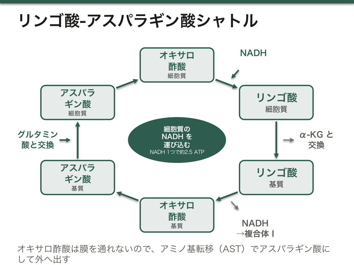
なぜ損をするシャトルがあるのか。グリセロール3-リン酸シャトルは電子を FAD に渡すので、エネルギーの一部を捨てる。その代わり不可逆で、細胞質の NADH が少なくても電子をどんどん引き込める。速く大量に解糖を回したい速筋や脳には、このほうが都合がよい。リンゴ酸-アスパラギン酸シャトルは収量が高いが、細胞質とミトコンドリアの NADH/NAD⁺ 比の差に頼るので、速さに限りがある。心筋では、こちらの運ぶ速さがグリセロール3-リン酸シャトルの10倍以上あるとされる。
ミトコンドリア輸送体ファミリー(SLC25)は、ヒトで最大の輸送体の一族で、53種類ある(3分の1は運ぶものがまだ分かっていない)。多くは似た分子どうしを1対1で交換する対向輸送体で、一方向に運ぶものや H⁺ と一緒に運ぶものもある。代表は、ATP/ADP 交換体(ATP を出し ADP を入れる)、リン酸輸送体(H⁺ と共輸送)、α-ケトグルタル酸/リンゴ酸交換体とアスパラギン酸/グルタミン酸交換体(どちらもリンゴ酸-アスパラギン酸シャトルの部品)、クエン酸輸送体(脂肪酸合成の材料を出す)、カルニチン/アシルカルニチン交換体(β酸化)、そして UCP1(褐色脂肪の脱共役蛋白)である。
肝のアスパラギン酸/グルタミン酸交換体(シトリン)の欠損がシトリン欠損症で、日本人に比較的多い。シャトルが回らないので肝の細胞質に NADH が溜まり、糖質を多くとると悪化する——患児が自然に糖質を嫌い、豆や肉を好むのはこのためである。カルニチン/アシルカルニチン交換体の欠損では長鎖脂肪酸を燃やせず、空腹時の低血糖と心筋症を起こす。
English version
Core
The inner mitochondrial membrane lets neither NADH nor NAD⁺ through. Cytosolic NADH from glycolysis is brought in by transferring only its electrons to another molecule, in two ways.
| Malate–aspartate shuttle | Glycerol-3-phosphate shuttle | |
|---|---|---|
| In the cytosol | Malate dehydrogenase reduces oxaloacetate with NADH → malate | Glycerol-3-phosphate dehydrogenase reduces DHAP with NADH → glycerol-3-phosphate |
| Crossing | Malate enters in exchange for α-ketoglutarate | Does not cross; an enzyme on the outer face of the inner membrane takes it |
| In the matrix | Malate → oxaloacetate regenerates NADH → complex I | The mitochondrial enzyme passes electrons to FAD → ubiquinone (bypassing complex I) |
| Return | Oxaloacetate cannot cross; transamination (AST) makes aspartate, which leaves in exchange for glutamate | DHAP stays in the cytosol; nothing to return |
| Per NADH | About 2.5 ATP | About 1.5 ATP |
| Direction / tissues | Enzyme steps reversible (the Asp/Glu carrier works one way); liver, heart, kidney | Irreversible; skeletal (fast) muscle, brain, brown fat |
| Per glucose | About 32 ATP | About 30 ATP |
Going further
Why keep a shuttle that loses energy? The glycerol-3-phosphate shuttle hands electrons to FAD and wastes some energy, but it is irreversible and keeps pulling electrons in even when cytosolic NADH is low — good for fast muscle and brain that run glycolysis hard. The malate–aspartate shuttle yields more but depends on the difference in NADH/NAD⁺ between cytosol and matrix, so its speed is limited. In heart it is said to carry reducing equivalents more than ten times faster than the glycerol-3-phosphate shuttle.
The mitochondrial carrier family (SLC25) is the largest transporter family in humans: 53 members, a third with unknown substrates. Most are antiporters that swap related molecules one-for-one; some are uniporters or carry H⁺ with the substrate. Examples: ATP/ADP translocase (ATP out, ADP in), phosphate carrier (with H⁺), α-ketoglutarate/malate carrier and aspartate/glutamate carrier (both parts of the malate–aspartate shuttle), citrate carrier (exports material for fatty acid synthesis), carnitine/acylcarnitine translocase (β-oxidation), and UCP1 (uncoupling protein of brown fat).
In the clinic
Deficiency of the liver aspartate/glutamate carrier (citrin) is citrin deficiency, relatively common in Japan. The shuttle cannot run, NADH collects in the hepatocyte cytosol, and sugar-rich food makes it worse — which is why affected children naturally avoid carbohydrate and prefer beans and meat. Carnitine/acylcarnitine translocase deficiency blocks long-chain fatty acid oxidation, causing fasting hypoglycaemia and cardiomyopathy.
ミトコンドリア病 — 母から伝わる病気
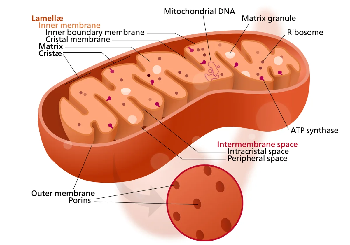
ミトコンドリアは独自のDNA(mtDNA)を持ち、電子伝達系の一部の蛋白をそこから作る。特徴は3つ。
- 母系遺伝——受精時に精子のミトコンドリアは伝わらない。母から子全員に伝わるが、父からは誰にも伝わらない
- ヘテロプラスミー——1つの細胞に正常と変異の mtDNA が混在する。変異の割合が閾値を超えた組織だけ発症する
- エネルギー需要の高い組織が侵される——脳・骨格筋・心筋・網膜・内耳・膵β細胞
ヘテロプラスミーが同じ変異でも家族内で症状も重症度も違う理由である。細胞分裂のたびにミトコンドリアが不均等に分配されるため、組織ごとに変異率が異なる。遺伝子型と表現型が1対1にならないという点で、メンデル遺伝と発想を切り替える必要がある。
代表的な疾患:
- MELAS——ミトコンドリア脳筋症・乳酸アシドーシス・脳卒中様発作。血管支配に一致しない病変が特徴
- MERRF——ミオクローヌスてんかんと赤色ぼろ線維
- Leber 遺伝性視神経症——若年男性の急性両眼視力低下
- Kearns-Sayre 症候群——外眼筋麻痺・網膜色素変性・心伝導障害
診断の手がかりは乳酸/ピルビン酸比の上昇(電子伝達が止まりNADHが溜まるため)と、筋生検のragged-red fibre(Gomori トリクローム染色で異常ミトコンドリアが集簇して赤く見える)。「多臓器にまたがる説明のつかない症状+乳酸高値」でこの群を思い出す。
English version
Core
Mitochondria have their own DNA (mtDNA) and make some of the proteins of the electron transport chain from it. Three features.
- Maternal inheritance — the sperm’s mitochondria are not transmitted at fertilisation. Passed by a mother to all her children and by a father to none.
- Heteroplasmy — normal and mutant mtDNA coexist in one cell. Only the tissues in which the proportion of mutant exceeds a threshold are affected.
- The tissues of highest energy demand are affected — brain, skeletal muscle, myocardium, retina, inner ear, pancreatic β cell.
Going further
Heteroplasmy is why the same mutation gives different symptoms and severity within a family. Mitochondria are distributed unequally at every cell division, so the mutant load differs between tissues. Genotype does not map one to one onto phenotype, and one must switch out of Mendelian thinking.
The representative diseases:
- MELAS — mitochondrial encephalomyopathy, lactic acidosis and stroke-like episodes. Characteristically the lesions do not follow a vascular territory.
- MERRF — myoclonic epilepsy with ragged-red fibres.
- Leber’s hereditary optic neuropathy — acute bilateral visual loss in a young man.
- Kearns–Sayre syndrome — external ophthalmoplegia, retinitis pigmentosa, cardiac conduction disease.
The diagnostic clues are a raised lactate/pyruvate ratio (electron transport is blocked and NADH accumulates) and ragged-red fibres on muscle biopsy (abnormal mitochondria clustered and staining red on Gomori trichrome). Think of this group when unexplained symptoms span several organs with a high lactate.
呼吸鎖を止めるもの — 作用点が違えば、症状も治療も違う
電子伝達系と ATP 合成を止める物質は、どこを止めるかで3つに分かれる。
| 型 | 作用点 | 例 | 酸素消費 | ATP | 熱 |
|---|---|---|---|---|---|
| 電子伝達阻害 | 複合体I | ロテノン、バルビツール酸 | 減る | 減る | 減る |
| 複合体III | アンチマイシンA | 減る | 減る | 減る | |
| 複合体IV | シアン・一酸化炭素・硫化水素・アジ化物 | 減る | 減る | 減る | |
| ATP合成酵素の阻害 | 複合体V | オリゴマイシン | 減る(勾配が飽和して電子も流れない) | 減る | — |
| 脱共役 | 内膜のH⁺勾配を短絡 | 2,4-ジニトロフェノール・UCP1(褐色脂肪) | 増える | 減る | 増える |
脱共役だけが「酸素消費が増える」——ここが試験の要になる。H⁺が ATP 合成酵素を通らずに漏れるので、勾配を保とうとして電子伝達が全速で回る。エネルギーはATP ではなく熱になる。
これが生理的に使われているのが褐色脂肪組織——UCP1〈サーモゲニン〉が意図的に脱共役を起こし、新生児の非ふるえ熱産生を担う。新生児は震えて熱を作れないので、この仕組みが体温維持の主役になる。肩甲間部・腎周囲・腋窩に分布し、ミトコンドリアが豊富で褐色に見える。
| 中毒 | 要点 |
|---|---|
| シアン中毒 | 複合体IVを阻害。細胞が酸素を使えないので静脈血が動脈血のように赤い(動静脈酸素較差の消失)。乳酸アシドーシス。ヒドロキソコバラミン(シアンを捕捉してシアノコバラミンに)、亜硝酸+チオ硫酸 |
| 一酸化炭素中毒 | ヘモグロビンへの結合(酸素の約250倍)に加え、複合体IVも阻害する。SpO2 は正常に見える——パルスオキシメータが CO-Hb と O2-Hb を区別できない。高濃度酸素・高気圧酸素療法 |
| 硫化水素中毒 | 同じく複合体IV。高濃度では嗅覚が麻痺して臭わなくなる——「臭わないから安全」は逆 |
| 2,4-ジニトロフェノール | かつて減量薬として売られ、高体温で死亡例が出て禁止された。「熱として捨てる」ことは痩せるが命に関わる |
共通する病態は「酸素はあるのに使えない〈組織性低酸素症〉」。PaO2 も SpO2 も正常なのに、細胞は嫌気性代謝に追い込まれて乳酸が溜まる。原因不明の高度な乳酸アシドーシスを見たら、この群を思い出す。
P/O比——NADH からは約2.5 ATP、FADH2 からは約1.5 ATP。FADH2 は複合体IIから入るので、複合体Iを飛ばす分だけ汲み出す H⁺ が少ない。入口の位置が収量を決める。
English version
Core
The substances that stop electron transport and ATP synthesis divide into three by where they act.
| Type | Site | Examples | O₂ use | ATP | Heat |
|---|---|---|---|---|---|
| Inhibition of electron transport | Complex I | Rotenone, barbiturates | Falls | Falls | Falls |
| Complex III | Antimycin A | Falls | Falls | Falls | |
| Complex IV | Cyanide, carbon monoxide, hydrogen sulphide, azide | Falls | Falls | Falls | |
| Inhibition of ATP synthase | Complex V | Oligomycin | Falls (the gradient saturates and electrons stop too) | Falls | — |
| Uncoupling | Short-circuits the H⁺ gradient | 2,4-dinitrophenol, UCP1 (brown fat) | Rises | Falls | Rises |
Going further
Only uncoupling increases oxygen consumption — and this is the crux of the examination. H⁺ leaks without passing through ATP synthase, so electron transport runs at full speed trying to maintain the gradient. The energy becomes heat instead of ATP.
Brown adipose tissue uses this physiologically — UCP1 (thermogenin) uncouples deliberately and provides the non-shivering thermogenesis of the newborn. The newborn cannot shiver to make heat, so this is the mainstay of its temperature. It lies between the scapulae, around the kidneys and in the axillae, and looks brown from its abundant mitochondria.
| Poisoning | The point |
|---|---|
| Cyanide | Inhibits complex IV. The cell cannot use oxygen, so venous blood is as red as arterial (the arteriovenous difference disappears). Lactic acidosis. Hydroxocobalamin (capturing cyanide as cyanocobalamin), or nitrite plus thiosulphate |
| Carbon monoxide | Besides binding haemoglobin (about 250 times more tightly than oxygen) it also inhibits complex IV. The SpO₂ looks normal — the pulse oximeter cannot distinguish CO-Hb from O₂-Hb. High-concentration or hyperbaric oxygen |
| Hydrogen sulphide | Complex IV likewise. At high concentrations it paralyses the sense of smell — "no smell, therefore safe" is the reverse of the truth |
| 2,4-dinitrophenol | Once sold for weight loss and banned after deaths from hyperthermia. Throwing energy away as heat does cause weight loss, and can kill |
The common state is "oxygen is present but cannot be used" — histotoxic hypoxia. The PaO₂ and the SpO₂ are normal yet the cells are driven to anaerobic metabolism and lactate accumulates. Think of this group on seeing a severe lactic acidosis of unknown cause.
The P/O ratio — about 2.5 ATP from NADH and about 1.5 from FADH₂. FADH₂ enters at complex II and skips complex I, so fewer H⁺ are pumped. The position of the entrance decides the yield.
まとめ
| TCA回路 — 電子を集める回転木馬 | ミトコンドリア基質で回る。アセチルCoA(2炭素)がオキサロ酢酸(4炭素)と結合してクエン酸(6炭素)になり、CO₂を2つ捨てながら一周してオキサロ酢酸に戻る。 |
|---|---|
| ピルビン酸脱水素酵素複合体 — 3つの酵素の順番と、リン酸化による開け閉め | ピルビン酸はミトコンドリア内膜のピルビン酸輸送体で基質に入り、酸化的脱炭酸を受けてアセチル CoA になる。 |
| クエン酸回路の8段階 — 酵素・クラス・反応の型・調節因子 | 段階反応酵素(クラス)反応の型出るもの・調節1アセチル CoA+オキサロ酢酸+H₂O → クエン酸+CoAクエン酸合成酵素(転移酵素)縮合(C–C 結合をつくる唯一の段階)不可逆。 |
| スクシニル CoA 合成酵素 — クエン酸回路の中の基質レベルのリン酸化 | α-ケトグルタル酸脱水素酵素が作ったスクシニル CoA は、チオエステル結合(–CO–S–CoA)に大きなエネルギーを持っている(加水分解の ΔG°′ 約 −33 kJ/mol、A… |
| 電子伝達系 — 電子を渡してH⁺を汲む | ミトコンドリア内膜に並ぶ4つの複合体。NADHとFADH₂が持ってきた電子を順に渡し、そのエネルギーでH⁺を基質側から膜間腔へ汲み出す。 |
| 電子の運び手 — フラビン・Fe-S クラスター・ユビキノン・シトクロムと Q サイクル | 電子伝達系の部品は、電子を2個まとめて運ぶものと、1個ずつ運ぶものに分かれる。 |
| ATP 合成酵素の回り方 — F₀ が回り、F₁ の3つの部屋が形を変える | ATP 合成酵素(複合体 V)は2つの部分でできている。 |
| ATP合成と脱共役 — 勾配を熱に変える | 電子伝達で作られたH⁺の勾配(プロトン駆動力)が、ATP合成酵素(複合体V)を回してATPを作る。 |
| ADP が呼吸の速さを決める — 呼吸調節とアデニル酸キナーゼ | ミトコンドリアの酸素の消費速度は、主に ADP(と Pᵢ)がどれだけあるかで決まる。 |
| 細胞質の NADH をミトコンドリアへ — 2つのシャトルと輸送体ファミリー | ミトコンドリア内膜は NADH も NAD⁺ も通さない。 |
| ミトコンドリア病 — 母から伝わる病気 | ミトコンドリアは独自のDNA(mtDNA)を持ち、電子伝達系の一部の蛋白をそこから作る。 |
| 呼吸鎖を止めるもの — 作用点が違えば、症状も治療も違う | 電子伝達系と ATP 合成を止める物質は、どこを止めるかで3つに分かれる。 |
このノートは医学生が授業の内容を自分の言葉でまとめたもので、診療の判断に使うものではありません。誤りに気づいたらお知らせください。