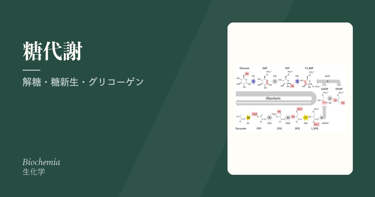
糖代謝は4本の経路が同じ材料を取り合う構図として読むと整理できる。解糖(壊してATP)、糖新生(作って血糖へ)、グリコーゲン合成/分解(蓄える/出す)、ペントースリン酸経路(NADPHと五炭糖を作る)。
解糖と糖新生はほぼ逆の道だが、3か所だけ別ルートを通る。そこが不可逆な段階であり、同時に調節点でもある。この3か所を押さえれば、糖代謝の調節はほぼ終わる。
- ヘキソキナーゼ/グルコキナーゼ ⇔ グルコース6ホスファターゼ
- PFK-1(解糖の律速) ⇔ フルクトース1,6ビスホスファターゼ
- ピルビン酸キナーゼ ⇔ ピルビン酸カルボキシラーゼ+PEPカルボキシキナーゼ
先天代謝異常はこの経路図のどこかが詰まった状態である。詰まれば手前に溜まり、先が足りなくなる——症状はこの2つの組み合わせでしかない。
- 解糖系 — 細胞質でグルコースを壊す
- 解糖系の10段階 — 酵素・中間体・反応の型を1枚に
- 解糖の調節 — 不可逆な3段階と、前向きの活性化
- 基質レベルのリン酸化 — 膜も酸素も使わずに ATP を作る
- ピルビン酸の3つの行き先
- 酵母のアルコール発酵と、解糖を止める毒 — フッ化物とヒ酸
- 糖新生 — 血糖を作る
- グリコーゲン — 蓄える/出す
- ペントースリン酸経路 — ATPを作らない糖の道
- ペントースリン酸経路の2つの相 — 非酸化的相のトランスケトラーゼとトランスアルドラーゼ
- Cori 回路 — 乳酸は老廃物ではなく、貸し借りである
解糖系 — 細胞質でグルコースを壊す
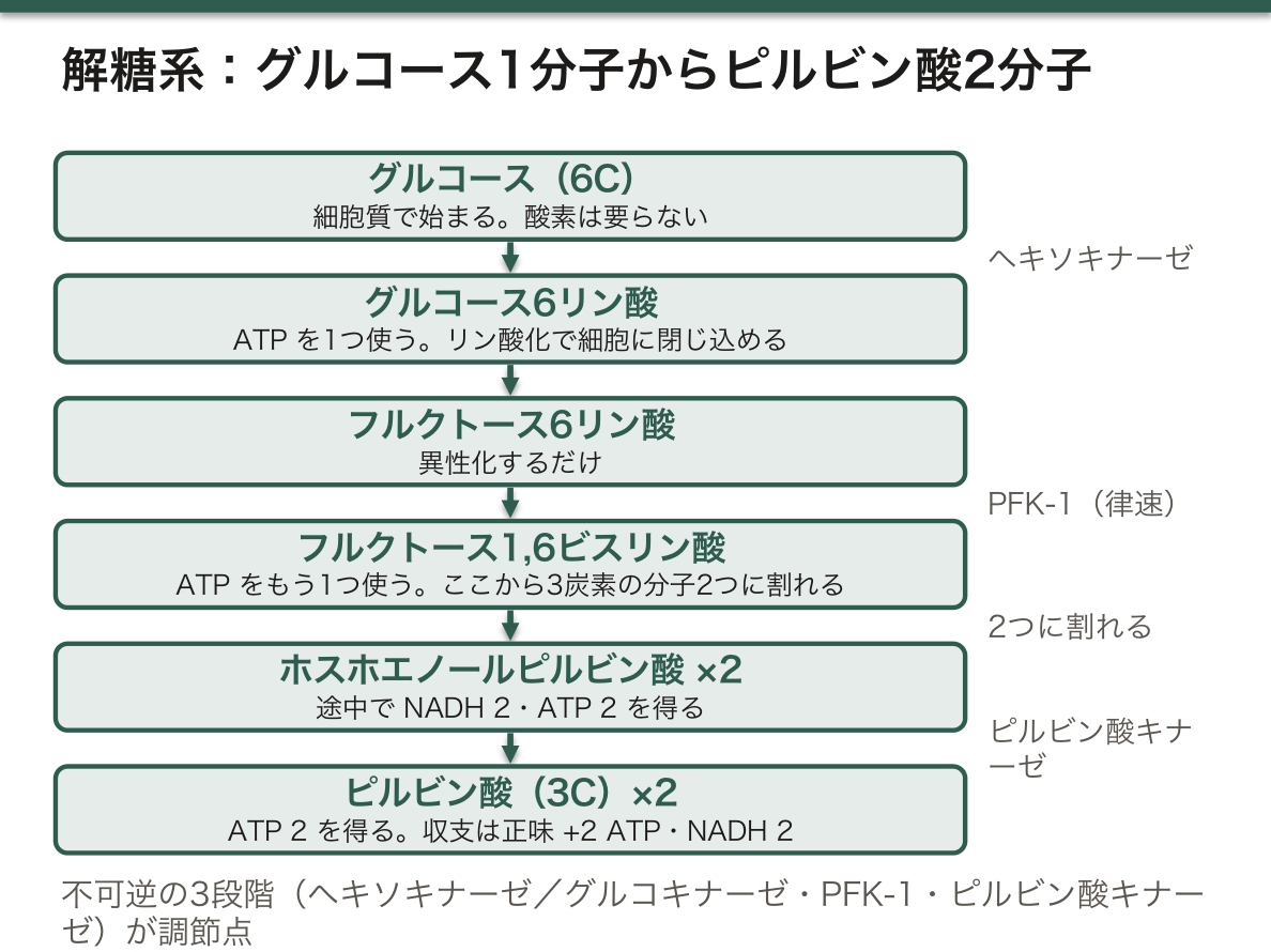
細胞質で、グルコース1分子 → ピルビン酸2分子。酸素は要らない。
収支:ATP を2つ使い、4つ作るので正味 +2 ATP、NADH 2。
不可逆な3段階=調節点
- ヘキソキナーゼ/グルコキナーゼ——グルコースをリン酸化して細胞に閉じ込める
- ホスホフルクトキナーゼ-1(PFK-1)——律速酵素。ここが最重要
- ピルビン酸キナーゼ——ATP を作る
酸素があればピルビン酸はミトコンドリアへ、無ければ乳酸になる。
PFK-1 の調節が糖代謝の中心にある。
- 促進——AMP(エネルギー不足)、フルクトース2,6ビスリン酸(最強の活性化因子)
- 抑制——ATP、クエン酸(TCAが飽和=もう要らない)、H⁺(アシドーシス)
フルクトース2,6ビスリン酸は PFK-2 が作る。この酵素はインスリンで脱リン酸化されると合成側、グルカゴンでリン酸化されると分解側に働く二機能酵素で、1つの分子で解糖と糖新生のスイッチを切り替えている。ホルモンが糖代謝の向きを決める仕組みの本体がここにある。
酸素が無くても回ることの意味も大きい。赤血球はミトコンドリアが無いので解糖しか使えず、必ず乳酸を出す。この乳酸を肝が受け取ってグルコースに戻すのがCori 回路である。激しい運動時の筋も同じ経路に頼る。
English version
Core
In the cytosol: one molecule of glucose → two of pyruvate. No oxygen is required.
Balance: two ATP used and four made, so net +2 ATP, plus 2 NADH.
Three irreversible steps = the control points
- Hexokinase / glucokinase — phosphorylates glucose and traps it in the cell.
- Phosphofructokinase-1 (PFK-1) — the rate-limiting enzyme; the most important point.
- Pyruvate kinase — makes ATP.
With oxygen the pyruvate goes to the mitochondrion; without it, it becomes lactate.
Going further
The regulation of PFK-1 is the centre of carbohydrate metabolism.
- Activated by — AMP (energy lacking), fructose 2,6-bisphosphate (the most powerful activator).
- Inhibited by — ATP, citrate (the TCA cycle is saturated = no more needed), H⁺ (acidosis).
Fructose 2,6-bisphosphate is made by PFK-2, a bifunctional enzyme which, dephosphorylated by insulin, works on the synthetic side and, phosphorylated by glucagon, on the degradative. One molecule switching between glycolysis and gluconeogenesis. Here is the substance of how hormones set the direction of carbohydrate metabolism.
That it turns without oxygen also matters greatly. The red cell has no mitochondria and can use only glycolysis, so it always produces lactate. The liver takes up that lactate and returns it as glucose — the Cori cycle. Muscle in vigorous exercise depends on the same pathway.
解糖系の10段階 — 酵素・中間体・反応の型を1枚に
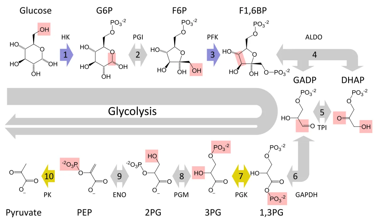
解糖は準備期(1〜5:ATP を2つ使ってグルコースを2つの三炭糖リン酸に割る)と報酬期(6〜10:三炭糖ごとに NADH 1つと ATP 2つを取り出す)に分かれる。報酬期は1分子のグルコースにつき2回回る。
| 段階 | 反応 | 酵素 | クラス・反応の型 | メモ |
|---|---|---|---|---|
| 1 | グルコース → グルコース6-リン酸 | ヘキソキナーゼ(肝・膵β細胞はグルコキナーゼ) | 転移酵素(リン酸化) | ATP を使う・不可逆・Mg²⁺ |
| 2 | G6P ⇄ フルクトース6-リン酸 | ホスホヘキソースイソメラーゼ | 異性化酵素(アルドース → ケトース) | 可逆 |
| 3 | F6P → フルクトース1,6-ビスリン酸 | ホスホフルクトキナーゼ-1(PFK-1) | 転移酵素(リン酸化) | ATP を使う・不可逆・最初の「後戻りしない」段階 |
| 4 | F1,6BP ⇄ ジヒドロキシアセトンリン酸+グリセルアルデヒド3-リン酸 | アルドラーゼ | 脱離酵素(C–C 結合を切る) | 可逆 |
| 5 | DHAP ⇄ GAP | トリオースリン酸イソメラーゼ | 異性化酵素 | 可逆。ここで2つとも GAP になる |
| 6 | GAP+NAD⁺+Pᵢ ⇄ 1,3-ビスホスホグリセリン酸+NADH+H⁺ | グリセルアルデヒド3-リン酸脱水素酵素(GAPDH) | 酸化還元酵素(酸化+リン酸の取り込み) | 最初のエネルギー獲得。活性部位に Cys |
| 7 | 1,3-BPG+ADP ⇄ 3-ホスホグリセリン酸+ATP | ホスホグリセリン酸キナーゼ | 転移酵素 | 基質レベルのリン酸化① |
| 8 | 3-PG ⇄ 2-ホスホグリセリン酸 | ホスホグリセリン酸ムターゼ | 異性化酵素(リン酸の位置を移す) | 可逆 |
| 9 | 2-PG ⇄ ホスホエノールピルビン酸+H₂O | エノラーゼ | 脱離酵素(脱水) | Mg²⁺。フッ化物で阻害 |
| 10 | PEP+ADP → ピルビン酸+ATP | ピルビン酸キナーゼ | 転移酵素 | 基質レベルのリン酸化②・不可逆 |
総反応式:グルコース+2 NAD⁺+2 ADP+2 Pᵢ → 2 ピルビン酸+2 NADH+2 H⁺+2 ATP+2 H₂O。
酵素の名前は反応の型を言っているので、名前から段階が復元できる。キナーゼ(1・3・7・10)は ATP とのリン酸の受け渡し、イソメラーゼとムターゼ(2・5・8)は形や位置の入れ替え、アルドラーゼとエノラーゼ(4・9)は水を使わずに結合を切る・水を外す、脱水素酵素(6)は酸化である。ホスホグリセリン酸キナーゼは ATP を作る向きに働いているが、名前は逆向き(ATP からリン酸を受け取る向き)で付けられている。
各段階には意味がある。1でリン酸を付けるのは、リン酸化された糖は膜を通れず細胞から逃げないから。2でケトースに変えるのは、C1 を3でリン酸化できるようにし、4で真ん中から対称に割れるようにするため。5で DHAP を GAP に変えるので、6以降は1本の道で進める。6で酸化のエネルギーを使ってリン酸を取り込み、高エネルギーのアシルリン酸(1,3-BPG)を作るので、7で ATP が作れる。8〜9で 2-PG から水を外すと、リン酸がエノールに付いた不安定な PEP になり、10で ATP をもう1つ作れる。
可逆な段階(2・4・5・6・7・8・9)は糖新生でもそのまま逆向きに使われ、不可逆な3つ(1・3・10)だけが別の酵素で迂回される。
エノラーゼのうち神経細胞に多いγ型は、神経特異エノラーゼ(NSE)として小細胞肺癌・神経芽腫の腫瘍マーカーに使われる。解糖の酵素の先天的な欠損で最も多いのはピルビン酸キナーゼ欠損症で、ミトコンドリアを持たない赤血球が ATP を作れずに壊れる溶血性貧血になる(手前に溜まった 2,3-BPG で酸素解離曲線は右へずれる)。
English version
Core
Glycolysis has a preparatory phase (steps 1–5: spend 2 ATP and split glucose into two triose phosphates) and a payoff phase (steps 6–10: from each triose, take 1 NADH and 2 ATP). The payoff phase runs twice per glucose.
| Step | Reaction | Enzyme | Class / reaction type | Note |
|---|---|---|---|---|
| 1 | Glucose → glucose-6-phosphate | Hexokinase (glucokinase in liver and β cells) | Transferase (phosphorylation) | Uses ATP; irreversible; Mg²⁺ |
| 2 | G6P ⇄ fructose-6-phosphate | Phosphohexose isomerase | Isomerase (aldose → ketose) | Reversible |
| 3 | F6P → fructose-1,6-bisphosphate | Phosphofructokinase-1 (PFK-1) | Transferase (phosphorylation) | Uses ATP; irreversible; first committed step |
| 4 | F1,6BP ⇄ dihydroxyacetone phosphate + glyceraldehyde-3-phosphate | Aldolase | Lyase (breaks a C–C bond) | Reversible |
| 5 | DHAP ⇄ GAP | Triose phosphate isomerase | Isomerase | Reversible; now both are GAP |
| 6 | GAP + NAD⁺ + Pᵢ ⇄ 1,3-bisphosphoglycerate + NADH + H⁺ | Glyceraldehyde-3-phosphate dehydrogenase (GAPDH) | Oxidoreductase (oxidation + phosphate uptake) | First energy gain; active-site Cys |
| 7 | 1,3-BPG + ADP ⇄ 3-phosphoglycerate + ATP | Phosphoglycerate kinase | Transferase | Substrate-level phosphorylation 1 |
| 8 | 3-PG ⇄ 2-phosphoglycerate | Phosphoglycerate mutase | Isomerase (moves the phosphate) | Reversible |
| 9 | 2-PG ⇄ phosphoenolpyruvate + H₂O | Enolase | Lyase (dehydration) | Mg²⁺; inhibited by fluoride |
| 10 | PEP + ADP → pyruvate + ATP | Pyruvate kinase | Transferase | Substrate-level phosphorylation 2; irreversible |
Overall: glucose + 2 NAD⁺ + 2 ADP + 2 Pᵢ → 2 pyruvate + 2 NADH + 2 H⁺ + 2 ATP + 2 H₂O.
Going further
Enzyme names state the reaction type, so the steps can be rebuilt from the names. Kinases (1, 3, 7, 10) move phosphate to or from ATP; isomerases and mutases (2, 5, 8) rearrange; aldolase and enolase (4, 9) split a bond or remove water without hydrolysis; the dehydrogenase (6) oxidises. Phosphoglycerate kinase works in the ATP-making direction but is named for the reverse.
Each step has a reason. Step 1: phosphorylated sugar cannot cross the membrane, so it is trapped. Step 2: the ketose lets C1 be phosphorylated in step 3 and lets aldolase split the molecule in the middle. Step 5: DHAP becomes GAP, so one road follows. Step 6: the energy of oxidation takes in phosphate and makes a high-energy acyl phosphate (1,3-BPG), so step 7 can make ATP. Steps 8–9: removing water from 2-PG gives PEP, an unstable enol phosphate, so step 10 makes another ATP.
The reversible steps (2, 4, 5, 6, 7, 8, 9) are used backwards in gluconeogenesis; only the three irreversible ones (1, 3, 10) are bypassed by other enzymes.
In the clinic
The γ form of enolase, abundant in neurons, is the tumour marker neuron-specific enolase (NSE) for small-cell lung cancer and neuroblastoma. The most common inherited glycolytic defect is pyruvate kinase deficiency: red cells, which have no mitochondria, cannot make ATP and are destroyed (haemolytic anaemia); 2,3-BPG builds up upstream and shifts the oxygen dissociation curve to the right.
解糖の調節 — 不可逆な3段階と、前向きの活性化
調節がかかるのは、大きく負の ΔG を持つ不可逆な3段階だけである(可逆な段階を止めても、平衡がずれるだけで流れは決まらない)。
| 酵素 | 活性化するもの | 阻害するもの | 意味 |
|---|---|---|---|
| ヘキソキナーゼ(I〜III) | — | グルコース6-リン酸(産物) | 使い切れない G6P が溜まれば取り込みを止める |
| グルコキナーゼ(肝) | インスリンで量が増える。フルクトース1-リン酸が GKRP から解放する | G6P では阻害されない | 血糖が高いときだけ働き、余りを肝に蓄える |
| PFK-1 | AMP・ADP・フルクトース2,6-ビスリン酸 | ATP・クエン酸・H⁺(・PEP) | エネルギーが足りないときに開く。TCA 回路が満ちていれば閉じる |
| ピルビン酸キナーゼ | フルクトース1,6-ビスリン酸(前向きの活性化)・PEP | ATP・アセチル CoA・アラニン・長鎖脂肪酸。肝型はグルカゴン → PKA のリン酸化で不活性化 | 上流が流れてきたら出口も開く。糖新生の材料が十分なら閉じる |
前向きの活性化(フィードフォワード)は、フィードバック阻害の逆向きの発想である。PFK-1 の産物フルクトース1,6-ビスリン酸が、2段階先のピルビン酸キナーゼを活性化する。上流が速く流れはじめたら、その知らせを下流に先回りして届けておくことで、途中の中間体が溜まらずに流れきる。
ピルビン酸キナーゼの阻害因子は「代わりの燃料や材料がある」という信号として読める。ATP とアセチル CoA は、エネルギーが足りている・脂肪酸からアセチル CoA が来ているという意味。アラニンは筋から糖新生の材料が届いているという意味で、肝ではピルビン酸を PEP 側(糖新生)へ回したいので、ピルビン酸キナーゼを止めて無駄な往復(PEP → ピルビン酸 → PEP)を防ぐ。
グルコキナーゼがヘキソキナーゼと違う理由も同じ考え方である。Km が高く(約 10 mM)、G6P で阻害されないので、血糖が上がったときだけ取り込み量が増え、上がった分を肝がグリコーゲンとして受け取れる。膵β細胞では同じ性質が「血糖のセンサー」として働く。
グルコキナーゼの機能が落ちる変異では、β細胞がインスリンを出し始める血糖の閾値が上がり、軽い高血糖が生まれつき続く(MODY2)。進行しにくく、多くは薬が要らない。逆に機能が上がる変異は、低い血糖でもインスリンが出る先天性高インスリン血症の原因になる。
English version
Core
Control acts only on the three irreversible steps, which have a large negative ΔG (blocking a reversible step only shifts an equilibrium).
| Enzyme | Activators | Inhibitors | Meaning |
|---|---|---|---|
| Hexokinase (I–III) | — | Glucose-6-phosphate (product) | If G6P is not being used, stop taking in glucose |
| Glucokinase (liver) | More enzyme with insulin; fructose-1-phosphate releases it from GKRP | Not inhibited by G6P | Works only when glucose is high and stores the excess |
| PFK-1 | AMP, ADP, fructose-2,6-bisphosphate | ATP, citrate, H⁺ (and PEP) | Opens when energy is short; closes when the TCA cycle is full |
| Pyruvate kinase | Fructose-1,6-bisphosphate (feed-forward), PEP | ATP, acetyl-CoA, alanine, long-chain fatty acids; liver form inactivated by glucagon → PKA phosphorylation | Opens the exit when the upstream flows; closes when gluconeogenic material is plentiful |
Going further
Feed-forward activation is the mirror of feedback inhibition. The PFK-1 product fructose-1,6-bisphosphate activates pyruvate kinase two steps later. When the upstream speeds up, the downstream is warned in advance so that intermediates do not pile up.
Read the pyruvate kinase inhibitors as ‘another fuel or material is available’. ATP and acetyl-CoA say energy is sufficient and fatty acids are supplying acetyl-CoA. Alanine says muscle is sending gluconeogenic material; the liver wants pyruvate to go towards PEP, so it stops pyruvate kinase and prevents a futile PEP → pyruvate → PEP loop.
Why glucokinase differs from hexokinase: a high Km (about 10 mM) and no inhibition by G6P mean uptake rises only when blood glucose rises, so the liver takes up the excess as glycogen. In β cells the same property makes it a glucose sensor.
In the clinic
Loss-of-function glucokinase variants raise the glucose threshold for insulin release, giving mild lifelong hyperglycaemia (MODY2) that rarely progresses and often needs no drugs. Gain-of-function variants cause congenital hyperinsulinism, with insulin released at low glucose.
基質レベルのリン酸化 — 膜も酸素も使わずに ATP を作る
ATP の作り方は2つしかない。
| 基質レベルのリン酸化 | 酸化的リン酸化 | |
|---|---|---|
| 仕組み | リン酸を持つ高エネルギー化合物から ADP へ直接リン酸を渡す | 電子伝達で作った H⁺ の勾配で ATP 合成酵素を回す |
| 必要なもの | 可溶性の酵素と中間体だけ | ミトコンドリア内膜・電子伝達系・O₂ |
| 場所 | 細胞質(解糖)とミトコンドリア基質(TCA 回路) | ミトコンドリア内膜 |
| グルコース1分子あたり | 解糖で正味2・TCA 回路で2(GTP) | 約26〜28 |
グルコースの分解で基質レベルのリン酸化が起きるのは3か所:①ホスホグリセリン酸キナーゼ(1,3-ビスホスホグリセリン酸 → 3-ホスホグリセリン酸)、②ピルビン酸キナーゼ(ホスホエノールピルビン酸 → ピルビン酸)、③スクシニル CoA 合成酵素(スクシニル CoA → コハク酸、GTP または ATP)。
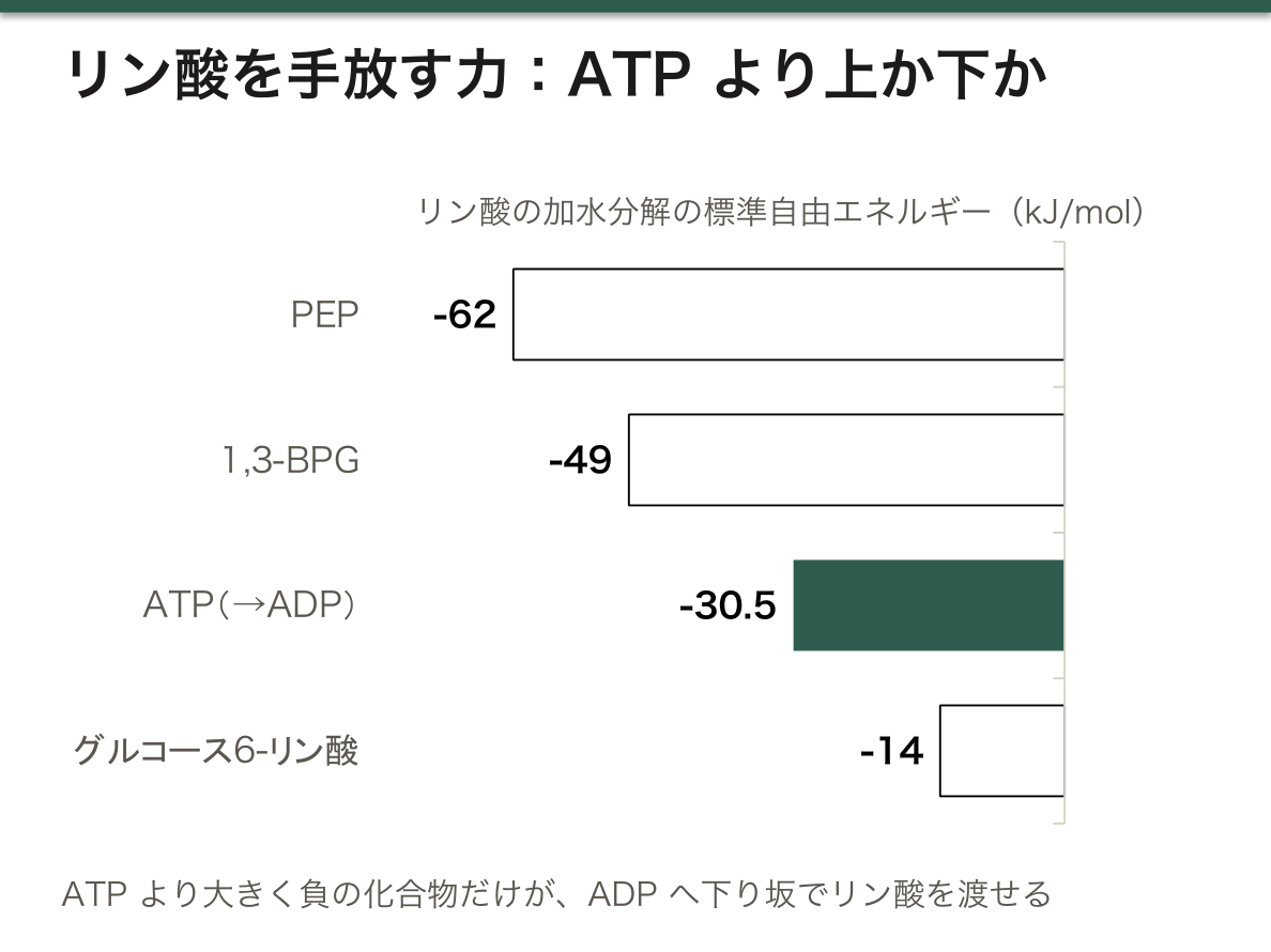
ADP にリン酸を渡せるのは、ATP よりリン酸を手放したがる化合物だけである。リン酸の加水分解の標準自由エネルギーは、PEP が約 −62 kJ/mol、1,3-BPG が約 −49 kJ/mol、ATP(→ADP)が約 −30.5 kJ/mol で、前の2つは ATP より「高い」位置にあるので ADP へ下り坂でリン酸を渡せる。グルコース6-リン酸(約 −14 kJ/mol)では ATP は作れない。
PEP がとくに高エネルギーなのは、リン酸が外れた後に起きる変化のためである。リン酸が外れると不安定なエノール形のピルビン酸ができ、すぐに安定なケト形に変わる(互変異性)。この変化が大きなエネルギーを放つので、反応は事実上一方向に進む。
嫌気的解糖の総反応式は グルコース+2 ADP+2 Pᵢ → 2 乳酸+2 ATP+2 H₂O で、NAD⁺ は乳酸脱水素酵素で再生されるので式に残らない。好気的に完全に酸化すれば約30〜32 ATP なので、酸素が無いときの取り分は1割に満たない。
赤血球は ATP のすべてを基質レベルのリン酸化に頼っている。赤血球には、1,3-BPG を 2,3-BPG に変えてからホスホグリセリン酸キナーゼを飛ばす迂回路(Rapoport-Luebering 経路)があり、ATP を1つ損する代わりに、ヘモグロビンの酸素の放しやすさを決める 2,3-BPG を作っている。高地や貧血で 2,3-BPG が増えるのはこの迂回路が太くなるためである。
English version
Core
There are only two ways to make ATP.
| Substrate-level phosphorylation | Oxidative phosphorylation | |
|---|---|---|
| How | A high-energy phosphate compound gives its phosphate directly to ADP | An H⁺ gradient made by electron transport turns ATP synthase |
| Needs | Only soluble enzymes and intermediates | Inner mitochondrial membrane, electron transport chain, O₂ |
| Where | Cytosol (glycolysis) and mitochondrial matrix (TCA cycle) | Inner mitochondrial membrane |
| Per glucose | Net 2 in glycolysis, 2 (GTP) in the TCA cycle | About 26–28 |
In glucose breakdown, substrate-level phosphorylation happens at three places: phosphoglycerate kinase (1,3-BPG → 3-PG), pyruvate kinase (PEP → pyruvate), and succinyl-CoA synthetase (succinyl-CoA → succinate, GTP or ATP).
Going further
Only compounds that give up phosphate more readily than ATP can phosphorylate ADP. Standard free energies of phosphate hydrolysis: PEP about −62 kJ/mol, 1,3-BPG about −49 kJ/mol, ATP (→ ADP) about −30.5 kJ/mol. The first two sit above ATP and pass phosphate ‘downhill’. Glucose-6-phosphate (about −14 kJ/mol) cannot make ATP.
PEP is so high in energy because of what happens after the phosphate leaves. The product is the unstable enol form of pyruvate, which at once becomes the stable keto form (tautomerisation). This releases much energy, so the reaction runs essentially one way.
The overall equation of anaerobic glycolysis is glucose + 2 ADP + 2 Pᵢ → 2 lactate + 2 ATP + 2 H₂O; NAD⁺ is regenerated by lactate dehydrogenase and does not appear. Full aerobic oxidation gives about 30–32 ATP, so without oxygen the cell gets less than a tenth.
In the clinic
Red cells make all their ATP by substrate-level phosphorylation. They have a bypass (the Rapoport-Luebering shunt) that turns 1,3-BPG into 2,3-BPG and skips phosphoglycerate kinase: one ATP is lost, but 2,3-BPG is made, which sets how easily haemoglobin releases oxygen. At altitude and in anaemia this bypass grows and 2,3-BPG rises.
ピルビン酸の3つの行き先
解糖でできたピルビン酸は、状況によって3方向へ分かれる。
- アセチルCoA(ピルビン酸デヒドロゲナーゼ/PDH)——好気的。TCAへ。不可逆
- 乳酸(乳酸脱水素酵素)——嫌気的。NAD⁺ を再生するために必要(解糖を回し続けるため)
- オキサロ酢酸(ピルビン酸カルボキシラーゼ)——糖新生の入口、またはTCA回路の補充
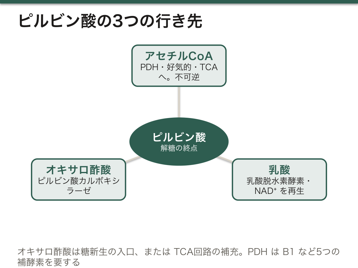
PDH は不可逆——ここが糖代謝の最大の一方通行である。だから脂肪酸から糖は作れない。
PDH は5つの補酵素を要し、そのうち4つがビタミン由来である:チアミン(B1)・リボフラビン(B2)・ナイアシン(B3)・パントテン酸(B5)+リポ酸。覚え方は「TLC For Nobody」。
ビタミンB1欠乏で PDH が止まると、ピルビン酸がアセチルCoA へ行けず乳酸へ流れる。結果として乳酸アシドーシスとATP不足が同時に起きる。エネルギー需要の高い組織から症状が出るため、心臓(脚気心=高拍出性心不全)と脳(Wernicke 脳症:眼球運動障害・失調・意識障害)が侵される。
臨床上きわめて重要な帰結——アルコール依存や低栄養の患者にブドウ糖を先に投与すると、残り少ないB1が解糖で消費されWernicke脳症を誘発する。ビタミンB1を先に、あるいは同時に投与するという原則は、この経路から直接導かれる。
English version
Core
The pyruvate from glycolysis goes three ways according to the situation.
- Acetyl-CoA (pyruvate dehydrogenase, PDH) — aerobic; to the TCA cycle. Irreversible.
- Lactate (lactate dehydrogenase) — anaerobic. Needed to regenerate NAD⁺ (to keep glycolysis turning).
- Oxaloacetate (pyruvate carboxylase) — the entrance to gluconeogenesis, or replenishment of the TCA cycle.
Going further
PDH is irreversible — the greatest one-way street of carbohydrate metabolism. This is why sugar cannot be made from fatty acids.
PDH requires five coenzymes, four of them vitamin-derived: thiamine (B1), riboflavin (B2), niacin (B3), pantothenate (B5), plus lipoic acid.
When PDH stops in thiamine deficiency, pyruvate cannot reach acetyl-CoA and overflows into lactate. The result is lactic acidosis and a shortage of ATP at the same time. The symptoms appear in the tissues of highest energy demand — the heart (wet beriberi, a high-output cardiac failure) and the brain (Wernicke’s encephalopathy: ophthalmoplegia, ataxia, confusion).
A consequence of the first importance clinically: giving glucose first to a patient who is alcohol-dependent or malnourished consumes what little thiamine remains in glycolysis and precipitates Wernicke’s encephalopathy. The rule that thiamine is given first, or together, follows directly from this pathway.
酵母のアルコール発酵と、解糖を止める毒 — フッ化物とヒ酸
発酵は、酸素を使わずにグルコースを分解して ATP を得ることの総称である。どの発酵も目的は同じで、GAPDH で使った NAD⁺ を取り戻すことにある。違うのは、ピルビン酸を何に変えて NADH の電子を捨てるかだけである。
| 乳酸発酵(ヒト・乳酸菌) | アルコール発酵(酵母・一部の細菌) | |
|---|---|---|
| 反応 | ピルビン酸+NADH → 乳酸+NAD⁺ | ①ピルビン酸 → アセトアルデヒド+CO₂ ②アセトアルデヒド+NADH → エタノール+NAD⁺ |
| 酵素 | 乳酸脱水素酵素 | ①ピルビン酸脱炭酸酵素(TPP・Mg²⁺)②アルコール脱水素酵素(Zn²⁺) |
| CO₂ | 出ない | 出る(パンが膨らむ・ビールの泡) |
ヒトはピルビン酸脱炭酸酵素を持たないので、エタノールを作れない。ヒトのアルコール脱水素酵素は、肝で逆向き(エタノール → アセトアルデヒド)に働いて、飲んだアルコールを処理している。
解糖を止める毒:フッ化物はエノラーゼを阻害する(活性部位で Mg²⁺・リン酸と複合体を作る)。ヒ酸は GAPDH でリン酸の代わりに取り込まれる。水銀などの重金属は GAPDH の活性部位の Cys の –SH に結合して不可逆に止める。
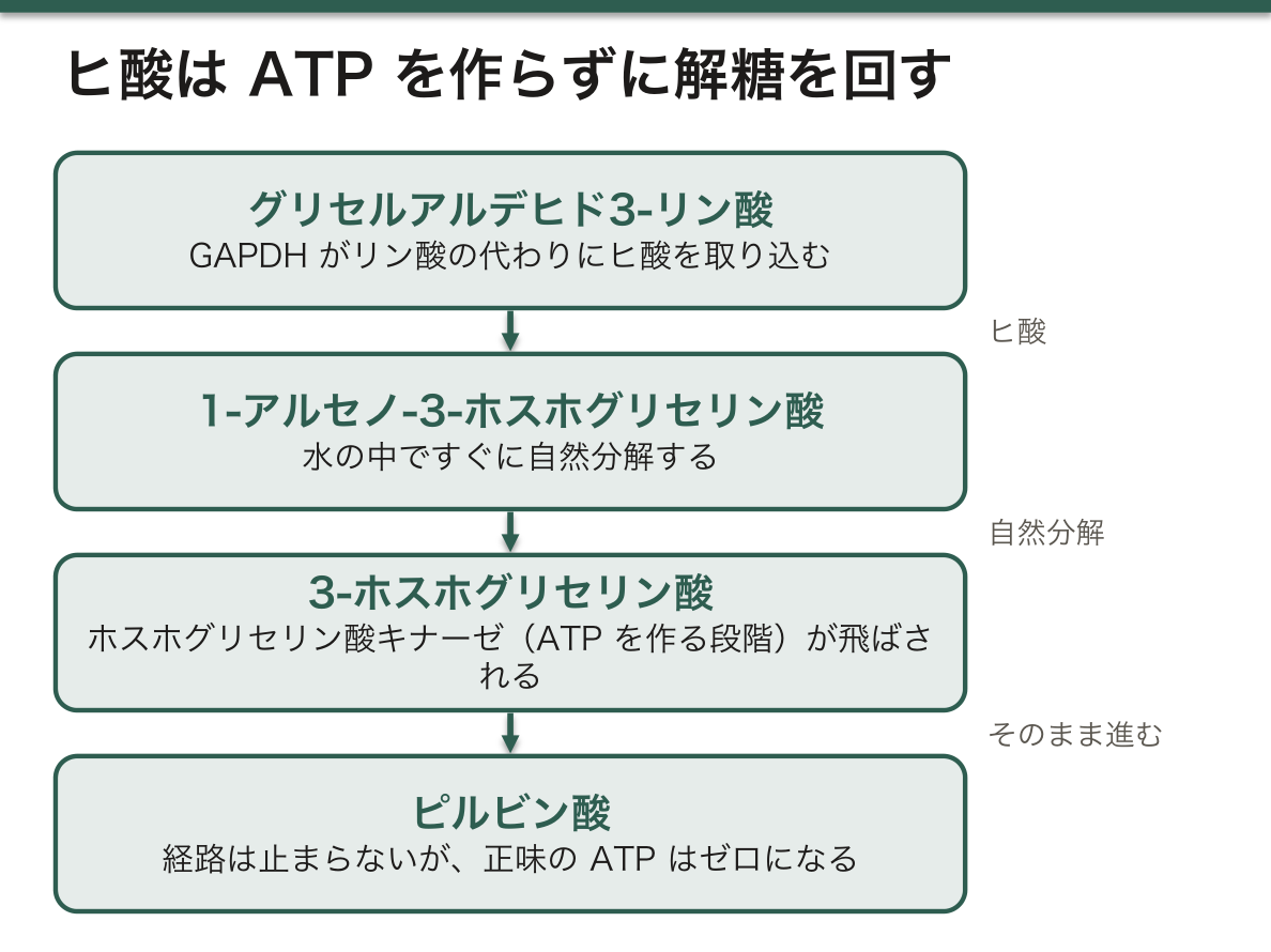
ヒ酸(AsO₄³⁻)は、ATP を作らずに解糖を回してしまう。ヒ酸はリン酸とほぼ同じ形なので GAPDH はこれを取り込み、1-アルセノ-3-ホスホグリセリン酸を作る。この化合物は水の中ですぐに自然分解して 3-ホスホグリセリン酸になるので、ホスホグリセリン酸キナーゼの段階(ATP を作る段階)が飛ばされる。経路は止まらないのに、報酬期の最初の ATP が消え、正味の ATP はゼロになる。これを「脱共役」と同じ発想で理解するとよい。
一方、亜ヒ酸(As³⁺)は別の場所を止める。2つの –SH を持つリポ酸に結合するので、ピルビン酸脱水素酵素と α-ケトグルタル酸脱水素酵素が止まり、乳酸アシドーシスが起きる。解毒薬のジメルカプロール(BAL)は –SH を2つ持ち、ヒ素を奪い返す。
TPP(チアミンピロリン酸)は、ピルビン酸脱炭酸酵素でもピルビン酸脱水素酵素でも「カルボキシ基を外して2炭素を運ぶ」役をしている。酵母が脱炭酸の後にアセトアルデヒドを手放すのに対し、ヒトの PDH はそれを酸化してアセチル CoA にする——同じ補酵素が、発酵と呼吸の分かれ道に立っている。
血糖を測る採血管には、フッ化ナトリウムを入れたものがある。採血後も赤血球と白血球が解糖を続けるので、室温で放置すると血糖は1時間に数%ずつ下がっていく。フッ化物がエノラーゼを止めることで、採血した時点の値を保てる。腸内の酵母が糖を発酵してエタノールを作り、飲酒していないのに酩酊する「腸内発酵症候群」という病態も報告されている。
English version
Core
Fermentation means breaking down glucose for ATP without oxygen. Every fermentation has one goal: to regenerate the NAD⁺ used by GAPDH. They differ only in what pyruvate is turned into to dump the NADH electrons.
| Lactic fermentation (humans, lactic acid bacteria) | Alcoholic fermentation (yeast, some bacteria) | |
|---|---|---|
| Reaction | Pyruvate + NADH → lactate + NAD⁺ | (1) pyruvate → acetaldehyde + CO₂ (2) acetaldehyde + NADH → ethanol + NAD⁺ |
| Enzymes | Lactate dehydrogenase | (1) pyruvate decarboxylase (TPP, Mg²⁺) (2) alcohol dehydrogenase (Zn²⁺) |
| CO₂ | None | Yes (bread rises, beer foams) |
Humans have no pyruvate decarboxylase, so they cannot make ethanol. Human alcohol dehydrogenase works the other way in the liver (ethanol → acetaldehyde) to clear the alcohol we drink.
Poisons of glycolysis: fluoride inhibits enolase (forming a complex with Mg²⁺ and phosphate in the active site). Arsenate replaces phosphate in the GAPDH reaction. Heavy metals such as mercury bind the –SH of the active-site Cys of GAPDH and block it irreversibly.
Going further
Arsenate (AsO₄³⁻) keeps glycolysis running without making ATP. It looks like phosphate, so GAPDH takes it in and makes 1-arseno-3-phosphoglycerate. This breaks down in water at once to 3-phosphoglycerate, skipping the phosphoglycerate kinase step that makes ATP. The pathway still flows, but the first payoff ATP is lost and net ATP becomes zero — think of it like uncoupling.
Arsenite (As³⁺) acts elsewhere. It binds the two –SH groups of lipoic acid, so pyruvate dehydrogenase and α-ketoglutarate dehydrogenase stop and lactic acidosis follows. The antidote dimercaprol (BAL) has two –SH groups and pulls the arsenic away.
TPP (thiamine pyrophosphate) removes a carboxyl group and carries a two-carbon unit in both pyruvate decarboxylase and pyruvate dehydrogenase. Yeast releases the acetaldehyde after decarboxylation; human PDH oxidises it to acetyl-CoA. The same coenzyme stands at the fork between fermentation and respiration.
In the clinic
Some blood glucose tubes contain sodium fluoride. Red and white cells keep doing glycolysis after sampling, so at room temperature glucose falls by a few per cent per hour; fluoride blocks enolase and keeps the value of the moment of sampling. ‘Auto-brewery syndrome’, in which gut yeast ferment sugar to ethanol and the patient becomes drunk without drinking, has also been reported.
糖新生 — 血糖を作る
主に肝(一部は腎皮質)で、糖以外の材料からグルコースを作る。空腹の血糖はこれが支える。
材料は乳酸・糖原性アミノ酸(とくにアラニン)・グリセロール。脂肪酸(偶数鎖)は材料にならない。
解糖の逆行だが、不可逆の3か所を迂回する。
- ピルビン酸 →(ピルビン酸カルボキシラーゼ、ミトコンドリア、ビオチン要求)→ オキサロ酢酸 →(PEPCK)→ PEP
- フルクトース1,6ビスホスファターゼ——糖新生の律速酵素
- グルコース6ホスファターゼ——肝と腎にしかない。だから筋は血糖を作れない
糖新生はエネルギーを食う(グルコース1分子あたりATP換算で6つ)。そのATPは脂肪酸のβ酸化から供給される。つまり「脂肪を燃やして糖を作る」——脂肪が糖の材料にならなくても、糖新生を動かす燃料にはなっている。この区別が問われる。
アラニン回路(グルコース-アラニン回路)——筋で分解されたアミノ酸のアミノ基をピルビン酸に渡してアラニンにし、肝へ運ぶ。肝でアミノ基は尿素回路へ、炭素骨格は糖新生へ。窒素の運搬と糖の材料供給を1つの分子で兼ねている。
アルコールが低血糖を起こす機序もここにある。アルコール代謝でNADH/NAD⁺ 比が上がると、乳酸→ピルビン酸、リンゴ酸→オキサロ酢酸の反応が逆向きに押され、糖新生の材料が枯れる。空腹での飲酒が重篤な低血糖を招くのはこのためで、同時に乳酸が溜まり、脂肪肝も進む。
フォン・ギールケ病(糖原病 I 型)——グルコース6ホスファターゼ欠損。グリコーゲンを分解しても糖新生をしても最後の1段階でグルコースを出せない。
- 重篤な空腹時低血糖(授乳間隔が空くと発作)
- 肝腫大(グリコーゲン蓄積)
- 乳酸アシドーシス(行き場を失った代謝物が乳酸へ)
- 高尿酸血症・高中性脂肪血症
「出口が塞がる」と、手前のすべてが渋滞するという詰まり方の典型例である。治療は頻回の経口摂取と生コーンスターチで低血糖を防ぐこと。
English version
Core
Chiefly in the liver (partly the renal cortex): glucose is made from non-carbohydrate material. The blood glucose of fasting rests on this.
The substrates are lactate, glucogenic amino acids (especially alanine) and glycerol. Fatty acids (even-chain) are not substrates.
It runs glycolysis backwards but bypasses the three irreversible steps.
- Pyruvate → (pyruvate carboxylase, mitochondrial, requiring biotin) → oxaloacetate → (PEPCK) → PEP
- Fructose 1,6-bisphosphatase — the rate-limiting enzyme of gluconeogenesis.
- Glucose 6-phosphatase — present only in liver and kidney. Hence muscle cannot make blood glucose.
Going further
Gluconeogenesis is expensive (six ATP equivalents per molecule of glucose), and that ATP is supplied by the β-oxidation of fatty acids. That is, fat is burnt to make sugar — fat is not the substrate of gluconeogenesis but it is the fuel that drives it. This distinction is asked.
The glucose–alanine cycle — in muscle, the amino group of degraded amino acids is handed to pyruvate to make alanine, which travels to the liver. There the amino group goes to the urea cycle and the carbon skeleton to gluconeogenesis. One molecule serving both to carry nitrogen and to supply substrate for sugar.
The mechanism by which alcohol causes hypoglycaemia lies here. Alcohol metabolism raises the NADH/NAD⁺ ratio, pushing lactate → pyruvate and malate → oxaloacetate backwards and exhausting the substrate for gluconeogenesis. This is why drinking on an empty stomach causes severe hypoglycaemia; lactate accumulates at the same time and fatty liver advances.
In the clinic
Von Gierke disease (glycogen storage disease type I) — glucose 6-phosphatase deficiency. Whether glycogen is broken down or gluconeogenesis is run, glucose cannot be released at the last step.
- Severe fasting hypoglycaemia (an attack whenever feeds are spaced out).
- Hepatomegaly (glycogen storage).
- Lactic acidosis (metabolites with nowhere to go pass to lactate).
- Hyperuricaemia and hypertriglyceridaemia.
The model case of "block the exit and everything behind it jams". Treatment is frequent feeding and uncooked cornstarch to prevent hypoglycaemia.
グリコーゲン — 蓄える/出す
グルコースを枝分かれした多糖として蓄える形。肝(約100g)と筋(約400g)にある。
- 肝のグリコーゲン——血糖の維持のため。約12〜24時間で枯れる
- 筋のグリコーゲン——筋自身のため。グルコース6ホスファターゼが無いので血糖にならない
合成はグリコーゲンシンターゼ(α1→4結合)と分枝酵素(α1→6)。分解はグリコーゲンホスホリラーゼ(律速)と脱分枝酵素。
枝分かれ構造には理由がある。非還元末端が増えるほど、同時に多くの酵素が働けるので分解が速い。加えて可溶性が高く、浸透圧の負担が小さい。「速く出し入れするための形」である。
調節は相反調節の典型で、cAMP → PKA がホスホリラーゼを活性化しシンターゼを不活性化する。
- 肝——グルカゴンとアドレナリンの両方に応じる(血糖のため)
- 筋——グルカゴン受容体が無く、アドレナリンと、収縮に伴うCa²⁺・AMP に応じる(自分のため)
この違いが、糖原病の症状が「低血糖型」と「運動不耐型」に分かれる理由である。
糖原病はどの臓器のどの酵素かで症状が決まる。
- I型(von Gierke)——グルコース6ホスファターゼ。肝・重症低血糖・乳酸アシドーシス
- II型(Pompe)——リソソームの酸性マルターゼ。細胞質の糖代謝は正常なので低血糖は起きない。心肥大と筋力低下で乳児期に致死的。「リソソーム病であって糖代謝異常ではない」点が鑑別の鍵
- III型(Cori)——脱分枝酵素。I型に似るが軽症で、乳酸アシドーシスは目立たない(糖新生は動くため)
- V型(McArdle)——筋のホスホリラーゼ。運動時の筋痛・こむら返り・ミオグロビン尿。低血糖は無い。休むと楽になり再開できる(second wind)
「肝型なら低血糖、筋型なら運動不耐」——臓器分布がそのまま症状になる。
English version
Core
The branched polysaccharide form in which glucose is stored: in liver (about 100 g) and muscle (about 400 g).
- Hepatic glycogen — to maintain the blood glucose. Exhausted in about 12–24 hours.
- Muscle glycogen — for the muscle itself. Having no glucose 6-phosphatase, it cannot become blood glucose.
Synthesis is by glycogen synthase (α1→4) and the branching enzyme (α1→6); breakdown by glycogen phosphorylase (rate-limiting) and the debranching enzyme.
Going further
There is a reason for the branched structure. The more non-reducing ends there are, the more enzymes can work at once and the faster it is broken down. It is also more soluble and imposes less osmotic load. It is a shape designed for putting in and taking out quickly.
The regulation is the classic reciprocal control: cAMP → PKA activates the phosphorylase and inactivates the synthase.
- Liver — responds to both glucagon and adrenaline (for the blood glucose).
- Muscle — has no glucagon receptor and responds to adrenaline and to the Ca²⁺ and AMP of contraction (for itself).
This difference is why the glycogen storage diseases divide into a hypoglycaemic and an exercise-intolerant type.
In the clinic
Which organ and which enzyme decides the symptoms of a glycogen storage disease.
- Type I (von Gierke) — glucose 6-phosphatase. Liver; severe hypoglycaemia; lactic acidosis.
- Type II (Pompe) — lysosomal acid maltase. Cytosolic carbohydrate metabolism is normal, so there is no hypoglycaemia. Cardiomegaly and weakness, fatal in infancy. That it is a lysosomal disease and not a disorder of carbohydrate metabolism is the key to the differential.
- Type III (Cori) — the debranching enzyme. Resembles type I but milder, with little lactic acidosis (gluconeogenesis still works).
- Type V (McArdle) — muscle phosphorylase. Muscle pain, cramps and myoglobinuria on exercise. No hypoglycaemia. Rest brings relief and exercise can be resumed (second wind).
"Hepatic type, hypoglycaemia; muscle type, exercise intolerance" — the distribution of the organ is directly the symptom.
ペントースリン酸経路 — ATPを作らない糖の道
細胞質で、グルコース6リン酸からNADPH とリボース5リン酸を作る。ATPは作らない。
- NADPH——還元力。脂肪酸・コレステロール・ステロイドの合成、グルタチオンの還元(抗酸化)、好中球の活性酸素産生
- リボース5リン酸——核酸の材料
律速酵素はグルコース6リン酸脱水素酵素(G6PD)で、NADPH によるフィードバック阻害を受ける。
NADH と NADPH を混同しないことが要点である。NADH は異化で作られATPを生む、NADPH は同化と抗酸化に使う。名前が似ているだけで役割は正反対に近い。
経路が盛んな組織は用途から導ける——肝・脂肪組織・乳腺・副腎皮質・精巣(合成のためのNADPH)、赤血球(抗酸化のためのNADPH)。
赤血球にとっては死活問題である。赤血球はミトコンドリアを持たず、酸素を運ぶために常に酸化ストレスに曝される。NADPH → グルタチオン還元 → 過酸化水素の除去という一本道しかない。
G6PD欠損症——世界で最も多い酵素異常(X連鎖潜性、マラリア流行地域に多い)。平時は無症状だが、酸化ストレスで急性溶血を起こす。
- 誘因——ソラマメ、抗マラリア薬(プリマキン)、サルファ剤、ニトロフラントイン、感染
- 所見——変性ヘモグロビンの沈着であるHeinz 小体、脾臓でそこをかじられたbite cell
慢性肉芽腫症は逆に、NADPHオキシダーゼの欠損で好中球が活性酸素を作れない病態である(NADPH自体は作れる)。カタラーゼ陽性菌(黄色ブドウ球菌・セラチア・アスペルギルス)による反復感染をきたす。同じ NADPH でも「作れない」のか「使えない」のかで病気が違う。
English version
Core
In the cytosol: from glucose 6-phosphate it makes NADPH and ribose 5-phosphate. It makes no ATP.
- NADPH — reducing power. Synthesis of fatty acids, cholesterol and steroids; reduction of glutathione (antioxidant defence); production of reactive oxygen by neutrophils.
- Ribose 5-phosphate — the raw material of nucleic acids.
The rate-limiting enzyme is glucose 6-phosphate dehydrogenase (G6PD), subject to feedback inhibition by NADPH.
Going further
The point is not to confuse NADH with NADPH. NADH is made in catabolism and yields ATP; NADPH is used for synthesis and antioxidant defence. The names are alike but the roles are almost opposite.
The tissues in which the pathway is active follow from the use — liver, adipose tissue, breast, adrenal cortex and testis (NADPH for synthesis); the red cell (NADPH for antioxidant defence).
For the red cell it is a matter of life and death. It has no mitochondria and, carrying oxygen, is constantly under oxidative stress. Its only road is NADPH → reduction of glutathione → removal of hydrogen peroxide.
In the clinic
G6PD deficiency — the commonest enzyme defect in the world (X-linked recessive, frequent where malaria is endemic). Silent at rest, it causes acute haemolysis under oxidative stress.
- Triggers — broad beans, antimalarials (primaquine), sulphonamides, nitrofurantoin, infection.
- Findings — Heinz bodies (deposits of denatured haemoglobin) and the bite cells left where the spleen has nibbled them out.
Chronic granulomatous disease is the converse: a defect of NADPH oxidase means the neutrophil cannot make reactive oxygen (NADPH itself can be made). It gives recurrent infection with catalase-positive organisms (Staphylococcus aureus, Serratia, Aspergillus).
With the same NADPH, the disease differs according to whether it cannot be made or cannot be used.
ペントースリン酸経路の2つの相 — 非酸化的相のトランスケトラーゼとトランスアルドラーゼ
ペントースリン酸経路は2つの相に分かれる。
| 酸化的相 | 非酸化的相 | |
|---|---|---|
| 反応 | G6P → 6-ホスホグルコノラクトン → 6-ホスホグルコン酸 → リブロース5-リン酸(+CO₂) | 五炭糖リン酸 ⇄ フルクトース6-リン酸・グリセルアルデヒド3-リン酸 |
| 主な酵素 | G6P 脱水素酵素(律速)・ラクトナーゼ・6-ホスホグルコン酸脱水素酵素 | イソメラーゼ・エピメラーゼ・トランスケトラーゼ(TPP)・トランスアルドラーゼ |
| 向き | 不可逆 | すべて可逆 |
| 産物 | NADPH 2・CO₂ 1 | NADPH は作らない。糖の炭素数を組み替える |
酸化的相の総反応:G6P+2 NADP⁺+H₂O → リボース5-リン酸+CO₂+2 NADPH+2 H⁺(最後にリブロース5-リン酸をイソメラーゼでリボース5-リン酸に変える)。リブロース5-リン酸はエピメラーゼでキシルロース5-リン酸にもなり、こちらが非酸化的相に入る。
非酸化的相では、トランスケトラーゼが2炭素、トランスアルドラーゼが3炭素の単位を糖から糖へ移す。差し引きすると、五炭糖3つ(15炭素)→ 六炭糖2つ(F6P)+三炭糖1つ(GAP)になり、解糖の中間体に戻る。
非酸化的相が可逆であることが、この経路を「必要に応じて形を変える道」にしている。細胞が何を必要としているかで、流れ方が4通りに変わる。
| 細胞の需要 | 流れ方 | 例 |
|---|---|---|
| リボースが要るが NADPH は要らない | 非酸化的相を逆向きに使い、F6P と GAP からリボース5-リン酸を作る | 分裂の盛んな細胞 |
| リボースも NADPH も同じくらい要る | 酸化的相だけ | — |
| NADPH がたくさん要り、リボースは要らない | 五炭糖を F6P に戻して G6P にし、もう一度酸化的相へ(最終的に G6P が CO₂ になる) | 脂肪酸合成の盛んな肝・脂肪組織、赤血球 |
| NADPH と ATP が要る | 五炭糖を F6P・GAP に変えて解糖へ流す | — |
キシルロース5-リン酸は信号としても働く。糖が多いと増え、蛋白脱リン酸化酵素を介して PFK-2 を合成側に傾け(フルクトース2,6-ビスリン酸が増えて解糖が進む)、脂肪合成の遺伝子も動かす。糖があり余ると脂肪に変わる仕組みの一部である。
トランスケトラーゼは TPP(ビタミン B1)を補酵素にするので、赤血球のトランスケトラーゼ活性が、TPP を足すとどれだけ上がるかを測ればビタミン B1 の不足を判定できる。B1 欠乏ではこの酵素も止まり、Wernicke 脳症の一因になると考えられている。
English version
Core
The pentose phosphate pathway has two phases.
| Oxidative phase | Non-oxidative phase | |
|---|---|---|
| Reactions | G6P → 6-phosphogluconolactone → 6-phosphogluconate → ribulose-5-phosphate (+ CO₂) | Pentose phosphates ⇄ fructose-6-phosphate and glyceraldehyde-3-phosphate |
| Main enzymes | G6P dehydrogenase (rate-limiting), lactonase, 6-phosphogluconate dehydrogenase | Isomerase, epimerase, transketolase (TPP), transaldolase |
| Direction | Irreversible | All reversible |
| Products | 2 NADPH, 1 CO₂ | No NADPH; reshuffles carbon chains |
Oxidative phase overall: G6P + 2 NADP⁺ + H₂O → ribose-5-phosphate + CO₂ + 2 NADPH + 2 H⁺ (an isomerase finally turns ribulose-5-phosphate into ribose-5-phosphate). An epimerase also turns ribulose-5-phosphate into xylulose-5-phosphate, which enters the non-oxidative phase.
In the non-oxidative phase transketolase moves 2-carbon and transaldolase moves 3-carbon units between sugars. The net result is 3 pentoses (15 C) → 2 hexoses (F6P) + 1 triose (GAP), back to glycolytic intermediates.
Going further
Because the non-oxidative phase is reversible, the pathway changes shape with the cell’s needs, in four modes.
| Cell’s need | Flow | Example |
|---|---|---|
| Ribose, not NADPH | Run the non-oxidative phase backwards, making ribose-5-phosphate from F6P and GAP | Dividing cells |
| Ribose and NADPH equally | Oxidative phase only | — |
| Much NADPH, no ribose | Turn pentoses back into F6P → G6P and run the oxidative phase again (G6P is finally burned to CO₂) | Liver and fat during fatty acid synthesis, red cells |
| NADPH and ATP | Turn pentoses into F6P and GAP and send them down glycolysis | — |
Xylulose-5-phosphate is also a signal. It rises when sugar is plentiful and, through a protein phosphatase, tilts PFK-2 to the synthesis side (more fructose-2,6-bisphosphate, faster glycolysis) and switches on lipogenic genes — part of how excess sugar becomes fat.
In the clinic
Transketolase uses TPP (vitamin B1), so thiamine status can be judged by how much red-cell transketolase activity rises when TPP is added. In B1 deficiency this enzyme also stops, which is thought to contribute to Wernicke encephalopathy.
Cori 回路 — 乳酸は老廃物ではなく、貸し借りである
激しい運動で筋の解糖が酸素供給を上回ると、ピルビン酸が乳酸へ還元される。目的はATP を作ることではなく、NAD⁺ を再生すること——NAD⁺ が無ければ解糖が止まるため。
乳酸は行き場を失った電子の一時的な置き場で、そのまま捨てられるのではなく肝へ送られる。
| 場所 | 起こること |
|---|---|
| 筋 | グルコース → 解糖 → 乳酸(2 ATP を得る) |
| 血流 | 乳酸が肝へ運ばれる |
| 肝 | 乳酸 → ピルビン酸 → 糖新生でグルコースへ戻す(6 ATP を消費) |
| 血流 | グルコースが筋へ戻る |
全体では ATP を失う(2 得て 6 使う)。それでも成立するのは、負担を「酸素の足りない筋」から「酸素のある肝」へ移しているから。臓器間で仕事を分担する仕組みになっている。
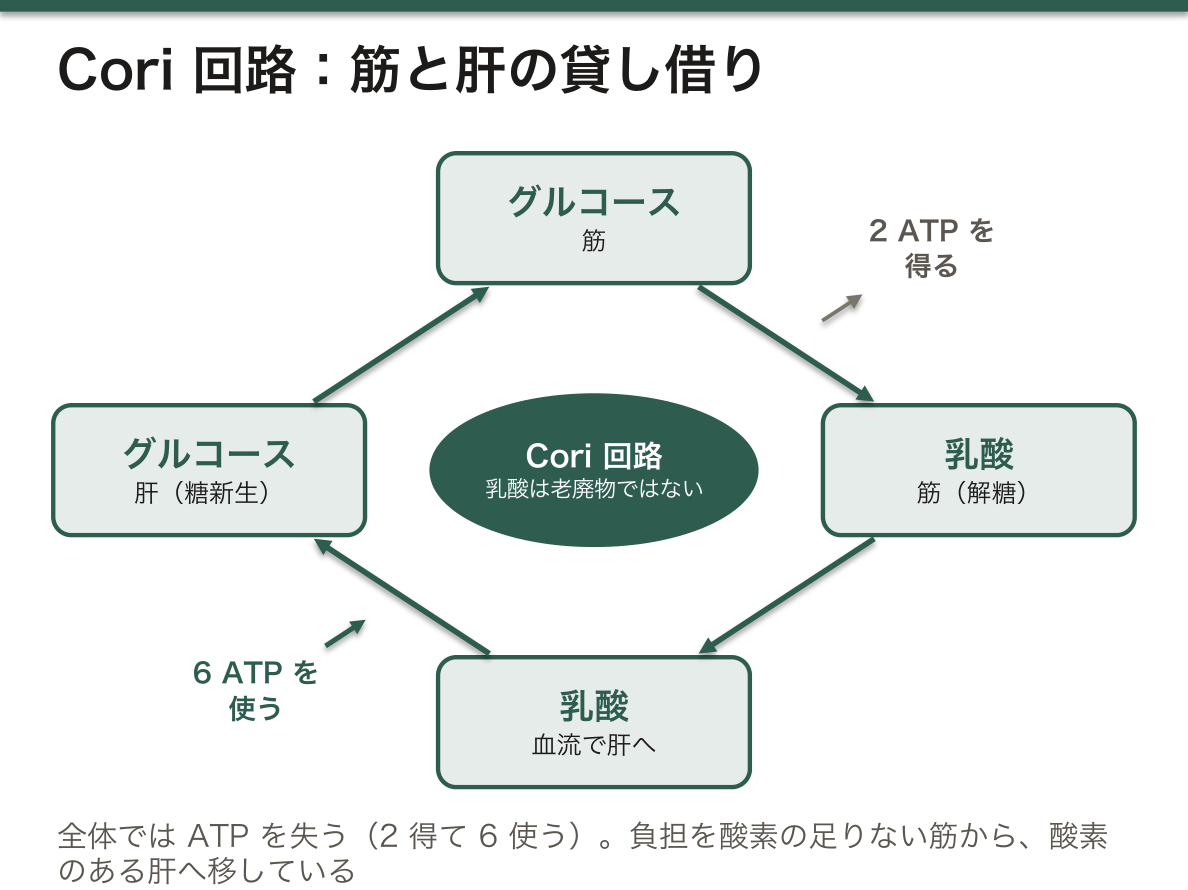
乳酸は代謝の行き止まりではない。 乳酸から作れるのはピルビン酸だけで、ピルビン酸へ戻せば TCA回路でも糖新生でも使える。近年は乳酸そのものが心筋・脳・遅筋のエネルギー源として積極的に利用されることが分かっており、「疲労物質」という古い理解は誤りとされる。
同じ骨格で、窒素を運ぶのがグルコース-アラニン回路——筋でピルビン酸にアミノ基を渡してアラニンとし、肝で外して尿素回路へ、炭素は糖新生へ。Cori 回路が炭素を、グルコース-アラニン回路が炭素と窒素を運ぶという関係になる。
| 乳酸アシドーシスの型 | 機序 | 例 |
|---|---|---|
| A型(組織低酸素あり) | 酸素が届かない | ショック・心停止・重症貧血・一酸化炭素中毒 |
| B型(組織低酸素なし) | 代謝そのものの異常 | メトホルミン(糖新生を抑え乳酸の消費を減らす)、肝不全(処理できない)、ビタミンB1欠乏(ピルビン酸脱水素酵素が止まる)、ミトコンドリア病、悪性腫瘍 |
ビタミンB1欠乏で乳酸が溜まるのが分かりやすい——ピルビン酸脱水素酵素の補酵素がチアミンピロリン酸なので、B1 が無いとピルビン酸がアセチルCoA へ進めず、乳酸へ溢れる。ブドウ糖を先に投与すると、残り少ないB1が一気に消費されて Wernicke 脳症を誘発する——この機序も同じ酵素の話。
アニオンギャップの開大した代謝性アシドーシスを見たら、乳酸・ケトン体・腎不全・中毒を考える。語呂 GOLDMARK / MUDPILES。
English version
Core
When glycolysis in muscle outruns the oxygen supply in vigorous exercise, pyruvate is reduced to lactate. The purpose is not to make ATP but to regenerate NAD⁺ — without it glycolysis would stop.
Lactate is a temporary parking place for electrons with nowhere to go, and is not simply discarded but sent to the liver.
| Place | What happens |
|---|---|
| Muscle | Glucose → glycolysis → lactate (gaining 2 ATP) |
| Blood | Lactate is carried to the liver |
| Liver | Lactate → pyruvate → back to glucose by gluconeogenesis (consuming 6 ATP) |
| Blood | Glucose returns to the muscle |
Overall ATP is lost (2 gained, 6 spent). It works nonetheless because the burden is shifted from muscle that lacks oxygen to liver that has it — a division of labour between organs.
Going further
Lactate is not a dead end of metabolism. All that can be made from lactate is pyruvate, and returned to pyruvate it can serve the TCA cycle or gluconeogenesis. It is now known that lactate itself is actively used as a fuel by cardiac muscle, brain and slow muscle fibres, and the old idea of a "fatigue substance" is regarded as wrong.
On the same skeleton, the glucose–alanine cycle carries nitrogen — the amino group is handed to pyruvate in muscle as alanine, removed in the liver for the urea cycle, and the carbon goes to gluconeogenesis. The Cori cycle carries carbon; the glucose–alanine cycle carries carbon and nitrogen.
| Type of lactic acidosis | Mechanism | Examples |
|---|---|---|
| Type A (tissue hypoxia present) | Oxygen does not arrive | Shock, cardiac arrest, severe anaemia, carbon monoxide poisoning |
| Type B (no tissue hypoxia) | A defect of metabolism itself | Metformin, liver failure, thiamine deficiency (pyruvate dehydrogenase stops), mitochondrial disease, malignancy |
Thiamine deficiency is the clearest — thiamine pyrophosphate being the coenzyme of pyruvate dehydrogenase, without B1 pyruvate cannot proceed to acetyl-CoA and overflows into lactate. Giving glucose first consumes what B1 remains at a stroke and precipitates Wernicke’s encephalopathy — the same enzyme again.
Seeing a metabolic acidosis with a widened anion gap, think of lactate, ketones, renal failure and poisoning (the mnemonics GOLDMARK / MUDPILES).
まとめ
| 解糖系 — 細胞質でグルコースを壊す | 細胞質で、グルコース1分子 → ピルビン酸2分子。 |
|---|---|
| 解糖系の10段階 — 酵素・中間体・反応の型を1枚に | 解糖は準備期(1〜5:ATP を2つ使ってグルコースを2つの三炭糖リン酸に割る)と報酬期(6〜10:三炭糖ごとに NADH 1つと ATP 2つを取り出す)に分かれる。 |
| 解糖の調節 — 不可逆な3段階と、前向きの活性化 | 調節がかかるのは、大きく負の ΔG を持つ不可逆な3段階だけである(可逆な段階を止めても、平衡がずれるだけで流れは決まらない)。 |
| 基質レベルのリン酸化 — 膜も酸素も使わずに ATP を作る | ATP の作り方は2つしかない。基質レベルのリン酸化酸化的リン酸化仕組みリン酸を持つ高エネルギー化合物から ADP へ直接リン酸を渡す電子伝達で作った H⁺ の勾配で ATP 合成… |
| ピルビン酸の3つの行き先 | 解糖でできたピルビン酸は、状況によって3方向へ分かれる。 |
| 酵母のアルコール発酵と、解糖を止める毒 — フッ化物とヒ酸 | 発酵は、酸素を使わずにグルコースを分解して ATP を得ることの総称である。 |
| 糖新生 — 血糖を作る | 主に肝(一部は腎皮質)で、糖以外の材料からグルコースを作る。 |
| グリコーゲン — 蓄える/出す | グルコースを枝分かれした多糖として蓄える形。 |
| ペントースリン酸経路 — ATPを作らない糖の道 | 細胞質で、グルコース6リン酸からNADPH とリボース5リン酸を作る。 |
| ペントースリン酸経路の2つの相 — 非酸化的相のトランスケトラーゼとトランスアルドラーゼ | ペントースリン酸経路は2つの相に分かれる。 |
| Cori 回路 — 乳酸は老廃物ではなく、貸し借りである | 激しい運動で筋の解糖が酸素供給を上回ると、ピルビン酸が乳酸へ還元される。 |
このノートは医学生が授業の内容を自分の言葉でまとめたもので、診療の判断に使うものではありません。誤りに気づいたらお知らせください。