糖はエネルギー源としてだけ扱われがちだが、体の中では「情報」と「構造」の材料でもある。
- 情報——細胞表面の糖鎖が、細胞の身分証明になる。ABO 血液型がその例
- 構造——グリコサミノグリカンが水を抱え込み、軟骨と硝子体の弾力を作る
そして糖は勝手に蛋白に付く(糖化)——酵素を介さない非酵素的な反応で、これが HbA1c と糖尿病合併症の実体になる。
だから「糖鎖が付く」には2種類ある——酵素が付ける糖鎖修飾(グリコシル化)と、勝手に付く糖化(グリケーション)。前者は機能、後者は劣化になる。
- 単糖から多糖まで — 名前が示す構造
- 単糖の名前の付け方 — 「カルボニルの種類」×「炭素の数」
- D/L・エピマー・アノマー — 糖の「違い」を3つに切り分ける
- 糖が環になる理由 — ヘミアセタール・ピラノースとフラノース・いす形
- Fischer 投影式から Haworth 式への書き換え — 5つの手順
- グリコシド結合の書き方と還元糖 — アノマー炭素が空いているかどうか
- 糖の酸化と還元 — 糖酸と糖アルコール
- 糖鎖が付く2つの道 — 機能としての修飾と、劣化としての糖化
- ABO 式血液型の化学 — 酵素が1つ違うだけで、型が変わる
- 糖鎖が作られる仕組みと、その異常
- シアル酸 — 糖鎖の末端に置かれた、「自分である」という印
- 分泌型と非分泌型 — 血液型物質が体液に出るかどうかで、かかる病気が変わる
- 単糖から多糖まで — 名前が示す構造
- 単糖の名前の付け方 — 「カルボニルの種類」×「炭素の数」
- D/L・エピマー・アノマー — 糖の「違い」を3つに切り分ける
- 糖が環になる理由 — ヘミアセタール・ピラノースとフラノース・いす形
- Fischer 投影式から Haworth 式への書き換え — 5つの手順
- グリコシド結合の書き方と還元糖 — アノマー炭素が空いているかどうか
- 糖の酸化と還元 — 糖酸と糖アルコール
- 糖鎖が付く2つの道 — 機能としての修飾と、劣化としての糖化
- ABO 式血液型の化学 — 酵素が1つ違うだけで、型が変わる
- 糖鎖が作られる仕組みと、その異常
- シアル酸 — 糖鎖の末端に置かれた、「自分である」という印
- 分泌型と非分泌型 — 血液型物質が体液に出るかどうかで、かかる病気が変わる
- まとめ
単糖から多糖まで — 名前が示す構造
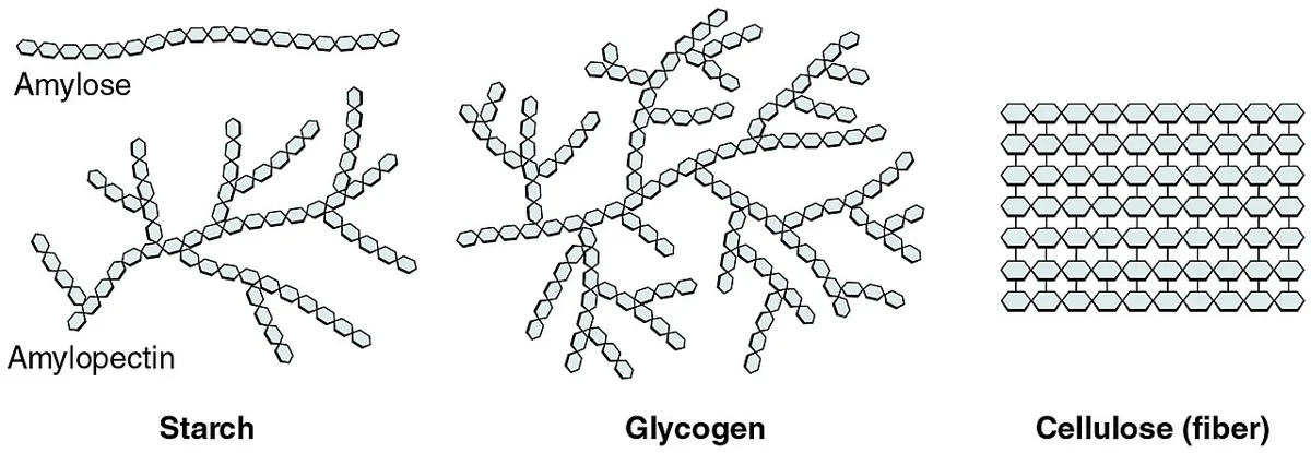
単糖——グルコース・ガラクトース・フルクトース(すべて C₆H₁₂O₆ の異性体)。
- アノマー——環になるときに生じる α と β。デンプンは α、セルロースは β。ヒトは β を切れない
二糖——マルトース(Glc+Glc)・ラクトース(Gal+Glc)・スクロース(Glc+Fru)。
多糖——デンプン・グリコーゲン(貯蔵)/セルロース(構造・食物繊維)。
グリコサミノグリカン(GAG)——二糖の繰り返し。強い陰性荷電で水を抱える。
- ヒアルロン酸——唯一、硫酸化されず蛋白と結合しない。関節液・硝子体
- コンドロイチン硫酸——軟骨。ケラタン硫酸——角膜。ヘパラン硫酸——基底膜
- ヘパリン——肥満細胞の顆粒。最も陰性荷電が強い
プロテオグリカン——核蛋白に GAG が多数結合したもの。アグリカンが軟骨の主役。
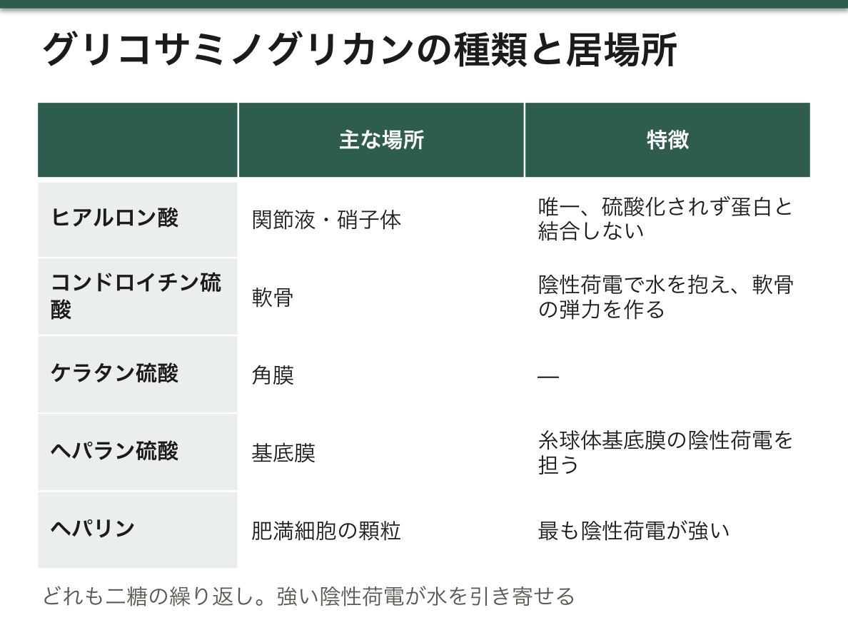
GAG の陰性荷電が水を引き寄せることが、軟骨の弾力の正体になる。荷重で水が押し出され、除荷で戻る(軟骨の章)——「関節が衝撃を吸収する仕組みが、電荷の話になっている」。
だから変形性関節症では、GAG の喪失が最初に起きる。軟骨が水を保てなくなり、力学的に弱くなる(変形性関節症の章)——「すり減る前に、含水率が落ちている」。
ヘパラン硫酸が糸球体基底膜の陰性荷電を担っている。これが失われると、陰性荷電のアルブミンが漏れる(微小変化型ネフローゼの章)——「孔の大きさではなく、電荷が壊れて蛋白が漏れる」。
ムコ多糖症は、GAG を分解できずに全身に溜まる病気になる。粗な顔貌・関節拘縮・角膜混濁・肝脾腫(蓄積症の章)——「同じ物質が溜まるので、臓器を越えて似た顔つきになる」。
β結合を切れないことが、食物繊維の定義そのものになる。ヒトの酵素では分解できず、腸内細菌が短鎖脂肪酸に変える(栄養の章)——「消化できないことに、意味がある」。
そして乳糖不耐症は、二糖類分解酵素の話になる。ラクターゼは小腸絨毛の先端にあり、感染性腸炎の後に一過性に失われる(小児の下痢の章)——「絨毛の先が削れると、まずこの酵素から失われる」。
English version
Core
Monosaccharides — glucose, galactose, fructose (all isomers of C₆H₁₂O₆).
- Anomers — the α and β that arise on ring formation. Starch is α, cellulose is β. Humans cannot cut β.
Disaccharides — maltose (Glc+Glc), lactose (Gal+Glc), sucrose (Glc+Fru).
Polysaccharides — starch and glycogen (storage) / cellulose (structure, dietary fibre).
Glycosaminoglycans (GAGs) — a repeating disaccharide. A strong negative charge holds water.
- Hyaluronan — the only one not sulphated and not bound to protein. Joint fluid, vitreous.
- Chondroitin sulphate — cartilage. Keratan sulphate — cornea. Heparan sulphate — basement membrane.
- Heparin — the granules of the mast cell. The most strongly negative.
Proteoglycan — a core protein with many GAGs attached. Aggrecan is the principal one of cartilage.
Going further
- That the negative charge of the GAGs draws water is the substance of the elasticity of cartilage. Water is pushed out under load and returns when unloaded (the chapter on cartilage) — the mechanism by which a joint absorbs shock turning out to be a matter of charge.
- Hence in osteoarthritis the loss of GAG comes first. Cartilage can no longer hold water and becomes mechanically weak (the chapter on osteoarthritis) — the water content falls before it wears away.
- Heparan sulphate carries the negative charge of the glomerular basement membrane. Lose it and negatively charged albumin leaks (the chapter on minimal change nephrotic syndrome) — protein leaking not because the pores are large but because the charge has broken.
- The mucopolysaccharidoses are diseases in which GAGs cannot be degraded and accumulate throughout the body. Coarse facies, joint contracture, corneal clouding, hepatosplenomegaly (the chapter on storage diseases) — the same substance accumulating, so the appearance resembles across organs.
- Not being able to cut the β linkage is the very definition of dietary fibre. Human enzymes cannot degrade it and the gut flora convert it into short-chain fatty acids (the chapter on nutrition) — there is a purpose in being indigestible.
- And lactose intolerance is a matter of the disaccharidases. Lactase sits at the tip of the small intestinal villus and is transiently lost after an infectious enteritis (the chapter on diarrhoea in children) — shave the tips of the villi and this enzyme is the first to go.
単糖の名前の付け方 — 「カルボニルの種類」×「炭素の数」
糖質の多くは一般式 Cn(H₂O)n で書けるので「炭水化物」と呼ばれるが、水が付いているわけではない。実体はヒドロキシ基を多数持つアルデヒドまたはケトンである。
大きさで4つに分ける:単糖(それ以上加水分解できない)・二糖(単糖2つ)・オリゴ糖(3〜10個。ラフィノース・デキストリンなど)・多糖(数百〜数千個。デンプン・グリコーゲン・セルロース)。
単糖の名前は、カルボニル基の種類と炭素の数を組み合わせて作る。
| アルデヒド基(–CHO)=アルドース | ケトン基(>C=O)=ケトース | |
|---|---|---|
| 3炭素(トリオース) | グリセルアルデヒド | ジヒドロキシアセトン |
| 5炭素(ペントース) | リボース・デオキシリボース | リブロース・キシルロース |
| 6炭素(ヘキソース) | グルコース・ガラクトース・マンノース | フルクトース |
たとえばグルコースはアルドヘキソース、フルクトースはケトヘキソース、リボースはアルドペントースである。炭素の番号は、カルボニル基に近い端から 1、2、3… と付ける。アルドースではアルデヒドの炭素が C1、ケトースではケトンの炭素が C2 になる。
不斉炭素の数で、異性体の数が決まる。4つの異なる基が付いた炭素(不斉炭素)が n 個あれば、立体異性体は最大 2n 個できる。アルドヘキソースは C2〜C5 の4つが不斉炭素なので 2⁴=16 種(D 型8種・L 型8種)、ケトヘキソースは C3〜C5 の3つなので 8 種である。グルコース・ガラクトース・マンノースはこの16種のうちの3つにすぎず、名前が違うのは「どの不斉炭素の –OH が左右どちらを向くか」が違うからである。
炭素の番号付けには、もう1つギリシャ文字の系統がある。主な官能基(カルボキシ基など)が付いた炭素の隣を α、その次を β、γ… と呼ぶ。α-アミノ酸、β-ヒドロキシ酪酸、γ-アミノ酪酸(GABA)がこの系統の名前である。数字の系統(C1 から数える)と1つずれる点に注意する。
English version
Core
Many carbohydrates fit the formula Cn(H₂O)n, hence ‘hydrates of carbon’, but there is no water in them. They are really aldehydes or ketones with many hydroxyl groups.
By size: monosaccharides (cannot be hydrolysed further), disaccharides (two units), oligosaccharides (3–10; raffinose, dextrins), polysaccharides (hundreds to thousands; starch, glycogen, cellulose).
A monosaccharide name combines the carbonyl type and the number of carbons.
| Aldehyde (–CHO) = aldose | Ketone (>C=O) = ketose | |
|---|---|---|
| 3 carbons (triose) | Glyceraldehyde | Dihydroxyacetone |
| 5 carbons (pentose) | Ribose, deoxyribose | Ribulose, xylulose |
| 6 carbons (hexose) | Glucose, galactose, mannose | Fructose |
Glucose is an aldohexose, fructose a ketohexose, ribose an aldopentose. Number the carbons from the end nearer the carbonyl group: the aldehyde carbon is C1 in an aldose; the ketone carbon is C2 in a ketose.
Going further
The number of chiral carbons sets the number of isomers. With n chiral carbons there are up to 2n stereoisomers. Aldohexoses have four (C2–C5): 16 isomers (8 D, 8 L). Ketohexoses have three (C3–C5): 8. Glucose, galactose and mannose are just three of the 16; their names differ because the –OH on some chiral carbon points the other way.
There is a second numbering system with Greek letters: the carbon next to the main functional group (such as a carboxyl group) is α, the next β, then γ. α-amino acid, β-hydroxybutyrate and γ-aminobutyrate (GABA) use this system. Note that it is shifted by one from the number system (which starts at C1).
D/L・エピマー・アノマー — 糖の「違い」を3つに切り分ける
| どこが違うか | 例 | 関係 | |
|---|---|---|---|
| D 型と L 型(鏡像異性体) | すべての不斉炭素 | D-グルコースと L-グルコース | 重ね合わせられない鏡像 |
| エピマー | 不斉炭素のうち1か所だけ | グルコースとガラクトース(C4)、グルコースとマンノース(C2) | 鏡像ではないジアステレオマー。別の糖 |
| アノマー(α と β) | 環になったときにできる新しい不斉炭素(アノマー炭素)だけ | α-D-グルコースと β-D-グルコース | 水の中で互いに入れ替わる |
D か L かは、カルボニル基から最も遠い不斉炭素(グルコースなら C5、リボースなら C4)の –OH が、Fischer 投影式で右にあれば D、左にあれば L と決める。体内の糖はほぼすべて D 型である。
エピマーは「1か所だけ違う」という関係なので、相手ごとに言う。ガラクトースもマンノースもグルコースのエピマーだが、ガラクトースとマンノースは2か所(C2 と C4)違うので、互いにはエピマーではない。D/L を決める最後の不斉炭素は同じなので、エピマーどうしは同じ D 型のままである。
D/L は形の記号で、光を回す向きとは別物である。もともとは右旋性(+)のグリセルアルデヒドを D と決めたことに由来し、ほかの糖は「グリセルアルデヒドと同じ向きの最後の不斉炭素を持つか」で D/L が決まる。実際の旋光の向きは測ってみないと分からず、D-グルコースは右旋性(+)だが、D-フルクトースは左旋性(−)である。D=右旋性と覚えると、フルクトースで間違える。
アノマーは水の中で入れ替わる(変旋光)。結晶の α-D-グルコースを水に溶かすと、環がいったん開いて鎖状になり、再び閉じるときに β にもなる。平衡では β が約64%、α が約36%で、鎖状のものは 0.02% 程度しか無い。β が多いのは、いす形で β-D-グルコースだけがすべての –OH と –CH₂OH を横向き(エカトリアル)に置けて、ぶつかり合いが最も少ないからである。
日本の教科書でも同じ3語を使うが、エピマーを「ジアステレオマーの特別な場合」、アノマーを「アノマー炭素についてのエピマー」と説明することもある。上の表のように、どの炭素が違うかで切り分ければ混乱しない。
ガラクトースをグルコースに変える酵素(UDP-ガラクトース 4-エピメラーゼ)は、C4 の向き1か所だけを入れ替える。ガラクトース血症ではこの経路のどこかが詰まり、使えないガラクトースが糖アルコール(ガラクチトール)に変わって水晶体に溜まり、白内障の原因になる。
English version
Core
| What differs | Example | Relationship | |
|---|---|---|---|
| D and L (enantiomers) | Every chiral carbon | D-glucose and L-glucose | Non-superimposable mirror images |
| Epimers | Only one chiral carbon | Glucose and galactose (C4); glucose and mannose (C2) | Diastereomers, not mirror images; different sugars |
| Anomers (α and β) | Only the new chiral carbon formed on ring closure (anomeric carbon) | α-D-glucose and β-D-glucose | Interconvert in water |
D or L is decided by the chiral carbon farthest from the carbonyl (C5 in glucose, C4 in ribose): –OH on the right in the Fischer projection = D, on the left = L. Almost all sugars in the body are D.
Epimer is a relationship between two named sugars. Galactose and mannose are both epimers of glucose, but they differ from each other at two carbons (C2 and C4), so they are not epimers of each other. The last chiral carbon is the same, so epimers stay D.
Going further
D/L describes shape, not the direction of optical rotation. It goes back to naming dextrorotatory (+) glyceraldehyde as D; other sugars are D or L according to their last chiral carbon. The actual rotation must be measured: D-glucose is (+) but D-fructose is (−). If you learn ‘D = dextrorotatory’, fructose will trick you.
Anomers interconvert in water (mutarotation). Dissolve crystalline α-D-glucose: the ring opens to the chain and closes again, sometimes as β. At equilibrium about 64% is β, 36% α, and only about 0.02% is open chain. β wins because only β-D-glucose can put every –OH and the –CH₂OH in equatorial positions in the chair, with the least crowding.
Japanese textbooks use the same three terms; some call an epimer a special diastereomer and an anomer an epimer at the anomeric carbon. Sorting by which carbon differs avoids confusion.
In the clinic
The enzyme that turns galactose into glucose (UDP-galactose 4-epimerase) flips only the C4 position. In galactosaemia this pathway is blocked; unused galactose becomes a sugar alcohol (galactitol) in the lens and causes cataract.
糖が環になる理由 — ヘミアセタール・ピラノースとフラノース・いす形
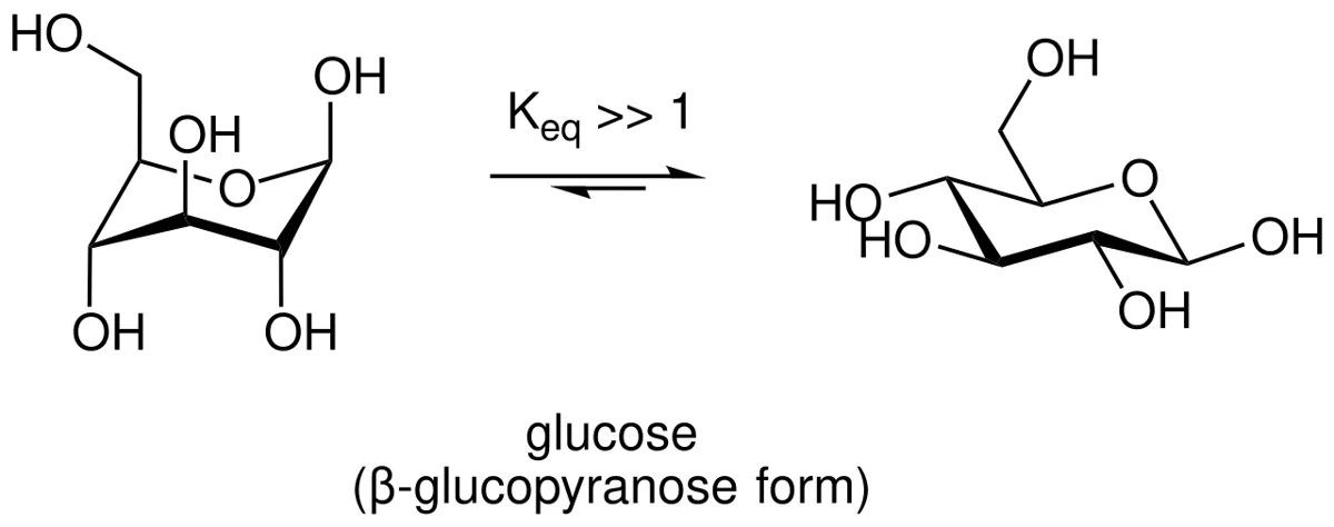
5炭素以上の糖は、水の中ではほとんどが環の形で存在する。分子の中の –OH が、同じ分子のカルボニル炭素を攻撃するからである。
–OH の酸素は電子が豊富(求核剤)、カルボニルの炭素は電子が不足している(求電子剤)。アルデヒドとアルコールが反応するとヘミアセタール、ケトンとアルコールならヘミケタールができる。糖では、カルボニルから最も遠い不斉炭素の –OH が攻撃する:
| 糖 | 攻撃する –OH | 攻撃される炭素 | 環 | 環の原子 |
|---|---|---|---|---|
| グルコース(アルドース) | C5 | C1 | ピラノース(6員環) | 炭素5つ+酸素1つ |
| フルクトース(ケトース) | C5 | C2 | フラノース(5員環) | 炭素4つ+酸素1つ |
| リボース(アルドース) | C4 | C1 | フラノース(5員環) | 炭素4つ+酸素1つ |
名前は、同じ形の環を持つ化合物ピラン(6員環)とフラン(5員環)から来ている。環になると、もとのカルボニル炭素に新しい –OH が生じ、その炭素は新しい不斉炭素(アノマー炭素)になる。炭素の番号は環になっても変わらない。
なぜ環のほうが安定なのか。5員環と6員環は結合の角度に無理がなく、C=O の二重結合1本が C–O の単結合2本に変わるので、エネルギー的に有利になる。逆に、反応のときには環が開いて鎖状のアルデヒドに戻る。糖の酸化や還元(還元糖の反応や糖アルコールの生成)は、ごく少量ある鎖状の形を通して進む。
いす形:6員環は平面ではなく、シクロヘキサンと同じようにいす形に折れている(Haworth 式は、それを平らに描いた略図である)。いす形では各炭素の置換基が、環の面から上下に出る「アキシアル」と、横に張り出す「エカトリアル」の2方向に分かれる。大きな基はエカトリアルのほうがぶつからず安定で、β-D-グルコピラノースはすべての大きな基をエカトリアルに置ける唯一のアルドヘキソースである。いす形どうしの入れ替わり(反転)にはエネルギーが要るので、ふつうは安定な1つの形に固定されている。
「グルコースがなぜ生物の主役の糖になったのか」の説明として、この安定性がよく挙げられる。いす形で最も安定なので、鎖状の形(反応性の高いアルデヒド)でいる割合が少なく、蛋白に勝手に結合する糖化が起きにくい。それでも高血糖が長く続けば糖化は進み、HbA1c や終末糖化産物として現れる。
English version
Core
Sugars with five or more carbons exist in water mostly as rings, because one –OH in the molecule attacks the carbonyl carbon of the same molecule.
The –OH oxygen is electron-rich (nucleophile); the carbonyl carbon is electron-poor (electrophile). Aldehyde + alcohol gives a hemiacetal; ketone + alcohol gives a hemiketal. In sugars, the –OH on the chiral carbon farthest from the carbonyl attacks:
| Sugar | Attacking –OH | Carbon attacked | Ring | Ring atoms |
|---|---|---|---|---|
| Glucose (aldose) | C5 | C1 | Pyranose (6-membered) | 5 C + 1 O |
| Fructose (ketose) | C5 | C2 | Furanose (5-membered) | 4 C + 1 O |
| Ribose (aldose) | C4 | C1 | Furanose (5-membered) | 4 C + 1 O |
The names come from pyran (6-ring) and furan (5-ring). On ring closure the old carbonyl carbon gets a new –OH and becomes a new chiral centre, the anomeric carbon. Carbon numbering does not change.
Going further
Why is the ring more stable? Five- and six-membered rings have comfortable bond angles, and one C=O double bond becomes two C–O single bonds, which is energetically favourable. For reactions the ring opens back to the chain aldehyde; oxidation and reduction of sugars go through the small amount of open chain.
Chair form: a six-membered ring is not flat; like cyclohexane it folds into a chair (the Haworth formula is a flat sketch of it). Each substituent points either up or down from the ring (axial) or out to the side (equatorial). Large groups are more stable equatorial, and β-D-glucopyranose is the only aldohexose that can put all large groups equatorial. Flipping between chairs needs energy, so the ring usually stays in its stable chair.
In the clinic
This stability is often used to explain why glucose became life’s main sugar: in its very stable chair, little is in the reactive open-chain aldehyde form, so non-enzymatic glycation of proteins is slow. Long hyperglycaemia still drives glycation, seen as HbA1c and advanced glycation end products.
Fischer 投影式から Haworth 式への書き換え — 5つの手順
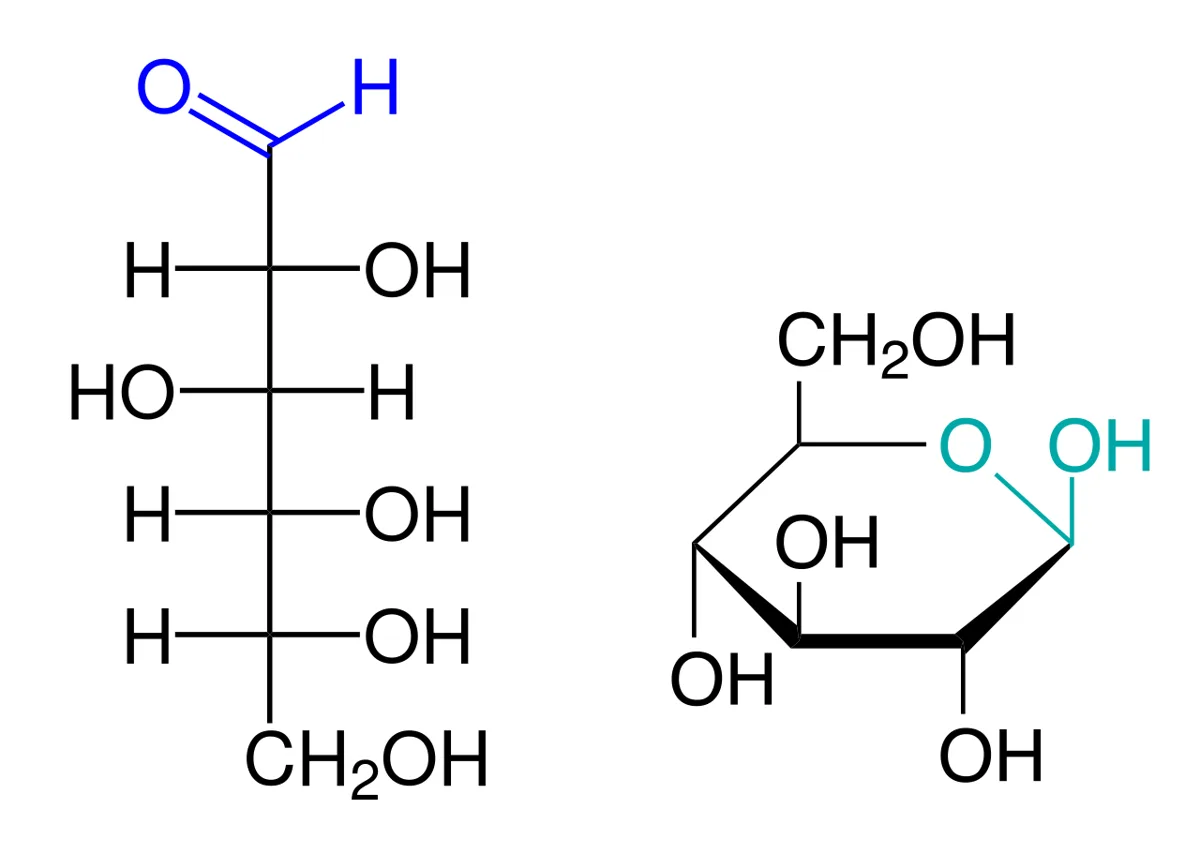
Fischer 投影式は鎖状の糖を縦に描いた図で、横の線は手前、縦の線は奥を向いている。Haworth 式は環になった糖を、環を水平に寝かせて斜め上から見た図である。一方から他方へは、次の5手順で機械的に書き換えられる。
①環を描く——ピラノースなら6員環、フラノースなら5員環。環の酸素を右奥に置く。②炭素に番号を振る——酸素の右隣(右端)がアノマー炭素(アルドースは C1、ケトースは C2)で、そこから手前を通って時計回りに番号が進む。③環に関わらない –OH を置く——Fischer 式で右にある –OH は Haworth 式で下、左にある –OH は上。④最後の炭素の基(–CH₂OH)を置く——D 型なら上、L 型なら下。⑤アノマー炭素に新しい –OH を置く——最後の炭素の基と反対側なら α、同じ側なら β。D 型では下向きが α、上向きが β になる。
| C1 | C2 | C3 | C4 | C5 の –CH₂OH | |
|---|---|---|---|---|---|
| D-グルコース(Fischer) | –CHO | 右 | 左 | 右 | (C5 の –OH は右=D) |
| β-D-グルコピラノース(Haworth) | –OH 上(β) | 下 | 上 | 下 | 上 |
| β-D-ガラクトピラノース(Haworth) | –OH 上(β) | 下 | 上 | 上(C4 だけ違う) | 上 |
「右は下」になる理由は、Fischer 式を右に90度倒してみると分かる。縦の鎖が横になり、右向きに出ていた基はすべて下を向く。そのままでは C5 の –OH が C1 に届かないので、C4–C5 の結合を回して –OH を環の側に向ける。このとき C5 に付いた –CH₂OH は上に持ち上がる——だから D 型では最後の炭素の基が上に来る。
α か β かは、Fischer 式からは決まらない。環が閉じるたびにどちら向きにもなりうるので、問題で「α」か「β」かを指定されて初めて描ける。逆に Haworth 式を読むときは、最後の炭素の基とアノマー炭素の –OH が同じ側か反対側かだけを見ればよい。図を上下にひっくり返して描かれていても、この「同じ側か反対側か」の関係は変わらない。
フルクトース(フラノース)では、アノマー炭素 C2 に –OH と C1 の –CH₂OH の2つが付く。C3 の –OH は Fischer 式で左なので上、C4 は右なので下、C5 の –CH₂OH(C6)は上。β-D-フルクトフラノースでは C2 の –OH が上(C6 と同じ側)、C1 の –CH₂OH が下になる。ショ糖の中のフルクトースがこの β-フラノースである。
English version
Core
A Fischer projection draws the open-chain sugar vertically: horizontal bonds come towards you, vertical bonds go away. A Haworth projection shows the ring lying flat, seen from slightly above. Five mechanical steps convert one into the other.
(1) Draw the ring: 6-membered for pyranose, 5-membered for furanose, ring oxygen at the back right. (2) Number the carbons: the carbon right of the oxygen (far right) is the anomeric carbon (C1 in aldoses, C2 in ketoses); numbering runs clockwise through the front. (3) Place the –OH groups not in the ring: right in Fischer = down in Haworth; left = up. (4) Place the last carbon group (–CH₂OH): up for D, down for L. (5) Place the new –OH on the anomeric carbon: opposite side to the last carbon group = α; same side = β. For D sugars, down is α and up is β.
| C1 | C2 | C3 | C4 | C5 –CH₂OH | |
|---|---|---|---|---|---|
| D-glucose (Fischer) | –CHO | right | left | right | (C5 –OH right = D) |
| β-D-glucopyranose (Haworth) | –OH up (β) | down | up | down | up |
| β-D-galactopyranose (Haworth) | –OH up (β) | down | up | up (only C4 differs) | up |
Going further
Why does right become down? Turn the Fischer formula 90° clockwise: the chain lies flat and every group that pointed right now points down. The C5 –OH cannot reach C1 yet, so the C4–C5 bond rotates to bring the –OH into the ring; this lifts the C5 –CH₂OH upward — that is why the last carbon group is up in D sugars.
α or β cannot be read from the Fischer formula. Each ring closure can give either, so you can only draw it once α or β is specified. When reading a Haworth formula, just ask whether the last carbon group and the anomeric –OH are on the same side or opposite sides; this does not change even if the drawing is flipped.
For fructose (furanose) the anomeric C2 carries both an –OH and the C1 –CH₂OH. C3 –OH is left in Fischer, so up; C4 is right, so down; the C5 –CH₂OH (C6) is up. In β-D-fructofuranose the C2 –OH is up (same side as C6) and the C1 –CH₂OH is down. The fructose in sucrose is this β-furanose.
グリコシド結合の書き方と還元糖 — アノマー炭素が空いているかどうか
2つの単糖の –OH どうしが脱水縮合すると、–O– でつながったグリコシド結合ができる。結合は「どの単糖が・α か β か・何番の炭素と何番の炭素か」の3つで書く。たとえば乳糖は Gal(β1→4)Glc——ガラクトースの C1(β 型)がグルコースの C4 とつながっている。
| 二糖 | 書き方 | 還元性 | 分解する酵素 |
|---|---|---|---|
| マルトース(麦芽糖) | Glc(α1→4)Glc | あり | マルターゼ・スクラーゼ |
| イソマルトース | Glc(α1→6)Glc | あり | イソマルターゼ |
| セロビオース | Glc(β1→4)Glc | あり | ラクターゼの β-グリコシダーゼ活性 |
| ラクトース(乳糖) | Gal(β1→4)Glc | あり | ラクターゼ |
| スクロース(ショ糖) | Glc(α1↔2β)Fru | なし | スクラーゼ |
| トレハロース | Glc(α1↔1α)Glc | なし | トレハラーゼ |
還元糖とは、アノマー炭素が結合に使われずに空いている糖である。空いていれば環が開いて鎖状のアルデヒド(またはα-ヒドロキシケトン)に戻れるので、Cu²⁺ を Cu⁺(赤褐色の Cu₂O)に、Ag⁺ を Ag に還元できる(Fehling 反応・Tollens 反応)。スクロースとトレハロースは2つのアノマー炭素どうしで結合しているので、どちらも開けず、還元性を持たない(↔ はその印)。
多糖の還元性:デンプンやグリコーゲンも、鎖の片端(還元末端)だけはアノマー炭素が空いている。ただし数千個の単位に対して1か所しかないので、ほとんど還元性を示さない。アミラーゼで切ると還元末端が一気に増えて還元性が強まる——デンプンの消化を還元糖の検出で追えるのはこのためである。
フルクトースは還元糖か。ケトースはアルデヒド基を持たないので、「アルデヒドを持つものを還元糖とする」狭い定義を使うヨーロッパの講義では、ケトースを非還元糖として扱うことがある。一方、Fehling 液のようなアルカリ性の試薬の中では、フルクトースはエンジオールを経てアルドースに異性化するので、反応は陽性になる。日本の教科書と国試では、アノマー炭素が空いているフルクトースは還元糖に分類される。どちらの立場で問われているかを確かめて答える。
トレハロースはキノコ・酵母・昆虫に多い二糖で、小腸のトレハラーゼで分解される。非還元性で安定なので、食品の保存料・保湿剤としても使われる。
尿糖の試験紙はグルコースオキシダーゼという酵素を使うのでグルコースにしか反応しないが、昔ながらの銅還元法は還元糖すべてに反応する。だから乳児のガラクトース血症や果糖尿は、試験紙では陰性で銅還元法だけが陽性になる、という食い違いで見つかることがある。
English version
Core
When –OH groups of two monosaccharides condense, a glycosidic bond (–O–) forms. Write it with three facts: which sugars, α or β, and which carbons. Lactose is Gal(β1→4)Glc: C1 of galactose (β) joins C4 of glucose.
| Disaccharide | Notation | Reducing? | Enzyme |
|---|---|---|---|
| Maltose | Glc(α1→4)Glc | Yes | Maltase, sucrase |
| Isomaltose | Glc(α1→6)Glc | Yes | Isomaltase |
| Cellobiose | Glc(β1→4)Glc | Yes | β-glycosidase activity of lactase |
| Lactose | Gal(β1→4)Glc | Yes | Lactase |
| Sucrose | Glc(α1↔2β)Fru | No | Sucrase |
| Trehalose | Glc(α1↔1α)Glc | No | Trehalase |
A reducing sugar is one whose anomeric carbon is free, not used in a bond. Then the ring can open to the chain aldehyde (or α-hydroxy ketone), which reduces Cu²⁺ to Cu⁺ (red-brown Cu₂O) or Ag⁺ to Ag (Fehling and Tollens tests). Sucrose and trehalose are joined anomeric carbon to anomeric carbon, so neither ring can open and they are non-reducing (↔ marks this).
Going further
Polysaccharides: starch and glycogen have one free anomeric carbon at one end (the reducing end), but only one per thousands of units, so they are barely reducing. Amylase creates many new reducing ends, so starch digestion can be followed by testing for reducing sugar.
Is fructose a reducing sugar? Ketoses have no aldehyde group, so European courses that define reducing sugars as those with an aldehyde sometimes call ketoses non-reducing. But in alkaline reagents such as Fehling’s solution, fructose isomerises to an aldose through an enediol, and the test is positive. Japanese textbooks and the national exam classify fructose, with its free anomeric carbon, as a reducing sugar. Check which definition the question uses.
Trehalose is common in mushrooms, yeast and insects and is split by intestinal trehalase. It is non-reducing and stable, so it is also used as a food preservative and moisturiser.
In the clinic
Urine glucose dipsticks use glucose oxidase and react only with glucose, but the old copper-reduction test reacts with every reducing sugar. So galactosaemia or fructosuria in infants can be found by the mismatch: dipstick negative, copper test positive.
糖の酸化と還元 — 糖酸と糖アルコール
糖の炭素は、端を酸化すればカルボン酸に、カルボニルを還元すればアルコールになる。どの炭素が変わったかで名前が決まる。
| 種類 | 変わる炭素 | グルコースからできるもの | 体での役割 |
|---|---|---|---|
| アルドン酸 | C1(アルデヒド)→ –COOH | グルコン酸(グルコノラクトン) | グルコースオキシダーゼの反応産物。血糖測定の原理 |
| ウロン酸 | C6(–CH₂OH)→ –COOH | グルクロン酸 | ビリルビン・薬物の抱合、グリコサミノグリカンの材料 |
| アルダル酸 | C1 と C6 の両方 | グルカル酸 | (体内での役割は小さい) |
| 糖アルコール | C1(カルボニル)→ –CH₂OH | ソルビトール(グルシトール) | アルドース還元酵素(NADPH)で作られる。ポリオール経路 |
ウロン酸がとくに重要なのは、C1 のアノマー炭素が空いたまま酸を持てるからである。グルクロン酸は UDP-グルクロン酸として活性化され、C1 で他の分子の –OH や –COOH にグリコシド結合する。結合した相手は –COO⁻ の電荷を背負うので水に溶けやすくなり、胆汁や尿に捨てられる。ビリルビンの抱合、多くの薬(アセトアミノフェン・モルヒネなど)の第2相反応がこれである。ヒアルロン酸・コンドロイチン硫酸も、グルクロン酸とアミノ糖の繰り返しでできている。
糖アルコールはカルボニルを失っているので環を作れず、還元性も無い。ソルビトールは細胞膜を通りにくいので細胞の中に溜まり、水を引き込む。マンニトールは同じ理由で血管の中に留まって水を引き出すので、浸透圧利尿薬(頭蓋内圧を下げる)として使われる。キシリトールは口の細菌が発酵できないので、虫歯になりにくい甘味料になる。
グルクロン酸抱合の酵素(UGT1A1)が弱いと、間接ビリルビンが上がる(Gilbert 症候群・Crigler-Najjar 症候群・新生児黄疸)。抗がん薬イリノテカンの活性代謝物も同じ酵素で抱合されるため、UGT1A1 の遺伝子多型によって重い下痢や好中球減少が起きやすく、投与前に多型を調べることがある。
English version
Core
Oxidise an end carbon of a sugar and you get a carboxylic acid; reduce the carbonyl and you get an alcohol. Which carbon changed decides the name.
| Type | Carbon changed | From glucose | Role in the body |
|---|---|---|---|
| Aldonic acid | C1 (aldehyde) → –COOH | Gluconic acid (gluconolactone) | Product of glucose oxidase; principle of glucose meters |
| Uronic acid | C6 (–CH₂OH) → –COOH | Glucuronic acid | Conjugation of bilirubin and drugs; building block of glycosaminoglycans |
| Aldaric acid | Both C1 and C6 | Glucaric acid | (minor role) |
| Sugar alcohol | C1 (carbonyl) → –CH₂OH | Sorbitol (glucitol) | Made by aldose reductase (NADPH); polyol pathway |
Going further
Uronic acids matter because the anomeric C1 stays free while the molecule carries an acid. Activated as UDP-glucuronate, glucuronic acid forms a glycosidic bond through C1 with an –OH or –COOH of another molecule. The partner now carries a –COO⁻ charge, dissolves in water and is excreted in bile or urine: bilirubin conjugation and phase II metabolism of many drugs (paracetamol, morphine). Hyaluronan and chondroitin sulphate are repeats of glucuronic acid and an amino sugar.
Sugar alcohols have lost the carbonyl, so they cannot form rings and are not reducing. Sorbitol crosses membranes poorly, collects in cells and draws in water. Mannitol for the same reason stays in the blood and draws water out, so it is an osmotic diuretic (to lower intracranial pressure). Mouth bacteria cannot ferment xylitol, so it is a tooth-friendly sweetener.
In the clinic
Weak glucuronidation (UGT1A1) raises unconjugated bilirubin (Gilbert syndrome, Crigler-Najjar syndrome, neonatal jaundice). The active metabolite of irinotecan is conjugated by the same enzyme, so UGT1A1 variants increase the risk of severe diarrhoea and neutropenia; the variant may be tested before treatment.
糖鎖が付く2つの道 — 機能としての修飾と、劣化としての糖化
糖鎖修飾(グリコシル化)——酵素が特定の場所に付ける。小胞体とGolgiで行われる。
- N結合型——アスパラギンに付く。小胞体で始まる
- O結合型——セリン/トレオニンに付く。Golgiで行われる。ムチンがこの型
- 働き——折り畳みの補助・安定性・細胞認識・行き先の指定(マンノース6-リン酸)
ABO 血液型——H 抗原に、A 転移酵素が N-アセチルガラクトサミンを、B 転移酵素がガラクトースを付ける。O型は転移酵素を欠き H 抗原のまま。
糖化(グリケーション)——酵素を介さず、糖のアルデヒド基が蛋白のアミノ基に結合する。濃度と時間に比例する。
- 初期は可逆(Schiff塩基)→ Amadori 転位で安定 → 終末糖化産物(AGEs)
HbA1c は、糖化が「濃度 × 時間」で決まることの応用そのものになる。赤血球の寿命が約120日なので、過去1〜2か月の平均を映す(HbA1cの章)——「酵素が関わらないから、平均が素直に反映される」。
だから赤血球の寿命が変われば、値がずれる。溶血で低く、鉄欠乏の未治療で高く出る——「時間の項が変わるので、同じ血糖でも違う数字になる」。
AGEs が、糖尿病合併症の共通の機序になる。膠原線維を架橋して硬くし、受容体(RAGE)を介して炎症を起こす(糖尿病合併症の章)——「血管が硬くなることと、腎が線維化することが、同じ反応でつながる」。
そして架橋は元に戻らない。だから血糖を下げても、既に起きた変化は残る——「早期の管理が意味を持つ理由が、可逆性の限界にある」。
ABO 血液型が糖転移酵素の違いであることが、輸血の抗体の性質を決めている。自然抗体は IgM で、常温で凝集する(輸血の章)——「ABO 不適合が即時型で激烈になるのは、この抗体の性質による」。
そして IgM は胎盤を通らない。だから ABO 不適合妊娠は、Rh 不適合ほど重症化しにくい(胎盤関門の章)——「抗体のクラスが、母児間で何が起きるかを決めている」。
English version
Core
Glycosylation — an enzyme puts it on at a particular place. Carried out in the endoplasmic reticulum and the Golgi.
- N-linked — attached to asparagine. Begins in the endoplasmic reticulum.
- O-linked — attached to serine or threonine. Carried out in the Golgi. The mucins are of this type.
- Roles — assisting folding, stability, cell recognition, specifying the destination (mannose 6-phosphate).
The ABO blood groups — on the H antigen, the A transferase puts N-acetylgalactosamine and the B transferase galactose. Group O lacks the transferase and remains H antigen.
Glycation — without an enzyme, the aldehyde group of a sugar binds an amino group of a protein. It is proportional to concentration and to time.
- Reversible at first (a Schiff base) → stabilised by the Amadori rearrangement → advanced glycation end-products (AGEs).
Going further
- HbA1c is the very application of glycation being set by "concentration × time". The red cell living about 120 days, it reflects the average of the past one to two months (the chapter on HbA1c) — no enzyme being involved, the average is reflected straightforwardly.
- Hence a change in red cell lifespan shifts the value. It reads low in haemolysis and high in untreated iron deficiency — the time term changing, so the same blood glucose gives a different number.
- AGEs are the common mechanism of the complications of diabetes. They cross-link collagen fibres and stiffen them, and cause inflammation through a receptor (RAGE) (the chapter on the complications of diabetes) — the stiffening of the vessel and the fibrosis of the kidney linked by the same reaction.
- And the cross-links do not come undone. So lowering the blood glucose leaves the change already made — the reason early control matters lies in the limit of reversibility.
- That the ABO groups are a difference of glycosyltransferases determines the character of the antibodies in transfusion. The natural antibody is IgM and agglutinates at room temperature (the chapter on transfusion) — ABO incompatibility being immediate and violent follows from the character of this antibody.
- And IgM does not cross the placenta. So ABO incompatibility in pregnancy is less severe than Rh incompatibility (the chapter on the placental barrier) — the class of the antibody deciding what happens between mother and child.
ABO 式血液型の化学 — 酵素が1つ違うだけで、型が変わる
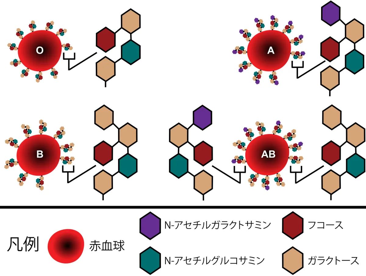
血液型の実体は、赤血球膜の糖脂質・糖蛋白に付いた糖鎖の末端になる。
共通の土台が H 抗原——H 遺伝子(FUT1)の産物がフコースを付けて作る。
| 型 | 付ける糖 | 酵素 |
|---|---|---|
| A | N-アセチルガラクトサミン | A 転移酵素 |
| B | ガラクトース | B 転移酵素 |
| AB | 両方 | 両方 |
| O | 何も付けない | 酵素が働かない(H 抗原のまま) |
A と B の転移酵素は、アミノ酸4個しか違わない。
血漿中には、自分が持たない抗原に対する IgM 型の自然抗体がある(規則抗体)。
Bombay 型——H 抗原そのものが作れない。ABO 検査では O 型に見えるが、O 型の血も輸血できない。
「O 型は、何も付けていない状態である」という理解が、輸血の原則を導く。O 型赤血球は A にも B にも認識される抗原を持たないので、誰にでも渡せる(輸血の章)——「引き算で理解すると、暗記が要らなくなる」。
ただし血漿は逆向きになる。O 型の血漿は抗A・抗B の両方を持つので、血漿製剤では AB 型が万能供血者になる——「赤血球と血漿で、万能な型が入れ替わる」。
自然抗体が IgM であることが、新生児溶血性疾患の型を決めている。IgM は胎盤を通らないので、ABO 不適合の溶血は Rh 不適合より軽く、初回の妊娠でも起こりうる(新生児黄疸の章)——「抗体のクラスが、病態の重さを決めている」。
Rh は蛋白抗原なので、免疫グロブリンのクラスが IgG になる(母子感染と血液型不適合の章)——「胎盤を通るので、2回目以降の妊娠で重くなる。だから抗D 免疫グロブリンを予防投与する」。
糖鎖抗原が、腫瘍マーカーにもなっている(腫瘍マーカーの項)——「CA19-9 は Lewis 式の糖鎖なので、Lewis 陰性の人では上がらない」。
English version
Core
The substance of a blood group is the terminus of the glycan attached to the glycolipids and glycoproteins of the red cell membrane.
The common foundation is the H antigen — made by the product of the H gene (FUT1) attaching fucose.
| Group | Sugar attached | Enzyme |
|---|---|---|
| A | N-acetylgalactosamine | A transferase |
| B | Galactose | B transferase |
| AB | Both | Both |
| O | Nothing attached | The enzyme does not work (it remains H antigen) |
The A and B transferases differ by only four amino acids.
The plasma carries IgM natural antibodies against the antigen one does not have (the regular antibodies).
The Bombay phenotype — the H antigen itself cannot be made. It looks like group O on ABO testing, but group O blood cannot be transfused either.
Going further
- The understanding that group O is the state of having nothing attached gives the principle of transfusion. The O red cell has no antigen recognised as A or B, so it can be given to anyone (the chapter on transfusion) — understood by subtraction, no memorising is needed.
- The plasma goes the other way, however. O plasma carries both anti-A and anti-B, so for plasma products group AB is the universal donor — the universal group swapping between red cells and plasma.
- That the natural antibody is IgM determines the pattern of haemolytic disease of the newborn. IgM does not cross the placenta, so ABO haemolysis is milder than Rh and can occur in a first pregnancy (the chapter on neonatal jaundice) — the class of the antibody deciding the severity.
- Rh being a protein antigen, the immunoglobulin class is IgG (the chapter on vertical transmission and blood group incompatibility) — it crosses the placenta, so second and later pregnancies are worse. Hence anti-D immunoglobulin is given prophylactically.
- Carbohydrate antigens serve as tumour markers too (the section on tumour markers) — CA19-9 is a Lewis glycan, so it does not rise in a Lewis-negative person.
糖鎖が作られる仕組みと、その異常
ヒトの蛋白の半分以上が糖鎖修飾を受ける。付き方は2通り。
| N結合型 | O結合型 | |
|---|---|---|
| 付く相手 | アスパラギン | セリン・トレオニン |
| 作られる場所 | 小胞体で共通の骨格を作り、Golgi で加工 | Golgi で1つずつ足す |
| 例 | 免疫グロブリン・多くの膜蛋白 | ムチン・プロテオグリカン |
糖鎖の役割——折り畳みの補助・分解からの保護・細胞どうしの認識・受容体としての標的。
先天性グリコシル化異常症(CDG)——糖鎖の合成や転移が障害される一群。多系統に症状が出る(発達の遅れ・肝障害・凝固異常・内斜視・脂肪の異常分布)。
スクリーニングには、血清トランスフェリンの等電点電気泳動が使われる。
「糖鎖が付かないと、蛋白は正しく働けない」という点が、CDG が多系統疾患になる理由になる。1つの酵素の異常が、何百もの蛋白に同時に影響する(翻訳後修飾の章)——「症状の組み合わせに、まとまりが出ない」。
糖鎖が半減期を決めていることは、薬の設計に使われている。エリスロポエチン製剤に糖鎖を足したダルベポエチンは、投与間隔を延ばせる(造血を促す薬の章)——「同じ蛋白でも、糖鎖の量で薬としての性質が変わる」。
糖鎖が細胞の目印になることも、多くの場面で効いてくる。肝の受容体はシアル酸が外れた糖蛋白を捕まえて処理する(古くなった蛋白の目印)——「末端の糖1つが、寿命を決めている」。
インフルエンザウイルスは、シアル酸を入口と出口の両方に使う(抗ウイルス薬の章)——「ノイラミニダーゼ阻害薬は、出るときにシアル酸を切る酵素を止めている」。
ムチンの O結合型糖鎖は、粘液の保水性そのものになる(粘液細胞の項)——「糖鎖が水を抱えるので、粘液は粘る」。
English version
Core
More than half of human proteins carry glycosylation. There are two ways of attachment.
| N-linked | O-linked | |
|---|---|---|
| Attached to | Asparagine | Serine, threonine |
| Made where | A common core is made in the endoplasmic reticulum and processed in the Golgi | Added one at a time in the Golgi |
| Examples | Immunoglobulin, many membrane proteins | Mucin, proteoglycan |
Roles of the glycan — assisting folding, protection from degradation, recognition between cells, being the target as a receptor.
Congenital disorders of glycosylation (CDG) — a group in which the synthesis or transfer of glycans is disturbed. Symptoms appear in many systems (developmental delay, liver disease, coagulopathy, esotropia, abnormal distribution of fat).
Isoelectric focusing of serum transferrin is used for screening.
Going further
- That a protein cannot work properly without its glycan is why CDG is a multisystem disease. An abnormality in one enzyme affects hundreds of proteins at once (the chapter on post-translational modification) — the combination of symptoms having no coherence.
- That the glycan sets the half-life is used in designing drugs. Darbepoetin, an erythropoietin preparation with added glycan, allows a longer dosing interval (the chapter on drugs promoting haematopoiesis) — the same protein taking on a different character as a drug according to the amount of glycan.
- That the glycan is a marker of the cell tells in many settings too. A hepatic receptor catches and processes glycoproteins from which sialic acid has been removed (the mark of an old protein) — a single terminal sugar setting the lifespan.
- Influenza virus uses sialic acid both as entrance and as exit (the chapter on antiviral drugs) — a neuraminidase inhibitor stops the enzyme that cuts sialic acid on the way out.
- The O-linked glycans of mucin are the very water-holding of mucus (the section on mucous cells) — the glycan holds water, so mucus is viscous.
シアル酸 — 糖鎖の末端に置かれた、「自分である」という印
細胞表面の糖鎖の末端には、しばしばシアル酸という負に荷電した糖が付いている。これが「自己」の目印として働いている。
- 補体を止める——シアル酸があると H 因子が結合し、補体第二経路の活性化が抑えられる。細菌にはシアル酸が乏しいので、そのまま攻撃される
- 免疫細胞を落ち着かせる——Siglec という受容体がシアル酸を読み取り、抑制の信号を出す
- 寿命の時計になる——血中の糖蛋白と赤血球は、シアル酸が外れると肝で捕まって除かれる。末端のシアル酸が「まだ新しい」という札になっている
荷電の効果も大きい。糸球体基底膜と足細胞の表面はシアル酸で強く負に帯電しており、同じく負に荷電したアルブミンを電気的に跳ね返している(荷電選択性)。
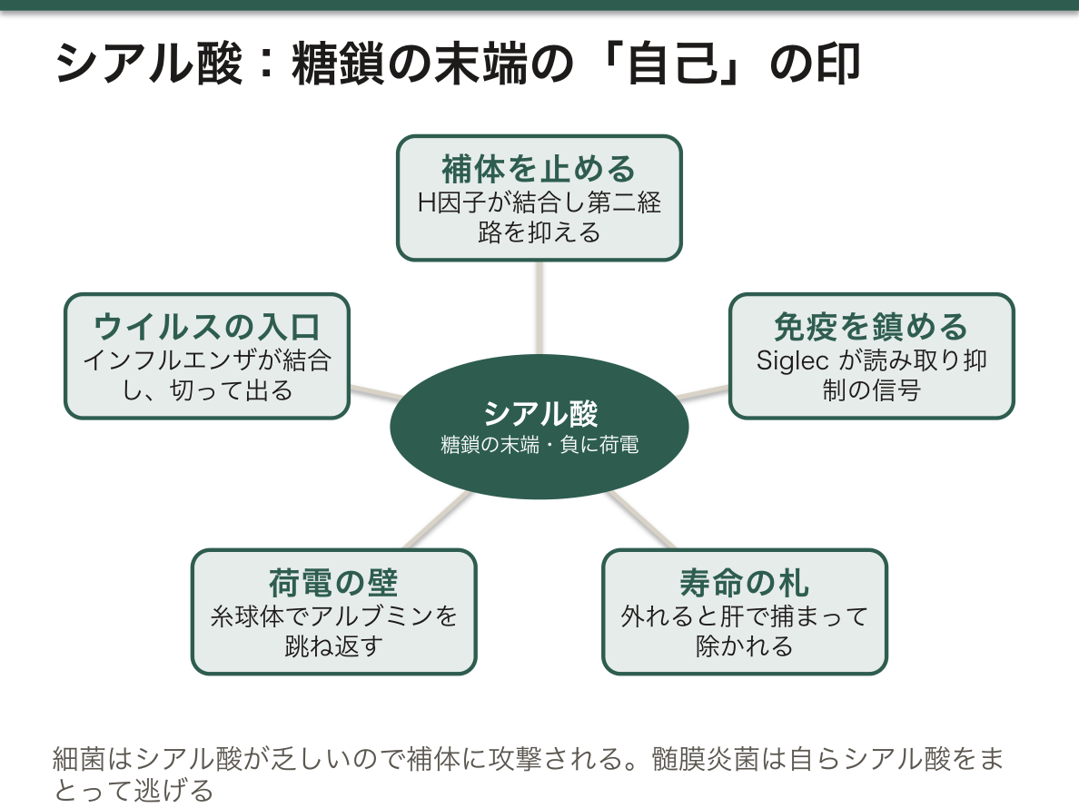
病原体は、この仕組みを逆手に取る。
| インフルエンザウイルス | ヘマグルチニンがシアル酸に結合して侵入し、ノイラミニダーゼがシアル酸を切って脱出する | オセルタミビル・ザナミビルはノイラミニダーゼを止め、ウイルスを細胞から離れられなくする |
| ヒトと鳥のウイルスの違い | 結合するシアル酸の繋がり方が違う(α2,6 と α2,3) | ヒトの上気道には α2,6 が多い。鳥インフルエンザがヒトに広がりにくい理由の1つ |
| 髄膜炎菌・B 群溶連菌 | 自らシアル酸で被膜を作る | 自己に化けて補体から逃げる |
シアル酸が外れる/減ることが、そのまま病気になる。
- 微小変化型ネフローゼ症候群——足細胞表面の陰性荷電が失われ、アルブミンだけが漏れる(選択性の高い蛋白尿)
- 発作性夜間ヘモグロビン尿症——補体の制御因子が膜に繋がらず、赤血球が壊される
- 癌——シアル酸を過剰に出して Siglec を刺激し、免疫から逃れる。腫瘍マーカーの CA19-9・CA125 はシアル酸を含む糖鎖抗原である
抗インフルエンザ薬はウイルスを殺さない。広がりを止めるだけなので、発症から 48 時間以内に始めないと意味が乏しい。
English version
Core
At the end of the glycans of the cell surface there is often a negatively charged sugar called sialic acid. It acts as the mark of "self".
- It stops complement — with sialic acid present, factor H binds and activation of the alternative pathway is suppressed. Bacteria are poor in sialic acid, so they are attacked as they are.
- It calms immune cells — receptors called Siglecs read the sialic acid and issue an inhibitory signal.
- It is a clock of lifespan — blood glycoproteins and red cells, once the sialic acid comes off, are caught and removed in the liver. The terminal sialic acid is the label saying "still new".
The effect of the charge is great too. The glomerular basement membrane and the surface of the podocyte are strongly negative with sialic acid and electrically repel albumin, which is negative as well (charge selectivity).
Going further
Pathogens turn this mechanism against us.
| Influenza virus | Haemagglutinin binds sialic acid to enter and neuraminidase cuts sialic acid to escape | Oseltamivir and zanamivir stop the neuraminidase and prevent the virus leaving the cell |
| Human and avian viruses differ | The linkage of the sialic acid they bind differs (α2,6 and α2,3) | The human upper airway is rich in α2,6. One reason avian influenza does not spread readily in people |
| Meningococcus, group B streptococcus | Make a capsule of sialic acid themselves | Disguised as self, they escape complement |
Sialic acid coming off, or diminishing, is itself a disease.
- Minimal change nephrotic syndrome — the negative charge on the podocyte surface is lost and albumin alone leaks (highly selective proteinuria).
- Paroxysmal nocturnal haemoglobinuria — the regulators of complement are not tethered to the membrane and red cells are destroyed.
- Cancer — releasing sialic acid in excess it stimulates Siglecs and escapes immunity. The tumour markers CA19-9 and CA125 are sialic acid-containing carbohydrate antigens.
In the clinic
Anti-influenza drugs do not kill the virus. They only stop its spread, so there is little point unless started within 48 hours of onset.
分泌型と非分泌型 — 血液型物質が体液に出るかどうかで、かかる病気が変わる
ABO 式の血液型物質は、赤血球だけでなく唾液・消化液・涙・精液にも出ている。ただし、出る人と出ない人がいる。
決めているのは FUT2 という別の遺伝子で、分泌腺の上皮で H 抗原(A・B の土台)を作る酵素をコードしている。
- 分泌型——FUT2 が働く。日本人の約 75%。体液に血液型物質が出る
- 非分泌型——FUT2 が働かない。赤血球の血液型は正常だが、体液には出ない
血液型は A 型なのに、唾液からは何も検出されないということが起こりうる。法医学で唾液から血液型を推定する手法に限界があるのはこのためである。
非分泌型であることが、感染症への抵抗性と結びついている。腸管の上皮に H 抗原が出ないので、それを足がかりにする病原体が取り付けなくなる。
| ノロウイルス | 非分泌型はほぼ感染しない型がある | ウイルスが H 抗原に結合して侵入するため。集団感染で一部の人だけ発症しない理由の 1 つ |
| ロタウイルス | 同様に型によって感受性が違う | ワクチンの効き方の差にも関わる |
| Helicobacter pylori | 分泌型で感染・潰瘍が多い | Lewis 抗原に結合して定着する |
| 尿路感染症 | 非分泌型で反復しやすい | こちらは逆向き。抗原の種類によって有利不利が入れ替わる |
1 つの多型が、あらゆる場面で有利になるわけではない。ある感染には強く、別の感染には弱い——だからどちらの型も集団に残っている。鎌状赤血球形質とマラリアのような「はっきりした取引」ではなく、多方向の綱引きの結果として多型が保たれている例として理解しておく。
ほかにも FUT2 は腸内細菌叢の構成に影響し、非分泌型はビタミン B12 の血中濃度が高い傾向があること、Crohn 病との関連が報告されている。「糖鎖 1 個の違い」が、消化管の生態系ごと変えているという見方ができる。
同じ食事で集団食中毒が起きても、発症しない人が一定数いる。「食べていない」と決めつけず、感受性の差を考慮して疫学調査を組む。
English version
Core
The ABO blood group substances appear not only on red cells but in saliva, digestive juice, tears and semen. Some people secrete them and some do not.
What decides it is a separate gene, FUT2, encoding the enzyme that makes the H antigen (the foundation of A and B) in the epithelium of the secretory glands.
- Secretor — FUT2 works. About 75 per cent of Japanese people. The blood group substance appears in body fluids.
- Non-secretor — FUT2 does not work. The red cell blood group is normal but nothing appears in the fluids.
So a person may be group A and yet nothing be detected in the saliva. This is why methods of inferring blood group from saliva in forensic medicine have their limits.
Going further
Being a non-secretor is linked to resistance to infection. With no H antigen on the intestinal epithelium, pathogens that use it as a foothold cannot attach.
| Norovirus | Some non-secretor types are almost never infected | Because the virus binds the H antigen to enter. One reason some people alone escape in an outbreak |
| Rotavirus | Susceptibility likewise differs by type | Bears on differences in how well the vaccine works |
| Helicobacter pylori | Infection and ulcer are commoner in secretors | It binds Lewis antigens to colonise |
| Urinary tract infection | Recurs more readily in non-secretors | Here the direction is reversed. Advantage and disadvantage swap with the kind of antigen |
One polymorphism is not advantageous in every setting. Strong against one infection and weak against another — which is why both types remain in the population. Understand it as an example of polymorphism maintained as the result of a tug of war in many directions rather than by a clear-cut bargain like sickle cell trait and malaria.
Besides this, FUT2 influences the composition of the gut flora, non-secretors tend to have a higher blood vitamin B12, and an association with Crohn’s disease has been reported. One can see "a difference of one sugar" changing the whole ecosystem of the gut.
In the clinic
Even in an outbreak of food poisoning from the same meal, a certain number of people do not fall ill. Do not conclude they "did not eat it"; design the epidemiological investigation allowing for differences in susceptibility.
まとめ
| 単糖から多糖まで — 名前が示す構造 | 単糖——グルコース・ガラクトース・フルクトース(すべて C₆H₁₂O₆ の異性体)。 |
|---|---|
| 単糖の名前の付け方 — 「カルボニルの種類」×「炭素の数」 | 糖質の多くは一般式 Cn(H₂O)n で書けるので「炭水化物」と呼ばれるが、水が付いているわけではない。 |
| D/L・エピマー・アノマー — 糖の「違い」を3つに切り分ける | どこが違うか例関係D 型と L 型(鏡像異性体)すべての不斉炭素D-グルコースと L-グルコース重ね合わせられない鏡像エピマー不斉炭素のうち1か所だけグルコースとガラクトース(C4… |
| 糖が環になる理由 — ヘミアセタール・ピラノースとフラノース・いす形 | 5炭素以上の糖は、水の中ではほとんどが環の形で存在する。 |
| Fischer 投影式から Haworth 式への書き換え — 5つの手順 | Fischer 投影式は鎖状の糖を縦に描いた図で、横の線は手前、縦の線は奥を向いている。 |
| グリコシド結合の書き方と還元糖 — アノマー炭素が空いているかどうか | 2つの単糖の –OH どうしが脱水縮合すると、–O– でつながったグリコシド結合ができる。 |
| 糖の酸化と還元 — 糖酸と糖アルコール | 糖の炭素は、端を酸化すればカルボン酸に、カルボニルを還元すればアルコールになる。 |
| 糖鎖が付く2つの道 — 機能としての修飾と、劣化としての糖化 | 糖鎖修飾(グリコシル化)——酵素が特定の場所に付ける。 |
| ABO 式血液型の化学 — 酵素が1つ違うだけで、型が変わる | 血液型の実体は、赤血球膜の糖脂質・糖蛋白に付いた糖鎖の末端になる。 |
| 糖鎖が作られる仕組みと、その異常 | ヒトの蛋白の半分以上が糖鎖修飾を受ける。 |
| シアル酸 — 糖鎖の末端に置かれた、「自分である」という印 | 細胞表面の糖鎖の末端には、しばしばシアル酸という負に荷電した糖が付いている。 |
| 分泌型と非分泌型 — 血液型物質が体液に出るかどうかで、かかる病気が変わる | ABO 式の血液型物質は、赤血球だけでなく唾液・消化液・涙・精液にも出ている。 |
このノートは医学生が授業の内容を自分の言葉でまとめたもので、診療の判断に使うものではありません。誤りに気づいたらお知らせください。