がん細胞は、酸素が十分にあっても解糖系を使い、乳酸を出す。これをWarburg 効果と呼ぶ。
効率だけを見れば不合理になる——解糖系は ATP を2つしか作らず、酸化的リン酸化は30以上を作る。
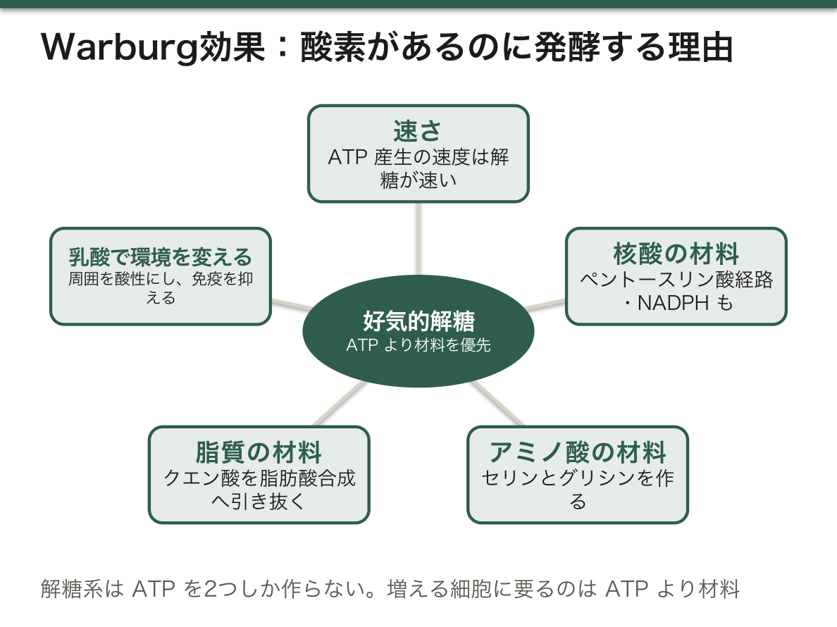
だが増える細胞に必要なのは ATP だけではない——核酸・アミノ酸・脂質という「材料」が要る。解糖系の中間体は、その材料の供給源になる。
つまり Warburg 効果は、エネルギーではなく材料を優先した結果になる——「増えるための代謝は、生きるための代謝と違う」。
そしてこの性質が、そのまま画像診断になった——FDG-PET は、糖の取り込みが多い場所を写している。
発癌の分子機構は発癌の章、糖代謝は糖代謝の章、核医学は核医学の章にある。
- Warburg効果と、それを画像にする
- 代謝を狙う薬と、治療が起こす代謝の異常
- 腫瘍マーカー — その分子は、もともと何をしていたのか
- グルタミン依存 — 癌は、糖以外の燃料も使っている
- 腫瘍の中は酸性である — 細胞の内と外で、pH が逆転している
- 増えるには膜が要る — 癌細胞が脂質を自分で作り始める
Warburg効果と、それを画像にする
Warburg 効果——酸素があっても解糖系を使い、乳酸を産生する(好気的解糖)。
なぜそうするか。
- 速い——ATP 産生の速度そのものは解糖系のほうが速い
- 材料が要る——ペントースリン酸経路へ回して核酸とNADPHを作り、セリンとグリシンを作り、脂質合成の材料を得る
- 環境を変える——乳酸が周囲を酸性にし、免疫細胞の働きを抑え、浸潤を助ける
関わる分子——HIF-1α(低酸素で安定化)・MYC・PI3K/AKT 経路・GLUT1 の増加・ヘキソキナーゼII。
FDG-PET——フルオロデオキシグルコースが GLUT で取り込まれ、リン酸化された後は先へ進めず細胞内に溜まる。
だから糖代謝の盛んな組織はすべて光る——脳・心筋・炎症・褐色脂肪・尿路(排泄)。
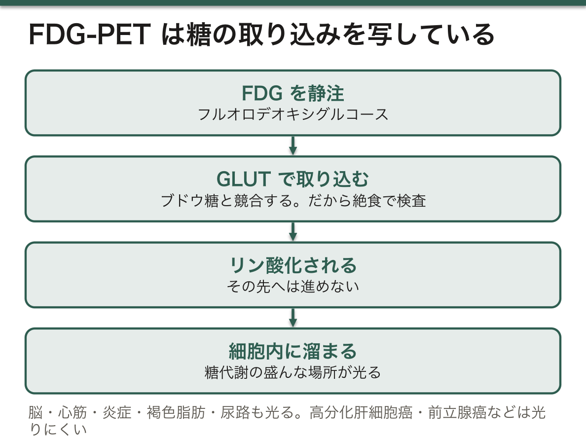
FDG-PET が「癌を写す検査」ではなく「糖代謝を写す検査」であることが、読影の要点になる(PETの章)——「炎症も、活動している筋も光る」。
だから偽陽性が多い。結核・サルコイドーシス・術後・感染——「集積があることは、悪性であることを意味しない」。
逆に偽陰性もある。糖代謝の低い腫瘍——高分化型肝細胞癌・腎細胞癌・前立腺癌・粘液産生腫瘍・小さい病変(病期診断の章)——「光らないことで否定できない癌がある」。
そして高血糖では感度が落ちる。ブドウ糖とFDGが取り込みを競合するので、検査前に絶食する——「準備の不備が、結果を変える」。
乳酸が免疫を抑えることが、腫瘍免疫の障壁の1つになっている(腫瘍免疫の章)——「代謝が、免疫療法の効きにくさを作っている」。
HIF-1α は、腎細胞癌で中心的な役割を果たす。VHL 遺伝子の異常で分解されず、血管新生が進む(腎細胞癌の章)——「だから血管新生阻害薬が、この癌でよく効く」。
English version
Core
The Warburg effect — using glycolysis and producing lactate even with oxygen present (aerobic glycolysis).
Why do it?
- It is fast — the rate of ATP production itself is greater by glycolysis.
- Material is needed — diverting into the pentose phosphate pathway to make nucleic acid and NADPH, making serine and glycine, and obtaining material for lipid synthesis.
- It changes the environment — lactate acidifies the surroundings, suppresses immune cells and assists invasion.
Molecules involved — HIF-1α (stabilised by hypoxia), MYC, the PI3K/AKT pathway, increased GLUT1, hexokinase II.
FDG-PET — fluorodeoxyglucose is taken up by GLUT and, once phosphorylated, can go no further and accumulates within the cell.
So every tissue with active glucose metabolism lights up — brain, myocardium, inflammation, brown fat, urinary tract (excretion).
Going further
- That FDG-PET is not a test picturing cancer but a test picturing glucose metabolism is the crux of reporting it (the chapter on PET) — inflammation lights up, and so does working muscle.
- Hence false positives are many. Tuberculosis, sarcoidosis, after an operation, infection — uptake does not mean malignancy.
- Conversely there are false negatives. Tumours of low glucose metabolism — well-differentiated hepatocellular carcinoma, renal cell carcinoma, prostate cancer, mucinous tumours, small lesions (the chapter on staging) — there are cancers that not lighting up cannot exclude.
- And sensitivity falls with hyperglycaemia. Glucose and FDG compete for uptake, so the patient fasts before the test — a failure of preparation changing the result.
- That lactate suppresses immunity is one of the barriers of tumour immunity (the chapter on tumour immunity) — metabolism creating the resistance to immunotherapy.
- HIF-1α plays a central role in renal cell carcinoma. With an abnormal VHL gene it is not degraded and angiogenesis advances (the chapter on renal cell carcinoma) — which is why angiogenesis inhibitors work well in this cancer.
代謝を狙う薬と、治療が起こす代謝の異常
核酸合成を止める薬(代謝拮抗薬)。
- メトトレキサート——ジヒドロ葉酸還元酵素を阻害する。救済にホリナート(活性型葉酸)
- 5-フルオロウラシル——チミジル酸合成酵素を阻害する
- ゲムシタビン・シタラビン——DNA合成に取り込まれて止める
- 6-メルカプトプリン——プリン合成を阻害。アロプリノールと併用すると中毒になる
腫瘍崩壊症候群——大量の腫瘍細胞が一気に壊れ、中身が血中に出る。
- 高尿酸血症・高カリウム血症・高リン血症・低カルシウム血症 → 急性腎障害・不整脈
- 危険が高い——急性白血病・Burkitt リンパ腫・腫瘍量が多いもの・治療感受性が高いもの
- 予防——十分な輸液・尿酸を下げる薬(アロプリノール/ラスブリカーゼ)
6-メルカプトプリンとアロプリノールの併用が危険なのは、代謝経路が交差するからになる。キサンチンオキシダーゼが 6-MP の分解にも関わる(プリン代謝の章)——「痛風の薬が、抗癌薬の血中濃度を数倍にする」。
だから腫瘍崩壊症候群の予防と、6-MP の投与が同時に必要な場面は要注意になる(支持療法の章)——「予防のための薬が、別の薬の中毒を作る」。
ラスブリカーゼは尿酸をアラントインに変える酵素製剤になる。ただし G6PD 欠損症では、副産物の H₂O₂ で溶血する(G6PD欠損症の章)——「解決のための酵素が、還元力の無い患者では毒になる」。
メトトレキサートの救済にホリナートを使うのは、阻害された段階の下流を補うからになる(葉酸代謝の章)——「阻害点より下に材料を入れれば、迂回できる」。
だから葉酸そのものでは救済にならない。還元されていないと使えない(代謝拮抗薬の章)——「似た名前の薬が、代替にならない場面になる」。
5-FU の代謝酵素(DPD)の欠損があると、重篤な毒性が出る(遺伝多型の章)——「標準量が、一部の人には致死量になる」。
English version
Core
Drugs that stop nucleic acid synthesis (antimetabolites).
- Methotrexate — inhibits dihydrofolate reductase. Rescue with folinate (the active folate).
- 5-Fluorouracil — inhibits thymidylate synthase.
- Gemcitabine, cytarabine — incorporated into DNA synthesis and stop it.
- 6-Mercaptopurine — inhibits purine synthesis. Given with allopurinol it becomes toxic.
Tumour lysis syndrome — a great many tumour cells break down at once and their contents enter the blood.
- Hyperuricaemia, hyperkalaemia, hyperphosphataemia, hypocalcaemia → acute kidney injury and arrhythmia.
- High risk — acute leukaemia, Burkitt lymphoma, a large tumour burden, high sensitivity to treatment.
- Prevention — ample fluid, a urate-lowering drug (allopurinol or rasburicase).
Going further
- 6-Mercaptopurine with allopurinol is dangerous because the metabolic pathways cross. Xanthine oxidase is also involved in the breakdown of 6-MP (the chapter on purine metabolism) — a drug for gout raising the blood level of an antitumour drug several-fold.
- So the setting where prevention of tumour lysis syndrome and 6-MP are needed together calls for care (the chapter on supportive care) — a drug given for prevention creating the toxicity of another drug.
- Rasburicase is an enzyme preparation converting urate to allantoin. But in G6PD deficiency the by-product H₂O₂ causes haemolysis (the chapter on G6PD deficiency) — an enzyme meant to solve the problem becoming a poison in a patient without reducing power.
- Folinate is used to rescue methotrexate because it supplies downstream of the blocked step (the chapter on folate metabolism) — put the material in below the point of inhibition and it can be bypassed.
- Hence folate itself does not rescue. Unless it is reduced it cannot be used (the chapter on antimetabolites) — a setting where a similarly named drug is no substitute.
- Deficiency of the enzyme metabolising 5-FU (DPD) gives severe toxicity (the chapter on genetic polymorphism) — a standard dose being a lethal dose for some people.
腫瘍マーカー — その分子は、もともと何をしていたのか
腫瘍マーカーは「癌だけが作る分子」ではない——正常でも作られる分子が、量として増えているだけになる。
| マーカー | 本来の正体 | 癌以外で上がるとき |
|---|---|---|
| AFP | 胎児の血漿蛋白(胎児型アルブミン) | 肝炎・肝硬変・妊娠 |
| CEA | 胎児の消化管の細胞接着分子 | 喫煙・炎症・糖尿病 |
| CA19-9 | 糖鎖抗原(Lewis 式血液型) | 胆道閉塞・膵炎。Lewis 陰性の約 5〜10% では上がらない |
| PSA | 精液を液化させる酵素 | 前立腺肥大・前立腺炎・射精・直腸診 |
| hCG | 胎盤のホルモン | 妊娠 |
使い道は主に、治療効果の判定と再発の監視になる——スクリーニングに向くものは少ない。
「もともと何をしていた分子か」を知ると、偽陽性が予測できる。AFP が胎児の蛋白なら妊娠と肝再生で上がるし、PSA が精液の酵素なら射精で上がる(精液の項)——「正体を知っていれば、丸暗記が要らない」。
癌胎児性抗原という言葉が示すとおり、多くは発生の途中で使われていた分子になる。腫瘍細胞が未分化になると、胎児期の遺伝子発現が戻る(脱分化)(腫瘍総論の章)——「マーカーの正体が、癌が『若返っている』ことの証拠になる」。
スクリーニングに向かない理由は、事前確率の低さにある。有病率の低い集団では、感度と特異度が高くても陽性的中率が低くなる(検査の性能の章)——「無症状の人に片端から測ると、偽陽性の検査が連鎖する」。
CA19-9 が Lewis 式血液型に依存することは、実務上の落とし穴になる(糖鎖の章)——「一部の人では、進行した膵癌でも上がらない」。
治療効果の判定に使うときは、半減期を考える(絨毛性疾患の章)——「hCG は半減期が短いので、下がり方の曲線そのものが治療の評価になる」。
English version
Core
A tumour marker is not a molecule made only by cancer — it is a molecule made normally too, increased in amount.
| Marker | What it really is | When it rises apart from cancer |
|---|---|---|
| AFP | A fetal plasma protein (fetal albumin) | Hepatitis, cirrhosis, pregnancy |
| CEA | A cell adhesion molecule of the fetal gut | Smoking, inflammation, diabetes |
| CA19-9 | A carbohydrate antigen (the Lewis blood group) | Biliary obstruction, pancreatitis. It does not rise in the 5–10 per cent who are Lewis-negative |
| PSA | The enzyme liquefying semen | Prostatic hypertrophy, prostatitis, ejaculation, rectal examination |
| hCG | A hormone of the placenta | Pregnancy |
Their use is chiefly in judging the effect of treatment and in watching for recurrence — few are suited to screening.
Going further
- Knowing what the molecule originally did lets false positives be predicted. AFP being a fetal protein it rises in pregnancy and liver regeneration, and PSA being the enzyme of semen it rises after ejaculation (the section on semen) — knowing what it is spares rote learning.
- As the term carcinoembryonic antigen indicates, most were molecules used in the course of development. When a tumour cell becomes undifferentiated, fetal gene expression returns (dedifferentiation) (the chapter on general tumour pathology) — the identity of the marker being evidence that the cancer has "grown young again".
- They are unsuited to screening because of the low prior probability. In a population of low prevalence the positive predictive value is low even with high sensitivity and specificity (the chapter on the performance of tests) — measure indiscriminately in asymptomatic people and a chain of false-positive investigations follows.
- That CA19-9 depends on the Lewis blood group is a practical pitfall (the chapter on glycans) — in some people it does not rise even in advanced pancreatic cancer.
- When using one to judge the effect of treatment, think of the half-life (the chapter on trophoblastic disease) — hCG has a short half-life, so the very curve of its fall is the assessment of treatment.
グルタミン依存 — 癌は、糖以外の燃料も使っている
増殖する細胞に必要なのは、エネルギーだけではない——核酸・アミノ酸・脂質という「材料」が要る。
グルタミンは、その材料の中心になる。
- グルタミナーゼでグルタミン酸へ → α-ケトグルタル酸として TCA 回路へ入る(anaplerosis)
- アミノ基を、プリン・ピリミジン・アミノ酸の合成へ供給する
- グルタチオンの材料になり、酸化ストレスに耐えさせる
Warburg 効果で解糖が乳酸へ流れてしまうぶん、TCA 回路は別の入口から補われている。
だから多くの腫瘍が、グルタミンを大量に消費する。
「なぜ酸素があるのに発酵するのか」という問いの答えが、この項で完成する。目的が ATP ではなく材料の確保なら、解糖の中間体を分岐路へ回すほうが合理的になる(この章のWarburg 効果の項)——「効率が悪いのではなく、目的が違う」。
不足する TCA 回路の中間体を、グルタミンが補っている(TCA 回路の章)——「回路から材料を抜き出すには、どこかから補充しなければ回らない」。
この依存が、治療の標的になる。アスパラギナーゼは急性リンパ性白血病でアスパラギンを枯渇させ、自分で合成できない腫瘍細胞を殺す(白血病の各論の章)——「栄養素を奪うという治療が、実際に成立している」。
代謝酵素そのものが、癌遺伝子として働く例も見つかっている。IDH1/2 の変異は 2-ヒドロキシグルタル酸を作り、エピゲノムを変えて分化を止める(エピジェネティクスの章)——「代謝産物が、遺伝子の読み方を変えている」。
だから神経膠腫や急性骨髄性白血病で、IDH 変異の有無が分類と治療に使われる(神経の病理の章)——「代謝の話が、そのまま病理診断の分類になった」。
English version
Core
A proliferating cell needs more than energy — it needs material: nucleic acid, amino acid, lipid.
Glutamine is the centre of that material.
- Glutaminase makes glutamate → it enters the TCA cycle as α-ketoglutarate (anaplerosis).
- It supplies amino groups to the synthesis of purine, pyrimidine and amino acids.
- It is the material of glutathione, letting the cell withstand oxidative stress.
To the extent that glycolysis runs off to lactate under the Warburg effect, the TCA cycle is replenished from another entrance.
Hence many tumours consume a great deal of glutamine.
Going further
- The answer to "why ferment when there is oxygen" is completed in this section. If the object is not ATP but securing material, diverting the intermediates of glycolysis into the branch paths is the rational course (the section on the Warburg effect in this chapter) — it is not that the efficiency is poor but that the object is different.
- Glutamine replenishes the missing intermediates of the TCA cycle (the chapter on the TCA cycle) — to draw material out of a cycle, it must be replenished from somewhere or it will not turn.
- This dependence becomes a target of treatment. Asparaginase exhausts asparagine in acute lymphoblastic leukaemia and kills the tumour cells that cannot make their own (the chapter on the individual leukaemias) — a treatment consisting of taking away a nutrient, actually working.
- Examples have been found of a metabolic enzyme itself acting as an oncogene. Mutant IDH1/2 makes 2-hydroxyglutarate, alters the epigenome and stops differentiation (the chapter on epigenetics) — a metabolite changing how the genes are read.
- Hence in glioma and acute myeloid leukaemia the presence of an IDH mutation is used in classification and treatment (the chapter on the pathology of the nervous system) — a matter of metabolism becoming, directly, a category of pathological diagnosis.
腫瘍の中は酸性である — 細胞の内と外で、pH が逆転している
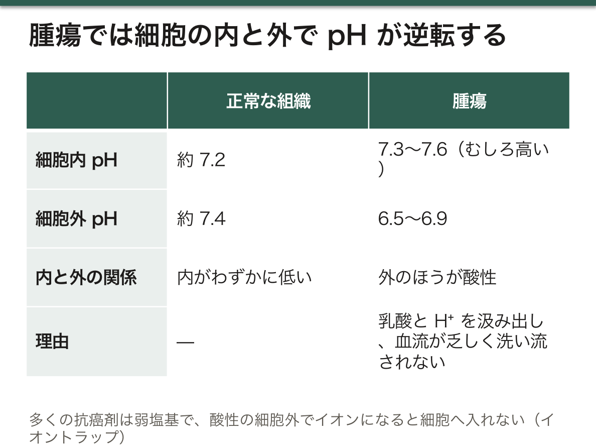
正常な組織では、細胞内 pH(約 7.2)は細胞外(約 7.4)よりわずかに低い。腫瘍ではこれが逆転している。
- 細胞外 pH は 6.5〜6.9 まで下がる——好気的解糖で乳酸と H⁺ を大量に出し、しかも血流が乏しいので洗い流されない
- 細胞内 pH はむしろ高い(7.3〜7.6)——H⁺ ポンプ・Na⁺/H⁺ 交換体・炭酸脱水酵素 IX を総動員して、酸を外へ汲み出しているため
細胞内をアルカリ側に保つことは、癌細胞にとって都合がよい——解糖系の酵素が活性化し、アポトーシスが起こりにくくなる。酸を外に捨てること自体が、増殖の条件になっている。
外が酸性であることが、腫瘍に3つの利益をもたらす。
- 浸潤を助ける——酸性の環境でプロテアーゼが働き、基質が分解される。正常細胞は酸に耐えられず死ぬので、腫瘍が場所を奪える
- 免疫を抑える——T 細胞と NK 細胞は酸性下で機能が落ちる。乳酸そのものが免疫細胞の代謝を妨げる
- 薬を効きにくくする——ここが臨床的に重要である
薬が効かなくなる仕組みは、単純な化学で説明できる(イオントラップ)。多くの抗癌剤は弱塩基である。酸性の細胞外でプロトン化してイオンになると、膜を通れなくなり細胞内へ入れない。逆に弱酸性の薬は入りやすい。「腫瘍まで薬が届いているのに効かない」という状況が、pH だけで起こりうる。
この性質を利用する試みもある。FDG-PET が腫瘍を映すのは糖の取り込みを見ているからだが、同じ代謝の帰結としてpH を画像化する手法や、酸性でだけ働く薬・酸性でだけ光る造影剤が研究されている。炭酸脱水酵素 IX の阻害も標的の 1 つである。
「濃度が足りているのに効かない」薬剤耐性には、薬が細胞の中へ入れていないという形もある。輸送と pH を分けて考える。
English version
Core
In normal tissue the intracellular pH (about 7.2) is slightly lower than the extracellular (about 7.4). In a tumour this is reversed.
- The extracellular pH falls to 6.5–6.9 — aerobic glycolysis releases a great deal of lactate and H⁺, and with poor blood flow they are not washed away.
- The intracellular pH is if anything higher (7.3–7.6) — proton pumps, the Na⁺/H⁺ exchanger and carbonic anhydrase IX are all mobilised to pump the acid out.
Keeping the inside on the alkaline side suits the cancer cell — the enzymes of glycolysis are activated and apoptosis becomes less likely. Throwing the acid outside is itself a condition of proliferation.
Going further
The acidity outside brings the tumour three advantages.
- It assists invasion — proteases work in an acid environment and the matrix is degraded. Normal cells cannot tolerate the acid and die, so the tumour can take the ground.
- It suppresses immunity — T cells and NK cells lose function in acid. Lactate itself impedes the metabolism of immune cells.
- It makes drugs less effective — this is the clinically important part.
The mechanism of drug failure can be explained by simple chemistry (ion trapping). Many antitumour drugs are weak bases. Protonated into an ion in the acid exterior, they can no longer cross the membrane and do not enter the cell. Weakly acidic drugs conversely enter readily. The situation of "the drug reaches the tumour and does not work" can arise from pH alone.
There are attempts to exploit this property. FDG-PET pictures a tumour because it sees glucose uptake, but as a consequence of the same metabolism, methods of imaging pH and drugs that act only in acid, or contrast agents that light up only in acid, are under study. Inhibition of carbonic anhydrase IX is one target.
In the clinic
Drug resistance where "the concentration is adequate and it does not work" also takes the form of the drug not getting into the cell. Think of transport and pH separately.
増えるには膜が要る — 癌細胞が脂質を自分で作り始める
細胞が 1 個から 2 個になるには、膜を 2 倍にしなければならない。膜の材料はリン脂質、すなわち脂肪酸である。
正常な成人の細胞は、脂肪酸を血液から取り込んで使う(肝と脂肪組織を除けば、自分で作る必要がない)。ところが多くの癌では、脂肪酸合成酵素(FASN)が強く発現している。
- 取り込みでは足りない/質が選べない——自分で作れば供給が安定する
- 自分で作った脂肪酸は飽和度が高い——膜が酸化されにくくなり、フェロトーシス(脂質過酸化による細胞死)に抵抗性になる
- 脂質は信号分子の材料でもある——増殖の信号を自前で用意できる
Warburg 効果で解糖に傾くことと矛盾しない。解糖の中間体とクエン酸が脂質合成の材料として引き抜かれており、「ATP を作るためではなく、材料を作るために糖を使っている」という説明になる。
コレステロール合成の側も同じように亢進している。膜の流動性と、脂質ラフト(受容体が集まる足場)の形成に要る。スタチンが癌の発生・予後に関係するのではないかという議論は、この経路から出ている——ただし観察研究の結果は一貫しておらず、癌の予防目的でスタチンを使う根拠は無い。
治療の標的としての現状を正確に押さえておく。
| FASN 阻害 | 臨床試験の段階 | 正常組織への影響と、体重減少が課題 |
| ACC 阻害 | 同上 | 脂肪肝の治療薬としても開発されている |
| スタチン | 癌治療としては確立していない | 脂質異常症の治療としての適応で使う |
画像診断への応用は既に実用化されている。前立腺癌は FDG-PET で映りにくい(糖の取り込みが乏しい)が、コリンや酢酸を標識した PETでは映る——脂質合成が盛んな腫瘍は、脂質の材料を追えばよいという発想である。
PET で「光らない癌」があることを知っておく。前立腺癌・高分化肝細胞癌・粘液癌・腎細胞癌は FDG での検出に限界がある。
English version
Core
For a cell to become two from one, the membrane must be doubled. The material of the membrane is phospholipid, that is, fatty acid.
A normal adult cell takes fatty acid from the blood (apart from liver and adipose tissue it has no need to make its own). In many cancers, however, fatty acid synthase (FASN) is strongly expressed.
- Uptake is not enough, or the quality cannot be chosen — making its own gives a stable supply.
- Fatty acid it makes itself is more saturated — the membrane is less readily oxidised and it becomes resistant to ferroptosis (cell death by lipid peroxidation).
- Lipids are also the material of signalling molecules — it can prepare its own signals for proliferation.
This does not contradict the tilt towards glycolysis in the Warburg effect. The intermediates of glycolysis and citrate are drawn off as material for lipid synthesis, which gives the explanation that glucose is used not to make ATP but to make material.
Going further
Cholesterol synthesis is raised in the same way. It is needed for membrane fluidity and for forming lipid rafts (the platforms on which receptors gather). The argument that statins may bear on the occurrence and prognosis of cancer comes out of this pathway — but the observational results are inconsistent and there is no ground for using a statin to prevent cancer.
Note accurately where things stand as a target of treatment.
| FASN inhibition | At the stage of clinical trial | Effects on normal tissue and weight loss are the problems |
| ACC inhibition | The same | Also being developed as a treatment for fatty liver |
| Statins | Not established as a cancer treatment | Use them on the indication of treating dyslipidaemia |
The application to imaging is already in practical use. Prostate cancer shows poorly on FDG-PET (glucose uptake is low) but shows on PET with labelled choline or acetate — the idea that a tumour with active lipid synthesis can be followed by the material of lipid.
In the clinic
Know that there are cancers that do not light up on PET. Prostate cancer, well-differentiated hepatocellular carcinoma, mucinous carcinoma and renal cell carcinoma have limits to detection with FDG.
まとめ
| Warburg効果と、それを画像にする | Warburg 効果——酸素があっても解糖系を使い、乳酸を産生する(好気的解糖)。 |
|---|---|
| 代謝を狙う薬と、治療が起こす代謝の異常 | 核酸合成を止める薬(代謝拮抗薬)。メトトレキサート——ジヒドロ葉酸還元酵素を阻害する。 |
| 腫瘍マーカー — その分子は、もともと何をしていたのか | 腫瘍マーカーは「癌だけが作る分子」ではない——正常でも作られる分子が、量として増えているだけになる。 |
| グルタミン依存 — 癌は、糖以外の燃料も使っている | 増殖する細胞に必要なのは、エネルギーだけではない——核酸・アミノ酸・脂質という「材料」が要る。 |
| 腫瘍の中は酸性である — 細胞の内と外で、pH が逆転している | 正常な組織では、細胞内 pH(約 7.2)は細胞外(約 7.4)よりわずかに低い。 |
| 増えるには膜が要る — 癌細胞が脂質を自分で作り始める | 細胞が 1 個から 2 個になるには、膜を 2 倍にしなければならない。 |
このノートは医学生が授業の内容を自分の言葉でまとめたもので、診療の判断に使うものではありません。誤りに気づいたらお知らせください。