骨は止まった構造物ではなく、常に壊されて作り直されている。成人でも年に約10%が入れ替わる。この作り替えを担うのが破骨細胞と骨芽細胞の対で、両者は互いに信号を送り合っている。
骨粗鬆症の薬が「壊す側を止める薬」と「作る側を促す薬」に分かれるのは、この2細胞系の理解から直接に出てくる。
- 石灰化と骨基質 — 何でできているか
- RANKLとOPG — 壊す側と作る側の会話
- 骨代謝マーカー — 作る側と壊す側を、別々に測る
- 骨粗鬆症の薬 — 骨のどの段階を狙っているか
- 骨密度が同じでも、折れやすさは違う — 骨の「質」の化学
- 骨と脂肪は、同じ細胞から分かれる — 骨髄で起きている取り合い
石灰化と骨基質 — 何でできているか
骨は有機質35%・無機質65%(重量比)。
- 有機質の90%以上がI型コラーゲン。ほかにオステオカルシン(ビタミンK依存性にγカルボキシル化される)・オステオネクチン・プロテオグリカン
- 無機質は主にハイドロキシアパタイト
石灰化に必要な条件。
- カルシウムとリンの濃度積が十分
- アルカリホスファターゼがピロリン酸(石灰化の抑制因子)を分解する
- コラーゲン線維が足場になる
| 疾患 | 異常 | 特徴 |
|---|---|---|
| 骨軟化症・くる病 | 石灰化の障害(ビタミンD欠乏・低リン) | 類骨が増える・Looser帯 |
| 骨粗鬆症 | 骨の量が減る(質は正常) | 石灰化は正常 |
| 骨形成不全症 | I型コラーゲンの異常 | 青色強膜・易骨折・難聴 |
| 低ホスファターゼ症 | アルカリホスファターゼ欠損 | アルカリホスファターゼが低い |
骨軟化症と骨粗鬆症の違いを、組織で言えるようにしておく。骨軟化症では骨の「量」はあるが石灰化していない(類骨のまま)。骨粗鬆症では石灰化した骨が「量として」減っている。だから骨軟化症では骨の痛みと筋力低下が前面に出て、骨粗鬆症は骨折するまで無症状という違いが生じる。
検査でも分かれる。骨軟化症ではアルカリホスファターゼが上がり、カルシウムかリンが低い。骨粗鬆症では血液検査は基本的に正常——「骨粗鬆症でカルシウムが低いなら、別の病気を考える」。
低ホスファターゼ症は「アルカリホスファターゼが低いことが病気」という珍しい疾患である。ピロリン酸を分解できず石灰化が進まない。健診でアルカリホスファターゼの低値を異常と認識できるかどうかが、診断の分かれ目になる。ビタミンB6依存性けいれんを伴うこともある——この酵素はピリドキサールリン酸の脱リン酸化にも関わるためである。
骨代謝マーカーは、骨の「回転の速さ」を見るもので、骨量を測る骨密度とは別の情報である。
- 骨形成——骨型アルカリホスファターゼ、I型プロコラーゲンN末端プロペプチド
- 骨吸収——I型コラーゲン架橋Nテロペプチド、酒石酸抵抗性酸ホスファターゼ5b
治療の効果判定は骨密度より速く出る——骨密度の変化は年単位だが、マーカーは数か月で動く。服薬の継続を促す材料としても使える。
English version
Core
Bone is 35 per cent organic and 65 per cent inorganic (by weight).
- Over 90 per cent of the organic part is type I collagen. Besides it, osteocalcin (γ-carboxylated in a vitamin K-dependent way), osteonectin and proteoglycans.
- The inorganic part is chiefly hydroxyapatite.
Conditions needed for mineralisation.
- The product of the calcium and phosphate concentrations is sufficient.
- Alkaline phosphatase degrades pyrophosphate (the inhibitor of mineralisation).
- Collagen fibres provide the scaffold.
| Disease | Defect | Features |
|---|---|---|
| Osteomalacia and rickets | Disturbed mineralisation (vitamin D deficiency, low phosphate) | Osteoid increases, Looser zones |
| Osteoporosis | The amount of bone falls (the quality is normal) | Mineralisation is normal |
| Osteogenesis imperfecta | Abnormal type I collagen | Blue sclerae, fragility, deafness |
| Hypophosphatasia | Deficiency of alkaline phosphatase | Alkaline phosphatase is low |
Going further
Be able to state the difference between osteomalacia and osteoporosis in terms of the tissue. In osteomalacia the "amount" of bone is there but not mineralised (it remains osteoid). In osteoporosis mineralised bone has fallen "in amount". Hence bone pain and weakness come to the front in osteomalacia, while osteoporosis is silent until a fracture.
The tests divide too. In osteomalacia alkaline phosphatase rises and either calcium or phosphate is low. In osteoporosis the blood tests are essentially normal — if calcium is low in supposed osteoporosis, think of another disease.
Hypophosphatasia is the unusual disease in which a low alkaline phosphatase is itself the illness. Pyrophosphate cannot be degraded and mineralisation does not proceed. Whether a low alkaline phosphatase on a health check is recognised as abnormal is what decides the diagnosis. It may be accompanied by vitamin B6-dependent seizures — because this enzyme is also involved in the dephosphorylation of pyridoxal phosphate.
Markers of bone metabolism show the speed of turnover and are different information from the bone density that measures the amount.
- Formation — bone-specific alkaline phosphatase, N-terminal propeptide of type I procollagen.
- Resorption — N-telopeptide of type I collagen cross-links, tartrate-resistant acid phosphatase 5b.
They show the effect of treatment sooner than bone density — bone density changes over years while the markers move in months. They can also be used as material for encouraging adherence.
RANKLとOPG — 壊す側と作る側の会話
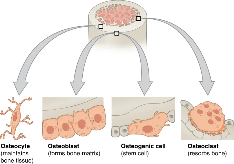
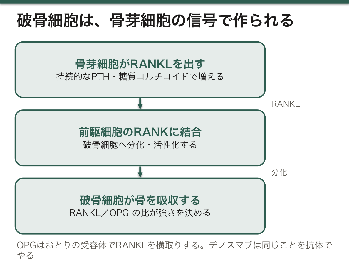
破骨細胞は単独では作られない。骨芽細胞・骨細胞が出す信号で分化する。
- RANKL——骨芽細胞側が出す。破骨細胞前駆細胞のRANKに結合して分化・活性化させる
- オステオプロテゲリン(OPG)——おとりの受容体。RANKLを横取りして破骨細胞の分化を止める
- RANKL / OPG の比が骨吸収の強さを決める
| 因子 | RANKL | 結果 |
|---|---|---|
| 副甲状腺ホルモン(持続) | ↑ | 骨吸収 |
| エストロゲン | ↓(OPG↑) | 閉経で骨吸収が進む |
| 糖質コルチコイド | ↑(かつ骨芽細胞を抑制) | 骨粗鬆症 |
- デノスマブはRANKLに対する抗体——オステオプロテゲリンと同じことを薬でやっている
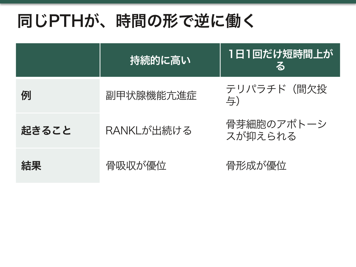
副甲状腺ホルモンが「持続か間欠か」で逆の作用をする——骨の生化学で最も面白く、かつ臨床に直結する事実である。
- 持続的に高い(副甲状腺機能亢進症)→ RANKLが出続けて骨吸収が優位
- 1日1回だけ短時間上がる(テリパラチド)→ 骨芽細胞のアポトーシスが抑えられ、骨形成が優位
同じホルモンが、投与の仕方だけで骨を壊す薬にも作る薬にもなる。「濃度」ではなく「時間の形」が結果を決める例として、これ以上分かりやすいものはない。
薬の作用点で整理すると、骨粗鬆症治療薬は3群になる。
| 群 | 薬 | 作用 |
|---|---|---|
| 骨吸収を抑える | ビスホスホネート・デノスマブ・エストロゲン・ラロキシフェン | 破骨細胞を止める |
| 骨形成を促す | テリパラチド・アバロパラチド | 骨芽細胞を増やす |
| 両方 | ロモソズマブ(スクレロスチン抗体) | 形成↑かつ吸収↓ |
デノスマブは中止すると急速に骨密度が失われ、多発椎体骨折を起こしうる——止めるときはビスホスホネートへ切り替える。抗体薬なので効果が完全に可逆で、その反動が出るのに対し、ビスホスホネートは骨に取り込まれて長く残る。薬物動態の違いが、中止時の扱いを分けている。
スクレロスチンは骨細胞が出す骨形成の抑制因子で、荷重がかかると減る。運動が骨を強くする分子的な機序であり、寝たきりで骨が失われる理由でもある。
English version
Core
The osteoclast is not made on its own. It differentiates on signals released by osteoblasts and osteocytes.
- RANKL — released by the osteoblast side. It binds RANK on the osteoclast precursor and drives differentiation and activation.
- Osteoprotegerin (OPG) — a decoy receptor. It intercepts RANKL and stops osteoclast differentiation.
- The RANKL/OPG ratio sets the strength of resorption.
| Factor | RANKL | Result |
|---|---|---|
| Parathyroid hormone (continuous) | ↑ | Bone resorption |
| Oestrogen | ↓ (OPG ↑) | Resorption advances after the menopause |
| Glucocorticoid | ↑ (and the osteoblast is suppressed) | Osteoporosis |
Denosumab is an antibody to RANKL — doing with a drug what osteoprotegerin does.
Going further
That parathyroid hormone acts in opposite directions according to whether it is continuous or intermittent is the most interesting fact in the biochemistry of bone, and directly clinical.
- Persistently high (hyperparathyroidism) → RANKL keeps being released and resorption predominates.
- Rising briefly once a day (teriparatide) → apoptosis of the osteoblast is suppressed and formation predominates.
The same hormone becomes a drug that destroys bone or one that builds it purely by how it is given. Nothing illustrates better that the shape in time, not the concentration, decides the result.
Arranged by their point of action, the drugs for osteoporosis fall into three groups.
| Group | Drug | Action |
|---|---|---|
| Suppress resorption | Bisphosphonate, denosumab, oestrogen, raloxifene | Stop the osteoclast |
| Promote formation | Teriparatide, abaloparatide | Increase the osteoblast |
| Both | Romosozumab (anti-sclerostin antibody) | Formation ↑ and resorption ↓ |
Stopping denosumab loses bone density rapidly and can cause multiple vertebral fractures — when stopping, change to a bisphosphonate. Being an antibody its effect is fully reversible and the rebound shows, whereas a bisphosphonate is taken into bone and remains a long time. A difference in pharmacokinetics divides how they are handled at withdrawal.
Sclerostin is an inhibitor of bone formation released by the osteocyte, and it falls under loading. This is the molecular mechanism by which exercise strengthens bone, and also the reason bone is lost when a patient is bedbound.
骨代謝マーカー — 作る側と壊す側を、別々に測る
| 骨形成マーカー | 骨吸収マーカー | |
|---|---|---|
| 由来 | 骨芽細胞の活動 | I型コラーゲンの分解産物 |
| 例 | 骨型ALP・オステオカルシン・P1NP | TRACP-5b・NTX・CTX・DPD |
| 検体 | 血清 | 血清・尿 |
日内変動がある——骨吸収マーカーは早朝に高い。採血の時刻と食事を揃える。
骨密度は変化が出るまで年単位かかるが、マーカーは数か月で動く——だから治療の効き始めを早く見る目的で使う。
骨型ALP は腎で排泄されない——腎機能が悪くても使える。
「作る」と「壊す」を別に測ることで、病態と薬の効き方が分けられる。
| 状態 | 形成 | 吸収 |
|---|---|---|
| 閉経後骨粗鬆症 | やや高い | 高い(高回転) |
| ステロイド性骨粗鬆症 | 低い(骨芽細胞が抑えられる) | やや高い |
| 骨軟化症 | 骨型ALP が高い | — |
(骨代謝の章)——「同じ骨粗鬆症でも、崩れている側が違えば選ぶ薬が違う」。
骨吸収と骨形成が連動していること(カップリング)が、薬の限界を作っている。吸収を止めると形成も落ちるので、骨密度の増加には上限がある(この章の RANKL の項)——「壊す細胞と作る細胞は、互いに話をしている」。
だから骨形成促進薬(テリパラチド・ロモソズマブ)が別枠で必要になる(骨の薬の章)——「テリパラチドは間欠投与でのみ骨を作る。持続では逆に骨を壊す」。
オステオカルシンは、骨が出すホルモンとしての側面も研究されている(FGF23 の項)——「骨が全身へ指令を出す器官だという見方が、複数の分子で裏づけられてきた」。
English version
Core
| Formation markers | Resorption markers | |
|---|---|---|
| Origin | Osteoblast activity | Degradation products of type I collagen |
| Examples | Bone-specific ALP, osteocalcin, P1NP | TRACP-5b, NTX, CTX, DPD |
| Sample | Serum | Serum, urine |
There is diurnal variation — resorption markers are high in the early morning. Align the hour of sampling and the meals.
Bone density takes years to show a change while the markers move in months — hence their use in seeing early that a treatment is beginning to work.
Bone-specific ALP is not excreted by the kidney — it can be used with poor renal function.
Going further
Measuring "building" and "destroying" separately allows the disease and the drug’s action to be separated.
| State | Formation | Resorption |
|---|---|---|
| Postmenopausal osteoporosis | Somewhat high | High (high turnover) |
| Steroid-induced osteoporosis | Low (the osteoblast is suppressed) | Somewhat high |
| Osteomalacia | Bone-specific ALP high | — |
(the chapter on bone metabolism) — with the same osteoporosis, a different side collapsing means a different drug.
- That resorption and formation move together (coupling) is what limits the drugs. Stop resorption and formation falls too, so there is a ceiling to the gain in bone density (the section on RANKL in this chapter) — the destroying cell and the building cell are talking to each other.
- Hence bone-forming drugs (teriparatide, romosozumab) are needed as a separate category (the chapter on drugs for bone) — teriparatide builds bone only when given intermittently. Continuously it destroys bone instead.
- Osteocalcin is also studied as a hormone released by bone (the section on FGF23) — the view that bone is an organ issuing instructions to the whole body has been supported by several molecules.
骨粗鬆症の薬 — 骨のどの段階を狙っているか
| 薬 | 作用点 | 効果 |
|---|---|---|
| ビスホスホネート | 骨に沈着し、破骨細胞に取り込まれてファルネシルピロリン酸合成酵素を阻害 | 吸収抑制 |
| デノスマブ | RANKL に対する抗体。破骨細胞の分化と生存を止める | 吸収抑制 |
| 選択的エストロゲン受容体調節薬 | 骨ではエストロゲン様、乳腺・子宮では拮抗 | 吸収抑制 |
| テリパラチド | 副甲状腺ホルモンの間欠投与 | 形成促進 |
| ロモソズマブ | スクレロスチンに対する抗体 | 形成促進+吸収抑制 |
| 活性型ビタミンD・カルシウム | 材料と吸収 | 基礎 |
骨折の危険が高いほど、形成促進薬から始めて吸収抑制薬へ繋ぐ。
ビスホスホネートが「骨に居座る薬」であることが、長所と短所の両方を作っている。骨に結合して長く残るので、休薬しても効果が続く(drug holiday が成り立つ)(骨の薬の章)——「一方で、長期使用で非定型大腿骨骨折と顎骨壊死が問題になる」。
デノスマブは骨に残らないので、性質が逆になる。中止すると骨吸収が跳ね返り、多発椎体骨折が起こりうる——「やめられない薬なので、次に何を繋ぐかを決めてから始める」。
スクレロスチンは、骨細胞が出す「作るのをやめろ」という信号になる。これを止めると形成が進む。硬結性骨化症の遺伝子研究から生まれた薬になる(この章の RANKL の項)——「稀な遺伝病の解明が、ありふれた病気の薬になった」。
骨は荷重がかからないと減るので、薬だけでは足りない(運動処方の章)——「臥床が続く患者では、薬の前に離床を考える」。
ステロイドを3か月以上使う予定なら、骨密度を待たずに予防を始める(脆弱性骨折の章)——「折れてから始めない、というのがこの領域の合言葉になる」。
English version
Core
| Drug | Point of action | Effect |
|---|---|---|
| Bisphosphonate | Deposits in bone, is taken up by the osteoclast and inhibits farnesyl pyrophosphate synthase | Suppresses resorption |
| Denosumab | Antibody to RANKL. Stops the differentiation and survival of the osteoclast | Suppresses resorption |
| Selective oestrogen receptor modulator | Oestrogen-like in bone, antagonist in breast and uterus | Suppresses resorption |
| Teriparatide | Intermittent parathyroid hormone | Promotes formation |
| Romosozumab | Antibody to sclerostin | Promotes formation and suppresses resorption |
| Active vitamin D and calcium | Material and absorption | The base |
The higher the fracture risk, the more one begins with a formation-promoting drug and follows with a resorption inhibitor.
Going further
That a bisphosphonate is a drug that settles into bone creates both its merits and its faults.
- Bound to bone it remains long, so the effect persists after stopping (a drug holiday is possible) (the chapter on drugs for bone) — on the other hand, with long use atypical femoral fracture and osteonecrosis of the jaw become a problem.
- Denosumab does not remain in bone, so its character is the reverse. Stopping it causes a rebound of resorption and multiple vertebral fractures can follow — a drug that cannot be stopped, so decide what will follow it before beginning.
- Sclerostin is the signal from the osteocyte saying "stop building". Blocking it advances formation. The drug came out of genetic research into sclerosteosis (the section on RANKL in this chapter) — elucidating a rare genetic disease became a drug for a common one.
- Bone is lost without loading, so drugs alone are not enough (the chapter on prescribing exercise) — in a patient confined to bed, think of getting them up before thinking of the drug.
- If steroids are planned for three months or more, begin prevention without waiting for the bone density (the chapter on fragility fracture) — "do not begin after the fracture" is the watchword of this field.
骨密度が同じでも、折れやすさは違う — 骨の「質」の化学
骨の強さは量(骨密度)だけでは決まらない。骨はハイドロキシアパタイト(硬いが脆い)と I 型コラーゲン(しなやか)の複合材料で、コラーゲンの側が「折れずに曲がる」性質を担っている。
コラーゲン線維は架橋で束ねられているが、架橋には性質の違う 2 種類がある。
| 酵素性架橋 | リシルオキシダーゼが規則正しく作る | 良い架橋。線維を正しく揃え、しなやかさを生む |
| 非酵素性架橋(AGEs) | 糖が非酵素的に結合してできる(ペントシジンなど) | 悪い架橋。無秩序に繋いでしまい、硬く脆くする |
骨密度が同じでも、悪い架橋が増えていれば折れやすい。
この考え方が、臨床の逆説を説明する。
2 型糖尿病では、骨密度がむしろ高いのに骨折が増える。体重が重いので骨密度は保たれるが、高血糖が続くとコラーゲンの糖化が進み、悪い架橋が蓄積する。骨密度を測るだけでは危険を見誤るので、糖尿病患者では骨密度の基準を1段階厳しく解釈するという考え方が広まっている。
同じ理屈が他の病態にも当てはまる。
- 慢性腎臓病——AGEs が排泄されず蓄積する。骨密度以上に骨折が多い
- ホモシスチン尿症——リシルオキシダーゼが阻害され、良い架橋が作れない。若年で骨粗鬆症になる
- ビタミン B6 欠乏——リシルオキシダーゼの補酵素なので、同じ経路が落ちる
- 骨形成不全症——コラーゲンそのものの異常。骨密度は測れるが質が根本的に悪い
薬の効き方の説明にもなる。ビスホスホネートは骨密度をあまり上げないのに骨折を減らす。一方で長期に使うと骨の代謝が過度に止まり、古い骨と AGEs が残り続けて非定型大腿骨骨折を起こしうる。骨は作り替えられ続けることで質を保っているので、止めすぎもまた質を落とす。
骨密度が正常な糖尿病・腎不全の患者でも、骨折歴があれば治療の対象になる。数値ひとつで安心しない。
English version
Core
The strength of bone is not set by the amount (bone density) alone. Bone is a composite of hydroxyapatite (hard but brittle) and type I collagen (supple), and the collagen side carries the property of bending without breaking.
Collagen fibres are bound by cross-links, and there are two kinds of different character.
| Enzymatic cross-links | Made regularly by lysyl oxidase | Good cross-links. They align the fibres correctly and give suppleness |
| Non-enzymatic cross-links (AGEs) | Formed when sugar binds non-enzymatically (pentosidine and others) | Bad cross-links. They tie at random and make the bone hard and brittle |
With the same bone density, more bad cross-links means readier fracture.
Going further
This way of thinking explains a clinical paradox.
In type 2 diabetes the bone density is if anything high and yet fractures increase. Body weight being greater the density is preserved, but with persistent hyperglycaemia the glycation of collagen advances and bad cross-links accumulate. Measuring the density alone misjudges the risk, so the view has spread that in a diabetic patient the density criterion should be read one step more strictly.
The same reasoning applies elsewhere.
- Chronic kidney disease — AGEs are not excreted and accumulate. More fractures than the density would suggest.
- Homocystinuria — lysyl oxidase is inhibited and good cross-links cannot be made. Osteoporosis at a young age.
- Vitamin B6 deficiency — being the coenzyme of lysyl oxidase, the same pathway falls.
- Osteogenesis imperfecta — the collagen itself is abnormal. The density can be measured but the quality is fundamentally poor.
It also explains how the drugs work. A bisphosphonate raises the bone density little and yet reduces fractures. Conversely used for a long time it stops bone metabolism excessively, old bone and AGEs remain, and atypical femoral fracture can follow. Bone keeps its quality by being continually rebuilt, so stopping it too much also lowers the quality.
In the clinic
Even with a normal bone density, a diabetic or renal-failure patient with a history of fracture is a candidate for treatment. Do not be reassured by a single number.
骨と脂肪は、同じ細胞から分かれる — 骨髄で起きている取り合い
骨芽細胞と骨髄の脂肪細胞は、どちらも同じ間葉系幹細胞から作られる。どちらへ進むかは転写因子の綱引きで決まる。
- Runx2 が働けば骨芽細胞へ——Wnt シグナルが押す
- PPARγ が働けば脂肪細胞へ——一方が上がれば他方が下がる関係にある
だから骨髄の脂肪が増えることと、骨が減ることは同じ現象の裏表である。加齢で赤色骨髄が黄色骨髄に置き換わることは、「造血が減る」だけでなく「骨を作る細胞が脂肪に取られている」という意味も持つ。
この分岐点を動かすものが、そのまま骨粗鬆症の危険因子になっている。
| ステロイド | PPARγ 側へ強く傾ける | 骨芽細胞が減り、アポトーシスも増える。ステロイド性骨粗鬆症は投与開始から数か月で進む |
| チアゾリジン薬 | PPARγ を直接活性化する | インスリン抵抗性は改善するが、骨折が増える(特に閉経後女性) |
| 加齢・不動 | Wnt シグナルが弱まる | 骨芽細胞側の押しが減る |
| 神経性やせ症 | 骨髄の脂肪が増える | 全身の脂肪は減っているのに骨髄では逆になる。低骨密度と骨折の一因 |
Wnt を押す方向が、新しい薬の標的になっている。骨細胞が出すスクレロスチンは Wnt を抑える蛋白で、これを抗体で中和すれば分岐が骨芽細胞側へ傾く。骨を作る薬(骨形成促進薬)が、この経路から生まれている。
⚠️ ステロイドを始めるときは、骨の対策を同時に始める。「骨密度が下がってから考える」では遅い——骨折の危険は骨密度が下がる前から上がっていることが知られている。
プレドニゾロン 5 mg 以上を 3 か月以上使う見込みなら、開始時点で骨粗鬆症の予防を検討する。後追いにしない。
English version
Core
The osteoblast and the adipocyte of the marrow are both made from the same mesenchymal stem cell. Which way it goes is settled by a tug of war between transcription factors.
- With Runx2 it goes to osteoblast — Wnt signalling pushes it.
- With PPARγ it goes to adipocyte — the two stand in a relation where one rises as the other falls.
So an increase in the fat of the marrow and a loss of bone are two sides of the same phenomenon. The replacement of red marrow by yellow marrow with age means not only "less haematopoiesis" but "the cells that build bone are being taken by fat".
Going further
What moves this branch point is, directly, a risk factor for osteoporosis.
| Steroids | Tilt it strongly towards PPARγ | Osteoblasts decrease and their apoptosis increases. Steroid-induced osteoporosis advances within months of starting |
| Thiazolidinediones | Activate PPARγ directly | Insulin resistance improves but fractures increase (especially in postmenopausal women) |
| Age and immobility | Wnt signalling weakens | The push on the osteoblast side falls |
| Anorexia nervosa | Marrow fat increases | The reverse of the whole-body fat, which is falling. One cause of the low bone density and fractures |
Pushing in the direction of Wnt has become a target for new drugs. Sclerostin, released by the osteocyte, is a protein that suppresses Wnt, and neutralising it with an antibody tilts the branch towards the osteoblast. The bone-building drugs (formation promoters) came out of this pathway.
⚠️ When starting steroids, start the measures for bone at the same time. "Think about it once the density falls" is too late — the fracture risk is known to rise before the density falls.
In the clinic
If prednisolone 5 mg or more is expected for three months or more, consider prevention of osteoporosis at the point of starting. Do not run after it.
まとめ
| 石灰化と骨基質 — 何でできているか | 骨は有機質35%・無機質65%(重量比)。 |
|---|---|
| RANKLとOPG — 壊す側と作る側の会話 | 破骨細胞は単独では作られない。骨芽細胞・骨細胞が出す信号で分化する。 |
| 骨代謝マーカー — 作る側と壊す側を、別々に測る | 骨形成マーカー骨吸収マーカー由来骨芽細胞の活動I型コラーゲンの分解産物例骨型ALP・オステオカルシン・P1NPTRACP-5b・NTX・CTX・DPD検体血清血清・尿日内変動がある… |
| 骨粗鬆症の薬 — 骨のどの段階を狙っているか | 薬作用点効果ビスホスホネート骨に沈着し、破骨細胞に取り込まれてファルネシルピロリン酸合成酵素を阻害吸収抑制デノスマブRANKL に対する抗体。 |
| 骨密度が同じでも、折れやすさは違う — 骨の「質」の化学 | 骨の強さは量(骨密度)だけでは決まらない。 |
| 骨と脂肪は、同じ細胞から分かれる — 骨髄で起きている取り合い | 骨芽細胞と骨髄の脂肪細胞は、どちらも同じ間葉系幹細胞から作られる。 |
このノートは医学生が授業の内容を自分の言葉でまとめたもので、診療の判断に使うものではありません。誤りに気づいたらお知らせください。