男女の生殖腺は同じ2つの仕事をする——配偶子を作ることと、ホルモンを出すこと。しかしやり方が正反対である。
- 精巣——幹細胞が残っており、思春期から生涯にわたって作り続ける。1日に数千万個
- 卵巣——胎児期に作り終えて止まっている。あとは減る一方で、一生に排卵するのは400個ほど
この違いが、加齢による変化の違い(男性の緩やかな低下と、女性の閉経という区切り)と、染色体異常の起こり方の違いを生む。
どちらも支配は同じ形をしている——下垂体の卵胞刺激ホルモンが「支持細胞」に、黄体形成ホルモンが「ステロイドを作る細胞」に効く。精巣では Sertoli 細胞と Leydig 細胞、卵巣では顆粒膜細胞と莢膜細胞。名前が違うだけで、配置も役割も対応している。
- 精細管 — Sertoli 細胞と血液精巣関門
- 精子形成 — 74日かけて、細胞質を捨てていく過程
- Leydig 細胞 — 精細管の外側で、ホルモンを作っている
- 精巣生検 — 造精機能を、組織で段階に分ける
- 精巣上体と精管 — 立体線毛と、厚い筋
- 前立腺と精嚢 — 域で分かれる病気
- 精子 — 3つの部分に、必要なものだけを積んでいる
- 陰茎の勃起組織 — 血を溜める海綿と、包む膜
- 精液 — 3つの腺が、別の目的で液を足している
- 受精能獲得 — 精子は、女性生殖路で最後の仕上げを受ける
- 陰嚢と精索 — 精巣を冷やすためだけに、これだけの仕掛けがある
- 前立腺癌の悪性度は、細胞ではなく腺の形で決まる
- 精細管 — Sertoli 細胞と血液精巣関門Tubulus seminifer contortus
- 精子形成 — 74日かけて、細胞質を捨てていく過程Spermatogenesis
- Leydig 細胞 — 精細管の外側で、ホルモンを作っているEndocrinocytus interstitialis
- 精巣生検 — 造精機能を、組織で段階に分ける
- 精巣上体と精管 — 立体線毛と、厚い筋Epididymis
- 前立腺と精嚢 — 域で分かれる病気Prostata
- 精子 — 3つの部分に、必要なものだけを積んでいるSpermatozoon
- 陰茎の勃起組織 — 血を溜める海綿と、包む膜Corpus cavernosum
- 精液 — 3つの腺が、別の目的で液を足しているSperma
- 受精能獲得 — 精子は、女性生殖路で最後の仕上げを受けるCapacitatio
- 陰嚢と精索 — 精巣を冷やすためだけに、これだけの仕掛けがあるPlexus pampiniformis
- 前立腺癌の悪性度は、細胞ではなく腺の形で決まる
- まとめ
精細管 — Sertoli 細胞と血液精巣関門Tubulus seminifer contortus
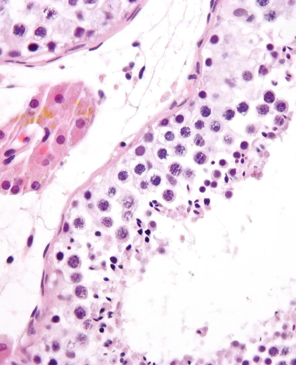
精子を作る管。壁を作るのは2種の細胞。
- 精上皮(生殖細胞)——基底側から内腔へ向かって成熟が進む。精祖細胞 → 一次精母細胞 → 二次精母細胞 → 精子細胞 → 精子
- Sertoli 細胞——基底膜から内腔まで貫く支持細胞。卵胞刺激ホルモンを受け、インヒビンを出す
管の外側の間質にLeydig 細胞があり、黄体形成ホルモンを受けてテストステロンを作る。
Sertoli 細胞どうしの密着結合が血液精巣関門を作り、管を基底部と管腔部に分ける。
血液精巣関門が必要な理由は免疫にある。減数分裂は思春期に初めて起こるので、精子は胸腺で自己として教え込まれた時期には存在しなかった。免疫系にとって精子は他人である。だから物理的に隔離するしかない。
この構造から2つのことが出る。
- 精管結紮術や精巣外傷のあとに抗精子抗体ができうる——関門が破れて精子が免疫系に晒されるため
- 薬も抗体も精細管の中へ入りにくい——精巣が白血病細胞の逃げ場(聖域)になり、再発部位になる
Sertoli 細胞は精子形成の律速である。1個が支えられる生殖細胞の数が決まっているので、Sertoli 細胞の数が精子産生の上限を決める。この数は思春期までに確定する。
Sertoli 細胞は胎児期にも決定的な仕事をする——抗ミュラー管ホルモンを出してミュラー管(女性の内性器になる管)を退縮させる。Leydig 細胞のテストステロンがウォルフ管を発達させるのと合わせて、内性器の性別が決まる。2つの細胞が別々のホルモンで別々の管を担当している。
停留精巣が問題になるのは温度である。精子形成には体温より2〜3℃低い環境が要り、陰嚢内に降りていないと精上皮が障害される。2歳までに手術するのは、不可逆になる前に降ろすためで、手術しても悪性化のリスク(精巣腫瘍)は残る。
Klinefelter 症候群(47,XXY)では精細管が硝子化して萎縮するが Leydig 細胞は比較的残るため、高身長・小さく硬い精巣・無精子症・女性化乳房という組み合わせになる。卵胞刺激ホルモンと黄体形成ホルモンが上がり(原発性)、テストステロンは低め。
精索静脈瘤は左に多い(左精巣静脈が左腎静脈へ直角に入るため)。陰嚢の温度が上がって精子形成が落ちる——男性不妊で治療可能な原因として最も多い。
English version
Core
The tubule in which sperm are made. Two cell types make the wall.
- Seminiferous (germinal) epithelium — maturation proceeds from the basal side towards the lumen: spermatogonium → primary spermatocyte → secondary spermatocyte → spermatid → spermatozoon.
- Sertoli cell — the supporting cell spanning from basal lamina to lumen. Responds to FSH and secretes inhibin.
In the interstitium outside the tubule are the Leydig cells, which respond to LH and make testosterone.
Tight junctions between Sertoli cells form the blood–testis barrier, dividing the tubule into a basal and an adluminal compartment.
Going further
The reason the blood–testis barrier is needed is immunological. Meiosis first occurs at puberty, so sperm did not exist at the time the thymus taught the immune system what is self. To the immune system, sperm are foreign. So they can only be isolated physically.
Two things follow from this structure.
- Anti-sperm antibodies can arise after vasectomy or testicular trauma — the barrier is broken and sperm are exposed to the immune system.
- Neither drugs nor antibodies enter the tubule readily — the testis becomes a sanctuary for leukaemic cells and a site of relapse.
The Sertoli cell is the rate-limiting step in spermatogenesis. The number of germ cells one can support is fixed, so the number of Sertoli cells sets the ceiling on sperm production. That number is settled by puberty.
The Sertoli cell also does decisive work in fetal life — it secretes anti-Müllerian hormone and causes the Müllerian ducts (which would become the female internal genitalia) to regress. Together with testosterone from the Leydig cells developing the Wolffian ducts, the sex of the internal genitalia is settled. Two cells, two hormones, each responsible for a different duct.
In the clinic
Undescended testis is a problem of temperature. Spermatogenesis requires an environment 2–3 °C below body temperature, and if the testis has not descended into the scrotum the germinal epithelium is damaged. Surgery by the age of two is to bring it down before the damage is irreversible; the risk of malignancy (testicular tumour) remains even after operation.
In Klinefelter syndrome (47,XXY) the tubules hyalinise and atrophy while the Leydig cells are comparatively spared, giving the combination of tall stature, small firm testes, azoospermia and gynaecomastia. FSH and LH are raised (primary) and testosterone is low.
Varicocele is commoner on the left (the left testicular vein enters the left renal vein at a right angle). The scrotal temperature rises and spermatogenesis falls — the commonest treatable cause of male infertility.
精子形成 — 74日かけて、細胞質を捨てていく過程Spermatogenesis
精細管の基底側から内腔側へ向かって、順に進む。
| 段階 | 細胞 | 核相 |
|---|---|---|
| 増殖期 | 精祖細胞(A型=幹細胞、B型=分化する) | 2n |
| 成長期 | 一次精母細胞(最大・最も目立つ) | 2n(4C) |
| 成熟期 | 二次精母細胞(すぐ分裂するので見つけにくい) | n |
| — | 精子細胞 | n |
| 精子完成 | 精子 | n |
精子完成で起きること——先体の形成・核の凝縮・鞭毛の伸長・ミトコンドリアの再配置・余分な細胞質の廃棄(残余小体)。残余小体は Sertoli 細胞が貪食する。
分裂しても細胞質が完全に分かれず、細胞間橋でつながったまま同調して進む。
全過程に約 74 日。精細管の一点を見ると、いくつかの段階が同時に並ぶ。
細胞間橋でつながることには、はっきりした理由がある。減数分裂で X か Y のどちらかしか持たなくなるのに、精子は同じ性能を保つ必要がある——「橋を通じて遺伝子産物を共有するので、Y を持つ精子でも X 上の遺伝子の産物を使える」。
A型精祖細胞が幹細胞として残ることが、精巣が一生作り続けられる理由になる。卵巣が出生時の在庫を使い切るのと、設計が正反対になる(この章の卵胞の項)——「作り続ける側は、分裂の回数が増えるぶん変異も溜まる」。
だから父親の年齢は、新生変異の数と相関する(単一遺伝子病の章)——「軟骨無形成症や Marfan 症候群の新生突然変異が、高齢の父で増える」。
分裂が盛んなことは、そのまま抗腫瘍薬と放射線への弱さになる(放射線生物学の章)——「精祖細胞が失われると、回復に年単位かかるか、戻らない」。
熱にも弱い。精巣が体外にあるのは、体温より 2〜3 ℃低く保つためになる(精巣の章)——「停留精巣を放置すると、造精機能が失われ、悪性化の危険も上がる」。
74日かかることが、不妊治療の時間軸を決める。精子形成の改善を評価するには、最低3か月あけて再検する。「先月の生活改善」は今日の精液所見に反映されていない。
熱に弱い——停留精巣・精索静脈瘤・長時間の高温環境で造精機能が落ちる。精索静脈瘤は左に多く、手術で改善しうる数少ない男性不妊の原因である。
抗癌薬と放射線は分裂中の精祖細胞を直撃する。治療前の精子凍結保存を必ず提示する——妊孕性温存は治療開始前にしか選べない。
Sertoli 細胞が血液精巣関門を作り、精子を免疫から隔てる。関門が破れると抗精子抗体ができる。精巣捻転・外傷・精管結紮後で問題になる。
Sertoli 細胞は AMH と インヒビン B を出す——インヒビン B は造精機能の指標になる。
English version
Core
It proceeds in order from the basal to the luminal side of the tubule.
| Phase | Cell | Ploidy |
|---|---|---|
| Proliferative | Spermatogonium (type A = stem cell, type B = differentiating) | 2n |
| Growth | Primary spermatocyte (the largest and most conspicuous) | 2n (4C) |
| Maturation | Secondary spermatocyte (divides at once, so hard to find) | n |
| — | Spermatid | n |
| Spermiogenesis | Spermatozoon | n |
What happens in spermiogenesis — formation of the acrosome, condensation of the nucleus, growth of the flagellum, redistribution of mitochondria, and discarding of surplus cytoplasm (the residual body), which the Sertoli cell phagocytoses.
At division the cytoplasm does not separate completely, and the cells remain joined by intercellular bridges and advance in synchrony. The whole process takes about 74 days. At one point in a tubule, several stages are seen side by side.
Going further
The intercellular bridges have a definite reason. After meiosis a cell carries either X or Y, yet every sperm must keep the same performance — gene products are shared through the bridges, so a Y-bearing sperm can use the products of genes on the X.
That type A spermatogonia remain as stem cells is why the testis can go on producing for life, the design being the exact opposite of the ovary using up the stock it had at birth — the side that keeps producing accumulates mutations along with the extra divisions.
So paternal age correlates with the number of new mutations — de novo mutations causing achondroplasia and Marfan syndrome increase with an older father.
Vigorous division is directly a vulnerability to cytotoxic drugs and radiation — once the spermatogonia are lost, recovery takes years or does not come.
It is also vulnerable to heat. The testis lies outside the body to be kept 2–3 °C below core temperature — an undescended testis left alone loses spermatogenesis, and the risk of malignancy rises.
In the clinic
That it takes 74 days sets the time-scale of fertility treatment. To assess improvement in spermatogenesis, repeat the test after at least three months. "The lifestyle change last month" is not reflected in today’s semen analysis.
It is vulnerable to heat — undescended testis, varicocele and prolonged high-temperature environments reduce production. Varicocele is commoner on the left and is one of the few causes of male infertility that surgery can improve.
Cytotoxic drugs and radiation strike the dividing spermatogonia directly. Always offer sperm cryopreservation before treatment — fertility preservation can only be chosen before treatment begins.
Sertoli cells form the blood–testis barrier and shield sperm from the immune system. If it is breached, anti-sperm antibodies form — a problem after torsion, trauma or vasectomy. Sertoli cells secrete AMH and inhibin B, which is an index of spermatogenesis.
Leydig 細胞 — 精細管の外側で、ホルモンを作っているEndocrinocytus interstitialis
精細管と精細管のあいだ(間質)にある。ステロイド産生細胞の典型的な姿をしている。
- 好酸性で、細胞質が泡状(脂肪滴)
- 滑面小胞体が発達し、ミトコンドリアのクリスタが管状
- ヒトでは Reinke 結晶という棒状の封入体が見られる(他の動物には無い)
LH の刺激でテストステロンを作る。Sertoli 細胞は FSH の刺激で働く——2つの細胞に、2つのゴナドトロピンが分かれて効く。
Sertoli 細胞はアンドロゲン結合蛋白を作り、精細管内のテストステロン濃度を血中の数十倍に保つ。
Sertoli 細胞が出すインヒビンが、FSH を抑える。
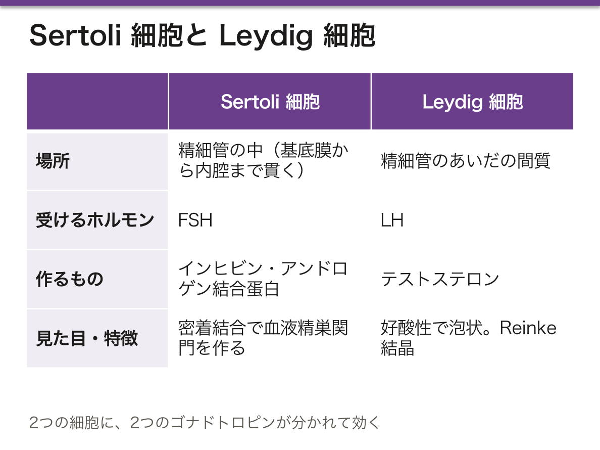
2つの細胞と2つのホルモンの対応を押さえると、検査の読み方が決まる。
| 所見 | 壊れている場所 |
|---|---|
| FSH が高い・テストステロンは正常 | Sertoli 細胞/造精機能(インヒビンが減った) |
| LH が高い・テストステロンが低い | Leydig 細胞(原発性性腺機能低下症) |
| LH も FSH も低い | 視床下部・下垂体(中枢性) |
(生殖の内分泌の章)——「ゴナドトロピンが高いか低いかで、上流か下流かが決まる」。
精細管内のテストステロン濃度が血中より桁違いに高いことは、治療に直結する。テストステロンを外から投与しても精細管内の濃度には届かず、むしろ LH を抑えて造精機能を落とす(ホルモン薬の章)——「男性ホルモンの補充が、不妊の原因になる」。
だから男性不妊では hCG や FSH 製剤を使う(不妊の章)——「補うのはホルモンそのものではなく、それを作らせる指令になる」。
Klinefelter 症候群では、Leydig 細胞が相対的に保たれ、精細管が硝子化する(染色体異常の章)——「だから FSH と LH がともに高く、女性化乳房が出る」。
Leydig 細胞は LH でテストステロンを作る。精細管の外にあるので、造精機能とテストステロン分泌は別々に障害されうる。
Klinefelter 症候群(47,XXY) — 精細管が硝子化して造精機能が失われ、テストステロンも低下する。高身長・女性化乳房・小さく硬い精巣・無精子症。FSH・LH が高く、テストステロンが低い(高ゴナドトロピン性性腺機能低下症)。
Kallmann 症候群はGnRH ニューロンの遊走障害で、嗅覚脱失を伴う低ゴナドトロピン性性腺機能低下症。ゴナドトロピンが高いか低いかで、障害が精巣にあるか中枢にあるかが分かる。
Leydig 細胞腫はホルモン産生腫瘍で、小児では思春期早発症、成人では女性化乳房を起こす。Reinke 結晶が組織学的な特徴である。
English version
Core
In the interstitium between the seminiferous tubules. It has the typical appearance of a steroid-producing cell.
- Eosinophilic, with foamy cytoplasm (lipid droplets).
- Developed smooth endoplasmic reticulum; mitochondria with tubular cristae.
- In humans, rod-shaped inclusions called Reinke crystals (not found in other animals).
It makes testosterone on LH stimulation; the Sertoli cell works on FSH stimulation — two gonadotrophins acting separately on two cells.
The Sertoli cell makes androgen-binding protein, holding the intratubular testosterone concentration at some tens of times that of the blood. Inhibin from the Sertoli cell suppresses FSH.
Going further
Fix the correspondence of two cells to two hormones and the interpretation of the tests follows.
| Finding | Where the fault lies |
|---|---|
| FSH high, testosterone normal | Sertoli cell / spermatogenesis (inhibin has fallen) |
| LH high, testosterone low | Leydig cell (primary hypogonadism) |
| Both LH and FSH low | Hypothalamus or pituitary (central) |
Whether the gonadotrophins are high or low decides whether the fault is upstream or downstream.
That the intratubular testosterone concentration is orders of magnitude above that of the blood bears directly on treatment. Testosterone given from outside does not reach the intratubular concentration and instead suppresses LH and reduces spermatogenesis — replacement of the male hormone becoming a cause of infertility. So in male infertility hCG or FSH preparations are used — what is supplied is not the hormone itself but the instruction to make it.
In Klinefelter syndrome the Leydig cells are relatively preserved while the tubules hyalinise — hence both FSH and LH are high and gynaecomastia appears.
In the clinic
The Leydig cell makes testosterone under LH. Lying outside the tubule, spermatogenesis and testosterone secretion can be impaired separately.
Klinefelter syndrome (47,XXY) — the tubules hyalinise, spermatogenesis is lost and testosterone falls. Tall stature, gynaecomastia, small firm testes, azoospermia. FSH and LH high with low testosterone (hypergonadotrophic hypogonadism).
Kallmann syndrome is a failure of GnRH neurone migration, giving hypogonadotrophic hypogonadism with anosmia. Whether the gonadotrophins are high or low tells you whether the fault is in the testis or centrally.
A Leydig cell tumour is hormone-producing, causing precocious puberty in a child and gynaecomastia in an adult. Reinke crystals are the histological feature.
精巣生検 — 造精機能を、組織で段階に分ける
無精子症では、作れていないのか、作れているのに出てこられないのかを分ける必要がある。組織がその答えを出す。
| 正常な造精 | 精細管に全段階の細胞が並ぶ | 閉塞性無精子症。精管や精巣上体の通過障害。精子は採れるので ICSI が可能 |
| 成熟停止 | 途中の段階で止まっている | 止まる段階によって、精子が採れるかどうかが変わる |
| Sertoli 細胞のみ | 胚細胞が完全に欠如し、Sertoli 細胞だけが並ぶ | 採取できる可能性は低いが、巣状に造精が残ることがある |
| 硝子化・線維化 | 精細管が潰れて索状になる | Klinefelter 症候群の典型像。Leydig 細胞は相対的に増える |
評価は Johnsen スコア(1〜10 点)で、精細管ごとに最も進んだ段階を点数化し、平均する。
「巣状に残っている」ことが、治療を変えた。
非閉塞性無精子症でも、精巣全体が一様に無精子なわけではない。顕微鏡下に精巣を開き、太くて白っぽい精細管を選んで採取する(顕微鏡下精巣内精子採取術)ことで、Sertoli 細胞のみと診断された症例でも 3〜5 割で精子が得られる。
ランダムに採るのではなく、組織の違いを見て選ぶ——組織学の知識が、そのまま手術の技術になっている例である。
⚠️ 生検そのものに害がある。血液精巣関門を破ると、精子が免疫系に曝される。抗精子抗体ができて、後の造精機能をさらに落としうる。だから診断目的だけの生検は避け、採取と同時に行うのが現在の考え方である。
採る前に調べておくことがある。
- 染色体検査——Klinefelter 症候群(47,XXY)は非閉塞性無精子症の 1 割強を占める
- Y 染色体微小欠失——AZFa・AZFb の欠失では精子が得られないので、手術を回避できる。AZFc なら得られる可能性がある
- FSH と精巣容積——FSH が高く精巣が小さければ、造精障害を示唆する
組織を見る前に、遺伝子で答えが出ることがある——検査の順序が、患者の負担を減らす。
男性不妊の精査は、採血と染色体検査が先である。生検は最後の手段で、しかも採取と同時に行う。
English version
Core
In azoospermia it is necessary to separate failure to produce from production that cannot get out. The histology gives the answer.
| Picture | What is seen | Meaning |
|---|---|---|
| Normal spermatogenesis | All stages present in the tubule | Obstructive azoospermia. Obstruction of the vas or epididymis. Sperm can be retrieved, so ICSI is possible |
| Maturation arrest | Stopped at some stage | Whether sperm can be retrieved depends on the stage at which it stops |
| Sertoli cell only | Germ cells entirely absent; only Sertoli cells line the tubule | Retrieval is unlikely, but spermatogenesis may persist focally |
| Hyalinisation and fibrosis | The tubules collapse into cords | The typical picture of Klinefelter syndrome. Leydig cells are relatively increased |
Assessment uses the Johnsen score (1–10): the most advanced stage in each tubule is scored and the scores averaged.
Going further
That it survives focally has changed the treatment. Even in non-obstructive azoospermia the testis is not uniformly without sperm. By opening the testis under the microscope and selecting the thicker, paler tubules (microdissection TESE), sperm are obtained in 30–50 per cent even of cases diagnosed as Sertoli cell only. Not sampling at random but choosing by looking at the difference in the tissue — an example of histological knowledge becoming directly a surgical technique.
⚠️ The biopsy itself does harm. Breaking the blood–testis barrier exposes sperm to the immune system; anti-sperm antibodies may form and reduce spermatogenesis further. So a biopsy for diagnosis alone is avoided, and current practice is to do it at the same time as retrieval.
There are things to investigate before taking any tissue.
- Karyotype — Klinefelter syndrome (47,XXY) accounts for a little over 10 per cent of non-obstructive azoospermia.
- Y chromosome microdeletions — with deletion of AZFa or AZFb no sperm are obtained, so the operation can be avoided. With AZFc there is a chance.
- FSH and testicular volume — a high FSH with small testes suggests a defect of spermatogenesis.
Sometimes the genes give the answer before the tissue is examined — the order of the investigations reduces the burden on the patient.
In the clinic
Investigation of male infertility begins with blood tests and karyotype. Biopsy is the last resort, and is done at the same time as retrieval.
精巣上体と精管 — 立体線毛と、厚い筋Epididymis
精巣輸出管——線毛細胞と無線毛細胞が交互に並び、内腔が波打って見える(鋸歯状)。液の大半をここで吸収する。
精巣上体管——1本の長い管が折り畳まれている(約6 m)。内腔は円滑。
- 多列円柱上皮+立体線毛(stereocilia)——実体はアクチンの長い微絨毛で、動かない
- 周囲に薄い平滑筋。頭部から尾部へ厚くなる
- ここで運動能と受精能を獲得し、尾部に蓄えられる
精管——内腔は狭く星形。粘膜にひだ。内縦・中輪・外縦の厚い3層の平滑筋が最大の特徴になる。
精管膨大部を経て、精嚢の導管と合流し射精管になる。
立体線毛が線毛ではないことが、この章で最も問われる混同になる。アクチンでできた長い微絨毛で、動かない。吸収に関わる(線毛と微絨毛の章)——「名前が構造を偽っているので、名称ではなく成分で覚える」。
同じ立体線毛が、内耳の有毛細胞にもある(コルチ器の章)——「聞くことと精子を熟させることが、同じ構造を共有している」。
精管の筋層が異常に厚いことが、標本の見分けになる。内腔の狭さと筋層の厚さの比が、他のどの管とも違う——「壁が内腔より厚い管は、精管を疑う」。
この筋層は交感神経で強く収縮する。だから交感神経を抑える薬で射精障害が起きる(α遮断薬の章)——「前立腺肥大症の薬の副作用が、この筋の話になる」。
精管結紮術の後も、精子は作られ続ける。吸収されるので溜まらない——「止めているのは輸送であって、産生ではない」。
先天性両側精管欠損は、嚢胞性線維症の変異と関連する。閉塞性無精子症の原因として重要になる(男性不妊の章)——「不妊の検査から、遺伝性疾患が見つかることがある」。
精巣上体を通るあいだに、精子は運動能と受精能を獲得する。精巣から採った精子はまだ泳げない——顕微授精(ICSI)が必要な理由の1つである。
精巣上体炎 — 若年では性感染症(クラミジア・淋菌)、高齢では尿路の腸内細菌が原因になる。年齢で起炎菌が変わるので抗菌薬の選択も変わる。挙上すると痛みが和らぐ(Prehn 徴候陽性)ことが精巣捻転との鑑別の手がかりになるが、捻転を否定できなければ緊急で手術に踏み切る。
立体線毛は動かない——名前に反して運動性は無く、吸収と分泌を担う。
精管の厚い筋層が射精時の強い蠕動を作る。触診で索状に硬く触れるので精管結紮術の確認に使える。
English version
Core
- Efferent ductules — ciliated and non-ciliated cells alternate, so the lumen looks wavy (scalloped). Most of the fluid is absorbed here.
- Duct of the epididymis — a single long duct folded on itself (about 6 m). The lumen is smooth. Pseudostratified columnar epithelium with stereocilia — in fact long actin microvilli, which do not move. A thin layer of smooth muscle around it, thickening from head to tail. Here sperm acquire motility and the capacity to fertilise, and are stored in the tail.
- Ductus deferens — a narrow, star-shaped lumen with mucosal folds. Its chief feature is the thick three-layered smooth muscle (inner longitudinal, middle circular, outer longitudinal).
Through the ampulla it joins the duct of the seminal vesicle to form the ejaculatory duct.
Going further
That stereocilia are not cilia is the confusion most often examined in this chapter. They are long microvilli made of actin and do not move; they take part in absorption — the name misrepresents the structure, so learn it by its composition, not its name.
The same stereocilia are found on the hair cells of the inner ear — hearing and the maturation of sperm sharing one structure.
The unusually thick muscle of the vas is how the slide is identified. The ratio of narrow lumen to thick muscle is unlike any other duct — a tube whose wall is thicker than its lumen should be suspected to be the vas.
That muscle contracts strongly under sympathetic control, so drugs that block the sympathetic cause disorders of ejaculation — an adverse effect of the drugs for prostatic hyperplasia that is a matter of this muscle.
After vasectomy, sperm continue to be made. They are absorbed, so they do not accumulate — what has been stopped is transport, not production.
Congenital bilateral absence of the vas is associated with cystic fibrosis mutations and is an important cause of obstructive azoospermia — a genetic disease found through an infertility investigation.
In the clinic
Passing through the epididymis, sperm acquire motility and the capacity to fertilise. Sperm taken from the testis cannot yet swim — one reason ICSI is needed.
Epididymitis — sexually transmitted organisms (chlamydia, gonococcus) in the young, urinary coliforms in the elderly. The organism changes with age, so the choice of antibiotic changes too. Relief of pain on elevation (a positive Prehn’s sign) is a clue distinguishing it from torsion, but if torsion cannot be excluded, proceed to emergency surgery.
Stereocilia do not move — despite the name they have no motility and serve absorption and secretion.
The thick muscle of the vas produces the strong peristalsis of ejaculation. It is palpable as a firm cord, which is used to confirm a vasectomy.
前立腺と精嚢 — 域で分かれる病気Prostata
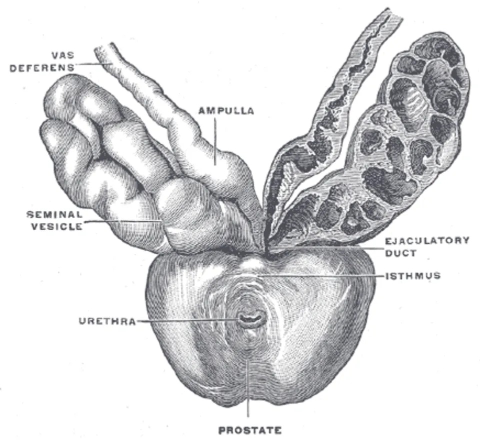
前立腺——30〜50個の管状胞状腺が、線維筋性間質に埋まる。腺腔に前立腺分泌顆粒(アミロイド小体)が見える。
- 上皮は2層——分泌細胞(円柱)と基底細胞。基底細胞が消えることが浸潤癌の指標になる
- 分泌——酸性ホスファターゼ・PSA・クエン酸・亜鉛・スペルミン
McNeal の域。
| 域 | 体積 | 病気 |
|---|---|---|
| 辺縁域 | 約70% | 癌の約7割。直腸診で触れる |
| 移行域 | 約5% | 前立腺肥大症 |
| 中心域 | 約25% | 射精管が通る。病変は少ない |
精嚢——粘膜が複雑に折れ込んで蜂巣状に見える。分泌は果糖(精子のエネルギー)とアルカリ、黄色色素。
どちらもアンドロゲン依存性になる。
基底細胞の有無が、良性と癌を分ける最も実用的な所見になる。免疫染色(p63・高分子量サイトケラチン)で基底層が消えていれば浸潤癌(免疫組織化学の章)——「1層の細胞が残っているかどうかで、診断が決まる」。
癌が辺縁域に多いことが、直腸診が有効な理由になる(前立腺癌の章)——「指で触れる場所に、癌が集まっている」。
逆に肥大症は移行域なので、大きくなっても直腸診で硬く触れない。そして尿道を内側から圧迫する——「症状を出しやすいのは肥大症、触れやすいのは癌になる」。
だから PSA の上昇は、癌だけを意味しない。肥大症・前立腺炎・射精・自転車・直腸診でも上がる(腫瘍マーカーの章)——「測る前に、上げる条件を除いておく」。
アンドロゲン依存性であることが、治療の骨格になる。5α還元酵素阻害薬が肥大症に、アンドロゲン遮断が癌に使われる(前立腺の薬の章)——「同じホルモンを、違う深さで止めている」。
精嚢の果糖は、精液検査で閉塞部位の推定に使われる。果糖が無ければ、精嚢か射精管より下流の閉塞になる——「成分の欠如が、詰まっている高さを教える」。
域が違えば病気が違う。移行域=前立腺肥大症、辺縁域=前立腺癌。
前立腺肥大症は内側から尿道を締めるので排尿症状(尿勢低下・残尿感・夜間頻尿)が主体。α1 遮断薬・5α還元酵素阻害薬で治療する。
前立腺癌は外側(辺縁域)から起こるので尿道から遠く、進行するまで無症状。直腸診で触れるのはこの辺縁域であり、PSA と直腸診で拾う。「症状が出ないから検診が要る」という論理が解剖から導ける。
基底細胞の有無が良悪の決め手になる。癌では基底細胞が消失する——p63・高分子サイトケラチンで確認する。
骨転移は造骨性で、Batson 静脈叢を介して脊椎へ広がる。
English version
Core
Prostate — 30–50 tubuloalveolar glands embedded in fibromuscular stroma. Corpora amylacea are seen in the lumina.
- The epithelium is two-layered — secretory (columnar) and basal cells. Loss of the basal cells is the marker of invasive carcinoma.
- Secretes acid phosphatase, PSA, citrate, zinc, spermine.
McNeal’s zones
| Zone | Volume | Disease |
|---|---|---|
| Peripheral | About 70% | About 70 per cent of cancers. Palpable per rectum |
| Transition | About 5% | Benign prostatic hyperplasia |
| Central | About 25% | The ejaculatory ducts pass through. Little disease |
Seminal vesicle — the mucosa folds intricately and looks honeycombed. It secretes fructose (energy for sperm), alkali and a yellow pigment. Both organs are androgen-dependent.
Going further
The presence or absence of basal cells is the most practical finding separating benign from malignant. If immunostaining (p63, high molecular weight cytokeratin) shows the basal layer has gone, it is invasive carcinoma — the diagnosis settled by whether one layer of cells remains.
That cancer is commonest in the peripheral zone is why the digital rectal examination works — the cancers gather where the finger can reach.
Hyperplasia, conversely, is in the transition zone, so it is not felt as hard per rectum however large it grows — and it compresses the urethra from within. Hyperplasia produces symptoms readily; cancer is readily palpable.
So a raised PSA does not mean cancer alone. It rises with hyperplasia, prostatitis, ejaculation, cycling and a rectal examination — exclude the conditions that raise it before measuring.
Androgen dependence is the framework of treatment. 5α-reductase inhibitors for hyperplasia, androgen deprivation for cancer — the same hormone blocked at different depths.
The fructose of the seminal vesicle is used in semen analysis to infer the site of an obstruction. No fructose means obstruction at or below the seminal vesicle or ejaculatory duct — the absence of a constituent telling you the level of the blockage.
In the clinic
A different zone means a different disease. Transition zone = benign hyperplasia; peripheral zone = carcinoma.
Benign prostatic hyperplasia squeezes the urethra from within, so voiding symptoms predominate (poor stream, incomplete emptying, nocturia). Treated with α1 blockers and 5α-reductase inhibitors.
Prostate cancer arises on the outside (peripheral zone), far from the urethra, and is asymptomatic until advanced. What the finger feels per rectum is that peripheral zone, and it is picked up by PSA and rectal examination. The logic that "screening is needed because it causes no symptoms" can be derived from the anatomy.
The presence or absence of basal cells is decisive for benign versus malignant — in cancer they are lost; confirm with p63 and high molecular weight cytokeratin.
Bone metastases are osteoblastic and spread to the spine through Batson’s venous plexus.
精子 — 3つの部分に、必要なものだけを積んでいるSpermatozoon
全長約60 μm。細胞質をほぼ捨てて軽くしてある。
| 部分 | 中身 | 役割 |
|---|---|---|
| 頭部 | 凝縮した核+先体 | 遺伝情報を運ぶ/透明帯を破る酵素 |
| 頸部 | 中心小体 | 頭と尾をつなぐ |
| 中片部 | らせん状のミトコンドリア鞘 | ATP を供給する |
| 主部・終部 | 軸糸(9+2)と外側緻密線維 | 運動 |
先体はゴルジ装置由来のリソソーム様顆粒——ヒアルロニダーゼ・アクロシンを含む。
核のヒストンはプロタミンに置き換わり、DNA が極度に凝縮している。
精子形成には約74日、精巣上体の通過にさらに約2週間かかる。
「細胞質を捨てる」という設計から、精子の弱点が全部出てくる。修復酵素も抗酸化系もほとんど積んでいないので、酸化ストレスに弱い(活性酸素の章)——「軽くした代償が、傷を直せないことになる」。
そして受精後、精子のミトコンドリアは分解される。だからミトコンドリア DNA は母親からしか伝わらない(母系遺伝)(ミトコンドリア病の章)——「父親由来のミトコンドリアは、受精卵の中で選択的に消される」。
軸糸が 9+2 であることは、線毛の病気とそのまま重なる。原発性線毛機能不全症では、気道の線毛と精子の尾が同じ蛋白の異常で動かない(線毛・鞭毛の章)——「慢性副鼻腔炎・気管支拡張症・不妊が並ぶ(Kartagener 症候群)」。
精子形成に74日かかることは、治療の評価の間隔を決めている。男性不妊で介入しても、精液所見が変わるのは3か月後になる(不妊の章)——「短い間隔で検査を繰り返しても、答えは出ない」。
同じ理由で、抗腫瘍薬や放射線の影響も遅れて現れ、遅れて戻る(放射線生物学の章)——「治療前の精子保存を、治療を始める前に切り出す必要がある」。
精液検査は形態も見る。濃度・運動率・正常形態率が基本項目になる。
先体の異常 — グロボゾオスペルミア(円形頭部精子症)では先体が無いため透明帯を破れず、自然受精ができない。顕微授精の適応になる。
中片部のミトコンドリアが運動のエネルギーを供給する。受精後に父方ミトコンドリアは分解されるのでミトコンドリア病は母性遺伝になる。
尾部(鞭毛)は 9+2 構造で、気道の線毛と同じ。だから原発性繊毛運動不全症では精子も泳げず不妊になる——慢性副鼻腔炎・気管支拡張症と男性不妊が同じ患者に揃う理由である。
細胞質をほぼ捨てているので修復機構に乏しく、DNA 損傷を受けやすい。喫煙・加齢・酸化ストレスが精子 DNA の断片化を増やす。
English version
Core
About 60 µm long. Almost all the cytoplasm has been discarded to make it light.
| Part | Contents | Role |
|---|---|---|
| Head | Condensed nucleus + acrosome | Carries the genetic information / enzymes to break the zona pellucida |
| Neck | Centrioles | Joins head to tail |
| Midpiece | A helical mitochondrial sheath | Supplies ATP |
| Principal and end pieces | Axoneme (9+2) and outer dense fibres | Movement |
The acrosome is a lysosome-like granule derived from the Golgi, containing hyaluronidase and acrosin. The histones of the nucleus are replaced by protamines and the DNA is extremely condensed.
Spermatogenesis takes about 74 days, and passage through the epididymis about two weeks more.
Going further
Every weakness of the sperm follows from the design of discarding the cytoplasm. It carries almost no repair enzymes and no antioxidant system, so it is vulnerable to oxidative stress — the price of being made light is being unable to mend damage.
And after fertilisation the sperm’s mitochondria are degraded. So mitochondrial DNA is transmitted only from the mother (maternal inheritance) — paternal mitochondria are selectively destroyed inside the zygote.
That the axoneme is 9+2 makes the diseases of cilia overlap exactly. In primary ciliary dyskinesia the cilia of the airway and the tail of the sperm are immobilised by the same protein defect — chronic sinusitis, bronchiectasis and infertility appear together (Kartagener syndrome).
That spermatogenesis takes 74 days sets the interval for assessing treatment. After an intervention for male infertility, the semen analysis changes three months later — repeating the test at short intervals gives no answer. For the same reason the effects of cytotoxic drugs and radiation appear late and recover late — sperm storage must be raised before treatment begins.
In the clinic
Semen analysis looks at morphology too. Concentration, motility and the proportion of normal forms are the basic items.
Abnormality of the acrosome — in globozoospermia (round-headed sperm) the acrosome is absent, the zona cannot be penetrated and natural fertilisation is impossible. An indication for ICSI.
The mitochondria of the midpiece supply the energy for movement. Paternal mitochondria are degraded after fertilisation, hence the maternal inheritance of mitochondrial disease.
The tail (flagellum) has the 9+2 structure, the same as the cilia of the airway. So in primary ciliary dyskinesia the sperm cannot swim and the man is infertile — the reason chronic sinusitis, bronchiectasis and male infertility occur in the same patient.
Having discarded almost all its cytoplasm it is poor in repair mechanisms and susceptible to DNA damage. Smoking, age and oxidative stress increase sperm DNA fragmentation.
陰茎の勃起組織 — 血を溜める海綿と、包む膜Corpus cavernosum
3本の海綿体からなる——陰茎海綿体(左右2本)と、尿道海綿体(1本、尿道を含む)。
組織は「内皮で裏打ちされた洞(腔)を、平滑筋と結合組織の梁が仕切る」構造になる。
陰茎海綿体だけが、厚く伸びにくい白膜に包まれている。尿道海綿体の白膜は薄い。
血の入り方——深陰茎動脈 → らせん動脈 → 洞。
神経支配——勃起は副交感神経(骨盤内臓神経、S2〜S4)、射精は交感神経(L1〜L2)。
勃起は「血を入れること」ではなく、「出さないこと」で成立している。洞が膨らむと、白膜との間で導出静脈が押しつぶされて流出が止まる(静脈閉塞機構)——「硬い膜に囲まれているから、内圧が上がる」。
だから尿道海綿体は硬くならない。白膜が薄く、圧が逃げるので尿道が潰れない——「射精の通り道を確保するために、わざと硬くしていない」。
分子の道筋は一本道になっている。副交感神経と内皮から一酸化窒素 → グアニル酸シクラーゼ → cGMP → 平滑筋弛緩 → 洞へ血が入る(セカンドメッセンジャーの章)。
PDE5 阻害薬(シルデナフィルなど)は、cGMP を分解する酵素を止めている(男性の性腺の章)——「作らせるのではなく、壊させない薬になる」。
だから硝酸薬との併用が禁忌になる。両方とも cGMP を増やす方向に働き、全身の血管が弛緩して血圧が落ちる(硝酸薬の章)——「同じ経路の上流と下流を、同時に押している」。
持続勃起症では、流出が止まったまま戻らない。虚血型は組織が壊死しうる救急で、鎌状赤血球症や薬剤(PDE5 阻害薬・抗精神病薬)が原因になる(鎌状赤血球症の章)——「閉じ込める仕組みがあるからこそ、開かなくなる病気が存在する」。
勃起は血管の現象である。副交感神経(S2〜S4、骨盤内臓神経)が NO を出し、cGMP が増えて海綿体平滑筋が弛緩し、洞に血が満ちる。膨らんだ海綿体が白膜の下の導出静脈を圧迫して、血が出られなくなる——入れて、閉じ込めるという2段構えである。
PDE5 阻害薬(シルデナフィル)は cGMP の分解を止める。硝酸薬との併用は致命的な低血圧を起こすので禁忌である。
「勃起は副交感、射精は交感」——Point and Shoot と覚える。骨盤内手術(直腸癌・前立腺癌)での神経損傷で勃起障害と逆行性射精が起こる。神経温存術式が意識される理由である。
勃起障害は血管内皮機能障害の early signでもある。陰茎の動脈は細いので、冠動脈疾患に先立って症状が出うる。
English version
Core
Three corpora — the paired corpora cavernosa and the single corpus spongiosum containing the urethra.
The tissue is endothelium-lined sinuses partitioned by trabeculae of smooth muscle and connective tissue.
Only the corpora cavernosa are enclosed in a thick, inextensible tunica albuginea; that of the corpus spongiosum is thin.
How the blood enters — deep artery of the penis → helicine arteries → sinuses.
Innervation — erection is parasympathetic (pelvic splanchnic nerves, S2–S4), ejaculation sympathetic (L1–L2).
Going further
An erection is achieved not by letting blood in but by not letting it out. As the sinuses fill, the emissary veins are crushed between them and the tunica and outflow ceases (the veno-occlusive mechanism) — the pressure rises because it is enclosed in a rigid membrane.
So the corpus spongiosum does not become rigid. Its tunica is thin and the pressure escapes, so the urethra is not crushed — deliberately not made rigid, in order to keep the passage for ejaculation open.
The molecular pathway is a single line: nitric oxide from parasympathetic nerves and endothelium → guanylate cyclase → cGMP → relaxation of smooth muscle → blood enters the sinuses. PDE5 inhibitors (sildenafil and others) block the enzyme that degrades cGMP — a drug that does not make it but prevents it being destroyed.
So combination with a nitrate is contraindicated. Both act to raise cGMP, the vessels of the whole body relax and the pressure falls — pressing on the upstream and the downstream of the same pathway at once.
In priapism the outflow stays shut and does not reopen. The ischaemic type is an emergency in which the tissue can necrose, caused by sickle cell disease and by drugs (PDE5 inhibitors, antipsychotics) — a disease of failing to open exists precisely because there is a mechanism for shutting in.
In the clinic
Erection is a vascular phenomenon. The parasympathetic (S2–S4, pelvic splanchnic nerves) releases NO, cGMP rises, the cavernous smooth muscle relaxes and the sinuses fill. The distended corpora compress the emissary veins beneath the tunica and the blood cannot leave — a two-stage arrangement of letting in and shutting in.
PDE5 inhibitors (sildenafil) stop the breakdown of cGMP. Combination with nitrates causes fatal hypotension and is contraindicated.
Erection parasympathetic, ejaculation sympathetic — remember as "point and shoot".
Nerve injury in pelvic surgery (rectal, prostatic) causes erectile dysfunction and retrograde ejaculation — the reason nerve-sparing techniques are pursued.
Erectile dysfunction is also an early sign of endothelial dysfunction. The penile arteries are small, so symptoms can precede coronary disease.
精液 — 3つの腺が、別の目的で液を足しているSperma
精子そのものは体積の 1% 以下。残りは付属腺の分泌液になる。
| 腺 | 割合 | 足しているもの | 目的 |
|---|---|---|---|
| 精嚢 | 約 60〜70% | 果糖・プロスタグランジン・アルカリ・凝固蛋白 | エネルギーと、酸の中和 |
| 前立腺 | 約 20〜30% | PSA・酸性ホスファターゼ・クエン酸・亜鉛 | 凝固した精液を再び溶かす |
| 尿道球腺(Cowper 腺) | わずか | 粘液 | 射精前に尿道を洗い、潤す |
射精直後に精液は凝固し、10〜20分で液化する——凝固は精嚢由来のセメノゲリン、液化は前立腺由来の PSA による。
精液の pH は 7.2〜8.0。腟の酸性を中和する。
「固めてから溶かす」という順序に意味がある。いったん固まることで精子がその場に留まり、そこから徐々に泳ぎ出す——「一度に流れ出さないための、時間差の仕掛けになる」。
PSA が液化のための酵素であることは、腫瘍マーカーとしての性質を説明する。本来は精液中に出る分泌蛋白なので、血中に多く出ること自体が「漏れている」という意味になる(前立腺の章)——「癌に特異的な分子ではなく、前立腺に特異的な分子になる」。
だから前立腺肥大・前立腺炎・射精・直腸診でも上がる——「採血の前に何があったかを聞かないと、数字が読めない」。
精液量が少ないときは、どの腺の液が足りないかを考える。精嚢が寄与の大半なので、量が少なく・果糖が無く・酸性なら、射精管の閉塞や精嚢の欠損を疑う(不妊の章)——「量と pH と果糖の3つで、詰まっている高さが絞れる」。
先天性両側精管欠損は、嚢胞性線維症の遺伝子の異常で起こる(チャネル病の章)——「肺症状が無くても、閉塞性無精子症としてだけ現れることがある」。
精液の成分が、どこが詰まっているかを教える。
果糖は精嚢が出すので、無精子症で精液中の果糖が無ければ、精管・精嚢の閉塞または欠損を意味する。先天性両側精管欠損は嚢胞性線維症(CFTR)と関連する——肺症状が無くても CFTR 変異を持つことがある。
精液量が少なく、酸性で、果糖が無いなら射精管の閉塞を考える。
アルカリ性であることが重要である。腟内は酸性なので、精嚢のアルカリが中和しなければ精子は生きられない。
前立腺液の PSA が凝固した精液を再び液化させる。液化不全は精子の移動を妨げる。
逆行性射精では射精後の尿に精子が出ることで診断できる。糖尿病性神経障害・α遮断薬・前立腺手術後が原因になる。
English version
Core
The sperm themselves are under 1 per cent of the volume; the rest is the secretion of the accessory glands.
| Gland | Share | What it adds | Purpose |
|---|---|---|---|
| Seminal vesicle | About 60–70% | Fructose, prostaglandins, alkali, coagulating proteins | Energy, and neutralisation of acid |
| Prostate | About 20–30% | PSA, acid phosphatase, citrate, zinc, spermine | Reliquefies the coagulated semen |
| Bulbourethral (Cowper’s) gland | A little | Mucus | Rinses and lubricates the urethra before ejaculation |
Semen coagulates immediately after ejaculation and liquefies in 10–20 minutes — coagulation by semenogelin from the seminal vesicle, liquefaction by PSA from the prostate.
The pH is 7.2–8.0, neutralising the acidity of the vagina.
Going further
The order — set first, then dissolve — has a meaning. Setting keeps the sperm in place, and they then swim out gradually — a timing device to prevent it all running out at once.
That PSA is the enzyme of liquefaction explains its properties as a tumour marker. It is a secreted protein whose place is in the semen, so its appearance in the blood in quantity itself means leakage — a molecule specific to the prostate, not to cancer. So it rises with hyperplasia, prostatitis, ejaculation and a rectal examination — without asking what happened before the blood was taken, the number cannot be read.
When the volume is low, consider which gland’s fluid is missing. The seminal vesicle contributes most, so a low volume, no fructose and an acid pH suggests obstruction of the ejaculatory duct or absence of the seminal vesicles — volume, pH and fructose together narrow the level of the obstruction.
Congenital bilateral absence of the vas arises from mutations of the cystic fibrosis gene — it may present only as obstructive azoospermia, with no pulmonary symptoms.
In the clinic
The constituents of the semen tell you where the obstruction is.
Fructose comes from the seminal vesicles, so in azoospermia the absence of fructose means obstruction or absence of the vas or the vesicles. Congenital bilateral absence of the vas is associated with cystic fibrosis (CFTR) — a CFTR mutation may be present with no pulmonary symptoms.
A low volume, acid, with no fructose, suggests ejaculatory duct obstruction.
Being alkaline matters. The vagina is acid, and without the alkali of the seminal vesicles the sperm could not survive.
PSA from the prostatic fluid reliquefies the coagulated semen; failure of liquefaction impedes the movement of sperm.
Retrograde ejaculation is diagnosed by finding sperm in the urine after ejaculation. Causes are diabetic neuropathy, α blockers and prostatic surgery.
受精能獲得 — 精子は、女性生殖路で最後の仕上げを受けるCapacitatio
精巣上体を出た精子は、動けるようになってはいるがまだ受精できない。女性生殖路で数時間かけて変化して初めて受精能を持つ。これを受精能獲得という。
- 膜からコレステロールと被覆蛋白が抜ける——精漿由来の「蓋」が外れ、膜が流動的になる
- 膜がカルシウムを通しやすくなる——CatSper というカルシウムチャネルが働き出す
- 運動が変わる——直進から、鞭を打つような大きな動き(超活性化)へ。卵丘を押し分けて進むために要る
ここまで済んで、透明帯に触れると先体反応が起きる。先体の酵素が放出されて透明帯を溶かし、精子頭部が通る。
順序が臨床の技術を決めている。
| 体外受精(IVF) | 培養液で人工的に受精能獲得させてから卵と合わせる | 精子が自力で透明帯を破る必要がある |
| 顕微授精(ICSI) | 精子を1個、針で卵細胞質へ直接入れる | 受精能獲得も先体反応も要らない。運動しない精子でも使える |
だから高度な精子減少症や無精子症(精巣内から採取)では ICSI が選ばれる——精子側の「仕上げ」の工程を全部飛ばせるからである。
逆に言えば、ICSI は自然なら受精できなかった精子を使うということでもある。受精能獲得や先体反応は本来「使える精子を選ぶ関門」でもあったので、この関門を外すことの影響は今も追跡されている。
精子の寿命は女性生殖路で 3〜5 日、卵子は排卵後 約 24 時間。妊娠しうる期間(fertile window)が排卵の前に寄るのは、この差による。
排卵日を狙って性交する指導は遅い。精子のほうが長く生きるので、排卵の 2 日前から当日までが最も妊娠しやすい。
English version
Core
Sperm leaving the epididymis have become able to move but still cannot fertilise. Only after some hours of change in the female tract do they acquire that capacity — capacitation.
- Cholesterol and coating proteins are stripped from the membrane — the "lid" derived from the seminal plasma comes off and the membrane becomes fluid.
- The membrane becomes more permeable to calcium — the calcium channel CatSper begins to work.
- The movement changes — from straight swimming to a large whipping motion (hyperactivation), needed to push through the cumulus.
Only then, on touching the zona pellucida, does the acrosome reaction occur: the acrosomal enzymes are released, dissolve the zona, and the head passes through.
Going further
The sequence decides the clinical technique.
| What is done | Consequence | |
|---|---|---|
| IVF | Capacitation is induced artificially in culture medium before the sperm meet the egg | The sperm must break the zona by itself |
| ICSI | A single sperm is injected into the ooplasm with a needle | Neither capacitation nor the acrosome reaction is needed. Even a non-motile sperm can be used |
So in severe oligospermia or azoospermia (with retrieval from the testis) ICSI is chosen — because every finishing step on the sperm’s side can be skipped.
Put the other way, ICSI uses a sperm that could not have fertilised naturally. Capacitation and the acrosome reaction were also a gate that selected usable sperm, so the consequences of removing that gate are still being followed up.
Sperm survive 3–5 days in the female tract; the egg about 24 hours after ovulation. The fertile window is skewed to before ovulation because of that difference.
In the clinic
Advising intercourse aimed at the day of ovulation is too late. Sperm live longer, so the two days before ovulation up to the day itself are the most fertile.
陰嚢と精索 — 精巣を冷やすためだけに、これだけの仕掛けがあるPlexus pampiniformis
精子形成は体温では進まない。精巣は体温より 2〜4 ℃ 低く保たれている。そのための構造が3つ重なっている。
- 肉様膜(平滑筋)——寒いと収縮して皮膚を縮め、精巣を体に寄せる。暑いと弛緩して表面積を増やす
- 挙睾筋(横紋筋)——精巣を引き上げる。大腿内側をこすると同側の精巣が上がる(挙睾筋反射、L1-L2)
- 蔓状静脈叢——精巣静脈が精巣動脈に絡みつき、対向流熱交換器として働く。動脈血が精巣に届く前に、静脈血に熱を渡して冷える
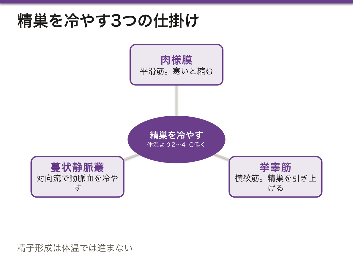
冷却の仕掛けが壊れると、そのまま不妊の原因になる。
精索静脈瘤は蔓状静脈叢の拡張で、男性不妊の原因として最も多い(不妊男性の約 40%)。左に約 90% が起きる——左精巣静脈が左腎静脈へ直角に注ぎ、右のように下大静脈へ斜めに入らないため、静水圧がそのまま伝わるからである。熱交換が働かず精巣温度が上がり、精子形成が落ちる。
⚠️ 急に出た右側の精索静脈瘤、あるいは臥位で消えない静脈瘤は、腹腔内の腫瘤による静脈の圧迫を疑う。腎細胞癌が腎静脈へ進展した像が典型で、精索静脈瘤が最初の徴候になることがある。
停留精巣も同じ理屈で問題になる。腹腔内に留まった精巣は温度が高く、精子形成が不可逆に障害されるうえに悪性化の危険が上がる。手術で降ろしても癌の危険は下がりきらないが、触れる位置に置くこと自体が早期発見の価値を持つ。
男性不妊の診察で立位・Valsalva をさせて陰嚢を触るのは、臥位では静脈瘤が消えて分からなくなるからである。
English version
Core
Spermatogenesis does not proceed at body temperature. The testis is held 2–4 °C below it, and three structures combine to do this.
- Dartos muscle (smooth) — contracts in the cold, wrinkling the skin and drawing the testis towards the body; relaxes in the heat to increase the surface area.
- Cremaster muscle (striated) — raises the testis. Stroking the inner thigh raises the testis on that side (cremasteric reflex, L1–L2).
- Pampiniform plexus — the testicular veins wrap around the testicular artery and work as a countercurrent heat exchanger: arterial blood gives up its heat to the venous blood and is cooled before reaching the testis.
Going further
When the cooling apparatus breaks, infertility follows directly.
Varicocele is dilatation of the pampiniform plexus and is the commonest cause of male infertility (about 40 per cent of infertile men). About 90 per cent occur on the left — the left testicular vein enters the left renal vein at a right angle rather than obliquely into the inferior vena cava as on the right, so the hydrostatic pressure is transmitted directly. The heat exchange fails, the testicular temperature rises and spermatogenesis falls.
⚠️ A varicocele that appears suddenly on the right, or one that does not disappear on lying down, should raise suspicion of an intra-abdominal mass compressing the vein. The typical picture is renal cell carcinoma extending into the renal vein, and the varicocele can be the first sign.
Undescended testis is a problem for the same reason. A testis remaining in the abdomen is hotter, spermatogenesis is irreversibly damaged and the risk of malignancy rises. Bringing it down surgically does not fully reduce the risk of cancer, but placing it where it can be palpated has value in itself for early detection.
In the clinic
In examining a man for infertility the scrotum is palpated standing and with a Valsalva manoeuvre, because lying down the varicocele disappears and is missed.
前立腺癌の悪性度は、細胞ではなく腺の形で決まる
多くの癌は細胞の異型で悪性度を判定するが、前立腺癌だけは腺の構造だけで決める——細胞の顔つき(核の大小不同など)は評価に入らない。
- Gleason パターン 3——大きさは不揃いだが、個々の腺は独立した輪郭を保っている
- パターン 4——腺が融合する・篩状になる・輪郭が崩れる
- パターン 5——腺構造がほぼ無い。索状・充実性・面疱壊死
1 つの腫瘍に複数のパターンが混在するので、最も多いものと、2 番目に多いもの(生検では最も悪いもの)を足す。3+4=7 と 4+3=7 は、同じ 7 でも予後が違う——前に書くほうが優勢なパターンだからである。
この「足し算」が分かりにくいため、現在は 1〜5 のグループ分類が併記される。
| グループ 1 | Gleason 6(3+3) | 転移することがほぼ無い。監視療法の対象になりうる |
| グループ 2 | 3+4=7 | 同じ 7 でも扱いが分かれる |
| グループ 3 | 4+3=7 | |
| グループ 4 | 8 | 積極的な治療の対象 |
| グループ 5 | 9〜10 |
グループ 1 に「癌」という名前を付けるべきかという議論があるほど、振る舞いが穏やかである。見つけても治療しないという選択(監視療法)が標準の1つになっており、過剰診断・過剰治療の代表例として PSA 検診の議論と結びついている。
診断の助けになる組織の特徴も、構造から来ている。
- 基底細胞が消える——正常な前立腺の腺には基底細胞の層があるが、癌では失われる。p63・高分子量サイトケラチンで染めて確認する
- 核小体が目立つ——補助的な所見
- 神経周囲浸潤——前立腺癌が被膜外へ出る主要な経路
⚠️ 前立腺癌は辺縁域に多く、肥大症は移行域に起きる。だから肥大症の手術で削った組織には、癌が含まれないことがある。癌を狙うなら、直腸から辺縁域に針を刺す——組織の解剖が、生検の方法を決めている。
前立腺癌の告知では、グループ 1 とグループ 5 では別の病気に近いことを伝える。「癌」という一語だけでは、患者の受け取り方を誤らせる。
English version
Core
Most cancers are graded by the atypia of the cells, but prostate cancer alone is graded by the architecture of the glands — the appearance of the cells (variation in nuclear size and so on) does not enter the assessment.
Gleason patterns
- Pattern 3 — irregular in size, but each gland retains an independent outline.
- Pattern 4 — glands fuse, become cribriform, or lose their outline.
- Pattern 5 — essentially no glandular structure. Cords, sheets, comedonecrosis.
Several patterns coexist in one tumour, so the commonest and the second commonest are added (in a biopsy, the commonest and the worst).
3+4=7 and 4+3=7 have different prognoses although both are 7 — because the pattern written first is the predominant one.
Going further
Because that addition is confusing, a grade group of 1–5 is now reported alongside.
| Group | Gleason | Meaning |
|---|---|---|
| 1 | 6 (3+3) | Essentially never metastasises. May be managed by active surveillance |
| 2 | 3+4=7 | The same 7, managed differently |
| 3 | 4+3=7 | |
| 4 | 8 | For active treatment |
| 5 | 9–10 |
Group 1 behaves so quietly that there is debate over whether it should be given the name "cancer" at all. Finding it and not treating it (active surveillance) is one of the standards of care, and it is bound up with the argument over PSA screening as the classic example of overdiagnosis and overtreatment.
The histological features that help the diagnosis also come from the architecture.
- The basal cells disappear — a normal prostatic gland has a layer of basal cells, lost in cancer. Confirm with p63 and high molecular weight cytokeratin.
- Prominent nucleoli — a supporting finding.
- Perineural invasion — the main route by which prostate cancer leaves the capsule.
⚠️ Prostate cancer is commonest in the peripheral zone; hyperplasia occurs in the transition zone. So tissue removed at an operation for hyperplasia may contain no cancer. To target the cancer, the needle is passed into the peripheral zone from the rectum — the anatomy of the tissue deciding the method of biopsy.
In the clinic
When telling a patient he has prostate cancer, convey that group 1 and group 5 are nearer to being different diseases. The single word "cancer" alone will make the patient take it wrongly.
まとめ
| 精細管 — Sertoli 細胞と血液精巣関門 | 精子を作る管。壁を作るのは2種の細胞。 |
|---|---|
| 精子形成 — 74日かけて、細胞質を捨てていく過程 | 精細管の基底側から内腔側へ向かって、順に進む。 |
| Leydig 細胞 — 精細管の外側で、ホルモンを作っている | 精細管と精細管のあいだ(間質)にある。ステロイド産生細胞の典型的な姿をしている。 |
| 精巣生検 — 造精機能を、組織で段階に分ける | 無精子症では、作れていないのか、作れているのに出てこられないのかを分ける必要がある。 |
| 精巣上体と精管 — 立体線毛と、厚い筋 | 精巣輸出管——線毛細胞と無線毛細胞が交互に並び、内腔が波打って見える(鋸歯状)。 |
| 前立腺と精嚢 — 域で分かれる病気 | 前立腺——30〜50個の管状胞状腺が、線維筋性間質に埋まる。 |
| 精子 — 3つの部分に、必要なものだけを積んでいる | 全長約60 μm。細胞質をほぼ捨てて軽くしてある。 |
| 陰茎の勃起組織 — 血を溜める海綿と、包む膜 | 3本の海綿体からなる——陰茎海綿体(左右2本)と、尿道海綿体(1本、尿道を含む)。 |
| 精液 — 3つの腺が、別の目的で液を足している | 精子そのものは体積の 1% 以下。残りは付属腺の分泌液になる。 |
| 受精能獲得 — 精子は、女性生殖路で最後の仕上げを受ける | 精巣上体を出た精子は、動けるようになってはいるがまだ受精できない。 |
| 陰嚢と精索 — 精巣を冷やすためだけに、これだけの仕掛けがある | 精子形成は体温では進まない。精巣は体温より 2〜4 ℃ 低く保たれている。 |
| 前立腺癌の悪性度は、細胞ではなく腺の形で決まる | 多くの癌は細胞の異型で悪性度を判定するが、前立腺癌だけは腺の構造だけで決める——細胞の顔つき(核の大小不同など)は評価に入らない。 |
このノートは医学生が授業の内容を自分の言葉でまとめたもので、診療の判断に使うものではありません。誤りに気づいたらお知らせください。