生化学の最初の実験は、ほとんどが2つの色の反応だけで組み立てられている。Fehling 反応(青から赤褐色へ)は「還元糖ができたか」を、ヨウ素デンプン反応(青紫)は「長いデンプンがまだ残っているか」を教える。前者は生成物が現れたことを、後者は基質が消えたことを見ている。この2つを組み合わせるだけで、糖の構造の違い(還元糖と非還元糖)、酵素が温度と pH にどう反応するか、酵素がどの結合を見分けているか、消化液のどこにどの酵素があるかまで、試験管の色から読み取れる。
この章では、それぞれの実験を何を混ぜたか → 何が見えたか → なぜそうなるか → 結果から何が言えるかの順に説明する。いちばん大事なのは対照(コントロール)の読み方である。「色が変わらなかった試験管」が何を証明しているのかを言えれば、その実験の意味はほぼつかめている。糖の構造、アミラーゼの機構、温度と pH の理論そのものは糖と酵素の章にあるので、ここでは実験の組み立てと結果の読み方に集中する。
- Fehling 反応 — 銅(II)が糖から電子を受け取り、赤い沈殿になる
- 還元糖と非還元糖 — 6本の試験管で、自由なアノマー炭素を探す
- ヨウ素デンプン反応 — 化学反応ではなく、らせんの中に並ぶヨウ素
- 温度と唾液アミラーゼ — 冷やした酵素は戻り、煮た酵素は戻らない
- pH と唾液アミラーゼ — 緩衝液を変えて、至適 pH を探す
- 酵素の特異性 — 6つの基質と2つの酵素、12本の試験管
- 消化液のグリコシダーゼと pH — 胃液だけがデンプンを分解しない理由
Fehling 反応 — 銅(II)が糖から電子を受け取り、赤い沈殿になる
Fehling 試薬は、使う直前に2つの液を混ぜて作る。Fehling I は青い硫酸銅(II)の水溶液、Fehling II は無色の酒石酸ナトリウムカリウム(ロッシェル塩)を濃い水酸化ナトリウムに溶かした液である。強アルカリの中では Cu2+ はそのままだと水酸化銅(II)として沈んでしまう。酒石酸イオン2つが銅を両側から抱え込むと濃い青の錯体になり、アルカリの中でも溶けたまま反応できる。混ぜた液は日がたつと錯体が壊れてくるので、2液に分けて保存する。
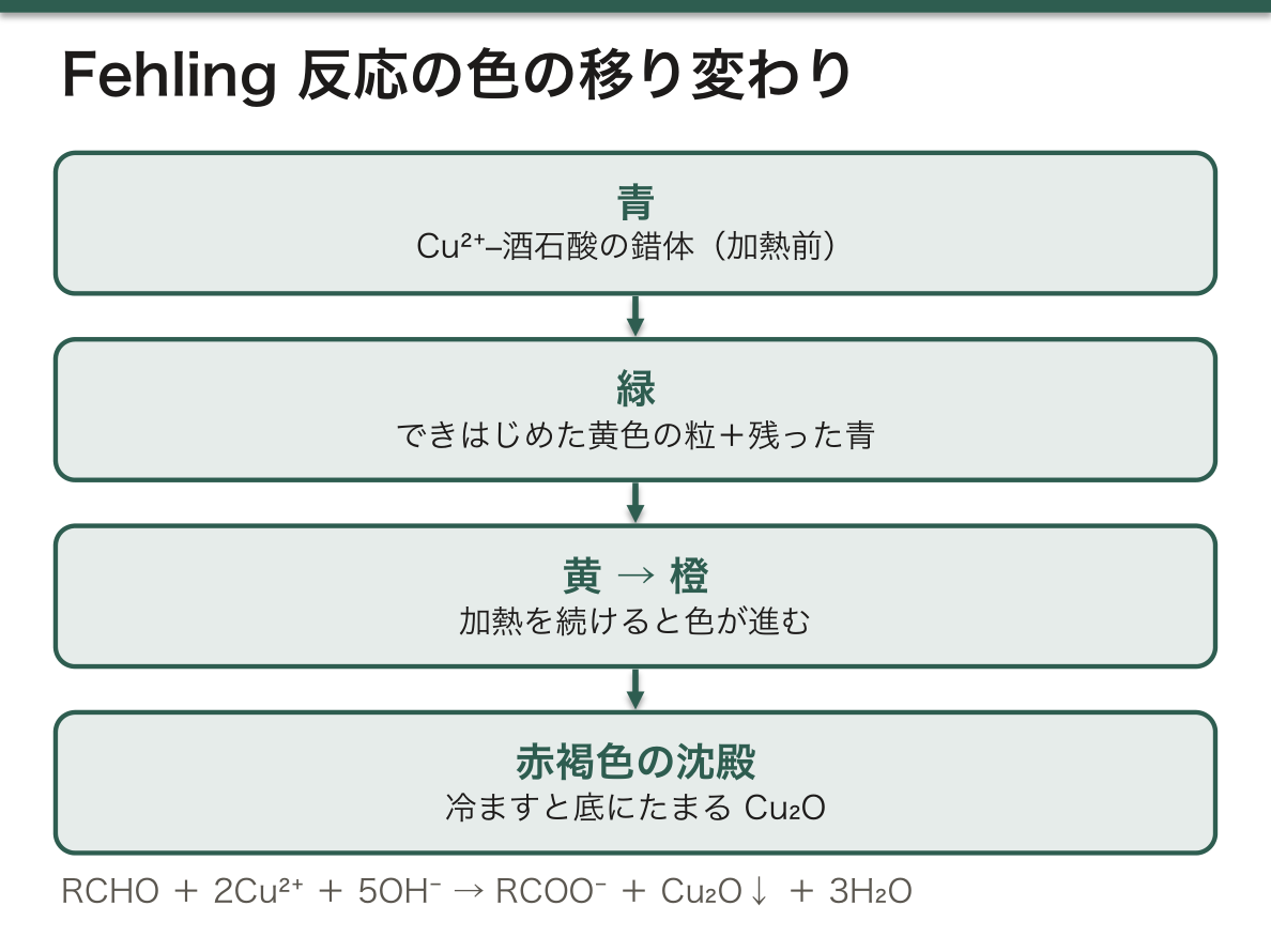
試料と2液を同じ量ずつ混ぜて静かに加熱する。還元糖があれば、液は青 → 緑 → 黄 → 橙と変わり、冷ますと底に赤褐色の酸化銅(I)(Cu2O)がたまる。途中の緑は、できはじめた黄色の粒と残っている青が重なって見える色で、別の物質ではない。
この反応で酸化剤は Cu2+ の錯体で、Cu+ に還元される。還元剤は糖で、鎖状になったときのアルデヒド基がカルボキシ基(アルカリ中ではカルボキシラート)に酸化される。ブドウ糖ならグルコン酸になる。まとめると RCHO + 2Cu2+ + 5OH− → RCOO− + Cu2O↓ + 3H2O である。
還元糖を見る試薬はほかにもあり、どれも「金属イオンを糖が還元する」という同じ考え方でできている。違いはどの金属を、どのくらいのアルカリ(または酸)で使うかである。
| 試薬 | 中身 | 陽性のしるし | 特徴 |
|---|---|---|---|
| Fehling | Cu2++酒石酸、強アルカリ | 赤褐色の Cu2O | 2液を直前に混ぜる |
| Benedict | Cu2++クエン酸、炭酸ナトリウムで弱アルカリ | 緑〜橙〜赤褐色 | 1液のまま安定。量に応じて色が段階的に変わる |
| Barfoed | 酢酸銅(II)、弱酸性 | 赤い沈殿 | 酸性では銅が還元されにくいので、反応の速い単糖だけが短時間で陽性になる。単糖と二糖を区別できる |
| Tollens | アンモニア性硝酸銀 [Ag(NH3)2]+ | ガラスの内側に銀の鏡 | Ag+ が金属の銀に還元される |
Fehling 反応はあるかないかを見る定性反応で、量を測るのには向かない。熱い強アルカリの中では糖そのものが分解されて、糖1分子あたりに還元する銅の数が一定にならないからである。
尿糖は、かつてこの銅の還元(Benedict 試薬や、それを錠剤にしたもの)で調べていた。銅還元法は還元糖ならどれにでも反応するので、ブドウ糖のほかにガラクトース・果糖・乳糖でも陽性になり、ビタミン C のような糖以外の還元物質でも偽陽性が出る。いまの尿試験紙はグルコースオキシダーゼという酵素を使い、ブドウ糖だけに反応する。この違いは逆に役に立ち、「試験紙は陰性なのに銅還元法は陽性」なら、尿に出ているのはブドウ糖以外の還元糖だと分かる。新生児のガラクトース血症を疑う手がかりとして使われてきたのはこのためである。
English version
Core
Fehling’s reagent is made just before use by mixing two solutions. Fehling I is blue copper(II) sulphate in water. Fehling II is colourless potassium sodium tartrate (Rochelle salt) in strong sodium hydroxide. In strong alkali, free Cu2+ would fall out as copper(II) hydroxide. Two tartrate ions hold the copper from both sides and form a deep blue complex, which stays dissolved and can react in alkali. The mixed reagent slowly breaks down, so the two parts are stored separately.
Equal volumes of sample and the two solutions are mixed and heated gently. If a reducing sugar is present, the liquid changes blue → green → yellow → orange, and on cooling a brick-red precipitate of copper(I) oxide (Cu2O) settles. The green is just yellow particles seen together with the remaining blue; it is not a different substance.
In this reaction the oxidising agent is the Cu2+ complex, which is reduced to Cu+. The reducing agent is the sugar: the aldehyde group of its open-chain form is oxidised to a carboxyl group (a carboxylate in alkali). Glucose becomes gluconic acid. Overall: RCHO + 2Cu2+ + 5OH− → RCOO− + Cu2O↓ + 3H2O.
Going further
Other reagents for reducing sugars use the same idea: the sugar reduces a metal ion. They differ in which metal is used and how alkaline (or acidic) the solution is.
| Reagent | Contents | Positive sign | Feature |
|---|---|---|---|
| Fehling | Cu2+ + tartrate, strong alkali | Brick-red Cu2O | Two solutions mixed just before use |
| Benedict | Cu2+ + citrate, mildly alkaline (sodium carbonate) | Green to orange to brick-red | Stable as one solution; colour changes in steps with the amount of sugar |
| Barfoed | Copper(II) acetate, weakly acidic | Red precipitate | Copper is harder to reduce in acid, so only fast-reacting monosaccharides turn positive in a short time; separates mono- from disaccharides |
| Tollens | Ammoniacal silver nitrate [Ag(NH3)2]+ | Silver mirror on the glass | Ag+ is reduced to metallic silver |
Fehling’s test is qualitative (present or absent); it is not good for measuring amounts. In hot strong alkali the sugar itself breaks down, so the number of copper ions reduced per sugar molecule is not fixed.
In the clinic
Urine sugar was once tested by this copper reduction (Benedict’s reagent, or a tablet version of it). Copper reduction reacts with any reducing sugar: glucose, but also galactose, fructose and lactose, and even non-sugar reducing substances such as vitamin C. Today’s urine strips use the enzyme glucose oxidase and react only with glucose. This difference is useful: if the strip is negative but copper reduction is positive, the urine contains a reducing sugar other than glucose. That is why it was used as a clue to galactosaemia in newborns.
還元糖と非還元糖 — 6本の試験管で、自由なアノマー炭素を探す
同じ Fehling 反応を6種類の糖の液で行うと、次のように分かれる。
| 試験管の中身 | 結果 | 理由 |
|---|---|---|
| ブドウ糖 | 陽性(赤褐色) | アルドース。C1 のアノマー炭素が自由で、環が開くとアルデヒド基が出る |
| 果糖 | 陽性 | ケトースだが、強アルカリの中でアルドースに変わる(下の「もう一歩」) |
| スクロース | 陰性(青のまま) | ブドウ糖の C1 と果糖の C2 が両方とも結合に使われている(α1↔β2)。どちらの環も開けない |
| 加水分解したスクロース | 陽性 | 結合が切れてブドウ糖と果糖になり、両方のアノマー炭素が自由になる |
| デンプン | 陰性 | 何千ものブドウ糖に対して還元末端は1分子に1つだけ。薄すぎて見えない |
| 加水分解したデンプン | 陽性 | マルトースやブドウ糖がたくさんでき、還元末端の数が一気に増える |
表をまとめる規則は1つである。還元性を決めるのは、グリコシド結合に使われていない「自由なアノマー炭素」(ヘミアセタール)が残っているかどうか。自由なアノマー炭素があれば、水の中で環が開いてカルボニル基が現れ、それが銅に電子を渡す。二糖でもマルトースや乳糖は片方のアノマー炭素が空いているので還元糖、スクロースやトレハロースは両方をふさいでいるので非還元糖になる。
この6本でいちばん大事なのは「加水分解の前は陰性、後は陽性」という組み合わせである。これは、試験管の中でグリコシド結合が切れたことの証拠になる。酵素の特異性や消化液の実験は、すべてこの読み方の上に組み立てられている。
なぜケトースの果糖が陽性になるのか。果糖のカルボニル基は C2 のケトンで、アルデヒドではない。しかし Fehling 試薬のような強アルカリの中では、C1 と C2 の間で水素が移り、エンジオール(C=C の両端に OH が付いた形)を経てブドウ糖やマンノースに組み替わる(ケト–エノール互変異性)。できたアルドースがアルデヒド基で銅を還元する。「アルデヒド基をもつものが還元糖」とだけ覚えると果糖を非還元糖と取り違えるが、日本の教科書でも果糖は還元糖として扱う。
この違いは食品の見分けにも使える。果糖ブドウ糖液糖で甘くした飲み物はそのまま Fehling 陽性、スクロースだけで甘くした飲み物は陰性で、酸を加えて煮てから試すと陽性に変わる。ただし酸性の飲み物の中ではスクロースが保存中に少しずつ加水分解されるので、古い飲み物では弱い陽性が出ることがある。比べるときは、作りたてのスクロース溶液を一緒に試して基準にする。ソーセージにつなぎのデンプンが入っているかは、ゆでた汁にヨウ素液を落とせば分かる。
血液の中の糖はほぼすべてブドウ糖で、いまは銅の還元ではなく酵素法(グルコースオキシダーゼやヘキソキナーゼ)で特異的に測る。糖尿病の診断には空腹時血糖 126 mg/dL(7.0 mmol/L)以上や HbA1c 6.5% 以上といった基準を使う。HbA1c そのものも「還元糖」の性質から生まれる。鎖状になったブドウ糖のアルデヒド基が、ヘモグロビンの β 鎖の N 末端のアミノ基と酵素なしで結合する(糖化)。Fehling 反応で銅に電子を渡していたのと同じ、開いたカルボニル基の反応しやすさが、体の中では蛋白を少しずつ変えている。
English version
Core
Running Fehling’s test on six sugar solutions gives this pattern.
| Tube contents | Result | Reason |
|---|---|---|
| Glucose | Positive (brick-red) | An aldose. The anomeric carbon C1 is free; when the ring opens, an aldehyde group appears |
| Fructose | Positive | A ketose, but it turns into an aldose in strong alkali (see below) |
| Sucrose | Negative (stays blue) | C1 of glucose and C2 of fructose are both used in the bond (α1↔β2). Neither ring can open |
| Hydrolysed sucrose | Positive | The bond is cut into glucose and fructose; both anomeric carbons become free |
| Starch | Negative | Only one reducing end per molecule of thousands of glucose units; too dilute to see |
| Hydrolysed starch | Positive | Many maltose and glucose molecules form, so the number of reducing ends rises sharply |
One rule explains the table. Reducing power depends on whether a free anomeric carbon (a hemiacetal) remains, not used in a glycosidic bond. If it is free, the ring can open in water, a carbonyl group appears, and it gives electrons to copper. Among disaccharides, maltose and lactose have one free anomeric carbon and are reducing; sucrose and trehalose block both and are non-reducing.
The key point of these six tubes is the pair “negative before hydrolysis, positive after”. It proves that a glycosidic bond was cut in the tube. The experiments on enzyme specificity and digestive juices are all built on this way of reading.
Going further
Why is fructose, a ketose, positive? Its carbonyl is a ketone at C2, not an aldehyde. But in strong alkali such as Fehling’s reagent, a hydrogen moves between C1 and C2, and through an enediol (OH on both ends of a C=C) fructose rearranges into glucose and mannose (keto–enol tautomerism). The aldose formed then reduces copper with its aldehyde group. If you learn only “sugars with an aldehyde are reducing”, you will wrongly call fructose non-reducing; Japanese textbooks also treat fructose as a reducing sugar.
This difference can identify foods. A drink sweetened with high-fructose corn syrup is Fehling-positive as it is; a drink sweetened only with sucrose is negative, and turns positive after boiling with acid. But in an acidic drink sucrose is slowly hydrolysed during storage, so an old drink may give a weak positive. When comparing, test a freshly made sucrose solution as a reference. Whether a sausage contains starch as a filler can be checked by adding iodine solution to the water it was boiled in.
In the clinic
Almost all sugar in the blood is glucose, and today it is measured specifically by enzymatic methods (glucose oxidase or hexokinase), not by copper reduction. Diabetes is diagnosed with criteria such as fasting glucose ≥ 126 mg/dL (7.0 mmol/L) or HbA1c ≥ 6.5%. HbA1c itself comes from the “reducing” property. The aldehyde group of open-chain glucose binds, without any enzyme, to the amino group at the N-terminus of the haemoglobin β chain (glycation). The same reactivity of the open carbonyl group that gave electrons to copper in the tube slowly changes proteins in the body.
ヨウ素デンプン反応 — 化学反応ではなく、らせんの中に並ぶヨウ素Amylum
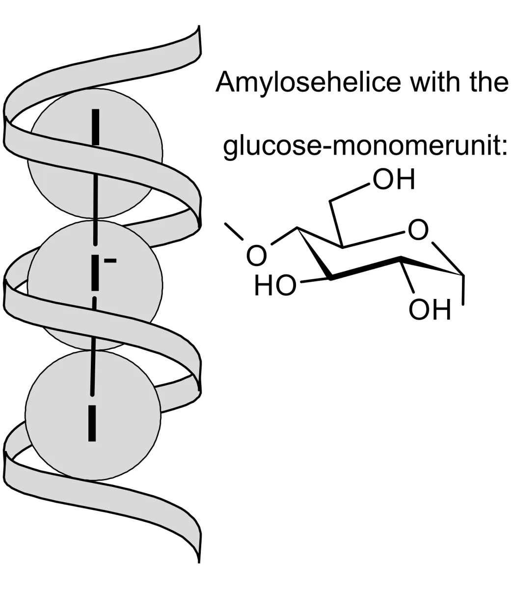
使うのはルゴール液(ヨウ素をヨウ化カリウム水溶液に溶かした褐色の液)である。ヨウ素 I2 は水にほとんど溶けないが、ヨウ化物イオンと結びついて三ヨウ化物イオン I3− になると溶ける。デンプンの液に数滴落とすと、加熱しなくてもすぐに濃い青紫(青黒)になる。
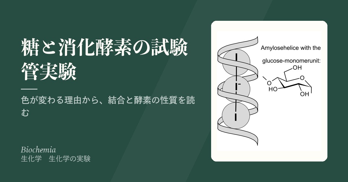
色のもとはアミロースである。α(1→4) 結合だけでつながったブドウ糖の長い鎖は、1巻きにおよそ6残基のらせんを作り、その内側は糖の −OH が外を向いているため水を嫌う細い筒になっている。ヨウ素はこの筒の中に一列に並んで入り込み(I3− や I5− の鎖)、橙〜赤の光を強く吸うので、残った光が青く見える。共有結合はできていないので、これは「反応」というより包み込まれてできる複合体である。
デンプンを加水分解した液では、鎖が短く切られてらせんが保てないので、ヨウ素は入る場所を失う。色はルゴール液そのものの黄褐色のままで、これが陰性である。枝分かれの多いアミロペクチンは枝の1本1本が短いので赤紫、さらに枝が短く密なグリコーゲンは赤褐色になる。つまりヨウ素の色は、まっすぐな鎖がどれだけ長いかを映している。
結果を読み違えやすい点が3つある。
1つ目は温度。ヨウ素デンプンの青は温めると消え、冷ますと戻る。熱でらせんがゆるみ、ヨウ素が筒から出てしまうからである。だからこの反応は室温で読む。「加熱は要らない」は手間の話ではなく、加熱すると読めなくなるという意味である。
2つ目はアルカリ。強いアルカリ性ではヨウ素そのものが OH− と反応して(I2 + 2OH− → I− + IO− + H2O)色を出せなくなる。pH の高い試験管で青が弱いとき、それが酵素の働きなのかヨウ素が消費されただけなのかを区別する必要がある。
3つ目は何を見ている反応か。ヨウ素で陰性なのは「長い鎖が無い」という意味で、「糖が無い」という意味ではない。加水分解したデンプンはヨウ素で陰性、Fehling で陽性になる。ヨウ素は基質が消えたことを、Fehling は生成物ができたことを見ており、2つを並べると酵素の働きを両側から確かめられる。
ヨウ素がグリコーゲンを染める性質は内視鏡で使われている。食道の正常な扁平上皮は細胞にグリコーゲンをためているので、ルゴール液を散布すると茶褐色に染まる。食道癌や異形成の部分はグリコーゲンが少なく、染まらない領域(ヨード不染帯)として浮かび上がる。日本では食道癌の拾い上げの基本手技である。皮膚科・神経内科では、ヨウ素を塗った上にデンプンをまいて汗の出る場所を青黒く浮かび上がらせる発汗試験(Minor 法)にも同じ反応を使う。
English version
Core
The reagent is Lugol’s solution (iodine dissolved in potassium iodide solution; brown). Iodine I2 barely dissolves in water, but it dissolves once it joins an iodide ion to form triiodide I3−. A few drops added to starch solution give an immediate deep blue-black colour, with no heating.
The colour comes from amylose. A long chain of glucose joined only by α(1→4) bonds forms a helix of about six residues per turn. The sugar −OH groups face outward, so the inside is a narrow water-repelling tube. Iodine lines up inside this tube (as chains of I3− and I5−) and strongly absorbs orange-red light, so the light left over looks blue. No covalent bond forms, so this is less a “reaction” than an inclusion complex.
In hydrolysed starch the chains are cut short and cannot hold a helix, so iodine has nowhere to go. The colour stays the yellow-brown of Lugol’s solution itself: this is negative. Amylopectin, with many short branches, gives red-violet; glycogen, with even shorter, denser branches, gives red-brown. So the iodine colour shows how long the straight chains are.
Going further
There are three common mistakes in reading the result.
First, temperature. The starch–iodine blue disappears on warming and returns on cooling, because heat loosens the helix and iodine leaves the tube. So the test is read at room temperature. “No heating required” is not about saving time; it means heating makes the test unreadable.
Second, alkali. In strongly alkaline solution iodine itself reacts with OH− (I2 + 2OH− → I− + IO− + H2O) and can no longer give colour. If the blue is weak in a high-pH tube, you must decide whether the enzyme worked or the iodine was simply used up.
Third, what the test shows. A negative iodine test means “no long chains”, not “no sugar”. Hydrolysed starch is iodine-negative but Fehling-positive. Iodine sees the substrate disappearing and Fehling sees the product appearing; using both checks the enzyme from two sides.
In the clinic
The way iodine stains glycogen is used in endoscopy. Normal squamous epithelium of the oesophagus stores glycogen, so sprayed Lugol’s solution stains it brown. Cancer and dysplasia contain little glycogen and appear as unstained areas. In Japan this is a basic method for finding oesophageal cancer. In dermatology and neurology, the same reaction is used in the starch–iodine sweat test (Minor’s test): iodine is painted on the skin, starch is dusted on, and sweating areas turn blue-black.
温度と唾液アミラーゼ — 冷やした酵素は戻り、煮た酵素は戻らない
酵素の源は自分の唾液で、中の α-アミラーゼがデンプンを切る。唾液は水で10倍に薄めて使う(唾液 1 mL + 水 9 mL)。薄めないと酵素が強すぎて、どの条件でも数分でデンプンが消えてしまい、差が見えないからである。
薄めた唾液 0.5 mL を3本の試験管に分け、① 38 ℃ の恒温器、② いったん沸騰させてから室温で冷ます、③ 氷水、に置く。10分たってから 1% デンプン液 0.5 mL を加え、それぞれの温度のまま10分置き、ヨウ素液を2滴落とす。先に酵素だけを置いておくのは、基質が入った瞬間から反応が始まるので、その時点で酵素がもう目的の温度になっていなければ比べられないからである。
| 試験管 | ヨウ素の色 | デンプン | 活性 | さらに室温で20分後 |
|---|---|---|---|---|
| 38 ℃ | 黄褐色(青くならない) | 無い | 高い | 変わらない(もう分解し終わっている) |
| 煮沸 → 冷却 | 濃い青 | 残っている | 無い | 変わらない(青のまま) |
| 氷水 | 青(煮沸よりやや淡い) | 残っている | 低い | 青が薄れていく |
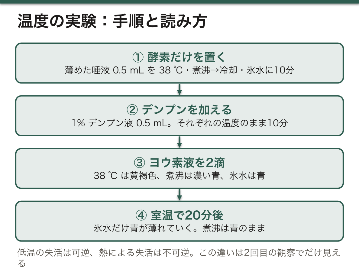
なぜ氷の試験管だけが後から変わるのか。低温では分子の動きが遅く、酵素と基質がぶつかる回数が減っているだけで、酵素の立体構造は壊れていない。室温に戻れば動きが戻り、残っていたデンプンを分解しはじめるので、青が薄れる。煮沸した酵素では、立体構造を保っていた水素結合・イオン結合・疎水性相互作用が切れてポリペプチド鎖がほどけ、互いに絡み合って固まる。冷ましても元の形には戻らない。低温は可逆、高温は不可逆という違いが、2回目の観察でだけ見える仕組みになっている。
2本の「失敗した」試験管にも意味がある。煮沸した試験管は陰性対照で、「働く酵素が無ければ、同じ時間・同じ量のデンプンはちゃんと青く残る」ことを示している。これがあるから、38 ℃ の試験管で青が消えたのは時間がたったせいではなく酵素のせいだと言える。氷の試験管は、同じ1本の試験管で温度だけを変えた前後比較になっていて、「酵素が壊れていたのではなく、遅くなっていただけ」を証明している。
検査室ではこの2つの性質を使い分けている。酵素の活性を測る血液や組織の検体は、冷やして運び、長く置くなら凍らせる。低温による失活は可逆なので、温め直せば測れるからである。逆に、熱による失活は意図的にも使われる。血清を 56 ℃ で30分温めると補体(熱に弱い蛋白の一群)だけが働かなくなるので、補体の影響を除きたい血清反応の前処理として行われる(非働化)。
English version
Core
The enzyme source is your own saliva, whose α-amylase cuts starch. Saliva is diluted ten times with water (1 mL saliva + 9 mL water). Undiluted saliva is so active that starch vanishes within minutes in every tube, and no difference can be seen.
0.5 mL of diluted saliva goes into each of three tubes: (1) a 38 °C thermostat, (2) boiled and then cooled at room temperature, (3) an ice bath. After 10 minutes, 0.5 mL of 1% starch is added, each tube stays at its temperature for 10 minutes, and 2 drops of iodine are added. The enzyme is placed first because the reaction starts the moment the substrate is added; if the enzyme is not yet at the test temperature, the tubes cannot be compared.
| Tube | Iodine colour | Starch | Activity | After 20 more minutes at room temperature |
|---|---|---|---|---|
| 38 °C | Yellow-brown (no blue) | Absent | High | No change (already fully digested) |
| Boiled → cooled | Deep blue | Present | None | No change (stays blue) |
| Ice bath | Blue (slightly paler than boiled) | Present | Low | The blue fades |
Going further
Why does only the ice tube change later? At low temperature molecules move slowly and enzyme and substrate meet less often, but the enzyme’s 3D structure is not damaged. Back at room temperature the movement returns, the enzyme starts digesting the remaining starch, and the blue fades. In the boiled enzyme, the hydrogen bonds, ionic bonds and hydrophobic interactions that held its shape are broken; the chain unfolds, tangles with others and clumps. Cooling does not bring the shape back. The difference — cold is reversible, heat is irreversible — becomes visible only in the second reading.
The two “failed” tubes also have a job. The boiled tube is the negative control: it shows that without working enzyme, the same amount of starch stays blue for the same time. Because of it, we can say that the blue disappeared at 38 °C because of the enzyme, not because time passed. The ice tube is a before-and-after comparison in the same tube with only the temperature changed, and proves that the enzyme was only slowed, not broken.
In the clinic
Clinical laboratories use both properties. Blood or tissue samples for enzyme activity are kept cold during transport and frozen for long storage, because loss of activity from cold is reversible and the enzyme works again when warmed. Heat inactivation is also used on purpose. Heating serum at 56 °C for 30 minutes stops only complement (a group of heat-sensitive proteins), so it is used before some serological tests to remove the effect of complement (heat inactivation).
pH と唾液アミラーゼ — 緩衝液を変えて、至適 pH を探す
5本の試験管にそれぞれ 5 mL のクエン酸–リン酸緩衝液を入れ、pH を 3.8・5.8・6.8・7.8・9.8 にそろえる。1% デンプン液 1 mL と薄めた唾液 0.5 mL を加えて 38 ℃ に15分置き、ヨウ素液を2滴落とす。試験管どうしで違うのは pH だけで、酵素・基質・温度・時間は同じにしてある。だから色の差は pH のせいだと言える。
ただの酸や塩基でなく緩衝液を使うのは、反応のあいだ pH を一定に保つためである。緩衝液の量(5 mL)は唾液(0.5 mL)の10倍あり、唾液自身の炭酸水素イオンが pH を動かすこともない。
| pH | ヨウ素の色 | デンプン | 活性 |
|---|---|---|---|
| 3.8 | 濃い青 | 残る | ほぼ無い |
| 5.8 | 青紫 | 一部残る | 低〜中 |
| 6.8 | 黄褐色 | 無い | 最大 |
| 7.8 | 淡い紫〜赤褐色 | 少し残る | 中 |
| 9.8 | 青 | 残る | ほぼ無い |
活性は pH 6.8 付近を頂点とする山なりになり、この至適 pH は唾液そのものの pH とほぼ一致する。
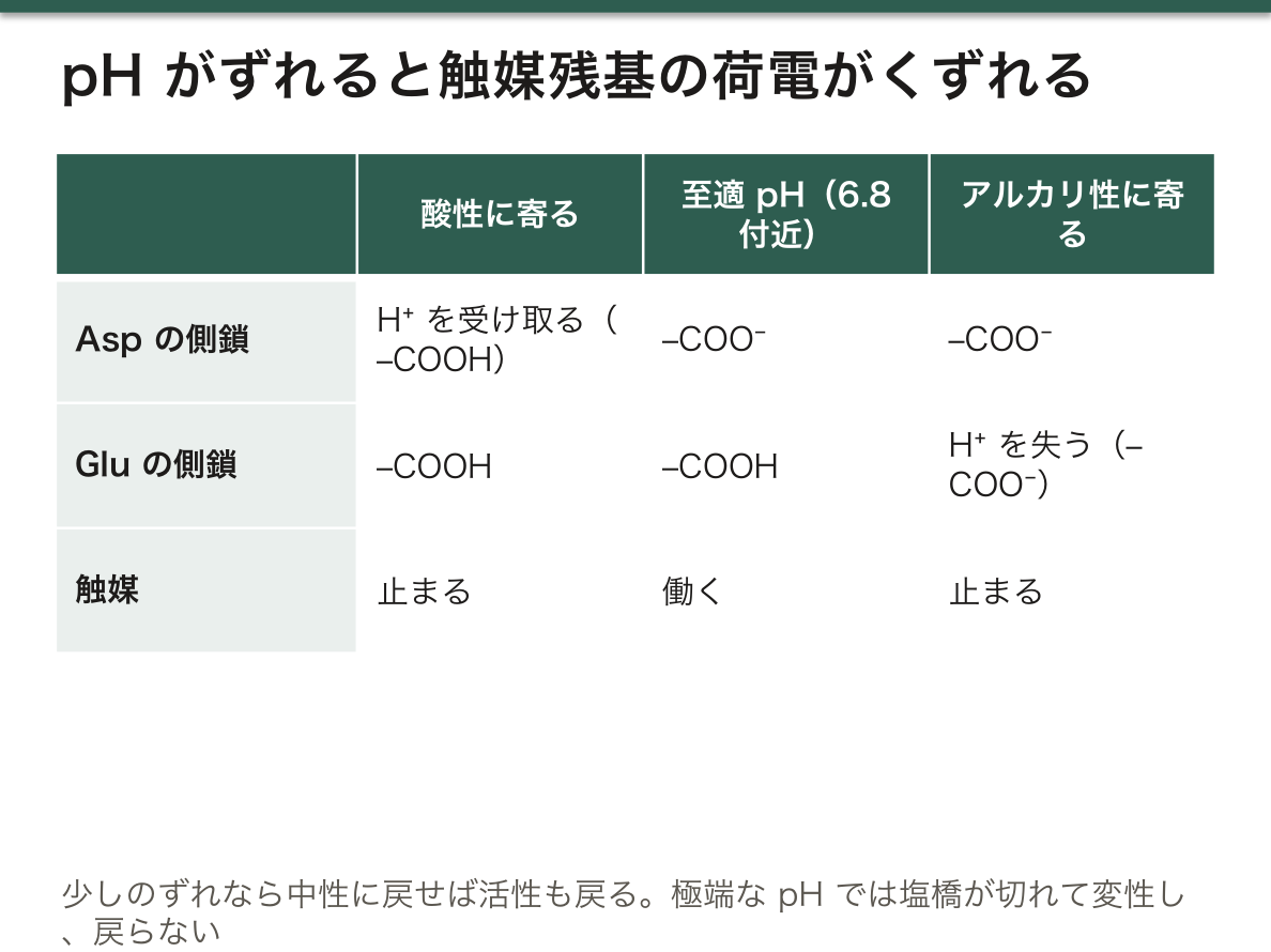
なぜ山なりになるのか。アミラーゼの活性部位では、アスパラギン酸の側鎖が −COO−、グルタミン酸の側鎖が −COOH という逆の荷電状態でそろっていなければならない。酸性に寄るとアスパラギン酸まで H+ を受け取り、アルカリ性に寄るとグルタミン酸が H+ を失う。どちらの側でも触媒が止まる。少しずれただけなら中性に戻せば活性も戻るが、極端な pH では塩橋が切れて変性し、戻らなくなる。
読むときの注意が2つある。1つは pH 9.8 の試験管で、強いアルカリはヨウ素そのものを消費するので、青が実際より弱く出ることがある。色がはっきりしなければ、ヨウ素液を1滴足して青が出るかを見るとよい。もう1つは塩化物イオンで、α-アミラーゼは Cl− があると活性が上がる。どの試験管にも同じ唾液を同じ量入れているので比較には影響しないが、酵素の「本当の」活性の高さは塩の量でも変わる。
English version
Core
Five tubes each receive 5 mL of citrate–phosphate buffer at pH 3.8, 5.8, 6.8, 7.8 and 9.8. 1 mL of 1% starch and 0.5 mL of diluted saliva are added, the tubes stay at 38 °C for 15 minutes, and 2 drops of iodine are added. Only the pH differs between tubes; enzyme, substrate, temperature and time are the same. So any colour difference can be put down to pH.
A buffer is used, not plain acid or base, to keep the pH constant during the reaction. There is ten times more buffer (5 mL) than saliva (0.5 mL), so the saliva’s own bicarbonate cannot shift the pH.
| pH | Iodine colour | Starch | Activity |
|---|---|---|---|
| 3.8 | Deep blue | Remains | Almost none |
| 5.8 | Blue-violet | Partly remains | Low–medium |
| 6.8 | Yellow-brown | Absent | Maximum |
| 7.8 | Pale violet to red-brown | A little remains | Medium |
| 9.8 | Blue | Remains | Almost none |
Activity forms a bell-shaped curve with its peak near pH 6.8, which is almost the pH of saliva itself.
Going further
Why a bell shape? In the active site of amylase, an aspartate side chain must be −COO− and a glutamate side chain must be −COOH — opposite charge states at the same time. On the acidic side aspartate also takes H+; on the alkaline side glutamate loses its H+. Either way, catalysis stops. A small shift is reversible when the pH returns to neutral, but at extreme pH salt bridges break, the enzyme denatures and does not recover.
Two cautions when reading. First, the pH 9.8 tube: strong alkali uses up iodine itself, so the blue can look weaker than it really is. If the colour is unclear, add one more drop of iodine and see if blue appears. Second, chloride: α-amylase is more active when Cl− is present. Every tube has the same saliva in the same amount, so the comparison is not affected, but the enzyme’s absolute activity also depends on the salt content.
酵素の特異性 — 6つの基質と2つの酵素、12本の試験管
6種類の基質(1% のデンプン・グリコーゲン・スクロース・トレハロース・イヌリン、水に浸した脱脂綿=セルロース)を、それぞれ唾液 1 mL(α-アミラーゼの源)かパン酵母 0.5 g(スクラーゼの源)と組み合わせ、12本にする。38 ℃ に15分置いたあと Fehling I・II を5滴ずつ加えて加熱し、黄〜橙に変わるかを見る。酵母の試験管は、底に沈んだ酵母ではなく上の澄んだ層を温める。
この設計の要は、6つの基質がどれも、そのままでは Fehling 陰性(非還元糖か、還元末端が薄すぎる多糖)だということである。だから陽性になれば、酵素がその結合を切って還元糖を出したことになる。
| 基質 | 単量体と結合 | アミラーゼ | 酵母スクラーゼ |
|---|---|---|---|
| デンプン | Glc の α(1→4)、枝に α(1→6) | + | − |
| グリコーゲン | Glc の α(1→4)、枝の α(1→6) が多い | + | − |
| スクロース | Glc(α1↔β2)Fru | − | + |
| トレハロース | Glc(α1↔1α)Glc | − | − |
| イヌリン | Fru の β(2→1) の鎖、端に Glc | − | +(弱いことが多い) |
| セルロース | Glc の β(1→4) | − | − |
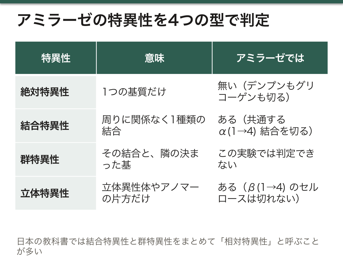
ヨーロッパの教科書では、基質特異性を絶対特異性(1つの基質だけ)・結合特異性(周りに関係なく1種類の結合)・群特異性(その結合と、隣の決まった基)・立体特異性(立体異性体やアノマーの片方だけ)の4つに分けることが多い。日本の教科書では、結合特異性と群特異性をまとめて相対特異性と呼び、絶対・相対・立体の3つで説明することが多い。
アミラーゼはデンプンとグリコーゲンの両方を切ったので絶対特異性ではない。両者に共通する α(1→4) 結合を切っているので結合特異性をもつ。セルロースは同じブドウ糖の同じ 1→4 結合で、違うのは α か β かだけなのに切れなかったので立体特異性もある。切った基質はすべてブドウ糖でできているので、隣の糖まで見分けているか(群特異性)はこの実験では判定できない。
酵母のスクラーゼ(インベルターゼ)はスクロースとイヌリンを切った。共通するのは果糖側の β-フルクトフラノシド結合で、この酵素は果糖の側をつかむβ-フルクトフラノシダーゼである。一方、ヒトの小腸のスクラーゼ(スクラーゼ–イソマルターゼ複合体)はブドウ糖の側をつかむ α-グルコシダーゼで、マルトースも切るがイヌリンは切らない。同じスクロースでも、どちら側の結合を認識しているかで切れる相手が変わる。イヌリンの試験管は陽性が弱く出やすく、ここが陰性なら「絶対特異性かどうか」は決められない。
この12本には酵素を入れない試験管が無い。酵母は自分の中に糖や還元物質をもち、市販のイヌリンには遊離の果糖が混じっていることがある。きちんと言い切るには、「基質+水」と「酵母+水」の2本を対照として足すとよい。
ヒトはセルロースの β(1→4) 結合やイヌリンの β(2→1) 結合を切る酵素をもたないので、これらは食物繊維として大腸まで届き、腸内細菌に発酵されてガスや短鎖脂肪酸になる。イヌリンは消化されず、体内で代謝もされず、糸球体で自由にろ過され、尿細管で再吸収も分泌もされない。このためイヌリンクリアランスは糸球体ろ過量(GFR)を測る基準の方法になっている。一方トレハロースは、ヒトの小腸の刷子縁にトレハラーゼがあるので消化できる。この酵素が欠けている人では、トレハロースを多く含むキノコを食べると下痢やおなかの張りが起こる。
English version
Core
Six substrates (1% starch, glycogen, sucrose, trehalose and inulin, and cotton wool in water = cellulose) are each paired with 1 mL of saliva (source of α-amylase) or 0.5 g of baker’s yeast (source of sucrase), making 12 tubes. After 15 minutes at 38 °C, 5 drops each of Fehling I and II are added, the tubes are heated, and you look for yellow to orange. In yeast tubes, heat the clear upper layer, not the yeast at the bottom.
The key to the design is that all six substrates are Fehling-negative by themselves (non-reducing sugars, or polysaccharides whose reducing ends are too dilute). So a positive result means the enzyme cut the bond and released a reducing sugar.
| Substrate | Monomers and bond | Amylase | Yeast sucrase |
|---|---|---|---|
| Starch | Glc α(1→4), branches α(1→6) | + | − |
| Glycogen | Glc α(1→4), more α(1→6) branches | + | − |
| Sucrose | Glc(α1↔β2)Fru | − | + |
| Trehalose | Glc(α1↔1α)Glc | − | − |
| Inulin | Chain of Fru β(2→1), Glc at one end | − | + (often weak) |
| Cellulose | Glc β(1→4) | − | − |
Going further
European textbooks often divide substrate specificity into four types: absolute (only one substrate), bond (one type of bond, whatever surrounds it), group (that bond plus a particular neighbouring group) and stereo (only one stereoisomer or anomer). Japanese textbooks often combine bond and group specificity as relative specificity and use three types: absolute, relative and stereo.
Amylase cut both starch and glycogen, so it is not absolutely specific. It cuts the α(1→4) bond they share, so it has bond specificity. Cellulose has the same glucose and the same 1→4 bond, differing only in α versus β, and was not cut, so it is also stereospecific. Every substrate it cut is made of glucose, so whether it also recognises the neighbouring sugar (group specificity) cannot be decided from this experiment.
Yeast sucrase (invertase) cut sucrose and inulin. What they share is the β-fructofuranoside bond on the fructose side; this enzyme is a β-fructofuranosidase that grips the fructose side. Human intestinal sucrase (the sucrase–isomaltase complex), by contrast, is an α-glucosidase that grips the glucose side: it also cuts maltose but not inulin. Even for the same sucrose, which side of the bond is recognised decides what else can be cut. The inulin tube is often only weakly positive; if it is negative, you cannot decide whether the enzyme is absolutely specific.
These 12 tubes have no tube without enzyme. Yeast contains its own sugars and reducing substances, and commercial inulin may contain free fructose. To be sure, add two control tubes: “substrate + water” and “yeast + water”.
In the clinic
Humans have no enzyme that cuts the β(1→4) bond of cellulose or the β(2→1) bond of inulin, so these reach the colon as dietary fibre and are fermented by gut bacteria into gas and short-chain fatty acids. Inulin is not digested or metabolised, is freely filtered by the glomerulus, and is neither reabsorbed nor secreted by the tubules. This is why inulin clearance is the reference method for measuring glomerular filtration rate (GFR). Trehalose, on the other hand, can be digested because the human small-intestinal brush border has trehalase. People who lack this enzyme get diarrhoea and bloating after eating mushrooms, which are rich in trehalose.
消化液のグリコシダーゼと pH — 胃液だけがデンプンを分解しない理由
唾液・胃液・膵液(パンクレアチン溶液)・腸液を 1 mL ずつ4本の試験管にとり、1% デンプン液 2 mL を加えて 38 ℃ に20分置く。Fehling I・II を加えて上の層を温め、還元糖ができたかを見る。38 ℃ にするのは、ヒトの消化酵素が体温で最もよく働くからである。同じ消化液に pH 試験紙を浸して pH も読む。
| 消化液 | Fehling | デンプンに働く酵素 | pH | pH を決めるイオンと出どころ |
|---|---|---|---|---|
| 唾液 | + | 唾液 α-アミラーゼ(内側から切る) | 約 6.5〜7 | HCO3−・リン酸イオン(唾液腺) |
| 胃液 | − | 無い | 約 1〜2 | H+(壁細胞の H+/K+-ATPase が汲み出し、Cl− が続いて HCl になる) |
| 膵液 | + | 膵 α-アミラーゼ | 約 8 | HCO3−(膵管の細胞。セクレチンで分泌が増える) |
| 腸液 | + | マルターゼ–グルコアミラーゼ、スクラーゼ–イソマルターゼ(端から切る) | 約 7.5 | HCO3−(膵液・胆汁・十二指腸腺) |
胃液だけが陰性になる理由は、重要な順に2つある。1つ目は、胃液にはグリコシド結合を切る酵素が無いこと。胃液の酵素はペプシン(蛋白)と胃リパーゼ(脂肪)だけである。2つ目は、pH 1〜2 では飲み込んだ唾液アミラーゼも働けないこと。至適 pH が 7 付近の酵素は、この酸性で触媒残基の荷電がくずれ、やがて変性する。
アミロペクチンの1分子を食べたとすると、分解はこう進む。口では唾液 α-アミラーゼが鎖の内側の α(1→4) 結合を切るが、枝の α(1→6) 結合とその近くは切れない。生じるのはマルトース・マルトトリオースと、枝の付け根を含むα-限界デキストリンである。胃では分解はほぼ止まる(食塊の内側に酸がしみこむまでのあいだだけ続く)。十二指腸では膵 α-アミラーゼが同じ特異性で大部分を切る。最後に小腸の刷子縁で、グルコアミラーゼが非還元末端からブドウ糖を1つずつはずし、イソマルターゼが α(1→6) の枝を切り、スクラーゼとマルターゼがマルトースやマルトトリオースを切る。ここで初めてすべてがブドウ糖になり、SGLT1 で吸収される。
腸液が陽性になるのは、本来は刷子縁の膜に付いている二糖類分解酵素が、はがれ落ちた上皮細胞とともに液に出てくるからである。胃酸が強いことの利点は、食物とともに入る微生物を殺すことと、蛋白をほどいてペプシノゲンをペプシンに変えることの2つである。
刷子縁の α-グルコシダーゼを止める薬がα-グルコシダーゼ阻害薬(アカルボース・ボグリボース・ミグリトール)である。二糖やオリゴ糖がブドウ糖になるのを遅らせて食後の血糖の上がり方をなだらかにする。分解されなかった糖は大腸で発酵されるので、おなかの張りやおならが副作用になる。この薬を飲んでいる人が低血糖を起こしたら、砂糖ではなくブドウ糖をとらせる。スクロースは切られるのが遅れて、すぐには血糖を上げないからである。膵液のアミラーゼは急性膵炎で血中に漏れ出て高くなるが、唾液腺の炎症(流行性耳下腺炎など)でも血中アミラーゼは上がる。どちら由来かは、膵型(P 型)と唾液腺型(S 型)のアイソザイムに分けて調べる。
English version
Core
1 mL each of saliva, gastric juice, pancreatic juice (pancreatin solution) and intestinal juice goes into four tubes, 2 mL of 1% starch is added, and the tubes stay at 38 °C for 20 minutes. Fehling I and II are added, the upper layer is heated, and you look for reducing sugar. 38 °C is used because human digestive enzymes work best at body temperature. The pH of each juice is read with indicator paper.
| Juice | Fehling | Enzyme acting on starch | pH | Ions that set the pH, and their source |
|---|---|---|---|---|
| Saliva | + | Salivary α-amylase (cuts inside the chain) | About 6.5–7 | HCO3−, phosphate (salivary glands) |
| Gastric juice | − | None | About 1–2 | H+ (pumped by the H+/K+-ATPase of parietal cells; Cl− follows to make HCl) |
| Pancreatic juice | + | Pancreatic α-amylase | About 8 | HCO3− (duct cells; increased by secretin) |
| Intestinal juice | + | Maltase–glucoamylase, sucrase–isomaltase (cut from the end) | About 7.5 | HCO3− (pancreatic juice, bile, duodenal glands) |
Gastric juice alone is negative, for two reasons in order of importance. First, gastric juice has no enzyme that cuts glycosidic bonds; its enzymes are only pepsin (protein) and gastric lipase (fat). Second, at pH 1–2 even swallowed salivary amylase cannot work: an enzyme with an optimum near pH 7 loses the correct charges on its catalytic residues in this acid and soon denatures.
Going further
Imagine eating one molecule of amylopectin. In the mouth, salivary α-amylase cuts α(1→4) bonds inside the chain, but not the α(1→6) branch bonds or the bonds near them. The products are maltose, maltotriose and α-limit dextrins containing the branch points. In the stomach digestion almost stops (it continues only until acid soaks into the food bolus). In the duodenum, pancreatic α-amylase, with the same specificity, does most of the cutting. Finally at the brush border of the small intestine, glucoamylase removes glucose one by one from the non-reducing end, isomaltase cuts the α(1→6) branches, and sucrase and maltase cut maltose and maltotriose. Only here does everything become glucose, which is absorbed by SGLT1.
Intestinal juice is positive because disaccharidases, normally attached to the brush-border membrane, come into the fluid with shed epithelial cells. Strong stomach acid has two benefits: it kills microbes that come with food, and it unfolds proteins and turns pepsinogen into pepsin.
In the clinic
Drugs that block the brush-border α-glucosidases are the α-glucosidase inhibitors (acarbose, voglibose, miglitol). They slow the conversion of di- and oligosaccharides to glucose and flatten the rise in blood glucose after meals. Undigested sugar is fermented in the colon, so bloating and flatulence are side effects. If a person taking this drug becomes hypoglycaemic, give glucose, not table sugar: sucrose is split too slowly to raise blood glucose quickly. Pancreatic amylase leaks into the blood and rises in acute pancreatitis, but blood amylase also rises with inflammation of the salivary glands (such as mumps). The source is found by separating the pancreatic (P-type) and salivary (S-type) isoenzymes.
まとめ
| Fehling 反応 — 銅(II)が糖から電子を受け取り、赤い沈殿になる | Fehling 試薬は、使う直前に2つの液を混ぜて作る。 |
|---|---|
| 還元糖と非還元糖 — 6本の試験管で、自由なアノマー炭素を探す | 同じ Fehling 反応を6種類の糖の液で行うと、次のように分かれる。 |
| ヨウ素デンプン反応 — 化学反応ではなく、らせんの中に並ぶヨウ素 | 使うのはルゴール液(ヨウ素をヨウ化カリウム水溶液に溶かした褐色の液)である。 |
| 温度と唾液アミラーゼ — 冷やした酵素は戻り、煮た酵素は戻らない | 酵素の源は自分の唾液で、中の α-アミラーゼがデンプンを切る。 |
| pH と唾液アミラーゼ — 緩衝液を変えて、至適 pH を探す | 5本の試験管にそれぞれ 5 mL のクエン酸–リン酸緩衝液を入れ、pH を 3.8・5.8・6.8・7.8・9.8 にそろえる。 |
| 酵素の特異性 — 6つの基質と2つの酵素、12本の試験管 | 6種類の基質(1% のデンプン・グリコーゲン・スクロース・トレハロース・イヌリン、水に浸した脱脂綿=セルロース)を、それぞれ唾液 1 mL(α-アミラーゼの源)かパン酵母 0.5… |
| 消化液のグリコシダーゼと pH — 胃液だけがデンプンを分解しない理由 | 唾液・胃液・膵液(パンクレアチン溶液)・腸液を 1 mL ずつ4本の試験管にとり、1% デンプン液 2 mL を加えて 38 ℃ に20分置く。 |
このノートは医学生が授業の内容を自分の言葉でまとめたもので、診療の判断に使うものではありません。誤りに気づいたらお知らせください。