遺伝子の数は約2万個だが、体内で働く蛋白の種類ははるかに多い。差を生むのが選択的スプライシングと翻訳後修飾である。
修飾は可逆なもの(リン酸化・アセチル化)と不可逆なもの(切断・γカルボキシル化)に分かれる。可逆な修飾はスイッチ、不可逆な修飾は完成の工程と考えると整理しやすい。
- リン酸化 — 体内で最も多用されるスイッチ
- 切断とγカルボキシル化 — 戻せない修飾
- 糖鎖・アセチル化・ユビキチン化 — 荷札と、寿命の指定
- 脂質による係留 — 蛋白を膜へ繋ぎ止める荷札
- 分泌される蛋白の道 — シグナルペプチドが行き先を決める
リン酸化 — 体内で最も多用されるスイッチ
キナーゼが付け、ホスファターゼが外す。ヒトの遺伝子の約2%がキナーゼで、その数は500を超える。
| 付く場所 | キナーゼの型 | 例 |
|---|---|---|
| セリン・トレオニン | 大多数 | プロテインキナーゼA・C、AMP活性化プロテインキナーゼ |
| チロシン | 少数だが重要 | 受容体型チロシンキナーゼ・非受容体型(ABL・JAK) |
- リン酸基は負電荷が大きく、蛋白の立体構造を変える
- 同じ蛋白でも、リン酸化される部位により活性化にも不活化にもなる
- チロシンキナーゼは細胞増殖の中心——だから癌の分子標的の多くがここにある
「リン酸化=活性化」ではないことを、具体例で確かめておく。グリコーゲン代謝では、同じアドレナリン刺激による同じリン酸化が、逆向きの結果を生む。
- グリコーゲンホスホリラーゼ——リン酸化で活性化(分解が進む)
- グリコーゲン合成酵素——リン酸化で不活化(合成が止まる)
1つの信号で分解を上げ合成を下げる——無益回路を避ける設計が、ここでもリン酸化で実装されている。
チロシンキナーゼが薬の標的として突出している理由は、異常な活性化が単独で発癌を駆動しうる点にある。BCR-ABL融合遺伝子(慢性骨髄性白血病)ではABLが常時オンになるため、イマチニブでそこを止めるだけで病勢が制御できる。「1つの分子異常が病気のすべてを説明する」数少ない例で、分子標的薬の成功例の原型になった。
耐性の生じ方も分子から予測できる。薬が結合する部位のアミノ酸が変わる(ゲートキーパー変異)と効かなくなるので、結合様式の違う次世代の薬へ切り替える。T315I変異にはポナチニブ、というように変異と薬が一対一で対応する。
AMP活性化プロテインキナーゼは「エネルギーが足りない」ことを検知するキナーゼで、ATPが減りAMPが増えると活性化し、消費する経路を止めて産生する経路を回す。メトホルミンの作用機序の一部と考えられている。
English version
Core
Kinases put it on and phosphatases take it off. About 2 per cent of human genes are kinases, more than 500 of them.
| Where it goes | Type of kinase | Examples |
|---|---|---|
| Serine, threonine | The great majority | Protein kinase A and C, AMP-activated protein kinase |
| Tyrosine | Few but important | Receptor tyrosine kinases, non-receptor kinases (ABL, JAK) |
- The phosphate group carries a large negative charge and changes the conformation of the protein.
- In the same protein it may activate or inactivate according to the site phosphorylated.
- Tyrosine kinases are central to cell proliferation — hence many of the molecular targets in cancer are here.
Going further
Confirm with an example that phosphorylation does not mean activation. In glycogen metabolism, the same phosphorylation from the same adrenaline stimulus gives opposite results.
- Glycogen phosphorylase — activated by phosphorylation (breakdown proceeds).
- Glycogen synthase — inactivated by phosphorylation (synthesis stops).
One signal raising breakdown and lowering synthesis — the design that avoids a futile cycle is implemented here too by phosphorylation.
The reason tyrosine kinases stand out as drug targets is that abnormal activation alone can drive carcinogenesis. In the BCR-ABL fusion gene (chronic myeloid leukaemia) ABL is permanently on, so stopping it with imatinib alone controls the disease. One of the few instances in which a single molecular abnormality explains the whole disease, it became the prototype of the successful molecularly targeted drug.
How resistance arises can be predicted from the molecule too. Change an amino acid in the site the drug binds (a gatekeeper mutation) and it stops working, so switch to a next-generation drug with a different binding mode. Ponatinib for the T315I mutation, and so on — mutation and drug corresponding one to one.
AMP-activated protein kinase is the kinase that senses "energy is short": as ATP falls and AMP rises it is activated, stopping the pathways that spend and turning those that produce. It is thought to be part of the mechanism of metformin.
切断とγカルボキシル化 — 戻せない修飾
限定分解による活性化は、前もって作っておいて、必要な瞬間に一斉に動かすための仕組みである。
| 前駆体 | 活性型 | 意味 |
|---|---|---|
| トリプシノーゲン | トリプシン | 膵内で活性化すると膵炎 |
| プロトロンビン | トロンビン | 凝固カスケード |
| プロインスリン | インスリン+Cペプチド | Cペプチドは内因性インスリンの指標 |
| プロカスパーゼ | カスパーゼ | アポトーシス |
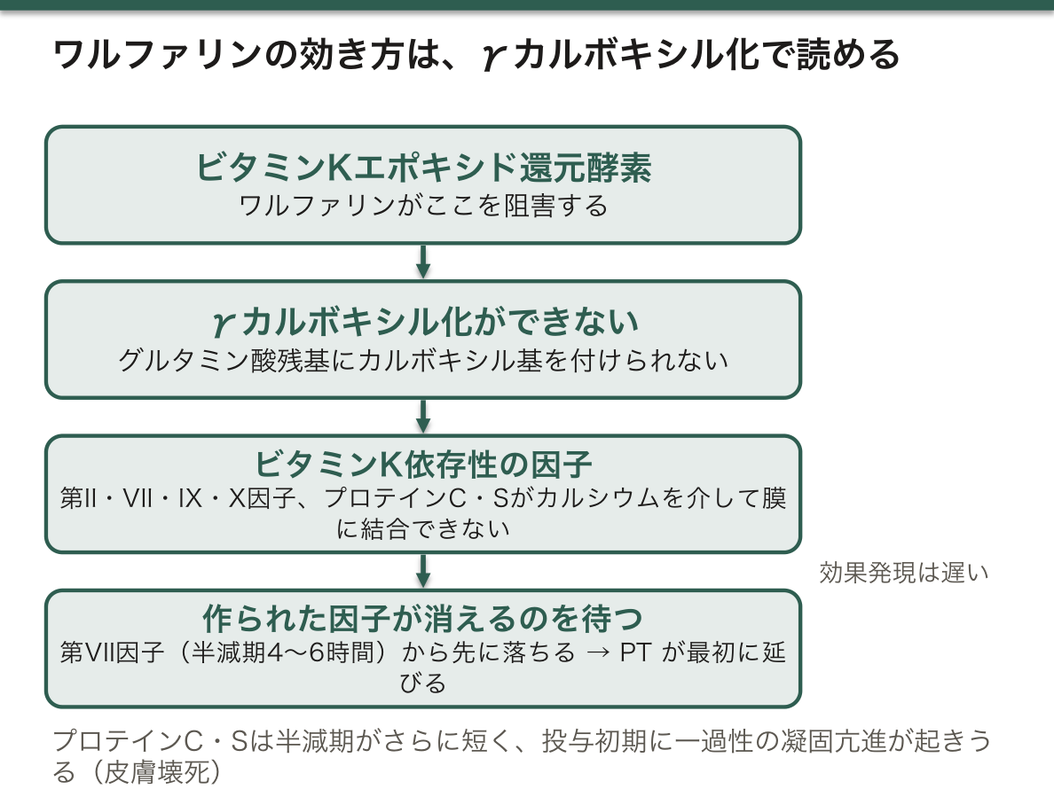
γカルボキシル化:ビタミンK依存性に、グルタミン酸残基へカルボキシル基を追加する。
- 第II・VII・IX・X因子、プロテインC・Sが対象
- カルシウムを介してリン脂質膜に結合できるようになる
- ワルファリンはビタミンKエポキシド還元酵素を阻害する
Cペプチドが臨床で重宝する理由は、インスリンと等モルで分泌されるのに、肝で初回通過効果を受けず、半減期が長いためである。
- 1型糖尿病の診断——内因性分泌が枯れているかが分かる
- インスリン自己注射による低血糖の見破り——インスリンは高いのにCペプチドは低い。外から入れた証拠になる
- インスリノーマでは両方とも高い
ワルファリンの効き方が、γカルボキシル化から全部説明できる。
- 効果発現が遅い——すでに作られた因子が消えるのを待つ必要がある。第VII因子(半減期4〜6時間)から先に落ちる
- プロトロンビン時間(外因系)が最初に延びる——第VII因子が外因系だから
- 投与初期に一過性の凝固亢進が起きうる——プロテインC・Sの半減期はさらに短く、抗凝固因子のほうが先に消える。これがワルファリン誘発性皮膚壊死の機序で、ヘパリンを重ねる根拠になる
- ビタミンKで拮抗できるが、効果が出るまで数時間かかる。緊急なら第IX因子複合体(プロトロンビン複合体濃縮製剤)を使う
納豆・青汁・クロレラが禁止されるのも同じ理屈で、納豆菌は腸内でビタミンK2を産生し続けるため、一度食べると数日効果が続く。
English version
Core
Activation by limited proteolysis is the mechanism for making something in advance and setting it all in motion at the needed moment.
| Precursor | Active form | Meaning |
|---|---|---|
| Trypsinogen | Trypsin | Activated within the pancreas it gives pancreatitis |
| Prothrombin | Thrombin | The coagulation cascade |
| Proinsulin | Insulin + C-peptide | C-peptide is the index of endogenous insulin |
| Procaspase | Caspase | Apoptosis |
γ-Carboxylation: vitamin K-dependent addition of a carboxyl group to glutamate residues.
- Factors II, VII, IX and X, and proteins C and S are the substrates.
- They become able to bind the phospholipid membrane through calcium.
- Warfarin inhibits vitamin K epoxide reductase.
Going further
The reason C-peptide is so useful clinically is that it is secreted equimolar with insulin, is not subject to first-pass extraction in the liver, and has a longer half-life.
- Diagnosis of type 1 diabetes — it shows whether endogenous secretion is exhausted.
- Detecting hypoglycaemia from self-injected insulin — the insulin is high and the C-peptide low. Evidence that it came from outside.
- In an insulinoma both are high.
The way warfarin acts can be explained entirely from γ-carboxylation.
- The onset is slow — one must wait for the factors already made to disappear. Factor VII (half-life 4–6 hours) falls first.
- The prothrombin time (extrinsic) lengthens first — because factor VII is in the extrinsic pathway.
- A transient hypercoagulable state can occur early in treatment — proteins C and S have even shorter half-lives, so the anticoagulant factors disappear first. This is the mechanism of warfarin-induced skin necrosis, and the ground for overlapping with heparin.
- It can be reversed with vitamin K, but that takes hours. In an emergency use prothrombin complex concentrate.
That natto, green juice and chlorella are prohibited follows the same reasoning: the natto organism goes on producing vitamin K2 in the gut, so one meal has an effect lasting days.
糖鎖・アセチル化・ユビキチン化 — 荷札と、寿命の指定
糖鎖修飾——小胞体とGolgi装置で付く。
- N型(アスパラギンに付く)とO型(セリン・トレオニンに付く)
- 役割は折り畳みの補助・安定性・細胞外での認識
- マンノース6リン酸はリソソームへの荷札(リソソーム蓄積症の章)
アセチル化——主にヒストン。正電荷が中和されてDNAとの結合が緩み、転写が進む。
ユビキチン化——ポリユビキチン鎖が分解の合図。プロテアソームへ送られる。
- 3段階の酵素(E1活性化・E2結合・E3リガーゼが基質を選ぶ)
糖鎖は「見た目」を決めるので、検査と免疫の両方に効く。
- ABO血液型は糖転移酵素の違いで、赤血球表面の糖鎖の末端が変わることによる。A型はN-アセチルガラクトサミン、B型はガラクトース、O型は酵素が働かず前駆体のまま
- 糖鎖の違いが腫瘍マーカーになる——CA19-9はLewis式血液型の糖鎖で、Lewis陰性者(日本人の約10%)では膵癌があっても上がらない
- ヘパリンとヘパラン硫酸はグリコサミノグリカンで、強い負電荷がアンチトロンビンを活性化する
CA19-9が陰性になる10%という数字は、実務で腫瘍マーカーを解釈するときに必ず要る。「マーカーが正常だから癌ではない」と言えない集団がいる。
ユビキチン・プロテアソーム系は治療標的になっている。ボルテゾミブ(プロテアソーム阻害薬)が多発性骨髄腫に効くのは、形質細胞が大量の免疫グロブリンを作り、不良品の処理にプロテアソームを酷使しているためである。処理能力を奪うと、不良蛋白が溜まって小胞体ストレスでアポトーシスに至る——細胞の「働きすぎ」を弱点として突いた薬である。
E3リガーゼが基質を選ぶという性質は、近年の創薬に直結している。目的の蛋白をE3リガーゼへ引き寄せて分解させる分子(蛋白分解誘導キメラ)という発想で、従来「薬で狙えない」とされた蛋白を標的にできる。
English version
Core
Glycosylation — added in the endoplasmic reticulum and the Golgi.
- N-linked (to asparagine) and O-linked (to serine and threonine).
- Roles: assisting folding, stability, recognition outside the cell.
- Mannose 6-phosphate is the label for the lysosome (the chapter on lysosomal storage disease).
Acetylation — chiefly of histones. The positive charge is neutralised, binding to DNA loosens, and transcription proceeds.
Ubiquitination — a polyubiquitin chain is the signal for degradation. It is sent to the proteasome. Three stages of enzymes (E1 activating, E2 conjugating, E3 ligase choosing the substrate).
Going further
Glycans decide "appearance", so they tell both in testing and in immunity.
- The ABO blood groups arise because a difference of glycosyltransferase changes the terminus of the glycan on the red cell surface. A adds N-acetylgalactosamine, B galactose, and in O the enzyme does not work and it remains the precursor.
- Differences in glycans become tumour markers — CA19-9 is a Lewis blood group glycan, and in Lewis-negative people (about 10 per cent of Japanese) it does not rise even with pancreatic cancer.
- Heparin and heparan sulphate are glycosaminoglycans, and their strong negative charge activates antithrombin.
The figure "10 per cent in whom CA19-9 is negative" is always needed when interpreting a tumour marker in practice. There is a population of whom one cannot say "the marker is normal, so it is not cancer".
The ubiquitin–proteasome system is a target of treatment. Bortezomib (a proteasome inhibitor) works in multiple myeloma because the plasma cell makes a great deal of immunoglobulin and works its proteasome hard disposing of defective product. Take away that capacity and defective protein accumulates, giving endoplasmic reticulum stress and apoptosis — a drug that strikes at the cell’s "overwork" as a weakness.
That the E3 ligase chooses the substrate bears directly on recent drug discovery. The idea of a molecule that draws a target protein to an E3 ligase and has it degraded (a proteolysis-targeting chimera) makes it possible to target proteins hitherto considered undruggable.
脂質による係留 — 蛋白を膜へ繋ぎ止める荷札
水溶性の蛋白を膜へ留めるために、脂質が共有結合で付けられる。
| 修飾 | 付くもの | 例 |
|---|---|---|
| プレニル化 | ファルネシル基・ゲラニルゲラニル基(コレステロール合成経路の中間体) | Ras・Rho・ラミン |
| ミリストイル化・パルミトイル化 | 脂肪酸 | Src・Gタンパク質のα |
| GPI アンカー | 糖脂質。細胞膜の外側に繋ぎ止める | CD55・CD59・アルカリホスファターゼ・アセチルコリンエステラーゼ |
プレニル化されないと、Ras は膜へ行けず、信号を伝えられない。
GPI アンカーの合成には PIGA 遺伝子が要る。
「場所に着けなければ働けない」という点が、この修飾の意味になる。作られただけでは足りず、正しい膜に置かれて初めて信号になる(シグナル伝達の章)——「翻訳後修飾が、機能の最後の一歩を決めている」。
プレニル基がコレステロール合成経路の中間体であることが、スタチンの副作用につながる(スタチンの項)——「本流を止めると、支流のプレニル化も減る」。
ビスホスホネートも、同じ経路を止めている(骨粗鬆症の薬の項)——「破骨細胞で小さな G 蛋白がプレニル化できなくなり、細胞骨格が働かなくなる」。
PIGA の後天的な変異が、発作性夜間ヘモグロビン尿症になる(溶血の章)——「GPI で繋がる CD55・CD59 が失われ、補体から守られなくなる」。
だから治療は補体を止める薬になる(補体の章)——「エクリズマブは C5 を止める。莢膜を持つ細菌に弱くなるので、髄膜炎菌ワクチンが必須になる」。
English version
Core
To hold a water-soluble protein at a membrane, a lipid is attached covalently.
| Modification | What is attached | Examples |
|---|---|---|
| Prenylation | Farnesyl or geranylgeranyl group (intermediates of the cholesterol synthesis pathway) | Ras, Rho, lamins |
| Myristoylation, palmitoylation | Fatty acids | Src, the α subunit of G proteins |
| The GPI anchor | A glycolipid. Tethers to the outer face of the membrane | CD55, CD59, alkaline phosphatase, acetylcholinesterase |
Without prenylation, Ras cannot reach the membrane and cannot transmit a signal.
The synthesis of the GPI anchor requires the PIGA gene.
Going further
- The meaning of this modification is that a protein cannot work unless it reaches its place. Being made is not enough; only placed on the right membrane does it become a signal (the chapter on signal transduction) — post-translational modification deciding the last step to function.
- That the prenyl group is an intermediate of cholesterol synthesis leads to the adverse effects of statins (the section on statins) — stop the main stream and the tributary of prenylation thins too.
- Bisphosphonates stop the same pathway (the section on the drugs for osteoporosis) — in the osteoclast the small G proteins can no longer be prenylated and the cytoskeleton stops working.
- An acquired mutation of PIGA gives paroxysmal nocturnal haemoglobinuria (the chapter on haemolysis) — CD55 and CD59, which are tethered by GPI, are lost and the cell is no longer protected from complement.
- Hence the treatment is a drug that stops complement (the chapter on complement) — eculizumab stops C5. Resistance to encapsulated bacteria falls, so meningococcal vaccination is essential.
分泌される蛋白の道 — シグナルペプチドが行き先を決める
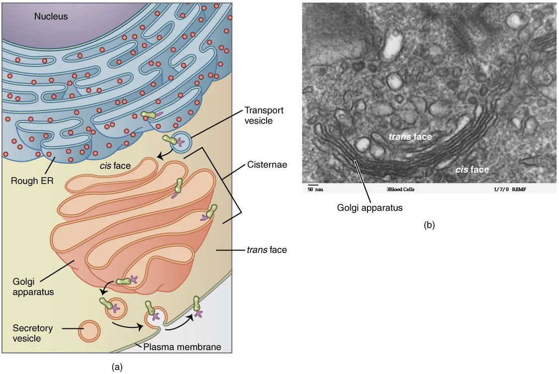
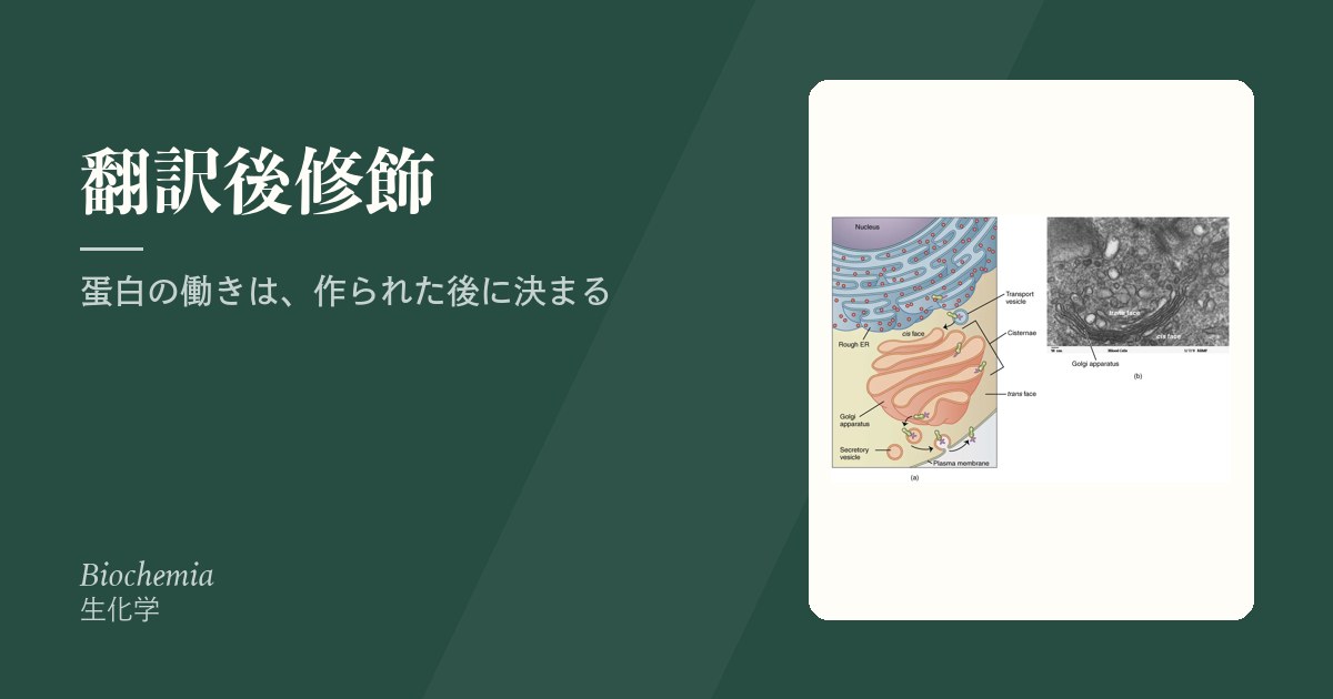
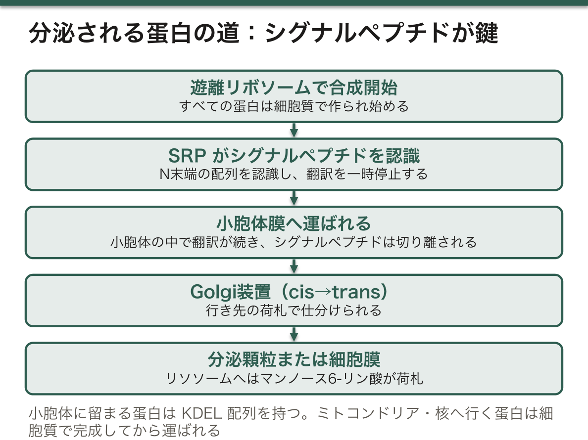
蛋白の合成は、すべて細胞質の遊離リボソームで始まる。N末端にシグナルペプチドがあると、そこから先が変わる。
- シグナル認識粒子(SRP)が認識 → 翻訳を一時停止 → 小胞体膜へ運ぶ
- 小胞体の中で翻訳が続き、シグナルペプチドは切り離される
- 小胞体 → Golgi(cis → trans)→ 分泌顆粒または細胞膜
行き先を決める荷札——リソソームへは マンノース6-リン酸、小胞体に留まる蛋白には KDEL 配列。
ミトコンドリア・核・ペルオキシソームへ行く蛋白は、別の配列を持ち、細胞質で完成してから運ばれる。
「どこで作るかではなく、どこへ送るかが配列で決まっている」という点が要点になる。荷札を間違えると、正しく作られた蛋白が届かない(小胞体ストレスの項)——「作れることと、届くことは別の問題になる」。
封入体細胞病〈I-cell disease〉(ムコリピドーシスII型)が、その典型になる(リソソーム病の章)——「マンノース6-リン酸を付ける酵素が欠け、リソソーム酵素が細胞外へ漏れる。血中の酵素活性が高いのに、細胞内では欠乏している」。
逆にこの荷札があるから、酵素補充療法が成立する(Pompe 病の項)——「マンノース6-リン酸を付けた酵素を点滴すれば、細胞が受容体で取り込んでくれる」。
粗面小胞体が発達した細胞は、分泌が仕事の細胞になる(小胞体の章)——「形質細胞・膵腺房細胞・肝細胞。染色像から機能が読める」。
ジフテリア毒素や志賀毒素は、この経路を逆流して細胞質へ入る(細菌の武器の章)——「運び出す仕組みが、そのまま入り込む道になる」。
English version
Core
All protein synthesis begins on free ribosomes in the cytosol. If there is a signal peptide at the N terminus, what follows changes.
- The signal recognition particle (SRP) recognises it → translation pauses → it is carried to the membrane of the endoplasmic reticulum.
- Translation continues inside the reticulum and the signal peptide is cut off.
- Endoplasmic reticulum → Golgi (cis → trans) → secretory granule or cell membrane.
The labels that decide the destination — mannose 6-phosphate for the lysosome, the KDEL sequence for proteins that stay in the endoplasmic reticulum.
Proteins bound for mitochondrion, nucleus and peroxisome have other sequences and are carried after being completed in the cytosol.
Going further
- The crux is that the sequence decides not where it is made but where it is sent. Mislabel it and a correctly made protein does not arrive (the section on endoplasmic reticulum stress) — being able to make it and its arriving are different problems.
- I-cell disease (mucolipidosis type II) is the type case (the chapter on lysosomal disease) — the enzyme that adds mannose 6-phosphate is missing and the lysosomal enzymes leak outside the cell. The enzyme activities in the blood are high while there is a deficiency within the cell.
- Conversely, it is because this label exists that enzyme replacement therapy works (the section on Pompe disease) — infuse an enzyme carrying mannose 6-phosphate and the cell takes it up through the receptor.
- A cell with well-developed rough endoplasmic reticulum is a cell whose work is secretion (the chapter on the endoplasmic reticulum) — plasma cell, pancreatic acinar cell, hepatocyte. The function can be read from the staining.
- Diphtheria toxin and Shiga toxin run this route backwards into the cytosol (the chapter on the weapons of bacteria) — the machinery for carrying things out becoming, directly, the way in.
まとめ
| リン酸化 — 体内で最も多用されるスイッチ | キナーゼが付け、ホスファターゼが外す。ヒトの遺伝子の約2%がキナーゼで、その数は500を超える。 |
|---|---|
| 切断とγカルボキシル化 — 戻せない修飾 | 限定分解による活性化は、前もって作っておいて、必要な瞬間に一斉に動かすための仕組みである。 |
| 糖鎖・アセチル化・ユビキチン化 — 荷札と、寿命の指定 | 糖鎖修飾——小胞体とGolgi装置で付く。 |
| 脂質による係留 — 蛋白を膜へ繋ぎ止める荷札 | 水溶性の蛋白を膜へ留めるために、脂質が共有結合で付けられる。 |
| 分泌される蛋白の道 — シグナルペプチドが行き先を決める | 蛋白の合成は、すべて細胞質の遊離リボソームで始まる。 |
このノートは医学生が授業の内容を自分の言葉でまとめたもので、診療の判断に使うものではありません。誤りに気づいたらお知らせください。