止血は「速く強く、しかしそこで止まる」ことを同時に満たさねばならない。カスケードによる増幅と、複数の抑制系がその両立を担う。
凝固因子の多くは不活性な前駆体として血中を流れている——必要な瞬間に一斉に動かすための設計である(翻訳後修飾の章)。
- カスケードと、検査が見ている場所
- 線溶と、出血傾向の読み方
- ビタミンK依存性の因子 — 1つの反応が、止血の要になっている
- 血小板の活性化 — 抗血小板薬が止めている場所
- 凝固は、膜の上でしか進まない — カルシウムとリン脂質の面
- 凝固と炎症は、同じ系の2つの面である
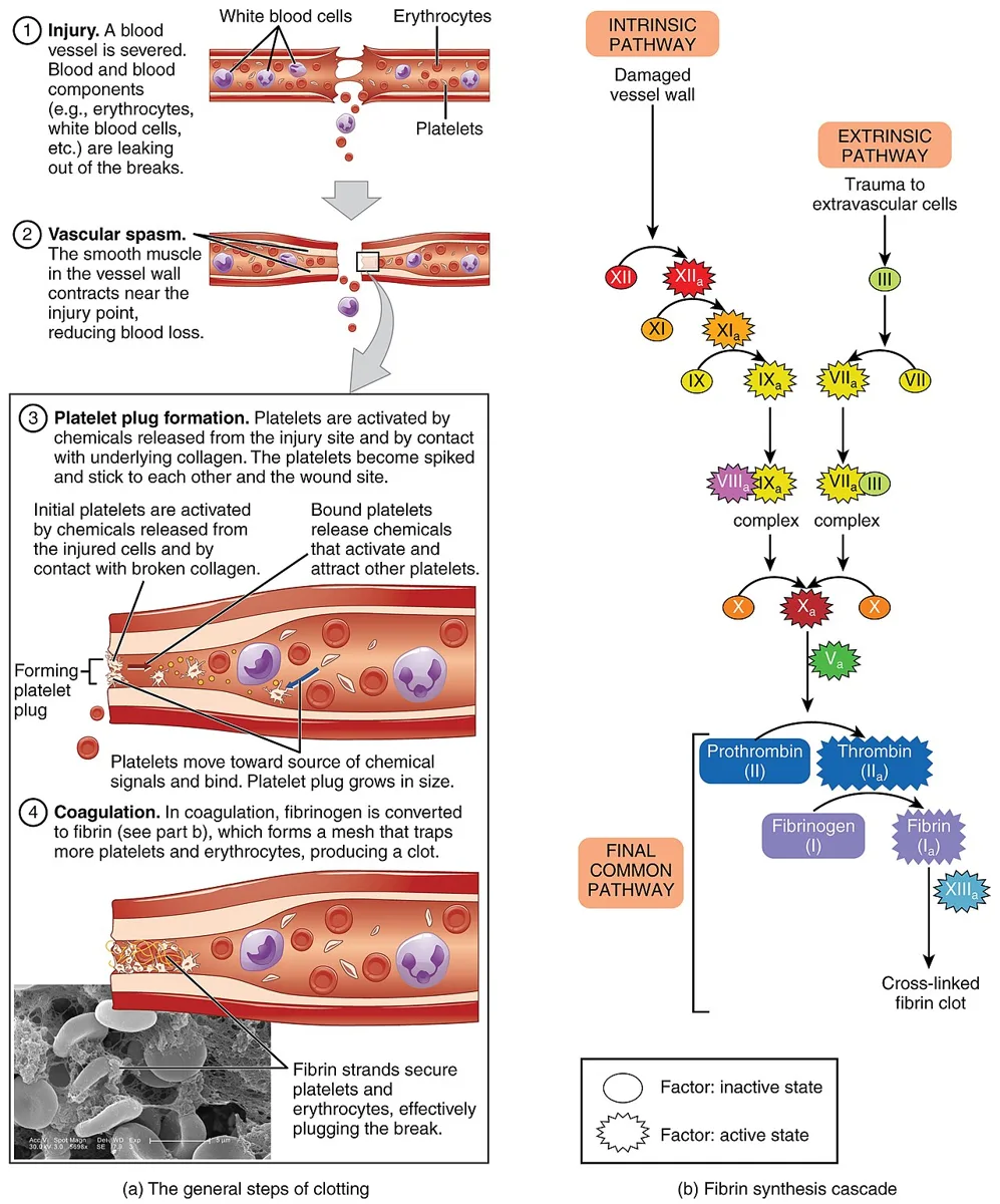
カスケードと、検査が見ている場所
| 系 | 因子 | 検査 |
|---|---|---|
| 外因系 | 組織因子・第VII因子 | プロトロンビン時間 |
| 内因系 | 第XII・XI・IX・VIII因子 | 活性化部分トロンボプラスチン時間 |
| 共通路 | 第X・V・II(プロトロンビン)・I(フィブリノゲン) | 両方 |
- ビタミンK依存性は第II・VII・IX・X因子とプロテインC・S
- 第VIII因子だけが肝以外(血管内皮)でも作られる → 肝硬変で他の因子が下がっても第VIII因子は下がらない
- 第XIII因子はフィブリンを架橋する。欠損しても両検査は正常——臍出血や創部の遅発性出血で疑う
プロトロンビン時間だけ延長 → 第VII因子(最も半減期が短い)。肝障害・ワルファリン・ビタミンK欠乏の初期。
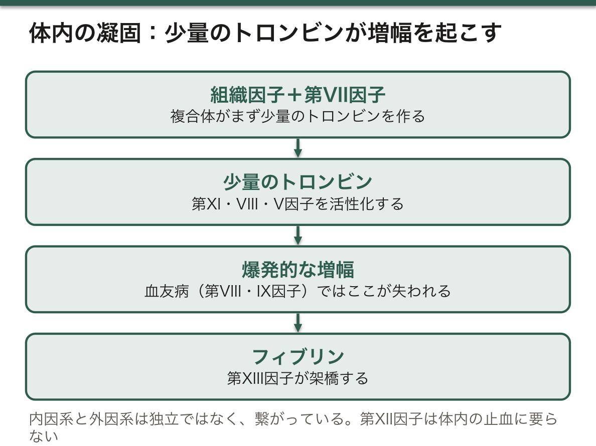
体内で実際に起きていることは、教科書のカスケードとは少し違う。生体内では組織因子と第VII因子の複合体がまず少量のトロンビンを作り、そのトロンビンが第XI・VIII・V因子を活性化して爆発的に増幅する。内因系と外因系は独立ではなく、繋がっている。
この理解が、いくつかの疑問に答える。
- 第XII因子が欠損しても出血しない——活性化部分トロンボプラスチン時間は著明に延長するのに、臨床的には無症状。体内の止血には第XII因子が要らないからである
- 血友病(第VIII・IX因子欠損)は重い出血を起こす——増幅の段階が失われるので、最初の少量のトロンビンだけでは止血が続かない。いったん止まったように見えて後から出血するのはこのため
抑制系が3つあることも押さえる。
- アンチトロンビン——トロンビンと第Xa因子を不活化。ヘパリンが作用を1000倍にする
- プロテインC・S——第Va・VIIIa因子を分解。トロンビンがトロンボモジュリンと結合して活性化する(トロンビンが自分を止める側にも回る)
- 組織因子経路インヒビター
第V因子Leiden変異は活性化プロテインCによる分解を受けにくい第V因子で、欧州人に多い遺伝性血栓性素因である。日本人では極めて稀で、日本ではプロテインS欠乏(徳島型)のほうが多い。
English version
Core
| System | Factors | Test |
|---|---|---|
| Extrinsic | Tissue factor, factor VII | Prothrombin time |
| Intrinsic | Factors XII, XI, IX, VIII | Activated partial thromboplastin time |
| Common | Factors X, V, II (prothrombin), I (fibrinogen) | Both |
- Vitamin K-dependent: factors II, VII, IX, X and proteins C and S.
- Factor VIII alone is also made outside the liver (vascular endothelium) → in cirrhosis the other factors fall but factor VIII does not.
- Factor XIII cross-links fibrin. Its deficiency leaves both tests normal — suspect it from umbilical bleeding or delayed bleeding from a wound.
PT prolonged alone → factor VII (the shortest half-life). Early liver disease, warfarin, vitamin K deficiency.
Going further
What actually happens in the body differs a little from the textbook cascade. In vivo the tissue factor–factor VII complex first makes a small amount of thrombin, and that thrombin activates factors XI, VIII and V for an explosive amplification. The intrinsic and extrinsic systems are not independent but joined.
This understanding answers several puzzles.
- Factor XII deficiency does not bleed — the APTT is markedly prolonged yet the patient is asymptomatic. Haemostasis in the body does not need factor XII.
- Haemophilia (factor VIII or IX deficiency) bleeds severely — the amplifying stage is lost, so the small initial amount of thrombin cannot sustain haemostasis. Bleeding that seems to have stopped and then recurs is explained by this.
Fix the three inhibitory systems too.
- Antithrombin — inactivates thrombin and factor Xa. Heparin multiplies its action a thousandfold.
- Proteins C and S — degrade factors Va and VIIIa. Activated when thrombin binds thrombomodulin (thrombin turning to the side that stops itself).
- Tissue factor pathway inhibitor.
Factor V Leiden is a factor V resistant to degradation by activated protein C and is the common hereditary thrombophilia of Europeans. It is extremely rare in Japanese people, where protein S deficiency (the Tokushima variant) is commoner.
線溶と、出血傾向の読み方
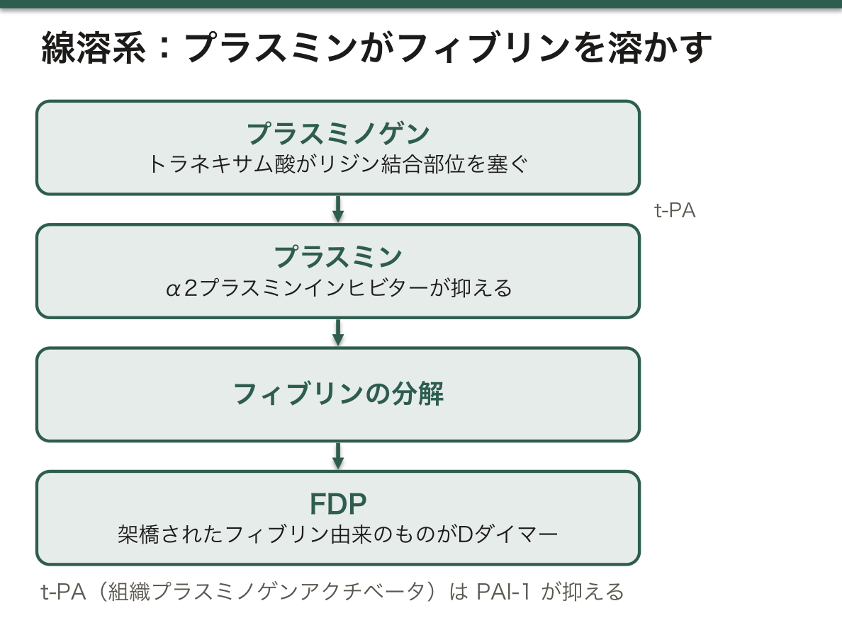
線溶系:プラスミノゲン →(組織プラスミノゲンアクチベータ)→ プラスミン → フィブリンを分解
- 分解産物がFDP。そのうち架橋されたフィブリン由来のものがDダイマー
- 抑制するのはα2プラスミンインヒビターとPAI-1
- トラネキサム酸はプラスミノゲンのリジン結合部位を塞ぐ
| 血小板の異常 | 凝固因子の異常 | |
|---|---|---|
| 出血の型 | 点状出血・紫斑・粘膜出血 | 関節内・筋肉内・深部の血腫 |
| 出血の時期 | 受傷直後 | 遅れて出る |
| 検査 | 血小板数・出血時間 | プロトロンビン時間・活性化部分トロンボプラスチン時間 |
出血の「見た目」で一次止血か二次止血かを分ける——これは問診と視診だけでできる、非常に効率のよい絞り込みである。皮膚の点状出血なら血小板・血管、関節内出血なら凝固因子。
播種性血管内凝固では、凝固と線溶が同時に亢進する。基礎疾患によってどちらが優位かが変わり、それが臨床像を決める。
| 線溶抑制型 | 線溶亢進型 | |
|---|---|---|
| 基礎疾患 | 敗血症 | 急性前骨髄球性白血病・腹部大動脈瘤・前立腺癌 |
| 主症状 | 臓器障害(微小血栓) | 出血 |
| PAI-1 | 著明高値 | 正常〜低値 |
敗血症では、内皮からPAI-1が大量に出て線溶が抑えられるので、できた血栓が溶けずに臓器を壊す。だから出血よりも臓器障害が前面に出る。
Dダイマーの解釈は「除外に使う」が原則である。感度は高いが特異度が低い——加齢・妊娠・悪性腫瘍・感染・術後・入院そのもので上がる。だから陰性なら深部静脈血栓症・肺血栓塞栓症を否定できるが、陽性でも診断にはならない。臨床的な事前確率が高い症例では、Dダイマーを飛ばして画像へ進む。
トラネキサム酸が外傷で死亡を減らすことが大規模試験で示されている。受傷から3時間以内という条件付きで、それ以降はむしろ有害でありうる。「線溶が亢進している時期に使う薬」だという理解が、投与のタイミングを決める。
English version
Core
Fibrinolysis: plasminogen → (tissue plasminogen activator) → plasmin → degrades fibrin.
- The degradation products are FDP; those derived from cross-linked fibrin are the D-dimer.
- Inhibited by α2-antiplasmin and PAI-1.
- Tranexamic acid blocks the lysine binding site of plasminogen.
| Platelet disorder | Coagulation factor disorder | |
|---|---|---|
| Type of bleeding | Petechiae, purpura, mucosal bleeding | Haemarthrosis, muscle haematoma, deep bleeding |
| Timing | Immediately after injury | Delayed |
| Tests | Platelet count, bleeding time | PT, APTT |
Going further
Separating primary from secondary haemostasis by the appearance of the bleeding — this can be done by history and inspection alone and is a very efficient narrowing. Petechiae in the skin mean platelets or vessels; haemarthrosis means clotting factors.
In disseminated intravascular coagulation, coagulation and fibrinolysis are both activated. Which predominates depends on the underlying disease and decides the clinical picture.
| Fibrinolysis-suppressed type | Fibrinolysis-enhanced type | |
|---|---|---|
| Underlying disease | Sepsis | Acute promyelocytic leukaemia, aortic aneurysm, prostate cancer |
| Main problem | Organ failure (microthrombi) | Bleeding |
| PAI-1 | Markedly raised | Normal to low |
In sepsis the endothelium releases a great deal of PAI-1 and fibrinolysis is suppressed, so the thrombi that form are not dissolved and destroy the organs. Hence organ failure rather than bleeding dominates.
The rule for interpreting the D-dimer is that it is used to exclude. Sensitive but not specific — raised by age, pregnancy, malignancy, infection, surgery and hospitalisation itself. So a negative result excludes deep vein thrombosis and pulmonary embolism, but a positive is not a diagnosis. Where the clinical pretest probability is high, skip the D-dimer and go to imaging.
Tranexamic acid has been shown in large trials to reduce mortality in trauma, on condition that it is given within three hours of injury; later it may be harmful. Understanding it as a drug used while fibrinolysis is enhanced decides the timing.
ビタミンK依存性の因子 — 1つの反応が、止血の要になっている
第 II・VII・IX・X 因子と、プロテイン C・プロテイン S は、作られた後にグルタミン酸残基を γ-カルボキシル化されて初めて働く。
この修飾で負電荷が並び、カルシウムを介してリン脂質膜に結合できるようになる——「膜の上に集まることで、反応が桁違いに速くなる」。
反応にはビタミンK が補酵素として要り、その過程で酸化される。ビタミンK エポキシド還元酵素(VKORC1)が元に戻して再利用する。
ワルファリンは、この VKORC1 を阻害する。
半減期が短い因子から先に落ちる——VII(4〜6 時間)< IX < X < II(60〜70 時間)。プロテインC はさらに短い。
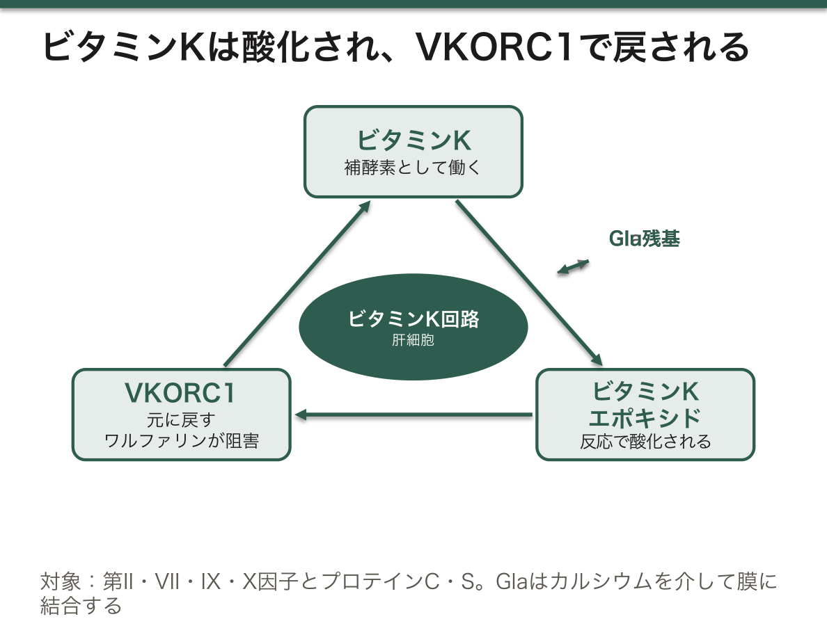
半減期の順序を知っていると、ワルファリンの臨床が全部説明できる。まず第 VII 因子が落ちるので PT(外因系)が先に延び、しかし抗凝固の実体である第 II 因子はまだ残っている(凝固検査の章)——「PT-INR が延びた時点では、まだ効いていない」。
だから開始直後にヘパリンを重ねる。さらにプロテインC が真っ先に落ちるので、開始直後はむしろ凝固に傾く(ワルファリン誘発性皮膚壊死)——「抗凝固薬が、最初の数日だけ血栓を促す」。
効果の判定と中和も、この図の上に置ける。ビタミンK の投与は肝に作らせ直すので数時間かかり、急ぐなら濃縮製剤で因子そのものを入れる(抗凝固薬の章)——「作らせるのか、入れるのかで、速さが違う」。
新生児のビタミンK欠乏性出血症も、同じ理屈になる。胎盤を通りにくく、母乳に少なく、腸内細菌がまだ作れない(新生児の章)——「3つの供給路が同時に細いので、予防投与を行う」。
閉塞性黄疸で PT が延びるのも、脂溶性ビタミンの吸収障害になる(胆汁酸の項)——「ビタミンK を入れて直るなら吸収の問題、直らないなら肝の合成能の問題になる」。
English version
Core
Factors II, VII, IX and X and proteins C and S work only after their glutamate residues have been γ-carboxylated after synthesis. The modification lines up negative charges so that, through calcium, they can bind a phospholipid membrane — gathering on a membrane makes the reaction orders of magnitude faster.
Vitamin K is the coenzyme of that reaction and is oxidised in the process. Vitamin K epoxide reductase (VKORC1) reduces it for reuse. Warfarin inhibits VKORC1.
The factors with the shortest half-lives fall first — VII (4–6 h) < IX < X < II (60–70 h). Protein C is shorter still.
Going further
Know the order of the half-lives and the whole clinical behaviour of warfarin is explained. Factor VII falls first, so the PT (extrinsic) is prolonged first, while factor II, the substance of the anticoagulation, is still present — when the PT-INR has lengthened, it is not yet working. Hence heparin is overlapped at the start.
And since protein C falls first of all, the first days are actually prothrombotic (warfarin-induced skin necrosis) — an anticoagulant promoting thrombosis for its first few days.
Assessment and reversal sit on the same diagram. Giving vitamin K has the liver make them again and takes hours; where haste is needed, concentrate gives the factors themselves — the difference in speed between making and giving.
Vitamin K deficiency bleeding of the newborn follows the same reasoning: it crosses the placenta poorly, breast milk contains little, and the gut flora cannot yet make it — three routes of supply narrow at once, hence prophylaxis.
That the PT lengthens in obstructive jaundice is malabsorption of a fat-soluble vitamin — if giving vitamin K corrects it, the problem is absorption; if not, it is hepatic synthesis.
血小板の活性化 — 抗血小板薬が止めている場所
一次止血は3段階で進む——粘着 → 活性化 → 凝集。
| 段階 | 分子 | 相手 |
|---|---|---|
| 粘着 | GPIb | von Willebrand 因子 → 露出したコラーゲン |
| 活性化 | トロンボキサンA₂・ADP・トロンビン | 自身と周囲の血小板 |
| 凝集 | GPIIb/IIIa | フィブリノゲンを介して血小板同士 |
薬の効く場所。
- アスピリン——シクロオキシゲナーゼ1 を不可逆的にアセチル化し、トロンボキサンA₂ を作らせない
- クロピドグレル・プラスグレル——ADP 受容体(P2Y12)を遮断
- アブシキシマブ・チロフィバン——GPIIb/IIIa を遮断(最終段階)
アスピリンの「不可逆」が、投与量と休薬期間の両方を決めている。血小板は核を持たず酵素を作り直せないので、効果は血小板の寿命(7〜10 日)だけ続く(血小板の章)——「薬の血中濃度が消えても、効果は消えない」。
だから手術前の休薬は 7〜10 日を目安にし、1日 1 回の少量で足りる(周術期管理の章)——「毎日、その日に新しく出てきた血小板だけを止めればよい」。
少量が有効で、増やすと逆に不利になる理由もここにある。血管内皮のシクロオキシゲナーゼ2 が作るプロスタサイクリンは、血小板を抑える側に働く。内皮は核を持つので酵素を作り直せるが、量を増やすとそちらまで抑えてしまう(エイコサノイドの章)——「低用量であることが、選択性を作っている」。
von Willebrand 因子が「粘着の橋渡し」であることは、出血の型を決める。von Willebrand 病では一次止血が障害され、皮膚粘膜の出血(点状出血・鼻出血・過多月経)になる(出血の型の章)——「関節内出血が主体の血友病とは、出方が違う」。
そして vWF は第 VIII 因子の運び屋でもあるので、二次止血にも一部かかる——「1つの分子が2つの系にまたがるので、検査が紛らわしくなる」。
English version
Core
Primary haemostasis proceeds in three stages — adhesion → activation → aggregation.
| Stage | Molecule | Partner |
|---|---|---|
| Adhesion | GPIb | von Willebrand factor → exposed collagen |
| Activation | Thromboxane A₂, ADP, thrombin | Itself and the surrounding platelets |
| Aggregation | GPIIb/IIIa | Platelet to platelet through fibrinogen |
Where the drugs act.
- Aspirin — irreversibly acetylates cyclo-oxygenase 1 so that thromboxane A₂ cannot be made.
- Clopidogrel, prasugrel — block the ADP receptor (P2Y12).
- Abciximab, tirofiban — block GPIIb/IIIa (the final step).
Going further
That aspirin is irreversible decides both the dose and the period of withdrawal. The platelet has no nucleus and cannot remake the enzyme, so the effect lasts the lifespan of the platelet (7–10 days) — the effect does not disappear when the blood level does.
So withdrawal before surgery is 7–10 days, and a small once-daily dose suffices — only the platelets newly released that day need be inhibited.
Here also is the reason a low dose is effective and a higher one counter-productive. The prostacyclin made by endothelial cyclo-oxygenase 2 acts to suppress platelets. The endothelium has a nucleus and can remake the enzyme, but raising the dose suppresses that too — the low dose is what creates the selectivity.
That von Willebrand factor is the bridge of adhesion decides the type of bleeding. In von Willebrand disease primary haemostasis is impaired, giving mucocutaneous bleeding (petechiae, epistaxis, menorrhagia) — a different picture from haemophilia, which is dominated by haemarthrosis.
And vWF is also the carrier of factor VIII, so it bears partly on secondary haemostasis as well — one molecule spanning two systems, which makes the tests confusing.
凝固は、膜の上でしか進まない — カルシウムとリン脂質の面
凝固因子は血漿に溶けたまま出会っても、ほとんど反応しない。反応は、負に荷電したリン脂質の面の上でだけ、爆発的に速くなる。
繋ぎ止めているのがカルシウムである。ビタミン K 依存性因子(II・VII・IX・X とプロテイン C・S)はγ-カルボキシル化されたグルタミン酸(Gla)を持ち、Gla がカルシウムを挟んで、リン脂質の面に結合する。
- 足場に集まると反応が数十万倍になる——酵素と基質と補因子が、同じ平面に並ぶため
- 足場は普段は隠れている——ホスファチジルセリンは細胞膜の内側にあり、外には出ていない
- 血小板が活性化すると外側へ裏返る(スクランブラーゼが働く)——そこで初めて凝固の反応台ができる
つまり凝固は「どこで起こすか」を膜で制御している。傷ついた場所の血小板の上だけで進み、血流の中では進まない。
この仕組みが分かると、日常の3つの事実が一度に説明できる。
| 採血管がクエン酸・EDTA を含む | カルシウムを奪う | Gla が膜に結合できなくなり、凝固が止まる。測るときはカルシウムを戻して開始する |
| 大量輸血で低カルシウム血症になる | 製剤のクエン酸が体内へ入る | 肝で代謝が追いつかない。凝固も心収縮も落ちるので補正する |
| Scott 症候群 | スクランブラーゼの異常でホスファチジルセリンが外へ出ない | 血小板数も凝固検査も正常なのに出血する。反応台が作れない |
抗リン脂質抗体症候群も、この面をめぐる話である。抗体はリン脂質そのものではなく、リン脂質に結合した蛋白(β2-グリコプロテイン I)を標的にする。試験管の中では反応台を奪い合うのでAPTT が延びるが、体内では内皮と血小板を活性化するので血栓ができる。検査が延びているのに血栓症になるという逆転は、「検査は面の上の反応を見ている」ことを知らないと理解できない。
ワルファリンの効き方も同じ枠組みで読める。因子そのものは作られているが、Gla が付いていないので膜に結合できない。この使えない因子(PIVKA-II)は測定でき、ビタミン K 欠乏やワルファリン内服の指標になる。肝細胞癌でも上がるので、腫瘍マーカーとしても使われている。
大量輸血の最中は、カルシウムを測って補う。「輸血しているのに血が止まらない」原因が、希釈ではなくクエン酸のこともある。
English version
Core
Clotting factors meeting in solution in the plasma scarcely react. The reaction becomes explosively fast only on a negatively charged phospholipid surface, and what tethers them there is calcium.
The vitamin K-dependent factors (II, VII, IX, X and proteins C and S) carry γ-carboxylated glutamate (Gla), and Gla binds the phospholipid surface with calcium between.
- Gathering on the scaffold multiplies the reaction hundreds of thousands of times — enzyme, substrate and cofactor lined up in the same plane.
- The scaffold is normally hidden — phosphatidylserine is on the inner leaflet and not exposed.
- When a platelet is activated it flips outwards (a scramblase acts) — and only then is the reaction table set.
That is, coagulation controls where it happens by means of the membrane. It proceeds only on the platelets at the site of injury, and not in the flowing blood.
Going further
Grasp the mechanism and three everyday facts are explained at once.
| What happens | Why | |
|---|---|---|
| Blood tubes contain citrate or EDTA | They take away the calcium | Gla cannot bind the membrane and clotting stops. Calcium is added back to start the assay |
| Massive transfusion causes hypocalcaemia | The citrate of the product enters the body | The liver cannot keep up with metabolising it. Both clotting and cardiac contraction fall, so it is corrected |
| Scott syndrome | A scramblase defect prevents phosphatidylserine from reaching the outer leaflet | The platelet count and the clotting tests are normal yet the patient bleeds — the reaction table cannot be set |
The antiphospholipid syndrome is also about this surface. The antibody targets not the phospholipid itself but a protein bound to it (β2-glycoprotein I). In the tube it competes for the reaction surface and the APTT is prolonged; in the body it activates endothelium and platelets and causes thrombosis. The inversion — a prolonged test with thrombosis — cannot be understood without knowing that the test looks at a reaction on a surface.
The action of warfarin reads within the same framework. The factors are made but carry no Gla and cannot bind the membrane. These useless factors (PIVKA-II) can be measured and are an index of vitamin K deficiency and of warfarin. They also rise in hepatocellular carcinoma and are used as a tumour marker.
In the clinic
During a massive transfusion, measure and replace the calcium. The reason "the bleeding will not stop although we are transfusing" may be citrate rather than dilution.
凝固と炎症は、同じ系の2つの面である
凝固と免疫は別々の仕組みに見えるが、進化的には1つの「傷への応答」から分かれたもので、今も互いに深く絡んでいる。
- 凝固因子が細胞に信号を送る——トロンビンはPAR 受容体を切って活性化する。内皮・血小板・白血球が「凝固が起きている」ことを知り、炎症性の反応を始める
- 炎症が凝固を始める——サイトカイン(IL-6・TNF-α)や内毒素が単球と内皮に組織因子を発現させる。血管の内側に、本来ないはずの引き金が現れる
- 炎症が抗凝固を弱める——内皮のトロンボモジュリンが減り、プロテイン C を活性化できなくなる。ブレーキが外れる
- 好中球が網を出す——ネトーシスで放出された DNA が足場になり、血小板と凝固因子を集める。DNA そのものが血栓の骨組みになる
だから重症感染では、必ず凝固が動く。
血栓は、もともと感染を封じ込めるための仕組みでもあった。微生物を線維素の網に閉じ込めて広がりを止める——この考え方を免疫血栓と呼ぶ。実際、フィブリノゲンを持たない動物は細菌感染で早く敗血症に至る。
問題は、この防御が全身で起きたときである。局所で有用だったものが血管全体で起これば、微小血栓で臓器が虚血に陥り、同時に因子を使い果たして出血する。DIC は防御の暴走として理解できる。
治療の考え方も、この枠組みから出てくる。
- 原因(感染)を叩くのが最優先——上流を止めない限り、下流の凝固は止まらない
- 抗凝固でブレーキを補う——アンチトロンビン製剤、トロンボモジュリン製剤。減っている生理的な抑制因子を戻すという発想である
- 抗炎症だけでは足りない——両方が同じ系である以上、片側だけを止めても回り続ける
⚠️ 動脈硬化も、この枠組みで見直されている。プラークの破綻は「詰まる」現象である前に炎症の現象であり、抗炎症薬(IL-1β 抗体・コルヒチン)が脂質を下げなくても心血管イベントを減らすことが示されている。血栓症を「配管の問題」としてだけ扱う見方は、もう古い。
敗血症で血小板が減り始めたら、DIC の入口として扱う。凝固検査を並べて追い、原因の制御を最優先にする。
English version
Core
Coagulation and immunity look like separate mechanisms, but in evolution they diverged from one response to injury and remain deeply entangled.
- Clotting factors signal to cells — thrombin cleaves and activates PAR receptors. Endothelium, platelets and leucocytes learn that coagulation is occurring and begin an inflammatory response.
- Inflammation starts coagulation — cytokines (IL-6, TNF-α) and endotoxin make monocytes and endothelium express tissue factor. A trigger appears on the inside of the vessel where it should not be.
- Inflammation weakens anticoagulation — endothelial thrombomodulin falls and protein C cannot be activated. The brake is released.
- Neutrophils cast nets — the DNA released in NETosis is a scaffold that gathers platelets and clotting factors. The DNA itself becomes the frame of the thrombus.
So in severe infection, coagulation always moves.
Going further
Thrombosis was originally also a mechanism for containing infection — trapping microorganisms in a fibrin net and stopping their spread. This idea is called immunothrombosis. Animals lacking fibrinogen do indeed reach sepsis sooner in bacterial infection.
The problem is when that defence occurs throughout the body. What was useful locally, occurring in every vessel, renders organs ischaemic with microthrombi and at the same time consumes the factors and causes bleeding. DIC can be understood as a defence running away.
The approach to treatment comes out of the same framework.
- Attacking the cause (the infection) comes first — the coagulation downstream will not stop unless the upstream is stopped.
- Supplement the brake with anticoagulation — antithrombin concentrate, thrombomodulin. The idea of restoring the physiological inhibitors that have been depleted.
- Anti-inflammatory treatment alone is not enough — being one system, stopping only one side leaves it turning.
⚠️ Atherosclerosis is being reconsidered in this framework too. Plaque rupture is a phenomenon of inflammation before it is one of blockage, and anti-inflammatory drugs (an IL-1β antibody, colchicine) have been shown to reduce cardiovascular events without lowering lipids. Treating thrombosis as a plumbing problem alone is out of date.
In the clinic
When the platelet count begins to fall in sepsis, treat it as the entrance to DIC. Follow the clotting tests together and put the control of the cause first.
まとめ
| カスケードと、検査が見ている場所 | 系因子検査外因系組織因子・第VII因子プロトロンビン時間内因系第XII・XI・IX・VIII因子活性化部分トロンボプラスチン時間共通路第X・V・II(プロトロンビン)・I(フィブリ… |
|---|---|
| 線溶と、出血傾向の読み方 | 線溶系:プラスミノゲン →(組織プラスミノゲンアクチベータ)→ プラスミン → フィブリンを分解分解産物がFDP。 |
| ビタミンK依存性の因子 — 1つの反応が、止血の要になっている | 第 II・VII・IX・X 因子と、プロテイン C・プロテイン S は、作られた後にグルタミン酸残基を γ-カルボキシル化されて初めて働く。 |
| 血小板の活性化 — 抗血小板薬が止めている場所 | 一次止血は3段階で進む——粘着 → 活性化 → 凝集。 |
| 凝固は、膜の上でしか進まない — カルシウムとリン脂質の面 | 凝固因子は血漿に溶けたまま出会っても、ほとんど反応しない。 |
| 凝固と炎症は、同じ系の2つの面である | 凝固と免疫は別々の仕組みに見えるが、進化的には1つの「傷への応答」から分かれたもので、今も互いに深く絡んでいる。 |
このノートは医学生が授業の内容を自分の言葉でまとめたもので、診療の判断に使うものではありません。誤りに気づいたらお知らせください。