体重の大部分は、細胞ではなく細胞が作った外の材料でできている。骨・軟骨・腱・血管壁・皮膚は、すべて細胞外基質が主役になる。
- 膠原線維(コラーゲン)——引っ張りに耐える。伸びない
- 弾性線維(エラスチン+フィブリリン)——伸びて戻る
- プロテオグリカン——水を抱えて圧縮に耐える
- 接着蛋白(フィブロネクチン・ラミニン)——細胞と基質をつなぐ
そして遺伝性の結合組織疾患は、この4つのどれが壊れたかで分類できる——「Ehlers-Danlos はコラーゲン、Marfan はフィブリリン、骨形成不全症はI型コラーゲン」。
基質を壊す側(マトリックスメタロプロテアーゼ)も同じくらい重要になる——「作ることと壊すことの均衡が、リモデリングそのものになる」。
- コラーゲンができるまで — 細胞の中と外で、6段階
- 基質を壊す側 — MMPと、線維化の均衡
- プロテオグリカンとヒアルロン酸 — 水を抱え込む分子
- 弾性線維とフィブリリン — 伸びて、戻る組織の材料
- インテグリン — 細胞が基質を掴む手であり、力を伝える線でもある
- 石灰化してはいけない場所が石灰化する — 抑える仕組みが働いている
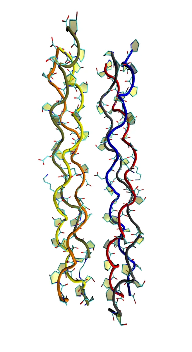
コラーゲンができるまで — 細胞の中と外で、6段階
細胞内。
- プレプロα鎖の合成(粗面小胞体)
- プロリンとリジンの水酸化——ビタミンCが必須。三重らせんの安定に要る
- 糖鎖の付加——水酸化リジンに
- 三重らせんの形成——プロコラーゲン。3残基ごとにグリシンが必要
細胞外。
- 両端のプロペプチドの切断——トロポコラーゲンになる
- 架橋——リシルオキシダーゼ(銅が必要)が、リジン由来の共有結合を作る
主な型——I型(骨・腱・皮膚・最多)・II型(硝子軟骨・硝子体)・III型(細網線維・血管・肉芽)・IV型(基底膜・らせんを作らず網目)。
弾性線維——エラスチン(デスモシン架橋)を、フィブリリンの微細線維が取り囲む。
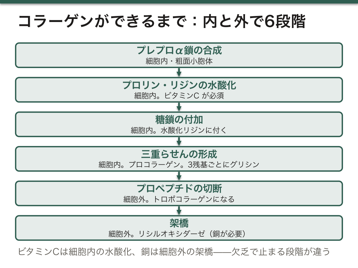
ビタミンCと銅が、それぞれ別の段階で必要になることが、欠乏症の違いを作る。ビタミンCは水酸化(細胞内)、銅は架橋(細胞外)——「同じコラーゲンの異常でも、止まった段階が違う」。
壊血病では、水酸化されないコラーゲンが分解される。歯肉出血・毛孔性の出血・創が開く・骨の異常(ビタミンCの章)——「新しく作るコラーゲンから先に不良になるので、傷の治りが最初に止まる」。
IV型コラーゲンの遺伝子異常が Alport 症候群になる。基底膜が薄く裂ける——血尿・腎不全・感音難聴・水晶体異常(遺伝性腎疾患の章)——「同じ基底膜を使う臓器が、まとめて障害される」。
そして抗糸球体基底膜抗体は、このIV型コラーゲンを標的にする。Goodpasture 症候群で腎と肺が同時に侵される(急速進行性糸球体腎炎の章)——「抗原が共通なので、離れた臓器が一緒に壊れる」。
I型コラーゲンの異常が骨形成不全症になる。易骨折・青色強膜・難聴・象牙質形成不全(小児の骨折の章)——「強膜が青いのは、薄くて脈絡膜が透けるからになる」。
フィブリリンの異常が Marfan 症候群になる。大動脈基部の拡張・水晶体の上方偏位・高身長(大動脈疾患の章)——「弾性線維に頼っている場所が、順に壊れていく」。
English version
Core
Inside the cell.
- Synthesis of the prepro-α chain (rough endoplasmic reticulum).
- Hydroxylation of proline and lysine — vitamin C is essential. Needed for the stability of the triple helix.
- Addition of glycan — to hydroxylysine.
- Formation of the triple helix — procollagen. A glycine is required every third residue.
Outside the cell.
- Cleavage of the propeptides at both ends — it becomes tropocollagen.
- Cross-linking — lysyl oxidase (copper required) makes covalent bonds derived from lysine.
The principal types — I (bone, tendon, skin; the commonest), II (hyaline cartilage, vitreous), III (reticular fibres, vessels, granulation), IV (basement membrane; forms a mesh, not a helix).
The elastic fibre — elastin (desmosine cross-links) surrounded by microfibrils of fibrillin.
Going further
- That vitamin C and copper are needed at different steps creates the difference between the deficiencies. Vitamin C for hydroxylation (inside the cell), copper for cross-linking (outside) — the same abnormal collagen, but the step at which it stopped differs.
- In scurvy, unhydroxylated collagen is degraded. Bleeding gums, perifollicular haemorrhage, wounds opening, abnormalities of bone (the chapter on vitamin C) — the newly made collagen goes bad first, so wound healing is the first thing to stop.
- An abnormality of the type IV collagen gene is Alport’s syndrome. The basement membrane is thin and splits — haematuria, renal failure, sensorineural deafness, lens abnormality (the chapter on inherited renal disease) — the organs using the same basement membrane damaged together.
- And the anti-glomerular basement membrane antibody targets this type IV collagen. In Goodpasture’s syndrome kidney and lung are attacked at once (the chapter on rapidly progressive glomerulonephritis) — the antigen being shared, distant organs break together.
- An abnormality of type I collagen is osteogenesis imperfecta. Fragility, blue sclerae, deafness, dentinogenesis imperfecta (the chapter on fractures in children) — the sclera is blue because it is thin and the choroid shows through.
- An abnormality of fibrillin is Marfan syndrome. Dilatation of the aortic root, upward dislocation of the lens, tall stature (the chapter on aortic disease) — the places that depend on elastic fibre breaking in turn.
基質を壊す側 — MMPと、線維化の均衡
マトリックスメタロプロテアーゼ(MMP)——亜鉛依存性の蛋白分解酵素。細胞外基質を分解する。
- コラゲナーゼ(MMP-1,8,13)・ゼラチナーゼ(MMP-2,9)・ストロメライシン
- 不活性型で分泌され、切断されて働く
- 阻害するもの——TIMP(組織性MMP阻害因子)
MMP と TIMP の均衡が、リモデリングを決める。
- MMP 優位——組織が壊れる。関節リウマチの骨破壊・動脈瘤・歯周病・癌の浸潤
- TIMP 優位——基質が溜まる。肝硬変・肺線維症・強皮症・ケロイド
線維化を進める中心的なサイトカインは TGF-β になる。
創傷治癒では、III型コラーゲンが先に作られ、時間をかけてI型に置き換わる——これが瘢痕の強度が徐々に上がる理由になる。
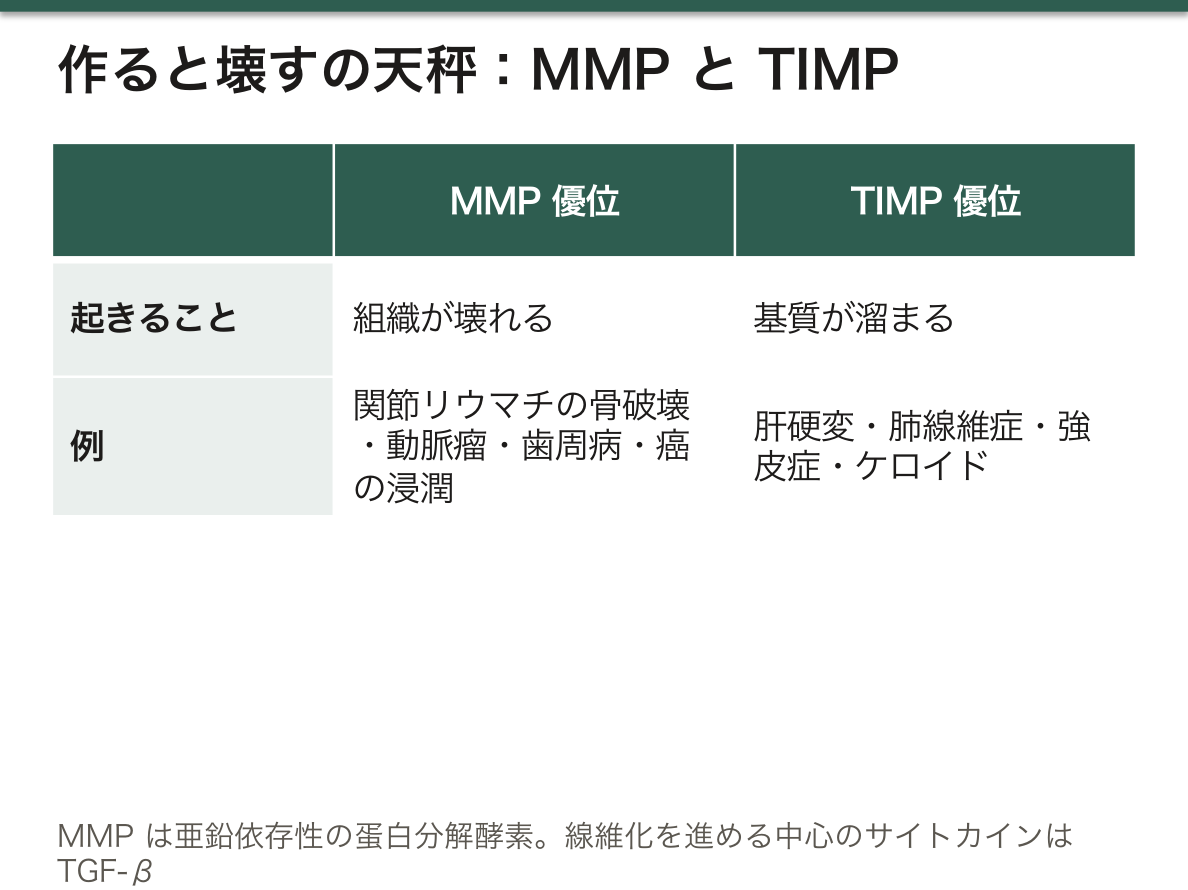
「作る」と「壊す」の均衡として捉えると、線維化と破壊が同じ枠に入る(修復と創傷治癒の章)——「別々に見える病気が、1本の天秤の両端になる」。
関節リウマチでは、滑膜のパンヌスが MMP を出して軟骨と骨を溶かす(関節リウマチの章)——「炎症を抑えることが、そのまま骨の保護になる」。
だから早期の治療が、不可逆な破壊を防ぐ根拠になる(治療戦略の章)——「溶けた骨は戻らないので、時間が予後を決める」。
腹部大動脈瘤では、中膜の弾性線維が MMP で分解される。喫煙が MMP 活性を上げる(大動脈瘤の章)——「禁煙が、拡大速度を落とす介入になる」。
TGF-β が線維化の中心であることが、抗線維化薬の標的になっている(特発性肺線維症の章)——「炎症を抑えるだけでは止まらない病気がある」。
創傷治癒でIII型がI型に置き換わるのに数か月かかる。だから抜糸の後も、瘢痕は伸びやすい時期が続く(創傷管理の章)——「見た目が閉じていることと、強度が戻っていることは違う」。
English version
Core
Matrix metalloproteinases (MMPs) — zinc-dependent proteases. They degrade the extracellular matrix.
- Collagenases (MMP-1, 8, 13), gelatinases (MMP-2, 9), stromelysins.
- Secreted in an inactive form and activated by cleavage.
- Inhibited by TIMPs (tissue inhibitors of metalloproteinases).
The balance of MMP and TIMP decides remodelling.
- MMP predominant — tissue is destroyed. Bone destruction in rheumatoid arthritis, aneurysm, periodontal disease, invasion by cancer.
- TIMP predominant — matrix accumulates. Cirrhosis, pulmonary fibrosis, systemic sclerosis, keloid.
The central cytokine driving fibrosis is TGF-β.
In wound healing type III collagen is made first and is replaced over time by type I — this is why the strength of a scar rises gradually.
Going further
- Taken as the balance of building and destroying, fibrosis and destruction fall into one frame (the chapter on repair and wound healing) — diseases that look separate being the two ends of one balance.
- In rheumatoid arthritis the synovial pannus releases MMPs and dissolves cartilage and bone (the chapter on rheumatoid arthritis) — suppressing the inflammation is, directly, protecting the bone.
- Hence the ground for early treatment preventing irreversible destruction (the chapter on treatment strategy) — dissolved bone does not come back, so time decides the outcome.
- In abdominal aortic aneurysm the elastic fibres of the media are degraded by MMPs. Smoking raises MMP activity (the chapter on aortic aneurysm) — stopping smoking being an intervention that slows the rate of expansion.
- That TGF-β is central to fibrosis has made it the target of the antifibrotic drugs (the chapter on idiopathic pulmonary fibrosis) — there are diseases that suppressing inflammation alone does not stop.
- The replacement of type III by type I in wound healing takes months. So the scar goes on stretching readily after the sutures are out (the chapter on wound management) — looking closed and having recovered its strength are different.
プロテオグリカンとヒアルロン酸 — 水を抱え込む分子
グリコサミノグリカン(GAG)は、二糖の繰り返し。強い負電荷を持つ。
| GAG | 硫酸基 | 主な場所 |
|---|---|---|
| ヒアルロン酸 | 無い(唯一) | 関節液・硝子体・皮膚・臍帯 |
| コンドロイチン硫酸 | あり | 軟骨・骨・血管 |
| ケラタン硫酸 | あり | 角膜・軟骨 |
| ヘパラン硫酸 | あり | 基底膜(糸球体の陰性荷電) |
| デルマタン硫酸 | あり | 皮膚・血管・心弁 |
負電荷が水とナトリウムを引き寄せ、膨潤圧を作る——これが軟骨の圧縮に対する強さになる。
ヒアルロン酸を軸に、多数のプロテオグリカン(アグリカン)が結合して巨大な集合体を作る。
「負電荷が水を抱える」という物理が、いくつもの臓器の機能になっている。軟骨は圧に耐え、硝子体は形を保ち、関節液は潤滑する(軟骨の章)——「支えているのは線維だけではなく、水になる」。
糸球体基底膜のヘパラン硫酸は、電荷による濾過の壁になっている。失われるとアルブミン(陰性荷電)が漏れる(糸球体疾患の章)——「微小変化型ネフローゼで、大きさの障壁は無事なのに蛋白が漏れる理由になる」。
分解できないと溜まる。ムコ多糖症は GAG の分解酵素の欠損で、粗な顔貌・関節拘縮・角膜混濁・肝脾腫・発達の遅れが出る(リソソーム病の章)——「壊せない分子が、少しずつ全身に溜まる」。
ヒアルロン酸は治療にも使われる(変形性関節症の章)——「関節内注射・眼科手術の粘弾性物質・皮膚の充填剤」。
血清ヒアルロン酸は、肝の線維化の指標にもなる(肝の章)——「間質の分子を測ることが、生検の代わりになりつつある」。
English version
Core
A glycosaminoglycan (GAG) is a repeating disaccharide with a strong negative charge.
| GAG | Sulphate | Chief location |
|---|---|---|
| Hyaluronan | None (the only one) | Joint fluid, vitreous, skin, umbilical cord |
| Chondroitin sulphate | Yes | Cartilage, bone, vessels |
| Keratan sulphate | Yes | Cornea, cartilage |
| Heparan sulphate | Yes | Basement membrane (the negative charge of the glomerulus) |
| Dermatan sulphate | Yes | Skin, vessels, cardiac valves |
The negative charge draws water and sodium and creates a swelling pressure — this is the strength of cartilage against compression.
On a backbone of hyaluronan, many proteoglycans (aggrecan) bind to form a huge aggregate.
Going further
- The physics of a negative charge holding water is the function of several organs. Cartilage bears pressure, the vitreous keeps its shape, joint fluid lubricates (the chapter on cartilage) — what supports is not fibre alone but water.
- The heparan sulphate of the glomerular basement membrane is a wall of filtration by charge. Lose it and albumin (negatively charged) leaks (the chapter on glomerular disease) — the reason protein leaks in minimal change nephrotic syndrome while the size barrier is intact.
- What cannot be degraded accumulates. The mucopolysaccharidoses are deficiencies of the enzymes degrading GAGs, with coarse facies, joint contracture, corneal clouding, hepatosplenomegaly and developmental delay (the chapter on lysosomal disease) — a molecule that cannot be broken accumulating little by little throughout the body.
- Hyaluronan is used in treatment too (the chapter on osteoarthritis) — intra-articular injection, the viscoelastic of ophthalmic surgery, dermal filler.
- Serum hyaluronan also serves as an index of hepatic fibrosis (the chapter on the liver) — measuring a molecule of the interstitium coming to replace a biopsy.
弾性線維とフィブリリン — 伸びて、戻る組織の材料
弾性線維は2つの部分でできている——中心のエラスチンと、それを取り囲む微細線維(フィブリリン-1)。
エラスチンは、リシルオキシダーゼ(銅が要る)によるデスモシン架橋で網になる。だから引き伸ばしても元に戻る。
ほとんど入れ替わらない——半減期は数十年。一度壊れると作り直されない。
| 疾患 | 壊れる分子 | 特徴 |
|---|---|---|
| Marfan 症候群 | フィブリリン-1(FBN1) | 高身長・クモ指・水晶体亜脱臼(上方)・大動脈基部拡張 |
| Ehlers-Danlos 症候群 | 主にコラーゲン | 皮膚の過伸展・関節の過可動・血管型は動脈破裂 |
| α1-アンチトリプシン欠損症 | エラスチンが分解される | 若年の肺気腫・肝硬変 |
「作り直さない材料」であることが、この分子群の病態を決めている。肺気腫で失われた肺胞壁が戻らないのは、エラスチンが再生されないからになる(慢性閉塞性肺疾患の章)——「禁煙は進行を止めるが、失ったものは戻さない」。
フィブリリン-1 の異常は、構造の話だけで終わらない。フィブリリンは TGF-β を微細線維に繋ぎ止めており、壊れると TGF-β が過剰に働く(シグナル伝達の章)——「Marfan 症候群は、支えの病気であると同時に信号の病気になる」。
だからアンジオテンシンII 受容体拮抗薬が、大動脈拡張の進行を抑えるという発想が生まれた(降圧薬の章)——「β遮断薬と合わせ、血圧と信号の両方を下げる」。
水晶体亜脱臼の向きが、鑑別の手がかりになる(前眼部の章)——「Marfan 症候群は上方、ホモシスチン尿症は下方へずれる」。
ホモシスチン尿症も、リシルオキシダーゼの架橋を妨げる(含硫アミノ酸の章)——「体型が似るのに、血栓症と知的障害を伴う点が違う」。
English version
Core
The elastic fibre is made of two parts — elastin at the centre and the microfibrils (fibrillin-1) that surround it.
Elastin is made into a net by desmosine cross-links formed by lysyl oxidase (copper required). Hence it returns after being stretched.
It scarcely turns over — the half-life is decades. Once broken it is not remade.
| Disease | Molecule affected | Features |
|---|---|---|
| Marfan syndrome | Fibrillin-1 (FBN1) | Tall stature, arachnodactyly, subluxation of the lens (upwards), dilatation of the aortic root |
| Ehlers–Danlos syndrome | Chiefly collagen | Hyperextensible skin, hypermobile joints; the vascular type ruptures arteries |
| α1-Antitrypsin deficiency | Elastin is degraded | Emphysema and cirrhosis in the young |
Going further
- Being a material that is not remade determines the diseases of this group of molecules. The alveolar wall lost in emphysema does not come back because elastin does not regenerate (the chapter on chronic obstructive pulmonary disease) — stopping smoking halts the progression but does not return what is lost.
- An abnormality of fibrillin-1 does not end as a matter of structure. Fibrillin tethers TGF-β to the microfibrils, and when it breaks TGF-β acts in excess (the chapter on signal transduction) — Marfan syndrome being a disease of signalling as much as of support.
- Hence the idea arose that an angiotensin II receptor blocker might slow the dilatation of the aorta (the chapter on antihypertensives) — with a β-blocker, lowering both the pressure and the signal.
- The direction of the lens subluxation is a clue in the differential (the chapter on the anterior segment) — Marfan syndrome upwards, homocystinuria downwards.
- Homocystinuria also impedes the cross-linking by lysyl oxidase (the chapter on the sulphur-containing amino acids) — the habitus resembles, but it differs in carrying thrombosis and intellectual disability.
インテグリン — 細胞が基質を掴む手であり、力を伝える線でもある
細胞は基質にただ乗っているのではなく、掴んでいる。その手がインテグリンで、膜を貫いて外の基質と中の細胞骨格を直接つなぐ受容体である。
- 認識する配列がある——多くのインテグリンは、フィブロネクチンなどにある RGD 配列(アルギニン・グリシン・アスパラギン酸)を掴む
- 信号が双方向に流れる——外の情報を中へ伝えるだけでなく、中からの信号で「掴む力」自体が変えられる(内から外への信号)
- 力を感じている——引っ張られると結合部に蛋白が集まり、硬い基質か柔らかい基質かで細胞の運命が変わる
掴めなくなった細胞は死ぬ——足場を失ったことによるアポトーシス(anoikis)。上皮細胞が本来いるべき場所から離れたときに増えないための、安全装置になっている。
インテグリンは、それぞれ別の場面で薬の標的になっている。
| GPIIb/IIIa | 血小板がフィブリノゲンを掴む手 | 阻害薬は最も強力な抗血小板薬。Glanzmann 血小板無力症はこの欠損 |
| 白血球のβ2 インテグリン | 血管内皮にしっかり接着して外へ出る | 白血球接着不全症——臍帯脱落の遅れと、膿の出ない反復感染 |
| α4 インテグリン | リンパ球が組織へ入る | ナタリズマブ(多発性硬化症・Crohn 病)。⚠️ 中枢の免疫監視が落ち、進行性多巣性白質脳症の危険がある |
| ヘミデスモソームのα6β4 | 表皮を基底膜に留める | 異常があると接合部型表皮水疱症 |
「掴む・離す」を制御することが、そのまま病態と治療になる。
癌の転移も同じ枠組みで読める。上皮間葉転換で接着の型を変え、anoikis に抵抗しながら移動し、転移先で別のインテグリンを使って定着する。どのインテグリンを出すかが、どこへ転移するかに関わっていると考えられている。
新生児で臍帯の脱落が2週間以上遅れ、感染を繰り返すのに膿ができないときは、白血球接着不全症を疑う。
English version
Core
A cell does not merely rest on the matrix; it grips it. That hand is the integrin, a receptor that spans the membrane and directly joins the matrix outside to the cytoskeleton within.
- There is a sequence they recognise — many integrins grip the RGD sequence (arginine–glycine–aspartate) found in fibronectin and others.
- Signals flow both ways — not only carrying information from outside inwards, but the strength of the grip itself being changed by a signal from within (inside-out signalling).
- They feel force — pulled, proteins gather at the junction, and the fate of the cell changes with whether the matrix is stiff or soft.
A cell that can no longer grip dies — apoptosis from loss of anchorage (anoikis). It is a safety device preventing an epithelial cell from proliferating once it has left the place where it belongs.
Going further
Integrins are drug targets, each in a different setting.
| GPIIb/IIIa | The hand with which the platelet grips fibrinogen | Inhibitors are the most powerful antiplatelet drugs. Glanzmann’s thrombasthenia is a deficiency of it |
| The β2 integrins of leucocytes | Adhere firmly to the endothelium and pass outside | Leucocyte adhesion deficiency — delayed separation of the cord and recurrent infection without pus |
| α4 integrin | Lymphocytes entering tissue | Natalizumab (multiple sclerosis, Crohn’s disease). ⚠️ Central immune surveillance falls, with a risk of progressive multifocal leukoencephalopathy |
| α6β4 of the hemidesmosome | Holds the epidermis to the basement membrane | An abnormality gives junctional epidermolysis bullosa |
Controlling "grip and let go" is, directly, disease and treatment.
Metastasis can be read in the same frame. In the epithelial–mesenchymal transition the type of adhesion changes, the cell moves while resisting anoikis, and settles at the destination using a different integrin. Which integrins are displayed is thought to bear on where it metastasises.
In the clinic
In a newborn whose cord separation is delayed beyond two weeks and who has repeated infections without forming pus, suspect leucocyte adhesion deficiency.
石灰化してはいけない場所が石灰化する — 抑える仕組みが働いている
血液はカルシウムとリンについてすでに過飽和である。つまり放っておけば、どこでも結晶ができる状態にある。血管が石灰化しないのは、偶然ではなく能動的に抑えているからである。
- ピロリン酸——結晶の成長を直接止める。アルカリホスファターゼが分解すると石灰化が進む
- マトリックス Gla 蛋白(MGP)——ビタミン K 依存性にγ-カルボキシル化されて働く
- フェチュイン A——肝が作る血中の阻害因子。カルシウムとリンを包んで運ぶ
石灰化は「防御が破れた結果」として起こる。
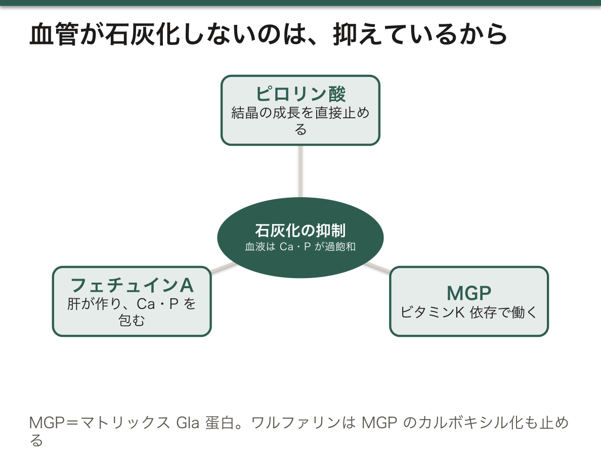
2つの型に分けると、原因の探し方が決まる。
| 異栄養性石灰化 | 血中 Ca・P は正常 | 傷んだ組織に起こる。壊死・瘢痕・粥腫・慢性膵炎・古い結核巣・腫瘍(乳癌の微細石灰化) |
| 転移性石灰化 | 血中 Ca・P が高い | 正常な組織に起こる。慢性腎臓病・副甲状腺機能亢進症・ビタミン D 中毒・悪性腫瘍 |
転移性石灰化が起こりやすい場所は決まっていて、局所がアルカリ性の組織——胃粘膜・腎・肺・血管・角膜である。酸を分泌する組織は、その裏側がアルカリに傾く。
臨床でいちばん重い形が、慢性腎臓病の血管石灰化である。リンが排泄されず、MGP は不活性型が増え、血管平滑筋細胞が骨芽細胞のような性質に変わって、自ら基質を石灰化させる。受動的に沈着するのではなく能動的に骨を作っているという点が近年の理解である。だからリン管理(食事・リン吸着薬)が予後に直結する。
⚠️ ワルファリンは MGP のカルボキシル化も止める。長期投与で血管の石灰化が進むことが指摘されており、DOAC が選ばれる理由の 1 つに挙げられることがある。ビタミン K の作用は、凝固だけではない。
画像で石灰化を見たら、まず血清 Ca と P を確認する。正常なら「そこの組織が傷んだ跡」、高ければ「全身の代謝の問題」と読み分けられる。
English version
Core
Blood is already supersaturated with respect to calcium and phosphate. That is, left alone, crystals would form anywhere. That vessels do not calcify is not chance but because it is actively suppressed.
- Pyrophosphate — stops the growth of crystals directly. Degraded by alkaline phosphatase, mineralisation advances.
- Matrix Gla protein (MGP) — works after vitamin K-dependent γ-carboxylation.
- Fetuin A — a circulating inhibitor made by the liver. It wraps calcium and phosphate and carries them.
Calcification occurs as the result of the defences being breached.
Going further
Dividing it into two types settles how to look for the cause.
| Dystrophic calcification | Blood Ca and P are normal | It occurs in damaged tissue. Necrosis, scar, atheroma, chronic pancreatitis, an old tuberculous focus, tumour (the microcalcification of breast cancer) |
| Metastatic calcification | Blood Ca and P are high | It occurs in normal tissue. Chronic kidney disease, hyperparathyroidism, vitamin D intoxication, malignancy |
The places where metastatic calcification occurs are fixed — tissues locally alkaline: gastric mucosa, kidney, lung, vessels, cornea. A tissue that secretes acid turns alkaline on its other side.
The gravest form in practice is the vascular calcification of chronic kidney disease. Phosphate is not excreted, the inactive form of MGP increases, and vascular smooth muscle cells take on the character of osteoblasts and mineralise their own matrix. The recent understanding is that they are actively making bone rather than passively depositing. Hence management of phosphate (diet, phosphate binders) bears directly on the outcome.
⚠️ Warfarin also stops the carboxylation of MGP. Long use has been noted to advance vascular calcification, and this is sometimes listed among the reasons a DOAC is chosen. The action of vitamin K is not confined to coagulation.
In the clinic
Seeing calcification on an image, first check the serum Ca and P. Normal reads as "the trace of damage in that tissue", high as "a problem of whole-body metabolism".
まとめ
| コラーゲンができるまで — 細胞の中と外で、6段階 | 細胞内。プレプロα鎖の合成(粗面小胞体)プロリンとリジンの水酸化——ビタミンCが必須。 |
|---|---|
| 基質を壊す側 — MMPと、線維化の均衡 | マトリックスメタロプロテアーゼ(MMP)——亜鉛依存性の蛋白分解酵素。 |
| プロテオグリカンとヒアルロン酸 — 水を抱え込む分子 | グリコサミノグリカン(GAG)は、二糖の繰り返し。 |
| 弾性線維とフィブリリン — 伸びて、戻る組織の材料 | 弾性線維は2つの部分でできている——中心のエラスチンと、それを取り囲む微細線維(フィブリリン-1)。 |
| インテグリン — 細胞が基質を掴む手であり、力を伝える線でもある | 細胞は基質にただ乗っているのではなく、掴んでいる。 |
| 石灰化してはいけない場所が石灰化する — 抑える仕組みが働いている | 血液はカルシウムとリンについてすでに過飽和である。 |
このノートは医学生が授業の内容を自分の言葉でまとめたもので、診療の判断に使うものではありません。誤りに気づいたらお知らせください。