女性の生殖路は、周期ごとに組織そのものが作り変えられる。だから「今どの時期か」を知らずに標本は読めない。
- 卵管——エストロゲンで線毛細胞が増え、分泌細胞と入れ替わる
- 子宮頸部——粘液の性質が周期で変わる(排卵期は薄く、黄体期は濃い)
- 腟——エストロゲンでグリコーゲンが蓄えられ、それが乳酸菌の餌になる
- 乳腺——妊娠と授乳で完成し、離乳で退縮する
そして子宮頸部の「扁平円柱上皮接合部」が、この領域で最も臨床的に重い一点になる——「子宮頸癌のほとんどが、この境目から出る」。
子宮内膜は生殖器の組織の章、月経周期は月経周期の章、子宮頸癌は婦人科腫瘍の章にある。
- 乳腺 — 完成するのは妊娠してから
- 乳腺の導管系 — 病気が起きる単位で捉える
- 乳汁分泌 — 2つのホルモンが、別々の細胞に効く
- 外陰の腺と粘膜 — 皮膚から粘膜へ移り変わる場所
- 乳腺は、間質のほうが病気を決めることがある
- 絨毛と胎盤関門 — 何が通り、何が通らないか
- 臍帯と卵膜 — 2本の動脈と1本の静脈
- 脱落膜 — 母体側が、受け入れるために作り変わる
- 栄養膜 — 2種類の細胞が、役割を分けている
- 胎盤のホルモン — 一時的にできた、最大の内分泌器官
- 胎児の造血は、場所を3回変える
- 胎盤が剥がれない — 脱落膜が欠けているということ
- 乳腺 — 完成するのは妊娠してからGlandula mammaria
- 乳腺の導管系 — 病気が起きる単位で捉えるDuctus lactifer
- 乳汁分泌 — 2つのホルモンが、別々の細胞に効くLactatio
- 外陰の腺と粘膜 — 皮膚から粘膜へ移り変わる場所Glandula vestibularis major
- 乳腺は、間質のほうが病気を決めることがある
- 絨毛と胎盤関門 — 何が通り、何が通らないかVillus chorialis
- 臍帯と卵膜 — 2本の動脈と1本の静脈Chorion
- 脱落膜 — 母体側が、受け入れるために作り変わるDecidua
- 栄養膜 — 2種類の細胞が、役割を分けているTrophoblastus
- 胎盤のホルモン — 一時的にできた、最大の内分泌器官Placenta
- 胎児の造血は、場所を3回変えるSanguis umbilicalis
- 胎盤が剥がれない — 脱落膜が欠けているということ
- まとめ
乳腺 — 完成するのは妊娠してからGlandula mammaria
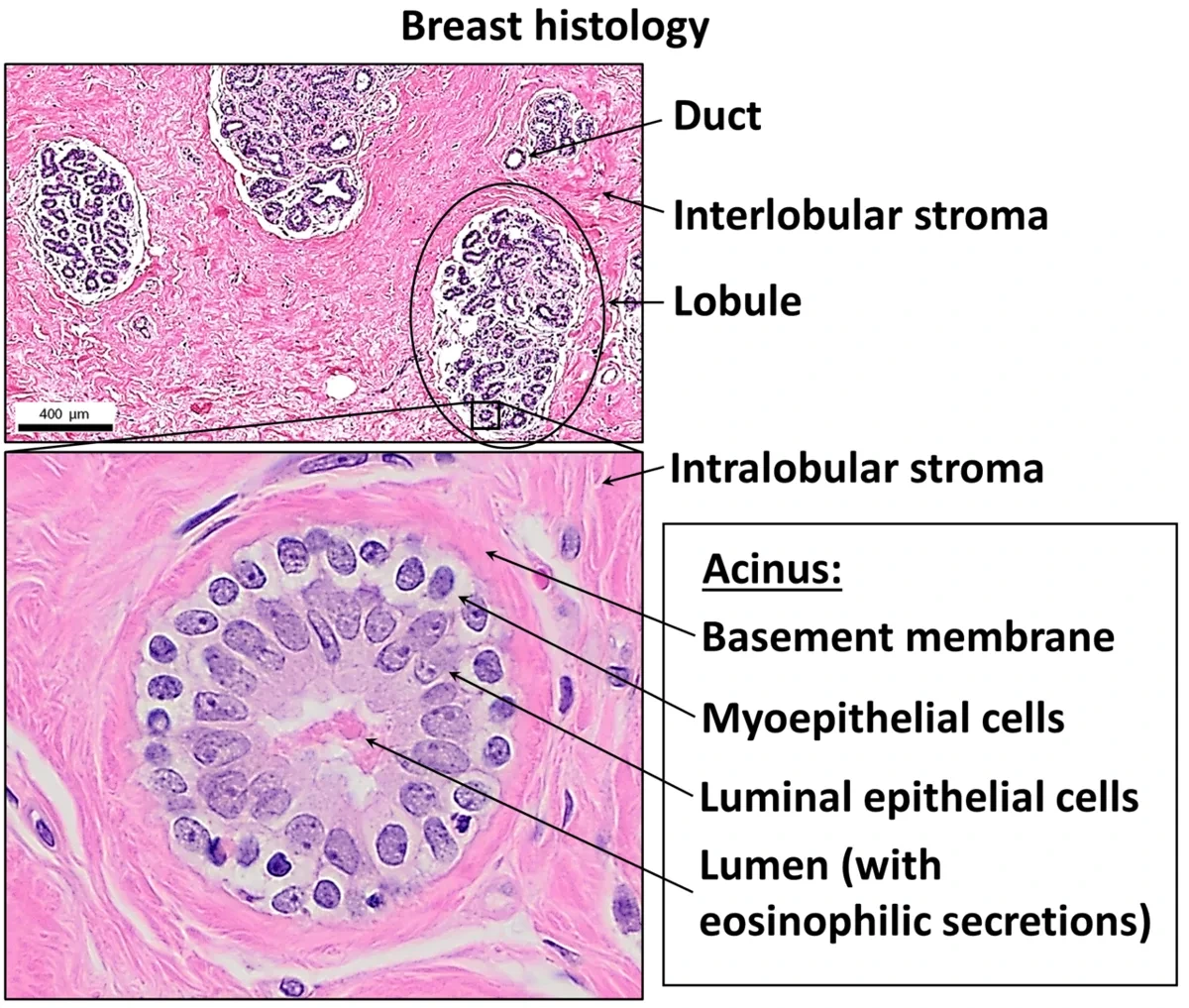
複合管状胞状腺。15〜20の葉に分かれ、それぞれが乳管洞を経て乳頭に開く。
- 終末乳管小葉単位(TDLU)——小葉と終末乳管の組。ほとんどの乳腺疾患がここから出る
- 上皮は2層——内側の腺上皮と、外側の筋上皮
非妊娠時——導管が主体で、腺房はほとんど無い。間質(線維と脂肪)が大部分を占める。
妊娠時——腺房が増殖して間質を置き換える。後期には初乳を含む。
授乳期——腺房が拡張。蛋白はメロクリン、脂肪はアポクリンで分泌される。
ホルモン——エストロゲンが導管を、プロゲステロンが小葉を発達させる。プロラクチンが産生を、オキシトシンが射出(筋上皮の収縮)を起こす。
TDLU がほとんどの乳腺疾患の発生母地になることが、この章の中心になる。線維腺腫も、乳管癌も小葉癌も、ここから出る(乳腺疾患の章)——「小葉癌という名前でも、出どころは同じ単位になる」。
筋上皮層が保たれているかどうかが、上皮内癌と浸潤癌を分ける(筋上皮細胞の章)——「基底膜と筋上皮が、浸潤の境界線になっている」。
プロラクチンとオキシトシンの役割の違いが、授乳の支援に直結する。吸啜が両方を刺激するので、飲ませることが最良の催乳になる(授乳の章)——「出ないから飲ませない、が悪循環を作る」。
そしてオキシトシンは、ストレスで抑えられる。射乳反射が働かないと、作られていても出ない——「環境を整えることが、薬より先の介入になる」。
妊娠中に腺房が間質を置き換えることが、妊娠期の乳癌を見つけにくくする。全体が硬く張るので、しこりが埋もれる(妊娠と合併症の章)——「見つかったときには進んでいることが多い」。
男性の乳腺も同じ構造を持ち、エストロゲン優位で発達する。女性化乳房は思春期・肝硬変・薬剤で起きる(肝硬変の身体所見の章)——「同じ組織が、ホルモン環境だけで動く」。
終末乳管小葉単位(TDLU)から、ほとんどの乳腺疾患が起こる。乳癌も線維腺腫も乳腺症も、この単位の病気である。
浸潤性乳管癌が最も多い。浸潤性小葉癌はE-カドヘリンを失って細胞がばらばらに一列で浸潤する(indian file)ため腫瘤を作りにくく、マンモグラフィで見つけにくい。両側性・多発性のことが多い。接着分子1つの違いが、画像所見と治療方針を変えている。
非浸潤性乳管癌(DCIS)は基底膜を越えていないので転移しない。マンモグラフィの微細石灰化で見つかる。
Paget 病 — 乳頭のびらん・湿疹様変化。下に乳管癌があるので「治らない乳頭の湿疹」は生検する。
English version
Core
A compound tubulo-alveolar gland, divided into 15–20 lobes, each opening at the nipple through a lactiferous sinus.
- Terminal duct lobular unit (TDLU) — the lobule with its terminal duct. Almost all breast disease arises here.
- The epithelium is two-layered — glandular cells within, myoepithelial cells without.
Non-pregnant — ducts predominate and there are almost no acini; stroma (fibrous tissue and fat) makes up most of it.
Pregnancy — the acini proliferate and replace the stroma; in late pregnancy they contain colostrum.
Lactation — the acini dilate. Protein is secreted merocrine, fat apocrine.
Hormones — oestrogen develops the ducts, progesterone the lobules. Prolactin drives production, oxytocin ejection (contraction of the myoepithelium).
Going further
That the TDLU is the site of origin of almost all breast disease is the centre of this chapter. Fibroadenoma, ductal carcinoma and lobular carcinoma all arise there — even the one called lobular comes from the same unit.
Whether the myoepithelial layer is preserved separates carcinoma in situ from invasive carcinoma — the basement membrane and the myoepithelium are the boundary of invasion.
The difference in the roles of prolactin and oxytocin bears directly on supporting breastfeeding. Suckling stimulates both, so feeding is the best way to bring in the milk — "it will not come, so I do not feed" creates the vicious circle. And oxytocin is suppressed by stress: without the let-down reflex, milk that has been made does not come out — arranging the environment is an intervention that comes before any drug.
That the acini replace the stroma in pregnancy makes breast cancer in pregnancy hard to find. The whole breast is firm and engorged, so a lump is buried — by the time it is found it is often advanced.
The male breast has the same structure and develops when oestrogen predominates. Gynaecomastia occurs at puberty, in cirrhosis and from drugs — the same tissue moving on the hormonal environment alone.
In the clinic
Almost all breast disease arises from the terminal duct lobular unit (TDLU) — carcinoma, fibroadenoma and fibrocystic change are all diseases of this unit.
Invasive ductal carcinoma is the commonest. Invasive lobular carcinoma loses E-cadherin and the cells infiltrate singly in single file (Indian file), so it forms no mass and is hard to see on mammography. It is often bilateral and multifocal. A difference in a single adhesion molecule changes the imaging findings and the management.
Ductal carcinoma in situ (DCIS) has not crossed the basement membrane and does not metastasise. It is found as microcalcification on mammography.
Paget’s disease — erosion or eczema-like change of the nipple. There is a ductal carcinoma beneath, so "eczema of the nipple that will not heal" is biopsied.
乳腺の導管系 — 病気が起きる単位で捉えるDuctus lactifer
乳房は15〜20個の腺葉が集まったもので、腺葉ごとに1本の乳管が乳頭へ開く。
乳管の枝分かれ——乳管洞(乳頭直下で拡張)→ 乳管 → 小葉間乳管 → 終末乳管 → 小葉(腺房)。
上皮は2層になっている——内側の腺上皮と、外側の筋上皮。その外を基底膜が包む。
終末乳管と、それに続く小葉をまとめて終末乳管小葉単位(TDLU)と呼ぶ。乳癌と線維嚢胞性変化の大半が、ここから発生する。
乳頭と乳輪の上皮は角化重層扁平上皮で、乳輪には Montgomery 腺(皮脂腺)がある。
「上皮が2層である」ことが、乳腺病理の読み方を決めている。筋上皮が保たれていれば良性、消えていれば浸潤癌を疑う(腫瘍総論の章)——「外側の層が残っているかどうかが、線を越えたかの指標になる」。
前立腺の基底細胞と、まったく同じ論理になる(前立腺の章)——「2層の腺は、どこでも同じ読み方ができる」。
TDLU という単位を知っていると、画像と病理が繋がる。石灰化が小葉に集まるか、乳管に沿って線状に並ぶかで、良性と非浸潤性乳管癌が分かれる(マンモグラフィの章)——「石灰化の並び方が、どの構造の中で起きたかを示している」。
乳管が乳頭へ開いていることは、症状の意味を変える。血性の乳頭分泌は、乳管内の病変(乳管内乳頭腫・非浸潤性乳管癌)が管腔とつながっていることを示す(良性腫瘍の章)——「分泌があるということは、道が通じているということになる」。
そして乳頭の Paget 病は、乳管内の癌細胞が表皮内へ這い上がった像になる。湿疹に見えるが、片側性で治らない(皮膚腫瘍の章)——「片側だけの、治らない乳頭のただれを湿疹と呼ばない」。
乳管を単位に考えると、症状の意味が決まる。
血性乳頭分泌は1本の乳管から出る(単孔性)ことが多く、乳管内乳頭腫か乳管内進展した癌を意味する。両側・多孔性・乳汁様なら内分泌性(高プロラクチン血症)を考える——「何本の孔から、何色が出るか」で鑑別が分かれる。
乳管造影・乳管内視鏡が使われるのは病変が乳管の中を進むからである。乳房部分切除では、乳管の走行に沿って十分な範囲を切る必要がある。
筋上皮細胞の有無が良悪の決め手になる。良性の腺管には筋上皮が残り、浸潤癌では失われる。p63・カルポニンの免疫染色で確認する。
乳房のリンパは大部分が腋窩へ流れるのでセンチネルリンパ節生検が行われる。
English version
Core
The breast is 15–20 lobes, each with one duct opening at the nipple.
Branching of the duct — lactiferous sinus (dilated just beneath the nipple) → duct → interlobular duct → terminal duct → lobule (acini).
The epithelium is two-layered — glandular cells within, myoepithelial cells without, with the basement membrane around them.
The terminal duct with the lobule that follows it is the terminal duct lobular unit (TDLU). Most breast cancers and fibrocystic changes arise here.
The epithelium of nipple and areola is keratinised stratified squamous, and the areola carries Montgomery’s glands (sebaceous).
Going further
That the epithelium is two-layered decides how breast pathology is read. Myoepithelium preserved suggests benign; lost, invasive carcinoma — whether the outer layer remains is the index of whether the line has been crossed. Exactly the same logic as the basal cells of the prostate — a two-layered gland is read the same way wherever it is.
Knowing the TDLU as a unit connects imaging to pathology. Whether the calcification is grouped in a lobule or lined up along a duct separates benign change from DCIS — the arrangement of the calcification shows within which structure it arose.
That the duct opens at the nipple changes the meaning of the symptom. Bloody nipple discharge shows that a lesion within the duct (intraductal papilloma, DCIS) is in continuity with the lumen — discharge means the road is open.
And Paget’s disease of the nipple is a picture of carcinoma cells from within the duct creeping up into the epidermis. It looks like eczema but is unilateral and does not heal — a unilateral, unhealing sore of the nipple is not called eczema.
In the clinic
Think in units of ducts and the meaning of the symptom is settled.
Bloody nipple discharge usually comes from one duct (single pore) and means an intraductal papilloma or a carcinoma spreading within the duct. Bilateral, multi-pore, milky discharge suggests an endocrine cause (hyperprolactinaemia) — the differential divides on how many pores and what colour.
Ductography and ductoscopy are used because the lesion travels inside the duct. In breast-conserving surgery a sufficient margin must be taken along the course of the duct.
The presence of myoepithelial cells is decisive for benign versus malignant. Benign ducts retain them; invasive carcinoma loses them. Confirm with p63 and calponin.
Most of the lymph of the breast drains to the axilla, hence sentinel node biopsy.
乳汁分泌 — 2つのホルモンが、別々の細胞に効くLactatio
作らせるホルモンと、出させるホルモンが別になっている。
| プロラクチン | オキシトシン | |
|---|---|---|
| 出る場所 | 下垂体前葉 | 下垂体後葉 |
| 働く細胞 | 腺上皮細胞 | 筋上皮細胞 |
| 作用 | 乳汁の産生 | 射乳(押し出す) |
| 調節 | ドパミンによる持続的な抑制 | 吸啜による正のフィードバック |
妊娠中は高いエストロゲンとプロゲステロンが、乳腺でのプロラクチンの作用を抑えている。胎盤娩出でそれが一気に落ちるので、産後2〜3日で乳汁が出始める。
初乳——分泌型 IgA・ラクトフェリン・リンパ球に富み、脂肪と乳糖は少ない。
プロラクチンだけがドパミンで抑えられている、という点が薬の副作用に直結する。ドパミン受容体を遮断する薬(抗精神病薬・メトクロプラミド・スルピリド)は高プロラクチン血症を起こす(向精神薬の章)——「妊娠していないのに乳汁が出る、月経が止まる」。
逆にプロラクチノーマの治療にドパミン作動薬(カベルゴリン・ブロモクリプチン)を使う(下垂体の章)——「抑制されている系は、抑制を強めれば治療になる」。
射乳が神経反射であることは、授乳の実際に効いてくる。乳首の吸啜刺激が脊髄から視床下部へ届き、オキシトシンが出る。痛みや不安は反射を止める(産褥の章)——「出ないという訴えの原因が、環境の側にあることがある」。
同じオキシトシンが子宮も収縮させるので、授乳は産後の子宮復古を早め、後陣痛を起こす(産後出血の章)——「授乳が、そのまま止血に働いている」。
そして授乳中の無月経も、プロラクチンが GnRH を抑えることで説明できる(生殖の内分泌の章)——「確実な避妊法ではないが、生理学としては筋が通っている」。
作らせるホルモンと出させるホルモンが別だから、介入点も別になる。
プロラクチンはドパミンによって常に抑制されている。だからドパミン拮抗薬(抗精神病薬・メトクロプラミド)で高プロラクチン血症になり、乳汁漏出・無月経・性腺機能低下を起こす。逆にプロラクチノーマの治療にはドパミン作動薬(カベルゴリン)を使う——下垂体腺腫で唯一、薬が第一選択である。
オキシトシンは射乳反射を起こす。吸啜刺激で反射的に分泌され、子宮も収縮させるので授乳が産後の子宮復古を助ける。分娩後出血にオキシトシンを使うのも同じ作用による。
妊娠中は高いエストロゲンとプロゲステロンが乳汁分泌を抑えている。胎盤が出た瞬間にこの抑制が外れて分泌が始まる——胎盤娩出が引き金である。
English version
Core
The hormone that makes the milk and the hormone that lets it out are different.
| Prolactin | Oxytocin | |
|---|---|---|
| Released from | Anterior pituitary | Posterior pituitary |
| Acts on | Glandular epithelial cell | Myoepithelial cell |
| Action | Production of milk | Ejection (pushing it out) |
| Regulation | Continuous inhibition by dopamine | Positive feedback from suckling |
During pregnancy, high oestrogen and progesterone suppress the action of prolactin on the breast. Delivery of the placenta removes that at a stroke, so milk begins to flow 2–3 days postpartum.
Colostrum — rich in secretory IgA, lactoferrin and lymphocytes; low in fat and lactose.
Going further
That prolactin alone is suppressed by dopamine bears directly on drug side effects. Drugs that block dopamine receptors (antipsychotics, metoclopramide, sulpiride) cause hyperprolactinaemia — milk without pregnancy, and amenorrhoea. Conversely, dopamine agonists (cabergoline, bromocriptine) treat a prolactinoma — a system held down by inhibition is treated by strengthening the inhibition.
That ejection is a neural reflex tells in the practice of breastfeeding. Suckling at the nipple travels through the spinal cord to the hypothalamus and oxytocin is released. Pain and anxiety stop the reflex — the cause of "no milk" is sometimes on the side of the environment.
The same oxytocin contracts the uterus, so breastfeeding hastens involution and causes after-pains — feeding acting directly as haemostasis.
And the amenorrhoea of lactation is explained by prolactin suppressing GnRH — not a reliable contraceptive, but coherent as physiology.
In the clinic
Because the hormone that makes and the hormone that ejects are different, the points of intervention differ too.
Prolactin is under continuous inhibition by dopamine. So dopamine antagonists (antipsychotics, metoclopramide) cause hyperprolactinaemia with galactorrhoea, amenorrhoea and hypogonadism. Conversely a prolactinoma is treated with a dopamine agonist (cabergoline) — the only pituitary adenoma for which a drug is first-line.
Oxytocin produces the let-down reflex. It is released reflexly on suckling and contracts the uterus too, so breastfeeding assists postpartum involution. Oxytocin is used in postpartum haemorrhage by the same action.
During pregnancy high oestrogen and progesterone suppress lactation. The moment the placenta is delivered that suppression is lifted and secretion begins — delivery of the placenta is the trigger.
外陰の腺と粘膜 — 皮膚から粘膜へ移り変わる場所Glandula vestibularis major
外陰は、外側から内側へ性質が変わる。
| 部位 | 上皮 | 付属器 |
|---|---|---|
| 大陰唇 | 角化重層扁平 | 毛・脂腺・汗腺・アポクリン腺(皮膚と同じ) |
| 小陰唇 | 薄く角化する重層扁平 | 毛が無い。脂腺はある |
| 腟前庭 | 非角化重層扁平 | 前庭腺 |
大前庭腺(Bartholin 腺)——腟前庭の後外側に開く、粘液を出す管状胞状腺。
小前庭腺(Skene 腺)——尿道の両脇に開く。
Hart 線——小陰唇の内側で、角化する上皮と角化しない上皮が切り替わる線。
「皮膚と粘膜の境目である」ことが、外陰の疾患の多様さを作っている。皮膚科の病気も、粘膜の病気も、どちらも出る(顔と粘膜の疾患の章)——「湿疹・扁平苔癬・硬化性苔癬・Behçet 病が同じ場所に並ぶ」。
Bartholin 腺の導管が詰まると嚢胞になり、感染すれば膿瘍になる(骨盤底と良性腫瘍の章)——「切開排膿だけでは再発するので、造袋術で開口部を作り直す」。
閉経後の女性で、この腺の腫大は腫瘍を疑う——「40 歳を超えて初発する Bartholin 腺の腫瘤は、生検の対象になる」。
硬化性苔癬は、外陰に特徴的な慢性の炎症になる(苔癬の章)——「白色の萎縮と瘙痒。長期には扁平上皮癌の危険が上がるので、経過を見る」。
アポクリン腺があることは、化膿性汗腺炎の好発部位を説明する(痤瘡と毛包の章)——「腋窩・鼠径・外陰という分布が、腺の分布とそのまま一致する」。
皮膚から粘膜へ移り変わる場所なので、皮膚の病気と粘膜の病気が両方出る。
硬化性苔癬 — 白色の萎縮性変化・掻痒・小陰唇の癒合。扁平上皮癌のリスクがあるので経過観察と生検が要る。ステロイド外用が治療。
Bartholin 腺(大前庭腺)は腟口の後外側に開く。導管の閉塞で嚢胞、感染で膿瘍。坐位困難な痛みで受診する。
アポクリン腺があるので化膿性汗腺炎が起こる——腋窩・鼠径・肛門周囲というアポクリン腺の分布に一致した部位に反復する。
外陰の Paget 病は境界明瞭な紅斑とびらんで、治らない湿疹として長く扱われることがある。難治性の外陰の皮疹は生検する。
English version
Core
The vulva changes in character from outside inwards.
| Site | Epithelium | Appendages |
|---|---|---|
| Labium majus | Keratinised stratified squamous | Hair, sebaceous, sweat and apocrine glands (as in skin) |
| Labium minus | Thinly keratinised stratified squamous | No hair; sebaceous glands present |
| Vestibule | Non-keratinised stratified squamous | Vestibular glands |
- Greater vestibular (Bartholin’s) gland — a mucous tubulo-alveolar gland opening posterolaterally in the vestibule.
- Lesser vestibular (Skene’s) glands — open beside the urethra.
- Hart’s line — the line on the inner aspect of the labium minus where keratinised epithelium changes to non-keratinised.
Going further
That it is the boundary of skin and mucosa creates the diversity of vulval disease — both dermatological and mucosal diseases occur there: eczema, lichen planus, lichen sclerosus and Behçet’s disease line up in the same place.
Obstruction of the duct of Bartholin’s gland gives a cyst, and infection an abscess — incision and drainage alone recurs, so marsupialisation is done to recreate an opening. In a postmenopausal woman, enlargement of this gland raises the suspicion of tumour — a Bartholin mass appearing first over the age of 40 is a subject for biopsy.
Lichen sclerosus is the chronic inflammation characteristic of the vulva — white atrophy and itching; over the long term the risk of squamous carcinoma rises, so it is followed up.
The presence of apocrine glands explains the sites of predilection of hidradenitis suppurativa — the distribution of axilla, groin and vulva coincides exactly with the distribution of the glands.
In the clinic
Being where skin passes into mucosa, both skin diseases and mucosal diseases appear.
Lichen sclerosus — white atrophic change, itching, fusion of the labia minora. There is a risk of squamous carcinoma, so follow-up and biopsy are required. Topical steroids are the treatment.
Bartholin’s (greater vestibular) gland opens posterolaterally at the introitus. Obstruction of the duct gives a cyst, infection an abscess. The patient presents with pain that makes sitting impossible.
Having apocrine glands, hidradenitis suppurativa occurs — recurring at sites that match the distribution of apocrine glands: axilla, groin and perianal region.
Extramammary Paget’s disease of the vulva is a sharply defined erythema with erosion, often treated for a long time as an unhealing eczema. Biopsy any refractory vulval eruption.
乳腺は、間質のほうが病気を決めることがある
乳腺の病変というと上皮を考えるが、乳腺は間質の割合が大きい臓器で、間質そのものが腫瘍になる。
- 小葉内間質——細胞成分に富み、粘液様で軟らかい。ホルモンに反応する
- 小葉間間質——密な線維と脂肪。乳房の大部分を占める
線維腺腫は、この小葉内間質の腫瘍である。上皮も一緒に増えるので「腺」の字が付くが、腫瘍性に増えているのは間質のほうだと考えられている。だから境界が明瞭で、よく動き、ホルモンに反応して妊娠中に大きくなる。
同じ間質の腫瘍でも、細胞密度が上がると別の名前になる。
| 線維腺腫 | 間質の細胞が少ない・均一 | 10〜30 代に多い。良性。経過観察でよい |
| 葉状腫瘍 | 間質の細胞が増え、葉状に突出する | 40〜50 代。急速に大きくなる。良性・境界・悪性に分かれる |
葉状腫瘍は、切除の幅が結果を決める。核出(腫瘤だけくり抜く)では再発するので、正常組織を付けて切除する。悪性型は肉腫として振る舞い、血行性に肺へ転移し、リンパ節転移は少ない——癌腫とは転移の経路が違うので、腋窩郭清の意味が乏しい。
⚠️ 「若い人の乳腺の腫瘤=線維腺腫」と決めつけない。急に大きくなった、3 cm を超える、年齢が合わないときは葉状腫瘍を考えて切除を検討する。針生検では両者の区別が難しい(採れた範囲の間質しか見えない)ことも知っておく。
間質は病気だけでなく画像にも効く。線維成分が多い乳房(高濃度乳房)ではマンモグラフィで腫瘤が隠れるため、若年者では超音波が優先される。組織の構成が、選ぶべき検査を決めている。
乳腺の腫瘤では、大きさの変化の速さを必ず聞く。同じ見た目でも、増大が速ければ扱いが変わる。
English version
Core
Lesions of the breast are thought of as epithelial, but the breast is an organ with a large proportion of stroma, and the stroma itself becomes a tumour.
- Intralobular stroma — rich in cells, myxoid and soft. Responds to hormones.
- Interlobular stroma — dense fibrous tissue and fat. Makes up most of the breast.
A fibroadenoma is a tumour of that intralobular stroma. The epithelium proliferates with it, hence the "adeno-" in the name, but it is thought that what is proliferating neoplastically is the stroma. So it is well circumscribed, freely mobile, and enlarges in pregnancy in response to hormones.
Going further
The same stromal tumour takes a different name when its cellularity rises.
| Stroma | Behaviour | |
|---|---|---|
| Fibroadenoma | Few, uniform stromal cells | Commonest in the teens to thirties. Benign; observation suffices |
| Phyllodes tumour | Stromal cellularity increased, projecting in leaf-like fronds | Forties to fifties. Enlarges rapidly. Benign, borderline or malignant |
In a phyllodes tumour the width of the excision decides the result. Enucleation (shelling out the mass alone) recurs, so it is excised with a cuff of normal tissue. The malignant form behaves as a sarcoma, metastasising haematogenously to the lung with few nodal metastases — the route of spread differs from a carcinoma, so axillary dissection has little meaning.
⚠️ Do not assume "a breast lump in a young woman = fibroadenoma". If it has enlarged suddenly, exceeds 3 cm, or the age does not fit, consider a phyllodes tumour and excision. Know too that core biopsy distinguishes them poorly (only the stroma within the sample is seen).
The stroma tells in the imaging as well as the disease. In a breast with much fibrous tissue (dense breast) a mass is hidden on mammography, so ultrasound takes priority in younger women. The composition of the tissue decides which investigation to choose.
In the clinic
With a breast lump, always ask how fast it has changed in size. The same appearance is managed differently if it is growing quickly.
絨毛と胎盤関門 — 何が通り、何が通らないかVillus chorialis
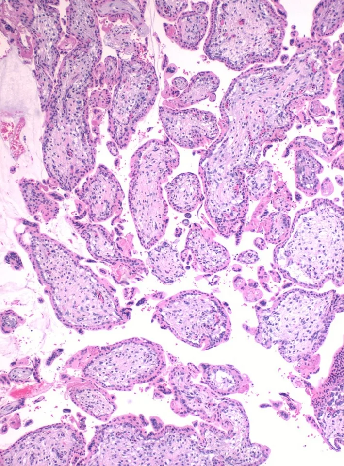
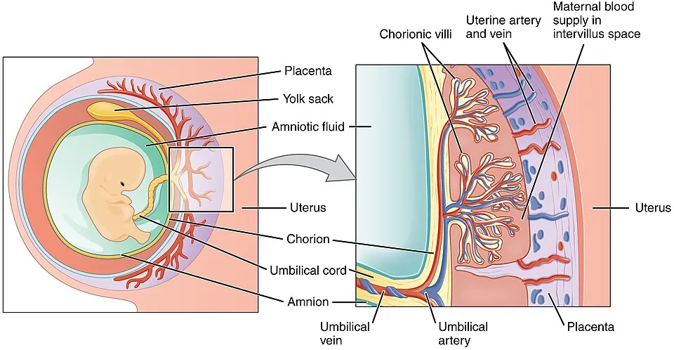
胎盤は、胎児側の絨毛膜有毛部と、母体側の脱落膜基底部からできる。
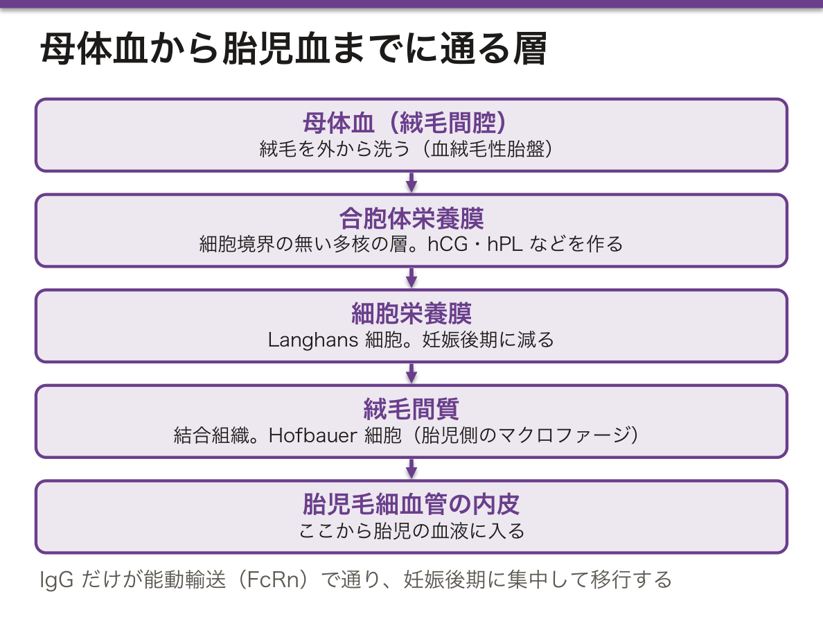
絨毛の構造(外→内)。
- 合胞体栄養膜(シンシチオトロホブラスト)——細胞境界が無い多核の層。母体血に接する。hCG・hPL・エストロゲン・プロゲステロンを作る
- 細胞栄養膜(Langhans 細胞)——妊娠後期に減る
- 絨毛間質——結合組織。Hofbauer 細胞(胎児側のマクロファージ)
- 胎児毛細血管の内皮
母体血は絨毛間腔を流れ、絨毛を外から洗う——血絨毛性胎盤。
IgG のみが能動輸送(FcRn)で通り、妊娠後期に集中して移行する。
IgG が通り IgM が通らないという1行が、繰り返し使われる。臍帯血や新生児血の IgM 高値=胎児が自分で作った=子宮内感染(先天感染の章)——「母から来た抗体と、自分で作った抗体を分けられる」。
そして IgG の移行が妊娠後期に集中することが、早産児の易感染性の理由になる(早産児の章)——「在胎週数が、そのまま免疫の量になっている」。
逆に、移行した IgG が病気を起こすこともある。Basedow 病の TSH 受容体抗体、重症筋無力症の抗体、特発性血小板減少性紫斑病の抗体(妊娠と自己免疫の章)——「母の抗体が、生まれてすぐの症状として現れる」。
その症状は、抗体が消えるとともに治まる。数週から数か月で自然に軽快する——「一過性であることを、あらかじめ家族に伝える」。
合胞体栄養膜が hCG を作ることが、妊娠検査と絨毛性疾患の診断の根拠になる(胞状奇胎の章)——「作る細胞が増えれば、値が異常に高くなる」。
そして hCG は構造が LH に似ており、さらに TSH 受容体も弱く刺激する(妊娠性一過性甲状腺機能亢進症の章)——「似た形をしたホルモンが、隣の受容体を押してしまう」。
通るか通らないかが、治療と診断を決める。
IgG は能動輸送で通るので母体の抗体が胎児へ移行する。新生児が最初の数か月守られるのもこれだが、母体の自己抗体が胎児を侵すこともある——新生児一過性重症筋無力症・新生児 Basedow 病・先天性心ブロック(抗 SS-A 抗体)。
IgM は通らないので、臍帯血の IgM 高値=胎内感染を意味する。
Rh 不適合 — 母体の抗 D 抗体(IgG)が胎盤を通って胎児赤血球を壊す。抗 D 免疫グロブリンの投与で感作を防ぐ。
TORCH(トキソプラズマ・風疹・サイトメガロウイルス・単純ヘルペス・梅毒)が通る。妊娠初期ほど重い。
薬は多くが通る——ヘパリンとインスリンは通らないので妊婦に使える。
English version
Core
The placenta is formed by the fetal chorion frondosum and the maternal decidua basalis.
Structure of a villus (outside in)
- Syncytiotrophoblast — a multinucleate layer with no cell boundaries, in contact with maternal blood. Makes hCG, hPL, oestrogen and progesterone.
- Cytotrophoblast (Langhans cells) — decrease in late pregnancy.
- Villous stroma — connective tissue with Hofbauer cells (fetal macrophages).
- Endothelium of the fetal capillary.
Maternal blood flows in the intervillous space and bathes the villi from outside — a haemochorial placenta.
Only IgG crosses, by active transport (FcRn), and the transfer is concentrated in late pregnancy.
Going further
The one line that IgG crosses and IgM does not is used again and again. A high IgM in cord or neonatal blood = the fetus made it itself = intrauterine infection — antibody from the mother can be told from antibody the baby made.
And that the transfer of IgG is concentrated in late pregnancy is why preterm infants are prone to infection — gestational age is directly the quantity of immunity.
Conversely, transferred IgG can cause disease — TSH receptor antibody in Graves’ disease, the antibody of myasthenia gravis, the antibody of immune thrombocytopenia — the mother’s antibody appearing as a symptom in the newborn. Those symptoms settle as the antibody disappears, resolving spontaneously over weeks to months — tell the family in advance that it is temporary.
That the syncytiotrophoblast makes hCG is the basis of the pregnancy test and of the diagnosis of gestational trophoblastic disease — when the cells that make it increase, the level becomes abnormally high. And hCG resembles LH in structure and also weakly stimulates the TSH receptor — a hormone of similar shape pressing the receptor next door.
In the clinic
What crosses and what does not decides both treatment and diagnosis.
IgG crosses by active transport, so maternal antibody reaches the fetus. This is what protects the newborn for the first months, but maternal autoantibody can also attack the fetus — transient neonatal myasthenia, neonatal Graves’ disease, congenital heart block (anti-SS-A antibody).
IgM does not cross, so a raised IgM in cord blood means intrauterine infection.
Rhesus incompatibility — maternal anti-D (IgG) crosses and destroys fetal red cells. Anti-D immunoglobulin prevents sensitisation.
TORCH (toxoplasma, rubella, cytomegalovirus, herpes simplex, syphilis) crosses; the earlier in pregnancy, the more severe.
Most drugs cross — heparin and insulin do not, and so can be used in pregnancy.
臍帯と卵膜 — 2本の動脈と1本の静脈Chorion
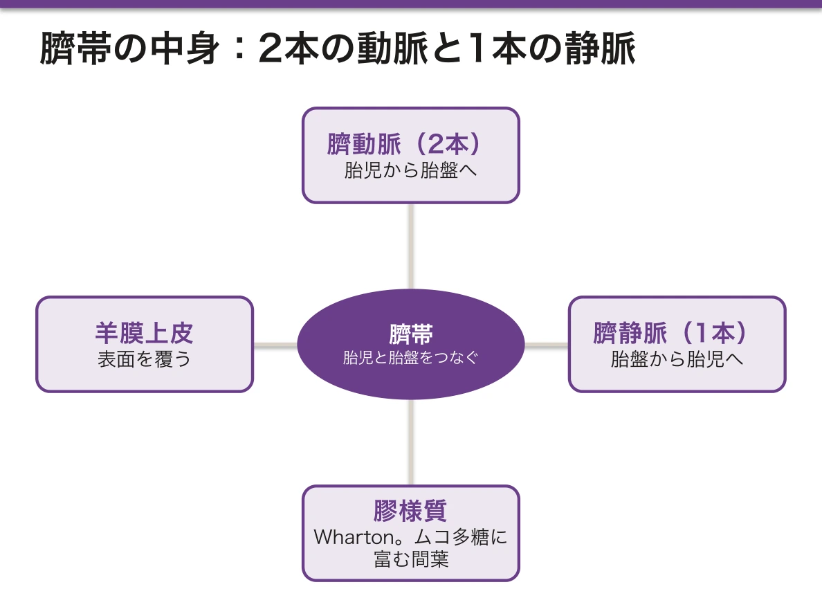
臍帯——臍動脈2本・臍静脈1本。Wharton 膠様質(ムコ多糖に富む間葉組織)に埋まる。表面は羊膜上皮。
- 臍動脈が胎児から胎盤へ(静脈血)、臍静脈が胎盤から胎児へ(動脈血)——名前と血の性質が逆になる
- 単一臍動脈——腎奇形と染色体異常の合併が増える
卵膜は3層——内から羊膜・絨毛膜・脱落膜。
羊水——前半は母体血漿由来、後半は胎児尿が主体。胎児が飲んで肺と腸から吸収する。
- 羊水過少——腎無形成・尿路閉塞・破水・胎盤機能不全
- 羊水過多——食道閉鎖・十二指腸閉鎖・無脳症(飲み込めない)・母体糖尿病
羊水の由来が、そのまま量の異常の鑑別になる。作る側(腎・尿路)が壊れれば過少、片付ける側(嚥下・消化管)が壊れれば過多(羊水異常の章)——「入りと出のどちらが止まったか、で分ける」。
だから羊水過少を見たら、まず腎と尿路を確認する(先天性腎尿路異常の章)——「羊水の量が、胎児の腎機能の代わりの指標になる」。
そして羊水が少ないと、肺が育たない。Potter 症候群では肺低形成が死因になる(腎の発生の章)——「腎の異常で、肺が原因で亡くなる」。
羊水過多で食道閉鎖を疑う筋道は、出生後の対応を変える。胃管が入らないことで確認し、誤嚥させない(新生児の嘔吐の章)——「生まれる前に疑えていれば、最初の授乳を止められる」。
臍帯の血管の名前と血の性質が逆であることは、胎児循環の理解の入口になる(胎児循環の章)——「動脈とは、心臓から出る血管という意味でしかない」。
臍帯血は造血幹細胞の供給源になる。移植に用いられ、HLA の一致度の要求が骨髄より緩い(造血幹細胞移植の章)——「捨てられていたものが、治療の資源になった」。
臍帯動脈は2本ある。単一臍帯動脈は他の奇形(とくに腎・心)を伴うことがあるので精査の契機になる。
臍帯動脈血ガスは分娩時の胎児のアシドーシスを客観的に評価する。pH と base excess で低酸素性虚血性脳症のリスクを見積もる。
Wharton 膠様質が臍帯を守っている。これが乏しいと臍帯が圧迫されやすい。臍帯卵膜付着・前置血管では破水時に胎児血管が破れて急速に失血する——出生前診断ができれば帝王切開で防げる。
臍帯脱出は臍帯が圧迫されて胎児が急速に低酸素になる産科緊急である。破水直後の徐脈で疑う。
臍静脈は新生児蘇生の血管路としても使える。
English version
Core
Umbilical cord — two umbilical arteries and one umbilical vein, embedded in Wharton’s jelly (mesenchyme rich in mucopolysaccharide) and covered by amniotic epithelium.
- The umbilical arteries run from fetus to placenta (venous blood), the umbilical vein from placenta to fetus (arterial blood) — the name and the nature of the blood are reversed.
- A single umbilical artery — associated with renal malformation and chromosomal abnormality.
The fetal membranes are three layers — amnion, chorion, decidua, from within.
Amniotic fluid — derived from maternal plasma in the first half, chiefly fetal urine in the second. The fetus swallows it and absorbs it from lung and gut.
- Oligohydramnios — renal agenesis, urinary obstruction, ruptured membranes, placental insufficiency.
- Polyhydramnios — oesophageal atresia, duodenal atresia, anencephaly (it cannot be swallowed), maternal diabetes.
Going further
The origin of the amniotic fluid is itself the differential of an abnormal volume. Break the producing side (kidney, urinary tract) and there is too little; break the clearing side (swallowing, gut) and there is too much — divide by whether the inflow or the outflow has stopped.
So on seeing oligohydramnios, check the kidneys and urinary tract first — the volume of fluid serves as a proxy for fetal renal function. And with too little fluid the lungs do not develop: in Potter sequence pulmonary hypoplasia is the cause of death — death from the lungs because of an abnormality of the kidney.
Reasoning from polyhydramnios to oesophageal atresia changes what is done after birth: confirm by failure to pass a nasogastric tube, and prevent aspiration — if it was suspected before delivery, the first feed can be withheld.
That the names of the cord vessels are the reverse of the nature of the blood is the entrance to understanding the fetal circulation — "artery" means no more than a vessel leaving the heart.
Cord blood is a source of haematopoietic stem cells, used in transplantation with a less stringent HLA match than marrow — what used to be discarded has become a therapeutic resource.
In the clinic
There are two umbilical arteries. A single umbilical artery may accompany other malformations (particularly renal and cardiac) and is a reason to investigate.
Umbilical artery blood gas gives an objective assessment of fetal acidosis at delivery. pH and base excess estimate the risk of hypoxic–ischaemic encephalopathy.
Wharton’s jelly protects the cord; where it is scanty the cord is compressed more easily. In velamentous cord insertion and vasa praevia, fetal vessels rupture when the membranes break and blood is lost rapidly — antenatal diagnosis allows prevention by caesarean section.
Cord prolapse is an obstetric emergency in which the cord is compressed and the fetus becomes hypoxic rapidly. Suspect it from bradycardia immediately after the membranes rupture.
The umbilical vein can also be used as vascular access in neonatal resuscitation.
脱落膜 — 母体側が、受け入れるために作り変わるDecidua
着床に伴い、子宮内膜の間質細胞が大きく円形になり、グリコーゲンと脂質を蓄える(脱落膜化)。プロゲステロンが起こす。
位置で3つに呼び分ける。
| 名称 | 場所 | その後 |
|---|---|---|
| 基底脱落膜 | 胚の下、絨毛膜有毛部と向き合う | 胎盤の母体面になる |
| 被包脱落膜 | 胚を子宮腔側から覆う | 伸びて薄くなり、消える |
| 壁側脱落膜 | 残りの子宮壁 | 被包脱落膜と癒合し子宮腔が閉じる |
胎盤の母体面は、基底脱落膜が絨毛の束を分けて 15〜20 個の分葉(cotyledon)に見せている。
栄養を蓄えることが、着床初期の胚への供給源になる。
脱落膜は「受け入れる」ためだけでなく、「入り込みすぎるのを止める」ためにある。栄養膜の浸潤は本来きわめて侵襲的で、脱落膜がその深さを制限している——「胎盤は、制御された浸潤である」。
だから脱落膜が欠けると、絨毛が筋層へ入り込む。癒着胎盤・嵌入胎盤・穿通胎盤は、この歯止めが無い場所(帝王切開の瘢痕・前置胎盤)で起こる(産後出血の章)——「前回の帝王切開が、次の妊娠の出血の危険になる」。
浸潤が制御されているという見方は、腫瘍と対比すると分かりやすい。絨毛性疾患(胞状奇胎・絨毛癌)は、その制御が外れた状態になる(絨毛性疾患の章)——「もともと浸潤する能力を持っている細胞なので、外れると速い」。
被包脱落膜が消えて子宮腔が閉じることは、超音波所見の暦になる。妊娠 14〜16 週頃に2枚の膜が癒合する(胎盤・胎膜の章)——「膜の枚数を数えられるのは、その前になる」。
脱落膜が無い/薄いところに着床すると、絨毛が深く入り込む。
癒着胎盤 — 脱落膜の欠損により絨毛が子宮筋層へ食い込む。侵入の深さで accreta/increta/percreta に分ける。前置胎盤+帝王切開の既往が最大の危険因子で、瘢痕部に脱落膜が乏しいことが理由である。分娩時に胎盤が剥がれず、大量出血になる。出生前に診断して、輸血と子宮摘出まで準備して臨む。
前置胎盤 — 無痛性の性器出血。内診は禁忌で、経腟超音波で診断する。
常位胎盤早期剥離 — 持続する腹痛と子宮の板状硬。出血が外に出ないことがある(後血腫)ので出血量と症状が合わない。DIC を合併する。「痛い出血=早剥、痛くない出血=前置」と対で覚える。
English version
Core
With implantation the stromal cells of the endometrium become large and rounded and store glycogen and lipid (decidualisation), driven by progesterone.
| Name | Position | Fate |
|---|---|---|
| Decidua basalis | Beneath the embryo, facing the chorion frondosum | Becomes the maternal surface of the placenta |
| Decidua capsularis | Covers the embryo on the side of the uterine cavity | Stretches, thins and disappears |
| Decidua parietalis | The rest of the uterine wall | Fuses with the capsularis, obliterating the cavity |
On the maternal surface of the placenta, the decidua basalis divides the bundles of villi into 15–20 cotyledons.
The stored nutrients are the source of supply to the embryo in early implantation.
Going further
The decidua exists not only to accept but to stop the invasion going too far. Trophoblastic invasion is by nature extremely aggressive, and the decidua limits its depth — the placenta is a controlled invasion.
So where the decidua is absent, the villi grow into the myometrium. Placenta accreta, increta and percreta occur where that brake is missing (a caesarean scar, placenta praevia) — a previous caesarean becoming the haemorrhagic risk of the next pregnancy.
The view that the invasion is controlled is clearest by contrast with tumour. Gestational trophoblastic disease (hydatidiform mole, choriocarcinoma) is that control coming off — the cells possess the capacity to invade in the first place, so once released they move fast.
That the capsularis disappears and the cavity closes is a calendar for ultrasound findings: the two membranes fuse at about 14–16 weeks — the number of membranes can be counted only before that.
In the clinic
Where implantation occurs on absent or thin decidua, the villi burrow deep.
Placenta accreta — an absence of decidua lets the villi bite into the myometrium. Divided by depth into accreta / increta / percreta. Placenta praevia with a previous caesarean is the greatest risk factor, because the scar bears little decidua. The placenta does not separate at delivery and massive haemorrhage follows. Diagnose antenatally and prepare for transfusion and hysterectomy.
Placenta praevia — painless vaginal bleeding. Digital examination is contraindicated; diagnose by transvaginal ultrasound.
Placental abruption — continuous abdominal pain and a board-like uterus. The bleeding may not appear externally (retroplacental haematoma), so the visible loss does not match the clinical state. DIC accompanies it.
Remember them as a pair: painful bleeding = abruption, painless bleeding = praevia.
栄養膜 — 2種類の細胞が、役割を分けているTrophoblastus
着床した胚の外側の細胞は、2層に分かれる。
| 細胞性栄養膜 | 合胞体性栄養膜 | |
|---|---|---|
| 位置 | 内側 | 外側(母体血に接する) |
| 細胞境界 | あり | 無い(多核の合胞体) |
| 分裂 | する(供給源) | しない |
| 働き | 予備・幹細胞 | ホルモン産生・物質交換・侵入 |
妊娠が進むと細胞性栄養膜は減り、関門が薄くなる——だから後期ほど物質が通りやすい。
絨毛外栄養膜は脱落膜へ侵入し、らせん動脈の壁を置き換えて低抵抗の血管に作り替える。
合胞体であることには、はっきりした利点がある。細胞間の隙間が無いので、母体血と接する面に「すき間からの通り道」が存在しない——「1枚の連続した膜が、そのまま関門になっている」。
そして細胞境界が無いことは、免疫からの回避にも働く。母体のリンパ球が攻撃する標的(古典的 HLA クラスI)を、この面はほとんど出していない(移植免疫の章)——「半分は他人の遺伝子なのに拒絶されない理由の一つになる」。
らせん動脈の作り替えが不十分だと、妊娠高血圧腎症になる。血管抵抗が下がらず胎盤が虚血に陥り、sFlt-1 などが母体循環へ出て全身の内皮を傷める(妊娠高血圧症候群の章)——「母体の高血圧と蛋白尿の起点が、胎盤側の血管の作り替えにある」。
関門が後期ほど薄くなることは、薬と感染の時期の話に直結する。器官形成期は催奇形性が問題になり、後期は移行量そのものが問題になる(臨界期の章)——「いつ曝露したかで、心配する内容が入れ替わる」。
合胞体栄養膜が hCG を作る。だからこの細胞が増える病態で hCG が異常高値になる。
胞状奇胎 — 全胞状奇胎は父性のみの二倍体で、胎児成分を欠く。hCG が異常高値・子宮が週数より大きい・両側の卵巣に黄体嚢胞。hCG が甲状腺を刺激して甲状腺機能亢進を起こすことがある。
絨毛癌 — 絨毛構造を作らずに栄養膜細胞が増殖する悪性腫瘍。血行性に肺・脳へ早期に転移するが、化学療法(メトトレキサート)に極めてよく反応する。hCG が治療効果の指標になるので娩出後も hCG を追跡する。
細胞性栄養膜が供給源で、妊娠後期には薄くなり、胎盤関門が薄くなる——後期ほど物質が通りやすい。
English version
Core
The outer cells of the implanted embryo divide into two layers.
| Cytotrophoblast | Syncytiotrophoblast | |
|---|---|---|
| Position | Inner | Outer (in contact with maternal blood) |
| Cell boundaries | Present | None (a multinucleate syncytium) |
| Divides | Yes (the source) | No |
| Function | Reserve, stem cells | Hormone production, exchange of substances, invasion |
As pregnancy advances the cytotrophoblast diminishes and the barrier thins — so substances cross more readily in late pregnancy.
The extravillous trophoblast invades the decidua and replaces the walls of the spiral arteries, remodelling them into low-resistance vessels.
Going further
Being a syncytium has a definite advantage. With no gaps between cells, the surface in contact with maternal blood has no route through a crack — a single continuous membrane serving as the barrier.
And the absence of cell boundaries also serves evasion of the immune system. This surface expresses almost none of the target of maternal lymphocytes (classical HLA class I) — one of the reasons half a set of foreign genes is not rejected.
If the remodelling of the spiral arteries is insufficient, pre-eclampsia follows. The vascular resistance does not fall, the placenta becomes ischaemic, and factors such as sFlt-1 enter the maternal circulation and damage the endothelium throughout the body — the origin of the mother’s hypertension and proteinuria lying in the remodelling of vessels on the placental side.
That the barrier thins in late pregnancy bears directly on the timing of drugs and infections. In organogenesis teratogenicity is the concern; later it is the quantity transferred — when the exposure occurred changes what one worries about.
In the clinic
The syncytiotrophoblast makes hCG. So in conditions where these cells increase, hCG becomes abnormally high.
Hydatidiform mole — a complete mole is diploid and wholly paternal, with no fetal parts. Abnormally high hCG, a uterus large for dates, bilateral theca lutein cysts. hCG may stimulate the thyroid and cause hyperthyroidism.
Choriocarcinoma — a malignant proliferation of trophoblast that forms no villi. It metastasises early and haematogenously to lung and brain but responds extremely well to chemotherapy (methotrexate). hCG is the index of response, so it is followed after delivery.
The cytotrophoblast is the source and thins in late pregnancy, so the placental barrier thins — substances cross more readily late.
胎盤のホルモン — 一時的にできた、最大の内分泌器官Placenta
合胞体性栄養膜が作る。妊娠中の内分泌の中心になる。
| ホルモン | 働き | 時期 |
|---|---|---|
| hCG(ヒト絨毛性ゴナドトロピン) | 黄体を維持しプロゲステロンを出させる | 8〜10週で頂点、以後低下 |
| hPL(ヒト胎盤性ラクトゲン) | 母体をインスリン抵抗性にし、胎児へブドウ糖を回す | 妊娠とともに増え続ける |
| プロゲステロン | 子宮の収縮を抑え、妊娠を維持 | 増え続ける |
| エストロゲン(主にエストリオール) | 子宮と乳腺の発育 | 増え続ける |
妊娠 7〜9週で、プロゲステロンの供給元が黄体から胎盤へ移る(黄体・胎盤移行)。
エストリオールは胎盤だけでは作れない——胎児副腎が出す DHEA-S を材料に、胎児肝と胎盤が分担する(胎児胎盤系)。
hCG が LH と α鎖を共有していることが、検査と症状の両方を説明する。だから hCG は LH 受容体を刺激して黄体を保ち、同時に TSH 受容体も弱く刺激する(ホルモンの化学の章)——「hCG が高い妊娠初期に、一過性の甲状腺機能亢進が起こる」。
hCG が極端に高い病態では、これが強く出る。胞状奇胎・多胎・重症妊娠悪阻(甲状腺の章)——「妊娠初期の甲状腺の数値を、そのまま Basedow 病と読まない」。
hPL の役割は、母体の代謝を犠牲にして胎児に糖を回すことにある。母体はインスリン抵抗性になり、脂肪をエネルギー源に切り替える(インスリン抵抗性の章)——「妊娠糖尿病が妊娠後期に出る理由が、胎盤の大きさと対応している」。
だから妊娠糖尿病は、分娩で胎盤が出れば速やかに改善する(妊娠糖尿病の章)——「原因の臓器が、体から出ていく病気になる」。
エストリオールが胎児と胎盤の共同作業であることは、かつて胎児の健康を測る指標に使われた(胎児評価の章)——「今は超音波と心拍に置き換わったが、成り立ちを知ると理屈が分かる」。
胎盤は一時的な内分泌器官として、母体の生理を作り変える。
hPL(ヒト胎盤性ラクトゲン)はインスリン抵抗性を作る。母体の糖を胎児へ回すための仕組みだが、耐えられなければ妊娠糖尿病になる。妊娠中期以降に発症するのは hPL が増える時期と一致するからである。
hCG は妊娠 8〜10週で頂点——つわりのピークと一致する。
プロゲステロンが子宮の収縮を抑え、妊娠を維持する。平滑筋を広く弛緩させるので便秘・胃食道逆流・尿管拡張(腎盂腎炎が増える)も起こる——1つのホルモンで妊婦のマイナートラブルの多くが説明できる。
エストロゲンは胎児副腎と胎盤が協働して作る(胎児胎盤系)ので、エストリオールは胎児の状態を映す。
English version
Core
Made by the syncytiotrophoblast. The centre of the endocrinology of pregnancy.
| Hormone | Action | Time course |
|---|---|---|
| hCG | Maintains the corpus luteum and keeps progesterone flowing | Peaks at 8–10 weeks, then falls |
| hPL (human placental lactogen) | Makes the mother insulin resistant, diverting glucose to the fetus | Rises continuously with the pregnancy |
| Progesterone | Suppresses uterine contraction and maintains the pregnancy | Rises continuously |
| Oestrogen (chiefly oestriol) | Development of uterus and breast | Rises continuously |
At 7–9 weeks the source of progesterone shifts from corpus luteum to placenta (the luteo-placental shift).
Oestriol cannot be made by the placenta alone — DHEA-S from the fetal adrenal is the raw material, and fetal liver and placenta share the work (the fetoplacental unit).
Going further
That hCG shares its α chain with LH explains both the test and the symptoms. hCG stimulates the LH receptor and preserves the corpus luteum, and weakly stimulates the TSH receptor as well — a transient hyperthyroidism in early pregnancy when hCG is high.
Where hCG is extremely high this is pronounced — hydatidiform mole, multiple pregnancy, hyperemesis gravidarum — do not read a thyroid result in early pregnancy straight off as Graves’ disease.
The role of hPL is to sacrifice maternal metabolism to divert glucose to the fetus. The mother becomes insulin resistant and switches to fat as her fuel — the reason gestational diabetes appears late corresponds to the size of the placenta. So gestational diabetes improves promptly once the placenta is delivered — a disease whose causative organ leaves the body.
That oestriol is a joint work of fetus and placenta was once used as an index of fetal wellbeing — now replaced by ultrasound and the heart rate, but knowing how it comes about makes the logic clear.
In the clinic
The placenta, as a temporary endocrine organ, remakes the mother’s physiology.
hPL creates insulin resistance. It is a mechanism for diverting maternal glucose to the fetus, but where it cannot be tolerated, gestational diabetes results. It appears from mid-pregnancy because that coincides with the rise in hPL.
hCG peaks at 8–10 weeks — coinciding with the peak of morning sickness.
Progesterone suppresses uterine contraction and maintains the pregnancy. It relaxes smooth muscle widely, so it also causes constipation, gastro-oesophageal reflux and ureteric dilatation (hence more pyelonephritis) — one hormone explaining most of the minor troubles of pregnancy.
Oestrogen is made jointly by fetal adrenal and placenta (the fetoplacental unit), so oestriol mirrors the state of the fetus.
胎児の造血は、場所を3回変えるSanguis umbilicalis
| 時期 | 場所 | 作られるもの |
|---|---|---|
| 3〜8 週 | 卵黄嚢(血島) | 有核の赤芽球。ヘモグロビンは Gower・Portland |
| 6 週〜出生前 | 肝(次いで脾) | 主に HbF(α₂γ₂) |
| 4〜5 か月以降 | 骨髄 | 出生後の主座。HbA(α₂β₂)へ切り替わる |
ヘモグロビンの鎖は、順に切り替わる——ε・ζ →(γ)→ β。出生時は HbF が約 70%、生後6か月でほぼ HbA になる。
臍帯血には造血幹細胞(CD34 陽性細胞)が多く含まれる。
α鎖は胎生期からずっと必要になる。
「α鎖はいつも要るが、β鎖は出生後から要る」という一点が、サラセミアの発症時期を決めている。
| α サラセミア | β サラセミア | |
|---|---|---|
| 発症 | 胎児期から(胎児水腫・Bart 水腫) | 生後 3〜6 か月(HbF が減ってから) |
(サラセミアの章)——「生まれてすぐは元気だった子が、半年で貧血になる理由が、この切り替えにある」。
だから HbF を再び増やす薬が、治療になる(ヒドロキシ尿素の項)——「胎児期の遺伝子を、もう一度使わせる」。
臍帯血移植は、この造血幹細胞を使う(造血幹細胞移植の章)——「HLA の一致がやや緩くてよいが、細胞数が限られるので生着が遅い」。
髄外造血は、胎児期の場所へ戻る現象になる(造血のニッチの項)——「骨髄が使えなくなると、肝と脾が再び造血を始める」。
造血の場所が3回変わることが、ヘモグロビンの型の変化と対応する。
HbF(α2γ2)は酸素親和性が高い——2,3-DPG と結合しにくいためで、母体から酸素を受け取るのに有利である。出生後 6か月頃までに HbA へ置き換わる。
だからβ鎖の異常症は生後数か月してから発症する。鎌状赤血球症・βサラセミアはγ鎖が減ってβ鎖に置き換わる時期に症状が出る。ヒドロキシウレアが HbF を増やして鎌状赤血球症を改善させるのはこの仕組みを逆用している。
α鎖の異常は胎児期から症状が出る——α鎖は HbF にも HbA にも要るからである。α サラセミアの最重症型(Hb Bart 胎児水腫)は子宮内で致死的。いつ発症するかが、どの鎖の異常かを教える。
肝と脾は髄外造血の場に戻れる。
English version
Core
| Time | Site | What is made |
|---|---|---|
| 3–8 weeks | Yolk sac (blood islands) | Nucleated erythroblasts. Haemoglobins Gower and Portland |
| 6 weeks to before birth | Liver (then spleen) | Chiefly HbF (α₂γ₂) |
| From 4–5 months | Bone marrow | The main site after birth. Switches to HbA (α₂β₂) |
The haemoglobin chains switch in order — ε, ζ → (γ) → β. At birth HbF is about 70 per cent, and by six months it is almost all HbA.
Cord blood is rich in haematopoietic stem cells (CD34-positive). The α chain is needed from fetal life onwards throughout.
Going further
The single point that the α chain is always needed while the β chain is needed only after birth decides when the thalassaemias present.
| α thalassaemia | β thalassaemia | |
|---|---|---|
| Onset | From fetal life (hydrops, Hb Bart’s) | At 3–6 months (once HbF has fallen) |
Here is why a child who was well at birth becomes anaemic at six months — it lies in the switch.
So a drug that raises HbF again is a treatment — making the fetal gene be used once more.
Cord blood transplantation uses these stem cells — the HLA match may be a little looser, but the cell number is limited, so engraftment is slow.
Extramedullary haemopoiesis is a return to the sites of fetal life — when the marrow cannot be used, liver and spleen begin to make blood again.
In the clinic
That the site of haemopoiesis changes three times corresponds to the changes in the type of haemoglobin.
HbF (α₂γ₂) has a high oxygen affinity — it binds 2,3-DPG poorly, which favours taking oxygen from the mother. It is replaced by HbA by about six months.
So disorders of the β chain present some months after birth. Sickle cell disease and β thalassaemia become symptomatic as the γ chain falls and is replaced by β. That hydroxyurea improves sickle cell disease by raising HbF turns this mechanism to advantage.
Disorders of the α chain are symptomatic from fetal life — because the α chain is needed in both HbF and HbA. The most severe form of α thalassaemia (Hb Bart’s hydrops fetalis) is lethal in utero.
When it presents tells you which chain is abnormal. Liver and spleen can return to being sites of haemopoiesis.
胎盤が剥がれない — 脱落膜が欠けているということ
分娩後に胎盤が剥がれるのは、脱落膜の海綿層で裂けるからである。ここに剥がれるための層があらかじめ用意されている。
脱落膜が欠けている場所では、絨毛が子宮筋層に直接食い込む。剥がれる面が無いので、無理に剥がすと大出血になる。
| 癒着胎盤 | 絨毛が子宮筋層に接している | 約 75% |
| 嵌入胎盤 | 筋層の中まで入る | 約 18% |
| 穿通胎盤 | 筋層を貫いて漿膜へ、時に膀胱まで | 約 7% |
危険因子ははっきりしている——前置胎盤と、帝王切開の既往。瘢痕には脱落膜が十分に作られないためである。帝王切開の回数が増えるほど危険が上がる。
だから「前置胎盤+帝王切開の既往」の組合せは、分娩前に必ず評価する。超音波と MRI で癒着を疑い、大量出血に備えた体制のある施設で、計画的に手術する。予期せぬ大出血として起きるか、準備された手術になるかで、母体の予後が変わる。
剥がそうとしないことが原則になる。無理に用手剥離すると子宮筋層ごと裂けて止血できなくなるので、胎盤を残したまま子宮全摘を選ぶことが多い。「胎盤が出ないから引っ張る」が最も危険な対応である。
剥離のしかたに関わる異常は、他にもある。
- 常位胎盤早期剥離——剥がれてはいけない時期に剥がれる。脱落膜の血管が破れて後血腫を作る。子宮が板のように硬くなり、外に出る血の量と病態が合わない
- 子宮内反——胎盤を強く牽引したときに子宮が裏返る。ショックと激痛を起こす
- 胎盤遺残——一部が残ると子宮が収縮できず出血が続く。娩出した胎盤は必ず広げて、欠けがないか確認する
分娩後の出血の原因は 4 つ(4 つの T)——子宮収縮不全・遺残・産道裂傷・凝固異常。組織のどこに問題があるかで、対応が変わる。
帝王切開の既往がある妊婦では、胎盤の付着部位を必ず確認する。前置なら、分娩の場所と体制をあらかじめ決めておく。
English version
Core
The placenta separates after delivery because it splits within the spongy layer of the decidua — a plane of separation prepared in advance.
Where the decidua is absent, the villi bite directly into the myometrium. There is no plane to separate along, so forcing it off causes massive haemorrhage.
| Depth | Share | |
|---|---|---|
| Accreta | Villi in contact with the myometrium | About 75% |
| Increta | Into the myometrium | About 18% |
| Percreta | Through the myometrium to the serosa, sometimes to the bladder | About 7% |
The risk factors are clear — placenta praevia and previous caesarean section, because a scar does not form adequate decidua. The more caesareans, the higher the risk.
Going further
So the combination of placenta praevia with a previous caesarean must always be assessed before delivery. Suspect accreta on ultrasound and MRI and operate electively in a unit prepared for massive haemorrhage. Whether it happens as an unexpected haemorrhage or as a prepared operation changes the maternal outcome.
The rule is not to try to separate it. Forced manual removal tears the myometrium with it and bleeding cannot be controlled, so hysterectomy with the placenta left in place is often chosen. "The placenta will not come, so pull" is the most dangerous response.
There are other abnormalities of separation.
- Placental abruption — separation at a time when it should not. The decidual vessels rupture and form a retroplacental haematoma. The uterus becomes board-like, and the visible blood loss does not match the clinical state.
- Uterine inversion — the uterus turns inside out when the placenta is pulled hard. Shock and severe pain.
- Retained placenta — a retained fragment prevents the uterus contracting and bleeding continues. Always spread the delivered placenta out and check that nothing is missing.
The causes of postpartum haemorrhage are four (the four Ts) — tone, tissue, trauma, thrombin. Where in the tissue the problem lies changes the response.
In the clinic
In a woman with a previous caesarean, always confirm where the placenta is attached. If it is praevia, decide in advance where and with what team she will deliver.
まとめ
| 乳腺 — 完成するのは妊娠してから | 複合管状胞状腺。15〜20の葉に分かれ、それぞれが乳管洞を経て乳頭に開く。 |
|---|---|
| 乳腺の導管系 — 病気が起きる単位で捉える | 乳房は15〜20個の腺葉が集まったもので、腺葉ごとに1本の乳管が乳頭へ開く。 |
| 乳汁分泌 — 2つのホルモンが、別々の細胞に効く | 作らせるホルモンと、出させるホルモンが別になっている。 |
| 外陰の腺と粘膜 — 皮膚から粘膜へ移り変わる場所 | 外陰は、外側から内側へ性質が変わる。部位上皮付属器大陰唇角化重層扁平毛・脂腺・汗腺・アポクリン腺(皮膚と同じ)小陰唇薄く角化する重層扁平毛が無い。 |
| 乳腺は、間質のほうが病気を決めることがある | 乳腺の病変というと上皮を考えるが、乳腺は間質の割合が大きい臓器で、間質そのものが腫瘍になる。 |
| 絨毛と胎盤関門 — 何が通り、何が通らないか | 胎盤は、胎児側の絨毛膜有毛部と、母体側の脱落膜基底部からできる。 |
| 臍帯と卵膜 — 2本の動脈と1本の静脈 | 臍帯——臍動脈2本・臍静脈1本。Wharton 膠様質(ムコ多糖に富む間葉組織)に埋まる。 |
| 脱落膜 — 母体側が、受け入れるために作り変わる | 着床に伴い、子宮内膜の間質細胞が大きく円形になり、グリコーゲンと脂質を蓄える(脱落膜化)。 |
| 栄養膜 — 2種類の細胞が、役割を分けている | 着床した胚の外側の細胞は、2層に分かれる。 |
| 胎盤のホルモン — 一時的にできた、最大の内分泌器官 | 合胞体性栄養膜が作る。妊娠中の内分泌の中心になる。 |
| 胎児の造血は、場所を3回変える | 時期場所作られるもの3〜8 週卵黄嚢(血島)有核の赤芽球。 |
| 胎盤が剥がれない — 脱落膜が欠けているということ | 分娩後に胎盤が剥がれるのは、脱落膜の海綿層で裂けるからである。 |
このノートは医学生が授業の内容を自分の言葉でまとめたもので、診療の判断に使うものではありません。誤りに気づいたらお知らせください。