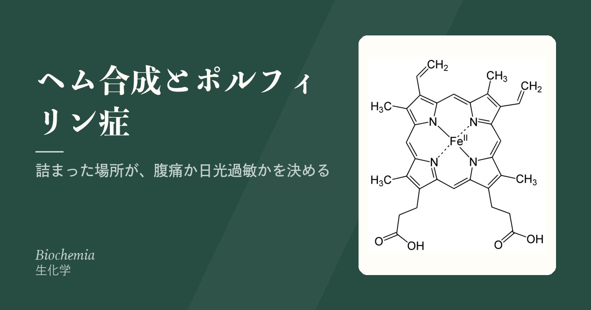
ヘム合成は8つの酵素で進み、ミトコンドリアで始まり、細胞質を経て、またミトコンドリアで終わる。この行き来のせいで、途中の中間体が漏れ出しやすい。
ポルフィリン症はどの酵素が詰まるかで、まったく違う顔を見せる。上流で詰まれば神経症状(腹痛)、下流で詰まれば皮膚症状(日光過敏)——溜まる分子の性質から、この振り分けが導ける。
- 8段階の経路と、律速のかかり方
- 急性型と皮膚型 — 溜まる分子の性質が症状を決める
- 急性ポルフィリン症の発作 — 何が誘発し、何で止めるか
- 鉄芽球性貧血 — ヘムが作れないと、鉄が行き場を失う
- ヘムは、赤血球だけのものではない
- ポルフィリン症を疑ったら、いつ・何を・どう採るか
8段階の経路と、律速のかかり方
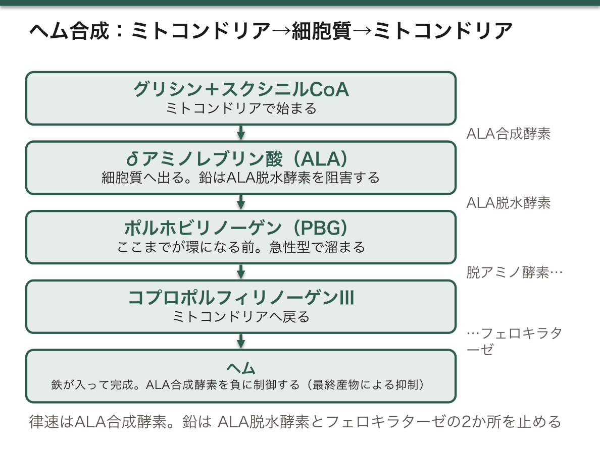
- ミトコンドリア:グリシン+スクシニルCoA →(δアミノレブリン酸合成酵素、律速)→ δアミノレブリン酸
- 細胞質:δアミノレブリン酸 →(δアミノレブリン酸脱水酵素)→ ポルホビリノーゲン →(ポルホビリノーゲン脱アミノ酵素)→ … → コプロポルフィリノーゲンIII
- ミトコンドリア:… →(フェロキラターゼ、鉄を入れる)→ ヘム
- ヘムがδアミノレブリン酸合成酵素を負に制御する(最終産物による抑制)
- 鉛は δアミノレブリン酸脱水酵素とフェロキラターゼを阻害する → 鉛中毒ではδアミノレブリン酸と遊離赤血球プロトポルフィリンが上がる
- 肝ではチトクロームP450の需要が、骨髄ではヘモグロビン合成が主な用途
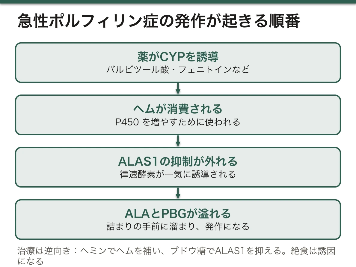
「律速酵素が最終産物で抑えられている」ことが、急性ポルフィリン症の発作の仕組みそのものである。
- 薬物などで肝のチトクロームP450が誘導される
- ヘムが消費される
- 抑えが外れてδアミノレブリン酸合成酵素が活性化する
- 詰まっている手前の中間体が大量に作られる → 発作
だから発作の治療は「ヘムを外から入れて、律速酵素を抑え込むこと」である(ヘミン製剤)。同時にブドウ糖を大量に投与する——糖はδアミノレブリン酸合成酵素を抑制するためで、絶食が発作の誘因になるのもここから説明される。
鉛中毒の所見も、この経路で全部説明できる。
- 小球性貧血——ヘムが作れない
- 好塩基性斑点——ピリミジン5ヌクレオチダーゼも阻害され、リボソームRNAが分解されずに残る
- 腹痛・末梢神経障害(下垂手)——δアミノレブリン酸の神経毒性
- 歯肉の鉛縁
鉄が足りないわけではないので、鉄剤は効かない。血清鉄はむしろ高い——フェロキラターゼが止まって鉄が使われないからである。治療はキレート(エデト酸カルシウムなど)と曝露源の除去である。
English version
Core
- Mitochondrion: glycine + succinyl-CoA →(δ-aminolevulinate synthase, rate-limiting)→ δ-aminolevulinate
- Cytosol: δ-aminolevulinate →(ALA dehydratase)→ porphobilinogen →(porphobilinogen deaminase)→ … → coproporphyrinogen III
- Mitochondrion: … →(ferrochelatase, inserting iron)→ haem
- Haem negatively regulates δ-aminolevulinate synthase (end-product inhibition).
- Lead inhibits ALA dehydratase and ferrochelatase → in lead poisoning δ-aminolevulinate and free erythrocyte protoporphyrin rise.
- In the liver the chief use is the demand of cytochrome P450, in the marrow haemoglobin synthesis.
Going further
That the rate-limiting enzyme is suppressed by the end product is the very mechanism of an attack of acute porphyria.
- A drug or the like induces hepatic cytochrome P450.
- Haem is consumed.
- The suppression is lifted and δ-aminolevulinate synthase is activated.
- A great deal of the intermediate before the block is made → the attack.
So the treatment of an attack is "to put haem in from outside and suppress the rate-limiting enzyme" (haemin preparations). Large amounts of glucose are given at the same time — because sugar suppresses δ-aminolevulinate synthase, which also explains why fasting is a trigger of attacks.
The findings of lead poisoning can all be explained on this pathway too.
- Microcytic anaemia — haem cannot be made.
- Basophilic stippling — pyrimidine 5′-nucleotidase is also inhibited, so ribosomal RNA remains undegraded.
- Abdominal pain and peripheral neuropathy (wrist drop) — the neurotoxicity of δ-aminolevulinate.
- A lead line on the gums.
Iron is not short, so iron preparations do not work. The serum iron is if anything high — because ferrochelatase has stopped and the iron is not used. Treatment is chelation (calcium edetate and others) and removal of the source of exposure.
急性型と皮膚型 — 溜まる分子の性質が症状を決める
| 急性型 | 皮膚型 | |
|---|---|---|
| 代表 | 急性間欠性ポルフィリン症 | 晩発性皮膚ポルフィリン症・骨髄性プロトポルフィリン症 |
| 詰まる場所 | 経路の上流 | 下流 |
| 溜まるもの | δアミノレブリン酸・ポルホビリノーゲン(環になる前) | ポルフィリン(環ができている) |
| 主症状 | 腹痛・精神症状・末梢神経障害・頻脈・高血圧 | 日光過敏・水疱・瘢痕・多毛 |
| 皮膚症状 | 無い | あり |
- 急性間欠性ポルフィリン症では尿が放置すると赤褐色になる(ポルホビリノーゲンが酸化重合する)
- 誘因:薬物(バルビツール酸・スルホンアミド・一部の抗てんかん薬)・飲酒・絶食・感染・月経周期
「なぜ環ができていると光に反応するのか」——ポルフィリン環は共役二重結合が閉じた構造で、可視光(約400 nm、Soret帯)を強く吸収して励起され、活性酸素を生じる。環になる前のδアミノレブリン酸とポルホビリノーゲンは光を吸収しない。
だから「上流で詰まる=皮膚症状が出ない」。分子の光学的性質が、そのまま臨床像を決めている。
急性間欠性ポルフィリン症は「腹痛の鑑別で最も見逃される疾患の一つ」である。
- 激しい腹痛なのに、腹膜刺激徴候が無く、圧痛も乏しい(内臓神経の障害であり、腹腔内に炎症が無い)
- 低ナトリウム血症を伴うことが多い(抗利尿ホルモン不適合分泌)
- 精神症状や四肢麻痺が加わる
- 若い女性に多く、月経周期と関連する
- 不要な開腹手術を受けてしまうことがある——そして麻酔薬が発作を悪化させる
診断は発作時の尿ポルホビリノーゲンである。発作中でなければ正常化しうるので、疑ったその場で採る。
晩発性皮膚ポルフィリン症は後天性の要因が強い——C型肝炎・飲酒・鉄過剰・エストロゲンが引き金になる。瀉血と少量のヒドロキシクロロキンが治療になるので、「手背の水疱と脆弱性」を見たら肝炎ウイルスを調べるという繋がりを覚えておく。
English version
Core
| Acute type | Cutaneous type | |
|---|---|---|
| Representative | Acute intermittent porphyria | Porphyria cutanea tarda, erythropoietic protoporphyria |
| Where the block is | Upstream in the pathway | Downstream |
| What accumulates | δ-Aminolevulinate, porphobilinogen (before the ring) | Porphyrins (the ring is formed) |
| Chief symptoms | Abdominal pain, psychiatric symptoms, peripheral neuropathy, tachycardia, hypertension | Photosensitivity, blisters, scarring, hypertrichosis |
| Skin symptoms | None | Present |
- In acute intermittent porphyria the urine turns red-brown on standing (porphobilinogen oxidises and polymerises).
- Triggers: drugs (barbiturates, sulphonamides, some antiepileptics), alcohol, fasting, infection, the menstrual cycle.
Going further
Why does having the ring make it react to light? The porphyrin ring is a closed structure of conjugated double bonds and absorbs visible light strongly (about 400 nm, the Soret band), is excited and generates reactive oxygen. δ-Aminolevulinate and porphobilinogen, before the ring, do not absorb light.
Hence "blocked upstream = no skin symptoms". The optical properties of the molecule decide the clinical picture directly.
Acute intermittent porphyria is one of the most-missed diseases in the differential of abdominal pain.
- Severe abdominal pain with no peritonism and little tenderness (it is a disorder of the visceral nerves and there is no inflammation in the abdomen).
- Hyponatraemia is often present (SIADH).
- Psychiatric symptoms or limb paralysis are added.
- Common in young women, related to the menstrual cycle.
- Patients sometimes undergo an unnecessary laparotomy — and the anaesthetic agents worsen the attack.
The diagnosis is urinary porphobilinogen during an attack. It can normalise outside an attack, so take the sample the moment it is suspected.
Porphyria cutanea tarda is strongly driven by acquired factors — hepatitis C, alcohol, iron overload and oestrogen are triggers. Venesection and low-dose hydroxychloroquine are the treatment, so remember the link "seeing blisters and fragility on the backs of the hands, test for hepatitis virus".
急性ポルフィリン症の発作 — 何が誘発し、何で止めるか
発作は、ヘム合成の律速酵素(ALA 合成酵素、ALAS1)が誘導されたときに起こる。詰まりの手前に ALA と PBG が溢れる。
| 誘発するもの | 機序 |
|---|---|
| 薬(バルビツール酸・フェニトイン・カルバマゼピン・スルホンアミド・リファンピシン・エストロゲン・プロゲステロン) | CYP を誘導し、ヘムが消費されて ALAS1 の抑制が外れる |
| 絶食・低炭水化物食 | 糖が減ると ALAS1 が誘導される(グルコース効果) |
| 感染・ストレス・手術・飲酒 | 異化の亢進 |
| 月経周期(黄体期) | プロゲステロン |
症状——激しい腹痛(腹部所見に乏しい)・嘔吐・便秘・頻脈・高血圧・低ナトリウム血症・精神症状・四肢の筋力低下(運動優位のニューロパチー)。
治療——誘因の除去・ブドウ糖の大量投与・ヘミン(ヘムアルギネート)。
「ヘムを入れると、律速が止まる」という負のフィードバックが、治療そのものになっている。足りないのはヘムなので、それを入れれば上流の産生も止まる(治療できる先天代謝異常の項)——「産物を補うことが、毒の産生を止めることになる」。
だからブドウ糖も同じ方向に働く(糖代謝の調節の章)——「糖が足りていれば、ALAS1 を誘導する必要が無い」。
「腹痛なのに、腹部所見が乏しい」という食い違いが、診断の入口になる(急性腹症の章)——「開腹術を受けてから診断される例が、いまも報告されている」。
尿を日光に当てると赤褐色になるのが、古典的な所見になる(尿を読む章)——「発作時の尿 PBG の測定が、最初の検査になる」。
使ってよい薬の一覧を、患者に持たせる(処方という実務の章)——「どの科でも処方が起こりうるので、本人が持っていることに意味がある」。
English version
Core
An attack occurs when the rate-limiting enzyme of haem synthesis (ALA synthase, ALAS1) is induced. ALA and PBG overflow before the block.
| Triggers | Mechanism |
|---|---|
| Drugs (barbiturates, phenytoin, carbamazepine, sulphonamides, rifampicin, oestrogen, progesterone) | They induce CYP; haem is consumed and the suppression of ALAS1 is lifted |
| Fasting, a low-carbohydrate diet | As sugar falls, ALAS1 is induced (the glucose effect) |
| Infection, stress, surgery, alcohol | Increased catabolism |
| The menstrual cycle (luteal phase) | Progesterone |
Symptoms — severe abdominal pain (with few abdominal findings), vomiting, constipation, tachycardia, hypertension, hyponatraemia, psychiatric symptoms, limb weakness (a predominantly motor neuropathy).
Treatment — removal of the trigger, large amounts of glucose, haemin (haem arginate).
Going further
- The negative feedback "put haem in and the rate-limiting step stops" is itself the treatment. What is short is haem, so putting it in stops the upstream production too (the section on treatable inborn errors of metabolism) — supplying the product stopping the production of the poison.
- Hence glucose works in the same direction (the chapter on the regulation of carbohydrate metabolism) — with sugar sufficient, there is no need to induce ALAS1.
- The discrepancy "abdominal pain with few abdominal findings" is the entrance to the diagnosis (the chapter on the acute abdomen) — cases diagnosed after a laparotomy are still reported.
- That the urine turns red-brown in sunlight is the classical finding (the chapter on reading the urine) — measuring the urinary PBG during an attack is the first test.
- Give the patient a list of drugs that may be used (the chapter on prescribing in practice) — a prescription can come from any department, so it matters that the patient carries it.
鉄芽球性貧血 — ヘムが作れないと、鉄が行き場を失う
鉄はあるのにヘムに組み込めず、赤芽球のミトコンドリアに沈着する。核の周りを取り巻く環状鉄芽球が見える(鉄染色)。
| 原因 | 内容 |
|---|---|
| 遺伝性 | ALAS2 の異常(X連鎖)。ビタミンB6 に反応することがある |
| 後天性・クローン性 | 骨髄異形成症候群(SF3B1 変異) |
| 薬・毒物 | イソニアジド(B6 を消費)・クロラムフェニコール・リネゾリド・鉛・亜鉛過剰による銅欠乏・アルコール |
血算——小球性が多いが、大小不同が強く二相性を示すことがある。
鉄の検査——血清鉄・フェリチン・トランスフェリン飽和度がいずれも高い。
「小球性貧血なのに、鉄が余っている」という組み合わせが、この病気の指紋になる。ヘモグロビンが作れない理由が、材料の不足ではなく組み立ての障害にある(貧血の各論の章)——「小球性を見たら、鉄欠乏・サラセミア・慢性疾患・鉄芽球性の4つを並べる」。
だから鉄剤を投与してはいけない(ヘプシジンの項)——「捨てる道が無いので、鉄過剰を悪化させる」。
薬剤性は、原因を止めれば治る(抗酸菌の章)——「イソニアジドではビタミンB6 の併用が、予防と治療の両方になる」。
鉛中毒でも同じ像になる(重金属の項)——「ヘム合成の2か所を止めるので、好塩基性斑点と合わせて現れる」。
骨髄異形成症候群の一型としての鉄芽球性貧血は、予後が比較的良い(骨髄異形成症候群の章)——「ルスパテルセプトのような、成熟を助ける薬が使われるようになった」。
English version
Core
Iron is present and cannot be incorporated into haem, and it deposits in the mitochondria of the erythroblast. Ring sideroblasts encircling the nucleus are seen (on iron staining).
| Cause | Content |
|---|---|
| Hereditary | An abnormality of ALAS2 (X-linked). It may respond to vitamin B6 |
| Acquired, clonal | Myelodysplastic syndrome (SF3B1 mutation) |
| Drugs and poisons | Isoniazid (it consumes B6), chloramphenicol, linezolid, lead, copper deficiency from excess zinc, alcohol |
The blood count — usually microcytic, but with marked anisocytosis it may be dimorphic.
Iron studies — serum iron, ferritin and transferrin saturation are all high.
Going further
- The combination "a microcytic anaemia with a surplus of iron" is the fingerprint of this disease. Haemoglobin cannot be made not for want of material but through a fault of assembly (the chapter on the individual anaemias) — seeing a microcytic anaemia, line up iron deficiency, thalassaemia, anaemia of chronic disease and sideroblastic anaemia.
- Hence iron must not be given (the section on hepcidin) — there is no route to discard it, so the iron overload is made worse.
- The drug-induced forms resolve when the cause is stopped (the chapter on the mycobacteria) — with isoniazid, vitamin B6 alongside is both prevention and treatment.
- Lead poisoning gives the same picture (the section on heavy metals) — it stops haem synthesis at two places and appears together with basophilic stippling.
- Sideroblastic anaemia as a form of myelodysplastic syndrome has a relatively good prognosis (the chapter on myelodysplastic syndrome) — drugs that assist maturation, such as luspatercept, have come into use.
ヘムは、赤血球だけのものではない
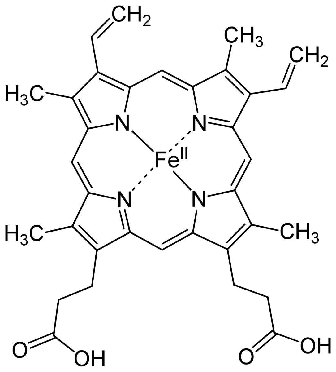
ヘムというと血色素を思い浮かべるが、体で作られるヘムの約 15% は赤血球以外で使われている。鉄を抱えたポルフィリン環は、電子をやり取りする部品として多くの酵素に組み込まれている。
| ヘモグロビン・ミオグロビン | 酸素を可逆的に結合する | 鉄は 2 価のまま保たれる |
| チトクロム P450 | 薬・ステロイド・毒物を酸化する | 肝で大量に要る |
| チトクロム a・b・c | 電子伝達系で電子を運ぶ | ミトコンドリア |
| カタラーゼ・ペルオキシダーゼ | 過酸化水素を分解する | ミエロペルオキシダーゼもこの仲間 |
| 一酸化窒素合成酵素・グアニル酸シクラーゼ | NO を作る/NO を受け取る | NO の受容体はヘムそのものである |
肝が P450 のためにヘムを必要とすることが、急性ポルフィリン症の発作の引き金を説明する。
ヘム合成の律速酵素 ALA 合成酵素は、肝の遊離ヘムの量による負のフィードバックで抑えられている。ここで薬(バルビツール酸・フェニトイン・スルホンアミド・経口避妊薬)を投与すると、P450 を増やすためにヘムが消費されて遊離ヘムが減り、抑えが外れて ALA 合成酵素が一気に誘導される。経路の途中が詰まっている患者では、その手前の中間体が大量に溜まり、発作になる。
治療のヘミン投与は、この論理をそのまま逆にたどっている——外からヘムを入れて遊離ヘムを満たし、フィードバックを効かせて上流を止める。「溜まったものを取り除く」のではなく「作るのをやめさせる」という発想である。
一酸化炭素の毒性も、ヘムが多用途であることの裏返しになる。CO はヘモグロビンに結合して酸素運搬を妨げるだけでなく、ミトコンドリアのチトクロム c 酸化酵素にも結合して呼吸そのものを止める。だから SpO2 が正常に見えても組織は低酸素であり、心筋と脳が先に障害される。
ポルフィリン症の患者に薬を出すときは、安全な薬かを必ず調べてから処方する。「よく使う薬だから大丈夫」は通用しない。
English version
Core
Haem calls to mind the blood pigment, but about 15 per cent of the haem made in the body is used outside the red cell. A porphyrin ring holding iron is built into many enzymes as a component that passes electrons.
| Haemoglobin, myoglobin | Bind oxygen reversibly | The iron is kept divalent |
| Cytochrome P450 | Oxidises drugs, steroids and poisons | Needed in quantity in the liver |
| Cytochromes a, b, c | Carry electrons in the electron transport chain | Mitochondria |
| Catalase, peroxidases | Degrade hydrogen peroxide | Myeloperoxidase belongs here |
| Nitric oxide synthase, guanylate cyclase | Make NO / receive NO | The receptor for NO is the haem itself |
Going further
That the liver needs haem for P450 explains the trigger of an attack of acute porphyria.
The rate-limiting enzyme of haem synthesis, ALA synthase, is suppressed by negative feedback from the amount of free haem in the liver. Give a drug here (barbiturate, phenytoin, sulphonamide, oral contraceptive) and haem is consumed to make more P450, the free haem falls, the suppression is lifted and ALA synthase is induced at a stroke. In a patient with a block partway along the pathway, the intermediate before it accumulates in quantity and there is an attack.
Giving haemin in treatment retraces this logic exactly — put haem in from outside, fill the free haem, restore the feedback and stop the upstream. The idea is not "remove what has accumulated" but "make it stop being produced".
The toxicity of carbon monoxide is the obverse of haem’s many uses. CO not only binds haemoglobin and impedes the carriage of oxygen but also binds mitochondrial cytochrome c oxidase and stops respiration itself. So the tissue is hypoxic while the SpO2 looks normal, and myocardium and brain are damaged first.
In the clinic
When prescribing for a patient with porphyria, always check whether the drug is safe before writing it. "It is a drug we use all the time, so it will be fine" does not apply.
ポルフィリン症を疑ったら、いつ・何を・どう採るか
ポルフィリン症の検査はタイミングと検体の扱いで結果が変わる。採り方を間違えると、発作中でも陰性に出る。
- 急性発作を疑うなら、発作中の尿でポルホビリノーゲン(PBG)を測る——発作が収まると正常化しうるので、症状のあるうちに採る
- 遮光して運ぶ——ポルフィリンは光で壊れる。アルミ箔で包む
- 随時尿でよい(クレアチニンで補正する)——24 時間尿を待つあいだに治療が遅れてはならない
- 尿を放置すると赤褐色に変わる——PBG が非酵素的に重合するため。古典的な所見だが、色が付かなくても否定できない
治療は検査結果を待たずに始める。疑った時点で検体を確保し、ブドウ糖とヘミンへ進む。
どの検体を測るかで、区別できる病型が変わる。
| 尿 | ALA・PBG・ウロポルフィリン | 急性型と晩発性皮膚ポルフィリン症を拾う。水に溶ける前駆体が出る |
| 便 | コプロポルフィリン・プロトポルフィリン | 水に溶けにくい後半の中間体。遺伝性コプロポルフィリン症と多様性ポルフィリン症の区別に要る |
| 血漿の蛍光 | 蛍光の極大波長 | 多様性ポルフィリン症に特徴的な波長がある |
| 赤血球 | プロトポルフィリン | 赤芽球性プロトポルフィリン症。鉛中毒・鉄欠乏でも上がる |
溜まる中間体が水に溶けるかどうかが、そのまま「症状」と「どこに出るか」を決めている。
- ALA と PBG は水溶性——尿へ出る。光では反応しない。だから皮膚症状は出ず、神経症状になる
- 環が閉じたポルフィリンは脂溶性で、光を吸収する——組織に留まり、日光で活性酸素を出して皮膚を壊す。便へ排泄される
⚠️ 鑑別の落とし穴。急性腹症・末梢神経障害・精神症状・低ナトリウム血症が揃った患者で、画像に異常が無いときにポルフィリン症を思い出す。開腹手術をされてしまう例が今も報告されている。
「原因不明の腹痛で何度も救急に来る若い女性」は、薬・月経周期との関連を聞くだけで診断に近づけることがある。
English version
Core
Tests for porphyria give different results according to the timing and the handling of the sample. Collected wrongly, they read negative even during an attack.
- To investigate an acute attack, measure porphobilinogen (PBG) in urine during the attack — it can normalise once the attack settles, so take it while the symptoms are present.
- Transport it protected from light — porphyrins are destroyed by light. Wrap in foil.
- A spot urine will do (corrected for creatinine) — treatment must not be delayed while waiting for a 24-hour collection.
- The urine turns red-brown on standing — PBG polymerises non-enzymatically. A classical finding, but the absence of colour does not exclude it.
Begin treatment without waiting for the result. Secure the specimen at the moment of suspicion and proceed to glucose and haemin.
Going further
Which specimen is measured changes which forms can be distinguished.
| Urine | ALA, PBG, uroporphyrin | Picks up the acute forms and porphyria cutanea tarda. The water-soluble precursors come out here |
| Stool | Coproporphyrin, protoporphyrin | The poorly soluble later intermediates. Needed to separate hereditary coproporphyria and variegate porphyria |
| Plasma fluorescence | The emission maximum | Variegate porphyria has a characteristic wavelength |
| Red cells | Protoporphyrin | Erythropoietic protoporphyria. It also rises in lead poisoning and iron deficiency |
Whether the intermediate that accumulates dissolves in water decides directly "the symptoms" and "where they appear".
- ALA and PBG are water-soluble — they come out in the urine. They do not react to light. Hence no skin symptoms, and neurological symptoms instead.
- A closed porphyrin ring is lipid-soluble and absorbs light — it stays in the tissue and generates reactive oxygen in sunlight and destroys the skin. It is excreted in the stool.
⚠️ The pitfall in the differential. In a patient with an acute abdomen, peripheral neuropathy, psychiatric symptoms and hyponatraemia together, and no abnormality on imaging, recall porphyria. Cases of laparotomy being performed are still reported.
In the clinic
With "a young woman coming repeatedly to the emergency department with abdominal pain of unknown cause", asking about the relation to drugs and to the menstrual cycle alone can bring the diagnosis close.
まとめ
| 8段階の経路と、律速のかかり方 | ミトコンドリア:グリシン+スクシニルCoA →(δアミノレブリン酸合成酵素、律速)→ δアミノレブリン酸細胞質:δアミノレブリン酸 →(δアミノレブリン酸脱水酵素)→ ポルホビリノ… |
|---|---|
| 急性型と皮膚型 — 溜まる分子の性質が症状を決める | 急性型皮膚型代表急性間欠性ポルフィリン症晩発性皮膚ポルフィリン症・骨髄性プロトポルフィリン症詰まる場所経路の上流下流溜まるものδアミノレブリン酸・ポルホビリノーゲン(環になる前)ポ… |
| 急性ポルフィリン症の発作 — 何が誘発し、何で止めるか | 発作は、ヘム合成の律速酵素(ALA 合成酵素、ALAS1)が誘導されたときに起こる。 |
| 鉄芽球性貧血 — ヘムが作れないと、鉄が行き場を失う | 鉄はあるのにヘムに組み込めず、赤芽球のミトコンドリアに沈着する。 |
| ヘムは、赤血球だけのものではない | ヘムというと血色素を思い浮かべるが、体で作られるヘムの約 15% は赤血球以外で使われている。 |
| ポルフィリン症を疑ったら、いつ・何を・どう採るか | ポルフィリン症の検査はタイミングと検体の扱いで結果が変わる。 |
このノートは医学生が授業の内容を自分の言葉でまとめたもので、診療の判断に使うものではありません。誤りに気づいたらお知らせください。