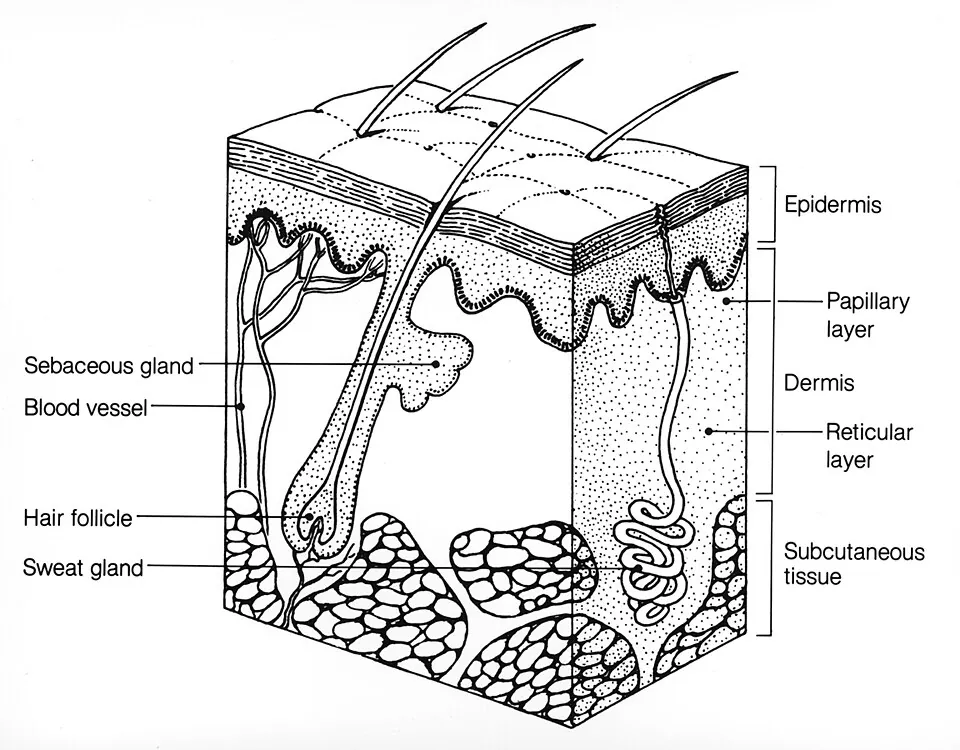
皮膚は体表面積1.6m²・重量約3kgの、体で最大の臓器である。組織学的には単純で、表皮(上皮)・真皮(結合組織)・皮下組織(脂肪)の3層しかない。しかしこの3層を数えられることが、そのまま診療になる。
- 水疱がどの層にできたか——表皮内か表皮下かで、疾患も見た目(弛緩性か緊満性か)も変わる
- 熱傷がどの深さまで届いたか——付属器が残っているかどうかで、自然に治るか植皮が要るかが決まる
- 腫瘍がどの細胞から出たか——角化細胞・メラノサイト・付属器で、予後がまったく違う
皮膚を作る細胞は角化細胞だけではない。表皮には由来の異なる3種の間借り人(メラノサイト・Langerhans 細胞・Merkel 細胞)が住んでいる。
臨床で特に重要なのは、創傷治癒と瘢痕である。真皮を越えて傷ついたときにだけ瘢痕が残るという境界線が、この章の実利になる。
- 表皮
- 表皮増殖単位 — 表皮は柱ごとに入れ替わる
- メラノサイト(と表皮の間借り人)
- メラニンの合成と色素沈着の調節 — 紫外線とホルモンが同じ受容体を押す
- 真皮
- 皮膚付属器 — 毛・脂腺・汗腺
- 細胞間の接着装置 — どこが壊れるかで、水疱の深さが決まる
- 角層 — レンガとモルタルで、水を逃がさない
- 毛周期 — 抜けるかどうかは、いまどの時期にいるかで決まる
- 毛包の層構造 — 毛と鞘を、内から外へ数える
- 爪 — 表皮が硬く変わった付属器
- 皮膚と付属器の発生 — 外胚葉の表皮と、中胚葉の真皮が組み合わさる
- 皮膚の加齢 — 年を重ねた皮膚と、日光を浴びた皮膚
- 表皮Epidermis
- 表皮増殖単位 — 表皮は柱ごとに入れ替わる
- メラノサイト(と表皮の間借り人)Melanocytus
- メラニンの合成と色素沈着の調節 — 紫外線とホルモンが同じ受容体を押すMelanosoma
- 真皮Dermis
- 皮膚付属器 — 毛・脂腺・汗腺Glandula sudorifera
- 細胞間の接着装置 — どこが壊れるかで、水疱の深さが決まるDesmosoma
- 角層 — レンガとモルタルで、水を逃がさないStratum corneum
- 毛周期 — 抜けるかどうかは、いまどの時期にいるかで決まるCyclus pili
- 毛包の層構造 — 毛と鞘を、内から外へ数えるFolliculus pili
- 爪 — 表皮が硬く変わった付属器Unguis
- 皮膚と付属器の発生 — 外胚葉の表皮と、中胚葉の真皮が組み合わさる
- 皮膚の加齢 — 年を重ねた皮膚と、日光を浴びた皮膚
- まとめ
表皮Epidermis
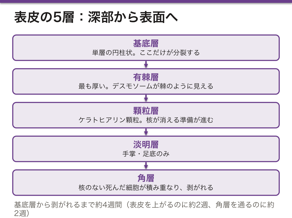
重層扁平上皮(角化型)。深部から順に。
- 基底層——単層の円柱状。ここだけが分裂する
- 有棘層——最も厚い。デスモソームが棘のように見える
- 顆粒層——ケラトヒアリン顆粒。ここで核が消える準備が進む
- 淡明層——手掌・足底のみ
- 角層——核のない死んだ細胞が積み重なる
基底層で生まれた細胞が角層まで上がって剥がれるまで約4週間(表皮を上がるのに約2週、角層を通るのに約2週)。
角層は死んだ細胞の層でありながら、皮膚の機能のほとんどを担っている。顆粒層で細胞は核と細胞小器官を捨て、層板顆粒から細胞間へ脂質(セラミド)を放出する。角化細胞(角質細胞)が煉瓦、脂質が漆喰という「煉瓦と漆喰」の構造ができ、これが水分の喪失を防ぎ、異物の侵入を防ぐ。
この漆喰が壊れることがアトピー性皮膚炎の出発点である。フィラグリン(ケラトヒアリン顆粒の主成分で、角層の天然保湿因子の材料でもある)の遺伝子変異があるとバリアが弱く、抗原が入りやすくなる。「乾燥しているから掻く」のではなく、バリアが壊れているから感作される——保湿が治療の基礎である理由がここにある。
乾癬では逆に増殖が暴走し、ターンオーバーが数日に短縮する。核を捨てる時間がないまま押し上げられるので、角層に核が残り(錯角化)、顆粒層が消える。厚い銀白色の鱗屑という肉眼所見が、この時間短縮の現れである。
水疱の深さが疾患を決める。
- 尋常性天疱瘡——デスモグレイン3(と1)に対する自己抗体。表皮内で細胞同士がほどける(棘融解)。弛緩性水疱ですぐ破れ、口腔粘膜のびらんで始まることが多い。Nikolsky 現象陽性
- 落葉状天疱瘡——デスモグレイン1のみ。角層下の浅い水疱で、粘膜は侵されない
- 水疱性類天疱瘡——ヘミデスモソーム(BP180・BP230)に対する抗体。表皮下で表皮ごと剥がれるので緊満性水疱。高齢者に多い
粘膜が侵されるかどうかがデスモグレイン1と3の分布で説明できる——粘膜には3が主に発現し、1は少ない。だから1だけを狙う落葉状天疱瘡では粘膜が無事で済む。抗原の分布地図が、そのまま症状の分布になる。
English version
Core
Stratified squamous, keratinised. From the depths upwards:
- Stratum basale — a single layer of columnar cells. Only this layer divides.
- Stratum spinosum — the thickest; the desmosomes look like spines.
- Stratum granulosum — keratohyalin granules. Here the preparation for losing the nucleus proceeds.
- Stratum lucidum — palms and soles only.
- Stratum corneum — dead cells without nuclei, piled up.
A cell born in the basal layer takes about four weeks to rise to the horny layer and be shed (about two weeks to ascend the epidermis and two more to traverse the horny layer).
Going further
The horny layer, though a layer of dead cells, carries out almost all the functions of the skin. In the granular layer the cell discards its nucleus and organelles and releases lipid (ceramides) into the intercellular space from the lamellar granules. The corneocytes are the bricks and the lipid the mortar, and this brick-and-mortar arrangement prevents the loss of water and the entry of foreign material.
Destruction of that mortar is the starting point of atopic dermatitis. A mutation of filaggrin (the chief component of the keratohyalin granules and the raw material of the natural moisturising factor) weakens the barrier and antigen enters readily. It is not that the skin is scratched because it is dry, but that sensitisation occurs because the barrier is broken — here is the reason moisturising is the foundation of treatment.
In psoriasis, by contrast, proliferation runs away and the turnover shortens to a few days. Pushed up without time to discard their nuclei, nuclei remain in the horny layer (parakeratosis) and the granular layer disappears. The thick silvery scale seen with the eye is the appearance of that shortened time.
In the clinic
The depth of the blister decides the disease.
- Pemphigus vulgaris — autoantibodies to desmoglein 3 (and 1). The cells come apart within the epidermis (acantholysis). Flaccid blisters that break at once, usually beginning with erosions of the oral mucosa. Nikolsky’s sign positive.
- Pemphigus foliaceus — desmoglein 1 only. Shallow, subcorneal blisters; the mucosa is spared.
- Bullous pemphigoid — antibodies to the hemidesmosome (BP180, BP230). The whole epidermis lifts off, so the blisters are tense. Commonest in the elderly.
Whether the mucosa is affected is explained by the distribution of desmogleins 1 and 3 — the mucosa expresses mainly 3 and little 1. So pemphigus foliaceus, which strikes only 1, leaves the mucosa intact. The map of the antigen’s distribution is directly the distribution of the symptoms.
表皮増殖単位 — 表皮は柱ごとに入れ替わる
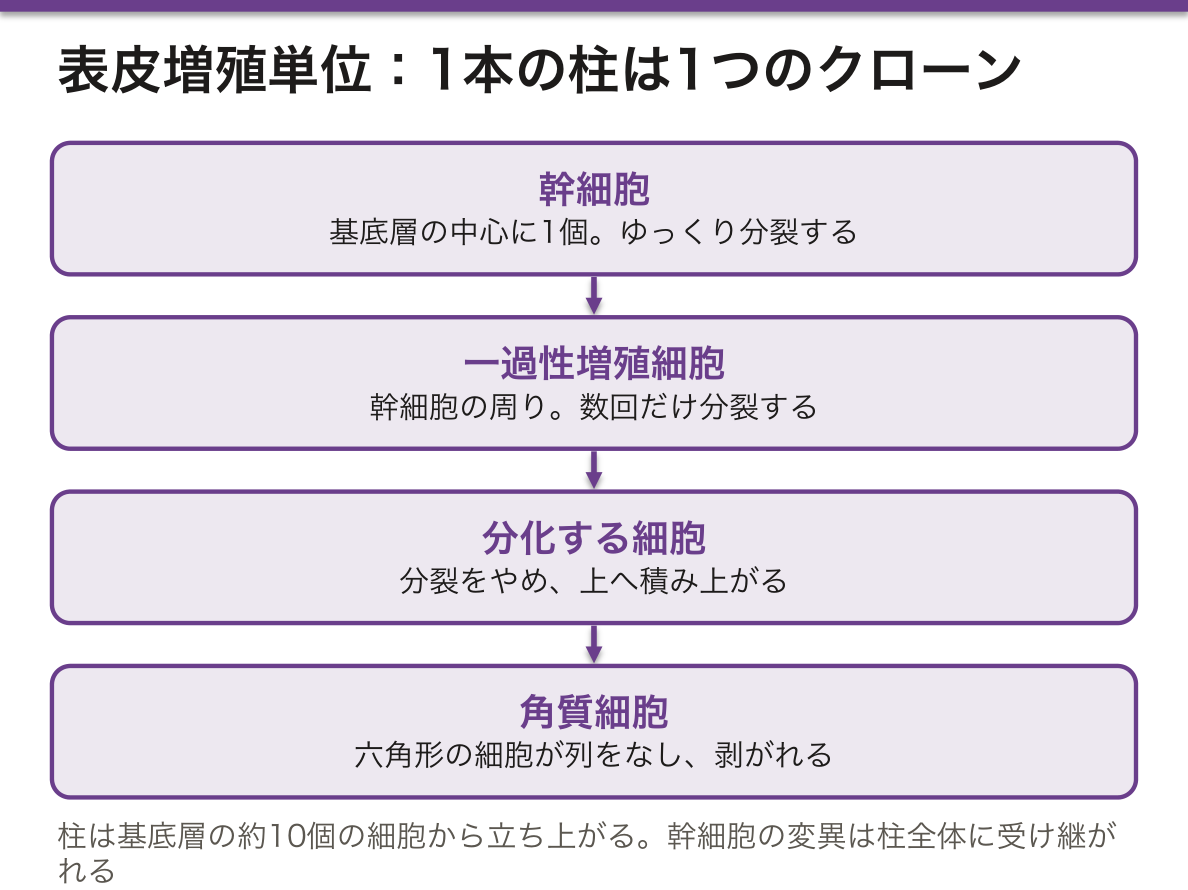
表皮の入れ替わりは、基底層のどの細胞もばらばらに分裂して起きるのではない。表皮増殖単位(epidermal proliferative unit、EPU)という縦の柱を単位にして進む、という考え方がある。
1つの柱は、
- 基底層の約10個の細胞——中心にゆっくり分裂する幹細胞が1個、その周りに数回だけ分裂する一過性増殖細胞(transit-amplifying cell)
- その上に積み上がる、分裂をやめて分化していく細胞
- いちばん上の、六角形の角質細胞が積み重なった列
からなる。マウスの薄い表皮でははっきり柱に見え、ヒトでは並びは不規則だが、幹細胞 → 増殖細胞 → 分化細胞という流れは同じである。
幹細胞は基底層のほか、毛包の膨隆部(bulge)にも控えている。
表皮の再生には2つの形がある。
- 生理的再生——基底層で生まれた細胞が約4週間で角層から剥がれる、毎日の入れ替わり
- 修復としての再生——傷の縁と、残った毛包・汗腺から角化細胞がヘミデスモソームを外して這い出し、傷を覆ってから分裂し、層を作り直す。上皮成長因子(EGF)や、真皮の線維芽細胞が出すケラチノサイト成長因子(KGF)が促す
柱という考え方が大事なのは、1つの柱が1個の幹細胞の子孫(クローン)だからである。幹細胞に起きた遺伝子の変化は、その柱全体に受け継がれ、消えずに残る。日光をよく浴びた皮膚には、p53 に変異を持つ細胞の小さな集まりが多数見つかる。癌はこうした変異クローンの中から生まれる。
傷を乾かさずに湿った状態で保つと早く治る(湿潤療法)。角化細胞は乾いたかさぶたの下を掘り進むより、湿った面の上を這うほうが速いからである。
広い熱傷では、患者自身の皮膚から角化細胞を培養して作る自家培養表皮が使われる。うまく生着するのは、培養の中に幹細胞が残っているときである。遺伝子を直した表皮幹細胞から培養表皮を作り、表皮水疱症の子どもの体表の約8割を置き換えた報告もある——幹細胞さえ残れば、表皮は丸ごと作り直せる。
English version
Core
The epidermis is not renewed by basal cells dividing at random. One model says renewal runs in vertical columns called epidermal proliferative units (EPU).
One column contains:
- about ten basal cells — one slowly dividing stem cell in the centre, surrounded by transit-amplifying cells that divide only a few times;
- above them, cells that have stopped dividing and are differentiating;
- at the top, a stack of hexagonal corneocytes.
In thin mouse epidermis the columns are clear. In humans the pattern is less regular, but the flow stem cell → amplifying cell → differentiated cell is the same.
Besides the basal layer, stem cells also wait in the bulge of the hair follicle.
Going further
Epidermal regeneration has two forms.
- Physiological renewal — daily turnover; a cell born in the basal layer is shed from the stratum corneum after about four weeks.
- Repair — keratinocytes at the wound edge and in surviving hair follicles and sweat glands release their hemidesmosomes and crawl out, cover the wound, then divide and rebuild the layers. Epidermal growth factor (EGF) and keratinocyte growth factor (KGF) from dermal fibroblasts help.
The column idea matters because one column is the offspring (clone) of one stem cell. A genetic change in that stem cell passes to the whole column and stays. Sun-exposed skin contains many small patches of cells with p53 mutations, and cancer arises from such mutant clones.
In the clinic
Wounds heal faster when kept moist instead of dry (moist wound healing). Keratinocytes crawl faster over a moist surface than they can dig under a dry scab.
For large burns, cultured epidermal autografts made from the patient’s own keratinocytes are used. They take well when stem cells remain in the culture. In one report, a cultured epidermis grown from gene-corrected epidermal stem cells replaced about 80% of the body surface of a child with epidermolysis bullosa — if stem cells survive, the whole epidermis can be rebuilt.
メラノサイト(と表皮の間借り人)Melanocytus
表皮には角化細胞のほかに由来の違う3種が住む。
- メラノサイト——神経堤由来。基底層にあり、樹状突起でメラノソームを周囲の角化細胞に手渡す。1個で30〜40個の角化細胞を担当
- Langerhans 細胞——骨髄由来の樹状細胞。主に有棘層。Birbeck 顆粒(テニスラケット状)を持つ。抗原提示
- Merkel 細胞——基底層。神経終末と接し、触覚(持続的な圧)を受け持つ
人種による肌の色の違いは、メラノサイトの数の差ではない。数はほぼ同じで、違うのはメラノソームの大きさ・数・分解されやすさである。色の濃い皮膚ではメラノソームが大きく単独で分布して分解されにくい。色は生産者の数ではなく、荷物の作りで決まる。
メラニンの役割は角化細胞の核の上に帽子のように配置され(核上キャップ)、紫外線からDNAを守ることである。だから色素が作れない眼皮膚白皮症(チロシナーゼ欠損など)では、若年から日光角化症・有棘細胞癌が多発する。
尋常性白斑はメラノサイトが自己免疫的に破壊される疾患で、境界明瞭な脱色素斑となる。神経堤由来という出自から、他の自己免疫疾患(橋本病・Addison 病・悪性貧血)を合併しやすい。
Langerhans 細胞は皮膚が免疫器官でもあることの証拠である。抗原を捕らえて所属リンパ節の傍皮質へ移動しT細胞に提示する。接触皮膚炎(IV型アレルギー)の感作相はこの移動そのものである。
悪性黒色腫はメラノサイト由来。ABCDE(Asymmetry 非対称・Border 辺縁不整・Color 色調不均一・Diameter 6mm以上・Evolution 変化)で疑う。
予後を決めるのは大きさではなくBreslow 厚(垂直方向の深さ)である。水平に広がっているあいだは転移しにくく、垂直に潜り始めると転移する——真皮に入って初めてリンパ管・血管に到達するためで、層構造がそのまま予後因子になっている。日本人では足底など末端黒子型が多く、足の裏のほくろに注意が要る。
Langerhans 細胞組織球症は Langerhans 細胞の腫瘍性増殖で、骨(頭蓋の打ち抜き像)・皮膚・尿崩症という一見ばらばらの症状が並ぶ。整形外科では好酸球性肉芽腫として単発の骨病変で出会う。
English version
Core
Besides the keratinocytes, three kinds of cell of different origin live in the epidermis.
- Melanocyte — neural crest derived. In the basal layer, handing melanosomes to the surrounding keratinocytes through its dendrites. One serves 30–40 keratinocytes.
- Langerhans cell — a bone marrow-derived dendritic cell, chiefly in the spinous layer. Has Birbeck granules (tennis-racket shaped). Antigen presentation.
- Merkel cell — in the basal layer, in contact with a nerve ending; responsible for touch (sustained pressure).
Going further
Racial differences in skin colour are not differences in the number of melanocytes. The number is much the same; what differs is the size, number and degradability of the melanosomes. In darker skin the melanosomes are larger, distributed singly, and resist degradation. Colour is decided by the make of the package, not by the number of producers.
Melanin’s role is to sit like a cap over the nucleus of the keratinocyte (the supranuclear cap) and protect the DNA from ultraviolet light. So in oculocutaneous albinism, where no pigment can be made (tyrosinase deficiency and others), actinic keratoses and squamous carcinomas appear in numbers from a young age.
Vitiligo is autoimmune destruction of the melanocytes, giving sharply defined depigmented patches. From its neural crest origin, it is associated with other autoimmune diseases (Hashimoto’s thyroiditis, Addison’s disease, pernicious anaemia).
The Langerhans cell is the evidence that the skin is also an immune organ. It captures antigen and migrates to the paracortex of the regional node to present it to T cells. The sensitisation phase of contact dermatitis (type IV allergy) is that migration itself.
In the clinic
Malignant melanoma is of melanocyte origin. Suspect it by ABCDE (asymmetry, irregular border, uneven colour, diameter over 6 mm, evolution).
The prognosis is decided not by the size but by the Breslow thickness (the depth vertically). While it is spreading horizontally it seldom metastasises; once it begins to burrow vertically it does — because only on entering the dermis does it reach lymphatics and blood vessels. The layered structure is itself the prognostic factor.
In Japanese people the acral lentiginous type, on the sole and elsewhere, is common, so a mole on the sole of the foot needs attention.
Langerhans cell histiocytosis is a neoplastic proliferation of Langerhans cells, in which bone (punched-out lesions of the skull), skin and diabetes insipidus appear together as apparently unrelated symptoms. In orthopaedics it is met as eosinophilic granuloma, a solitary bone lesion.
メラニンの合成と色素沈着の調節 — 紫外線とホルモンが同じ受容体を押すMelanosoma
メラニンはメラノサイトの中のメラノソーム(リソソームに近い小器官)で作られる。材料はチロシンで、鍵の酵素はチロシナーゼである。
チロシン →(チロシナーゼ)→ DOPA →(チロシナーゼ)→ ドーパキノンまで進み、そこで2つに分かれる。そのまま重合すれば黒褐色のユーメラニン、システインが加われば赤〜黄色のフェオメラニンになる。
メラノソームは中身の詰まり具合でI〜IV期に分かれる(I期は色素なし、IV期は電子密度の高いメラニンで満ちる)。成熟したものが樹状突起の先から角化細胞へ渡される。
色素細胞は表皮だけでなく、口腔などの粘膜の上皮、ぶどう膜、髄膜、内耳にもいる(いずれも神経堤由来)。網膜色素上皮は眼杯の外葉に由来する、別の系統の色素細胞である。
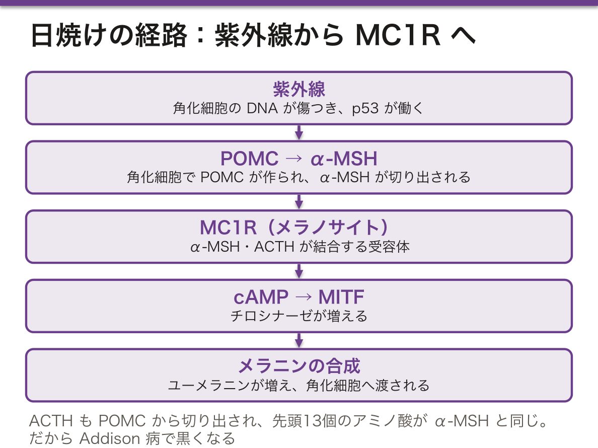
色素沈着を進める刺激は、ほとんどがメラノサイトの MC1R(メラノコルチン1受容体)に集まる。
| 刺激 | 経路 | 結果 |
|---|---|---|
| 紫外線(その場で) | すでにあるメラニンの酸化と移動 | 数分〜数時間で黒くなる |
| 紫外線(数日かけて) | 角化細胞の DNA 損傷 → p53 → POMC の発現 → α-MSH | メラニンが新しく作られる(日焼け) |
| α-MSH・ACTH | MC1R → cAMP → MITF → チロシナーゼ増加 | ユーメラニンが増える |
| エストロゲン・プロゲステロン | メラノサイトの反応性が上がる | 妊娠中の乳輪・外陰・腹部正中線の色素沈着、肝斑 |
ACTH で皮膚が黒くなる理由は、ACTH と α-MSH が同じ前駆体 POMC から切り出され、ACTH の先頭の13個のアミノ酸がα-MSH と同じ並びだからである。だから ACTH は MC1R にも結合する。
原発性副腎不全(Addison 病)では、コルチゾールが下がって負のフィードバックが外れ、ACTH が増える。その結果、皮膚と口腔粘膜、手のしわ、傷あとが黒くなる。一方、下垂体が原因の続発性副腎不全では ACTH が低いので、色はむしろ白い。色素沈着の有無が、どこが壊れたかを教える。
MC1R の機能が弱い型を持つ人は、フェオメラニンが多くなり、赤毛・色白・そばかすになりやすく、日焼けで黒くならず赤くなる。黒色腫の危険も高い。眼皮膚白皮症 1 型はチロシナーゼの欠損である。
治療もこの経路の上にある。ハイドロキノンはチロシナーゼを抑えて色素斑を薄くする。
English version
Core
Melanin is made in melanosomes (lysosome-related organelles) inside melanocytes. The material is tyrosine and the key enzyme is tyrosinase.
The pathway runs tyrosine → (tyrosinase) → DOPA → (tyrosinase) → dopaquinone, and then branches. If it polymerises directly, it becomes black-brown eumelanin; if cysteine is added, red-yellow pheomelanin.
Melanosomes are grouped into stages I–IV by how full they are (stage I has no pigment; stage IV is packed with electron-dense melanin). Mature ones are handed to keratinocytes from the tips of the dendrites.
Pigment cells live not only in the epidermis but also in the epithelium of mucosae such as the mouth, the uvea, the meninges and the inner ear (all from the neural crest). The retinal pigment epithelium comes from the outer layer of the optic cup and is a different line.
Going further
Most signals that increase pigment meet at one receptor on the melanocyte: MC1R (melanocortin 1 receptor).
| Stimulus | Route | Result |
|---|---|---|
| UV (immediate) | oxidation and movement of existing melanin | darkening within minutes to hours |
| UV (over days) | DNA damage in keratinocytes → p53 → POMC expression → α-MSH | new melanin (tanning) |
| α-MSH and ACTH | MC1R → cAMP → MITF → more tyrosinase | more eumelanin |
| Oestrogen and progesterone | melanocytes become more responsive | darker areolae, genitalia and linea nigra in pregnancy; melasma |
Why ACTH darkens the skin: ACTH and α-MSH are cut from the same precursor, POMC, and the first 13 amino acids of ACTH are the same as α-MSH. So ACTH can also bind MC1R.
In the clinic
In primary adrenal insufficiency (Addison disease), low cortisol removes negative feedback and ACTH rises. As a result skin, oral mucosa, palm creases and scars darken. In secondary insufficiency caused by the pituitary, ACTH is low and the skin is pale. Whether pigment is present tells where the failure is.
People with weak MC1R variants make more pheomelanin. They tend to have red hair, fair skin and freckles, burn instead of tanning, and have a higher melanoma risk. Oculocutaneous albinism type 1 is a lack of tyrosinase.
Treatment also sits on this pathway: hydroquinone inhibits tyrosinase and lightens pigmented spots.
真皮Dermis
表皮を支える結合組織。2層に分ける。
- 乳頭層——疎性結合組織。表皮へ乳頭状に突出し、毛細血管とMeissner 小体を入れる。表皮自体には血管が無いので、栄養はここから拡散で届く
- 網状層——密性結合組織。太いI型コラーゲンと弾性線維が編み目状に走り、皮膚の強さと弾力の実体
皮下組織にはPacini 小体(振動)がある。
付属器(毛包・汗腺)は真皮から皮下組織にかけて存在する。
真皮に達したかどうかが、瘢痕が残るかどうかの境界線である。表皮だけの損傷は基底層から完全に再生するが、真皮のコラーゲンが断たれると同じものには戻らず、線維芽細胞が作った瘢痕組織で埋められる。
熱傷の深度分類はこの原理そのものである。
- I度——表皮のみ。発赤。瘢痕なし
- II度 浅達性——真皮浅層まで。水疱。付属器が残るのでそこから上皮が広がり、1〜2週で瘢痕なく治る
- II度 深達性——真皮深層。付属器が乏しく治癒が遅れ、瘢痕を残す
- III度——全層。付属器が全滅するので上皮化の起点がなく、植皮が必要。神経も焼けるため痛みがない
「痛くない熱傷ほど重い」という逆説も、受容器が真皮にあるという組織学から出る。深さを問うことは、上皮の種を残せたかを問うことである。
治癒が過剰に働くとケロイド(元の創を越えて広がる)や肥厚性瘢痕(創内にとどまる)になる。前胸部・肩・恥骨上など張力のかかる部位に多い。
褥瘡の深達度分類(NPUAP)も同じ層の話である。持続的な圧迫でまず皮下組織と筋が虚血に陥るため、表面の見た目より深部の障害が大きいことが多い(deep tissue injury)。骨の突出部(仙骨・大転子・踵)に生じるのは、そこで組織が骨と外力に挟まれるからである。
加齢ではコラーゲンと弾性線維が減り、真皮乳頭が平坦化する。表皮と真皮の噛み合わせが浅くなるため、高齢者の皮膚は擦れただけで剥離する(スキンテア)。テープ剥離やベッド移乗での皮膚裂傷の多さは、この構造変化で説明できる。
English version
Core
The connective tissue supporting the epidermis, in two layers.
- Papillary layer — loose connective tissue, projecting as papillae into the epidermis and carrying capillaries and Meissner corpuscles. The epidermis itself has no vessels, so its nutrition arrives from here by diffusion.
- Reticular layer — dense connective tissue. Thick type I collagen and elastic fibres woven together: the substance of the strength and elasticity of the skin.
Pacinian corpuscles (vibration) lie in the subcutis. The appendages (hair follicles, sweat glands) extend from the dermis into the subcutis.
Going further
Whether the dermis has been reached is the boundary of whether a scar remains. Damage to the epidermis alone regenerates completely from the basal layer, but once the dermal collagen is cut it does not return the same and is filled by scar tissue made by fibroblasts.
The classification of burn depth is that principle itself.
- First degree — epidermis only. Erythema. No scar.
- Superficial second degree — into the superficial dermis. Blistering. The appendages survive, so epithelium spreads from them and it heals without scarring in 1–2 weeks.
- Deep second degree — deep dermis. Few appendages remain, healing is delayed and a scar remains.
- Third degree — full thickness. The appendages are wiped out so there is no starting point for epithelialisation and grafting is required. The nerves are burnt too, so it is painless.
The paradox that the more painless the burn the more severe it is also comes out of the histology: the receptors are in the dermis. To ask about depth is to ask whether the seed of the epithelium has been left.
When healing works excessively, a keloid (extending beyond the original wound) or a hypertrophic scar (staying within it) results, commonest where there is tension — anterior chest, shoulder, suprapubic region.
In the clinic
The staging of pressure ulcers (NPUAP) is the same story of layers. Sustained pressure makes the subcutis and muscle ischaemic first, so the damage in the depths is often greater than the surface suggests (deep tissue injury). They occur over bony prominences (sacrum, greater trochanter, heel) because there the tissue is caught between bone and the external force.
With age, collagen and elastic fibres decrease and the dermal papillae flatten. The interlocking of epidermis and dermis becomes shallow, so the skin of an elderly person peels away with mere friction (skin tears). The frequency of skin tears from removing tape or transferring a patient in bed is explained by this structural change.
皮膚付属器 — 毛・脂腺・汗腺Glandula sudorifera
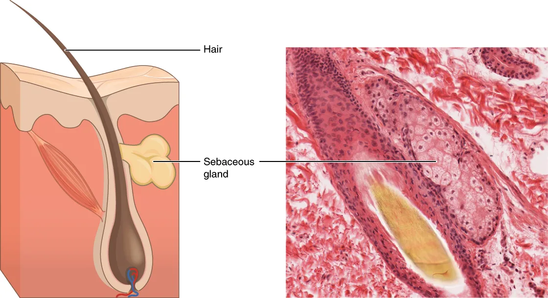
- 毛包——表皮が真皮へ落ち込んだ管。毛周期(成長期・退行期・休止期)を繰り返す。立毛筋(平滑筋)が付く
- 脂腺——毛包に開口。ホロクリン分泌(細胞ごと壊れて分泌物になる)。手掌・足底には無い
- エクリン汗腺——全身に分布。メロクリン分泌(開口分泌)。体温調節。支配は交感神経だが伝達物質はアセチルコリン
- アポクリン汗腺——腋窩・外陰・乳輪。毛包に開口。思春期以降に働く
付属器は表皮が真皮の中へ埋め込まれた「上皮の貯金」である。だから真皮の途中までの損傷では、残った毛包と汗腺の上皮が周囲へ這い出して表皮を再生する。II度浅達性熱傷や擦過傷が瘢痕なく治るのはこのためで、付属器の残存量が治癒能力そのものである。逆に、付属器の乏しい部位や深い損傷では治らない。
エクリン汗腺だけが交感神経でありながらアセチルコリンを使うという例外は、薬の副作用として現れる。抗コリン薬で汗が止まり、体温調節ができなくなって高齢者が熱中症を起こす。逆に原発性多汗症にボツリヌス毒素が効くのも、アセチルコリンの放出を止めるからである。
脂腺はアンドロゲン依存性に発達する。思春期に皮脂が増え、毛包の出口が角化で詰まり(面皰)、そこに Cutibacterium acnes が増えて炎症を起こすのが尋常性痤瘡である。ホルモン・角化・細菌の3つが同じ1本の管の中で起きている。
毛周期を知ると脱毛が読める。頭髪は約85%が成長期で、成長期は数年続く。
- 抗癌薬による脱毛——分裂の速い成長期の毛母細胞を叩くので、成長期脱毛として急速かつ広範に起こるが、毛包幹細胞は残るため再生する
- 分娩後脱毛・高熱後の脱毛——多くの毛が一斉に休止期へ移るため、2〜3か月遅れて抜ける(休止期脱毛)。自然に戻る
- 円形脱毛症——毛包に対する自己免疫。境界明瞭な脱毛斑
「いつ抜けるか」が原因を教える——直後なら成長期を直接叩いたもの、数か月後なら休止期へ移行させたものである。
English version
Core
- Hair follicle — a tube of epidermis sunk into the dermis. It repeats the hair cycle (anagen, catagen, telogen). The arrector pili (smooth muscle) is attached.
- Sebaceous gland — opens into the follicle. Holocrine secretion (the whole cell breaks down to become the secretion). Absent from palms and soles.
- Eccrine sweat gland — over the whole body. Merocrine secretion. Thermoregulation. Sympathetically innervated but the transmitter is acetylcholine.
- Apocrine sweat gland — axilla, external genitalia, areola. Opens into a follicle. Active from puberty.
Going further
The appendages are a savings account of epithelium buried in the dermis. So when injury reaches only part-way through the dermis, the epithelium of the surviving follicles and sweat glands creeps outwards and regenerates the epidermis. This is why a superficial second-degree burn or an abrasion heals without scarring — the amount of surviving appendage is the healing capacity itself. Conversely, where appendages are few or the injury deep, it does not heal.
The exception that the eccrine gland is sympathetic yet uses acetylcholine shows up as a drug effect. Anticholinergics stop sweating, thermoregulation fails and elderly people develop heat illness. Conversely botulinum toxin works in primary hyperhidrosis because it stops the release of acetylcholine.
The sebaceous gland develops under androgen. At puberty sebum increases, the outlet of the follicle blocks with keratin (a comedo), and Cutibacterium acnes multiplies there and causes inflammation: this is acne vulgaris. Hormone, keratinisation and bacteria all occurring within the same single duct.
In the clinic
Know the hair cycle and you can read hair loss. About 85 per cent of scalp hairs are in anagen, and anagen lasts some years.
- Chemotherapy-induced alopecia — the rapidly dividing matrix cells of anagen are struck, so anagen effluvium occurs rapidly and extensively; the follicular stem cells survive, so it regrows.
- Postpartum and post-fever hair loss — many hairs pass into telogen together, so shedding is delayed by 2–3 months (telogen effluvium). It recovers spontaneously.
- Alopecia areata — autoimmunity against the follicle. Sharply defined bald patches.
When it falls out tells you the cause — immediately, something struck anagen directly; some months later, something pushed hairs into telogen.
細胞間の接着装置 — どこが壊れるかで、水疱の深さが決まるDesmosoma
表皮の細胞は2種類の接着装置で繋がれている。どちらが壊れるかで、できる水疱の深さが変わる。
| 装置 | 繋ぐもの | 膜貫通蛋白 | 細胞内で繋がる線維 |
|---|---|---|---|
| デスモソーム | 角化細胞どうし | デスモグレイン・デスモコリン(カドヘリン科、Ca²⁺依存) | 中間径フィラメント(ケラチン) |
| ヘミデスモソーム | 基底細胞と基底膜 | BP180(XVII型コラーゲン)・BP230・インテグリンα6β4 | ケラチン |
ほかに密着結合〈タイト結合〉が顆粒層にあり、水と物質の透過を止める。接着結合(E-カドヘリン+アクチン)、ギャップ結合(コネキシン、細胞間の連絡)も存在する。
基底膜は4層——細胞膜/透明層〈lamina lucida〉/緻密層〈lamina densa、IV型コラーゲン〉/係留線維〈VII型コラーゲン〉で真皮へ繋がる。
「どの分子が標的か」で水疱症がすべて整理できる——組織学がそのまま診断学になる典型例。
| 疾患 | 標的 | 水疱の場所 | 蛍光抗体法 | 粘膜 |
|---|---|---|---|---|
| 尋常性天疱瘡 | デスモグレイン3(+1) | 表皮内(基底層直上) | 細胞間に網目状のIgG | 侵す(口腔から始まる) |
| 落葉状天疱瘡 | デスモグレイン1 | 表皮内(顆粒層=浅い) | 細胞間にIgG | 侵さない |
| 水疱性類天疱瘡 | BP180・BP230 | 表皮下 | 基底膜部に線状のIgG・C3 | 侵しにくい |
| 後天性表皮水疱症 | VII型コラーゲン | 表皮下 | 基底膜部に線状(真皮側) | — |
| SSSS・水疱性膿痂疹 | デスモグレイン1(細菌の表皮剝脱毒素が切断) | 表皮内(浅い) | — | 侵さない |
デスモグレイン1 と3 の分布が症状を決める——Dsg3 は粘膜と表皮深層に多く、Dsg1 は表皮浅層に多い。だからDsg3 を叩く尋常性天疱瘡は口腔のびらんで始まり深い水疱を作り、Dsg1 だけを叩く落葉状天疱瘡は粘膜を侵さず浅く剥がれる。
Nikolsky 現象(正常に見える皮膚を擦ると剥ける)は表皮内で結合が壊れている天疱瘡群・SSSS・TEN で陽性、表皮の下で剥がれる類天疱瘡では陰性——剥離の層がそのまま手技の所見になる。
どの接着装置に対する抗体かで、水疱の深さと臨床像が決まる。
尋常性天疱瘡 — デスモグレイン3(と1)に対する抗体。表皮の内部(基底層直上)で裂けるので水疱は弛緩性で破れやすく、びらんが目立つ。Nikolsky 現象陽性、口腔粘膜疹が先行する。組織では棘融解(acantholysis)、蛍光抗体法で表皮細胞間に網目状の IgG 沈着。
水疱性類天疱瘡 — ヘミデスモソーム(BP180・BP230)に対する抗体。表皮真皮境界で裂けるので緊満性の大きな水疱で破れにくい。高齢者に多く、粘膜疹は少ない。蛍光抗体法で基底膜部に線状の IgG・C3 沈着。
「弛緩性で口の中=天疱瘡、緊満性で高齢者=類天疱瘡」——接着装置の位置がそのまま鑑別点になっている。
ブドウ球菌性熱傷様皮膚症候群では毒素がデスモグレイン1を切るので、天疱瘡と同じ高さで裂ける。
English version
Core
The cells of the epidermis are joined by two adhesive devices, and which of them is destroyed changes the depth of the blister.
| Device | Joins | Transmembrane protein | Filament inside the cell |
|---|---|---|---|
| Desmosome | Keratinocyte to keratinocyte | Desmoglein, desmocollin (cadherins, Ca²⁺-dependent) | Intermediate filaments (keratin) |
| Hemidesmosome | Basal cell to basement membrane | BP180 (type XVII collagen), BP230, integrin α6β4 | Keratin |
In addition, tight junctions in the granular layer stop the passage of water and solutes; adherens junctions (E-cadherin + actin) and gap junctions (connexins) are also present.
The basement membrane is four layers — cell membrane / lamina lucida / lamina densa (type IV collagen) / anchoring fibrils (type VII collagen) joining it to the dermis.
Going further
Every blistering disease can be organised by which molecule is the target — the model example of histology becoming diagnostics.
| Disease | Target | Site of the blister | Immunofluorescence | Mucosa |
|---|---|---|---|---|
| Pemphigus vulgaris | Desmoglein 3 (+1) | Intraepidermal (just above the basal layer) | Net-like IgG between the cells | Affected (begins in the mouth) |
| Pemphigus foliaceus | Desmoglein 1 | Intraepidermal (granular layer — shallow) | IgG between the cells | Spared |
| Bullous pemphigoid | BP180, BP230 | Subepidermal | Linear IgG and C3 at the basement membrane | Seldom affected |
| Epidermolysis bullosa acquisita | Type VII collagen | Subepidermal | Linear at the basement membrane (dermal side) | — |
| SSSS, bullous impetigo | Desmoglein 1 (cleaved by the bacterial exfoliative toxin) | Intraepidermal (shallow) | — | Spared |
The distribution of desmogleins 1 and 3 decides the symptoms — Dsg3 predominates in mucosa and the deep epidermis, Dsg1 in the superficial epidermis. So pemphigus vulgaris, striking Dsg3, begins with oral erosions and makes deep blisters, while pemphigus foliaceus, striking only Dsg1, spares the mucosa and peels superficially.
Nikolsky’s sign (normal-looking skin peels when rubbed) is positive where the bonds are broken within the epidermis — the pemphigus group, SSSS, TEN — and negative in pemphigoid, where the split is beneath it. The layer of the split becomes directly a finding on examination.
In the clinic
Which adhesive device the antibody is against decides the depth of the blister and the clinical picture.
Pemphigus vulgaris — antibodies to desmoglein 3 (and 1). The split is within the epidermis (just above the basal layer), so the blisters are flaccid and break easily and erosions predominate. Nikolsky’s sign positive; oral lesions come first. Histologically acantholysis, with net-like IgG between the epidermal cells on immunofluorescence.
Bullous pemphigoid — antibodies to the hemidesmosome (BP180, BP230). The split is at the dermo-epidermal junction, so the blisters are large, tense and do not break easily. Commonest in the elderly, with few mucosal lesions. Linear IgG and C3 at the basement membrane.
"Flaccid and in the mouth = pemphigus; tense and in the elderly = pemphigoid" — the position of the adhesive device is itself the point of differentiation.
In staphylococcal scalded skin syndrome the toxin cleaves desmoglein 1, so the split is at the same level as in pemphigus.
角層 — レンガとモルタルで、水を逃がさないStratum corneum
角層は「レンガとモルタル」の模型で理解する。
- レンガ——角質細胞。核も小器官も失い、ケラチン線維とフィラグリン由来の天然保湿因子で満たされる
- モルタル——細胞間脂質。セラミド・コレステロール・遊離脂肪酸が層状に並ぶ
- 鉄筋——角質細胞の膜の内側を裏打ちする蛋白(ロリクリン・インボルクリン)
脂質は、顆粒層の層板顆粒(Odland 小体)から放出される。
角層のもう1つの守り——皮脂と汗が作る弱酸性の膜(pH 4.5〜6.0)。酸性であることが、常在菌叢と酵素の働きを保っている。
表皮の入れ替わりは約 4 週間(基底層から角層まで約2週間、角層内で約2週間)。
「バリアは細胞ではなく、細胞のあいだにある」という点が要点になる。水を逃がさないのは脂質の層で、細胞そのものではない(脂質の構造の章)——「だから保湿剤は、モルタルを補う設計になっている」。
フィラグリンの遺伝子異常が、アトピー性皮膚炎の最大の遺伝的要因になる(アトピー性皮膚炎の章)——「乾燥する皮膚から抗原が入り、感作が進む(経皮感作)」。
だから治療の順序が「まず保湿」になる——「炎症を抑えることと、壊れた壁を直すことは、別の仕事になる」。
経皮水分蒸散量(TEWL)が、バリアの機能を数字にする(熱傷の章)——「広範囲熱傷で、目に見えない水分喪失が膨大になる理由がここにある」。
角層が薄い部位ほど、薬が吸収されやすい(外用薬の章)——「同じステロイドでも、顔・陰部と手掌・足底では強さを変える」。
入れ替わりが 4 週間であることは、治療の評価の間隔を決めている——「角化異常症の治療で、効果を見るのに1か月は要る」。
バリアが壊れることが、アトピー性皮膚炎の出発点である。
フィラグリン遺伝子の変異があると天然保湿因子が作れず、角層が乾く。バリアが破れた皮膚から抗原が入り、感作が成立する——経皮感作である。食物アレルギーが皮膚から始まりうるという理解は離乳食の進め方と保湿の指導を変えた。
尋常性魚鱗癬もフィラグリンの異常による。
保湿は治療である。ステロイド外用で炎症を抑え、保湿でバリアを保つ——両輪であって、どちらか一方では足りない。
角層は薬の吸収も決める。角層が薄い部位(顔・陰部)ほど吸収が高いので、ステロイド外用薬のランクを部位で変える。手掌足底は角層が厚く吸収が悪い。密封療法(ODT)は角層を水和させて吸収を上げる手技である。
English version
Core
Understand the horny layer by the model of bricks and mortar.
- Bricks — corneocytes. Having lost nucleus and organelles, filled with keratin filaments and the natural moisturising factor derived from filaggrin.
- Mortar — intercellular lipid. Ceramides, cholesterol and free fatty acids arranged in lamellae.
- Reinforcement — the proteins lining the inside of the corneocyte membrane (loricrin, involucrin).
The lipid is released from the lamellar (Odland) bodies of the granular layer.
The other defence of the horny layer — the weakly acid film made by sebum and sweat (pH 4.5–6.0). Being acid maintains the resident flora and the action of the enzymes.
Turnover of the epidermis takes about four weeks (about two from basal layer to horny layer, about two within it).
Going further
The point is that the barrier is not the cells but what lies between them. It is the lipid layer that keeps water in, not the cells themselves — which is why a moisturiser is designed to replenish the mortar.
Mutation of filaggrin is the greatest genetic factor in atopic dermatitis — antigen enters through the drying skin and sensitisation proceeds (transcutaneous sensitisation). So the order of treatment is "moisturise first" — suppressing inflammation and repairing a broken wall are two different jobs.
Transepidermal water loss (TEWL) turns barrier function into a number — here is why the invisible loss of water in an extensive burn is so enormous.
The thinner the horny layer, the more readily a drug is absorbed — the same steroid is given at different strengths for the face and genitals than for palms and soles.
That turnover takes four weeks sets the interval for assessing treatment — a month is needed to see the effect of treatment in a disorder of keratinisation.
In the clinic
Breakdown of the barrier is the starting point of atopic dermatitis. With a filaggrin mutation the natural moisturising factor cannot be made and the horny layer dries. Antigen enters through the broken barrier and sensitisation is established — transcutaneous sensitisation. The understanding that food allergy can begin in the skin has changed advice on weaning and on moisturising. Ichthyosis vulgaris is also due to a filaggrin abnormality.
Moisturising is treatment. Topical steroids suppress the inflammation and emollients maintain the barrier — the two are a pair, and either alone is not enough.
The horny layer also decides drug absorption. Where it is thin (face, genitals) absorption is higher, so the potency class of a topical steroid is varied by site. Palms and soles have a thick horny layer and absorb poorly. Occlusive dressing therapy is a technique that hydrates the horny layer to raise absorption.
毛周期 — 抜けるかどうかは、いまどの時期にいるかで決まるCyclus pili
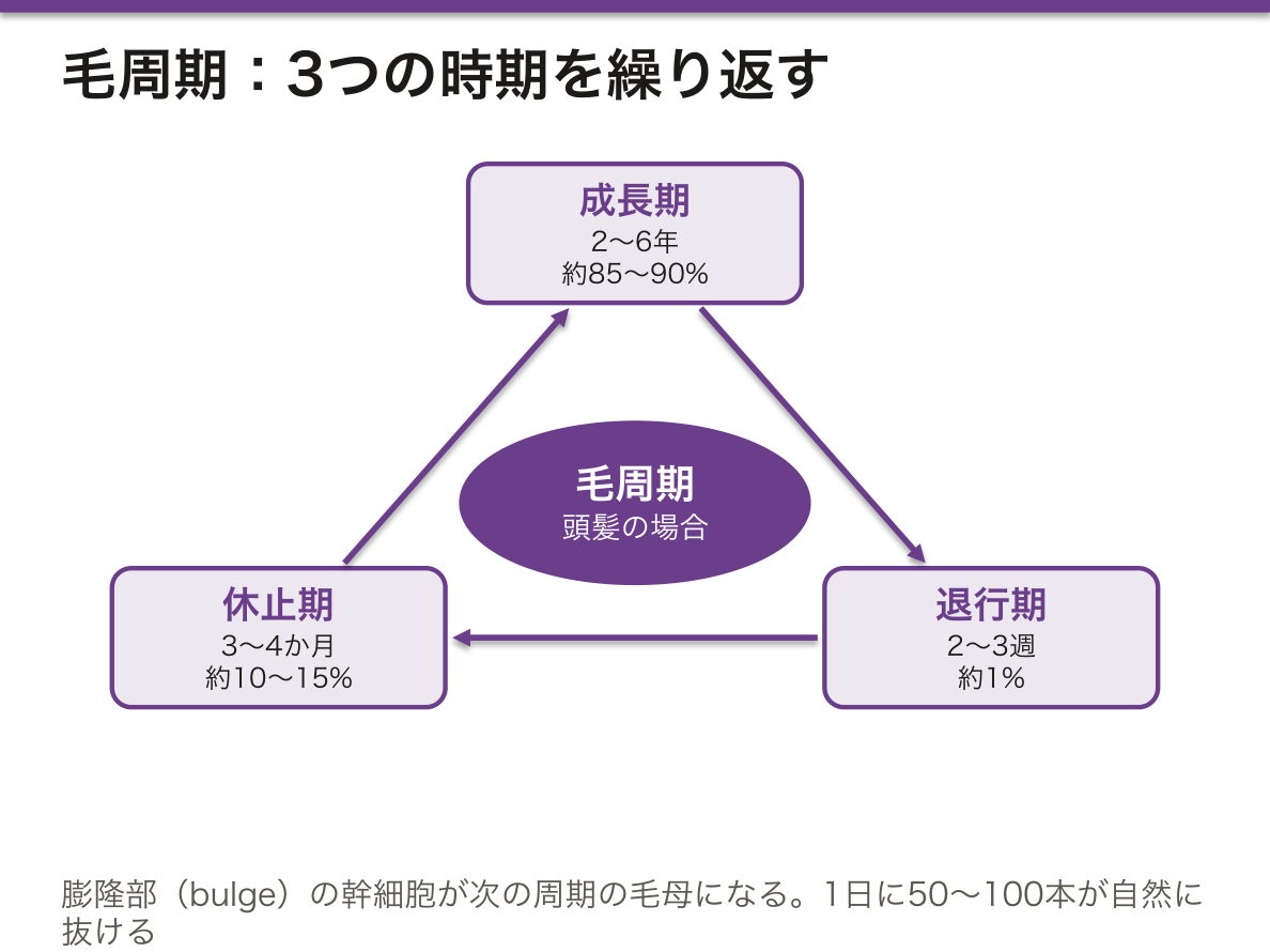
毛は連続して伸びるのではなく、3つの時期を繰り返す。
| 時期 | 状態 | 頭髪での期間と割合 |
|---|---|---|
| 成長期(anagen) | 毛母細胞が盛んに分裂する | 2〜6 年・約 85〜90% |
| 退行期(catagen) | 毛球が縮む | 2〜3 週・約 1% |
| 休止期(telogen) | 毛は毛包に留まるだけ | 3〜4 か月・約 10〜15% |
1日に 50〜100 本が自然に抜ける。
毛包の膨隆部(bulge)に幹細胞があり、次の周期の毛母と、表皮の再生の供給源になる。
部位で周期が違う——眉毛や睫毛は成長期が短いので、長くならない。
「いまどの時期にいるか」で、脱毛の型と可逆性が決まる。
| 型 | 機序 | 時期 |
|---|---|---|
| 成長期脱毛 | 分裂中の毛母が直接障害される | 抗腫瘍薬・放射線。1〜3週で急速に抜ける |
| 休止期脱毛 | 多くの毛が一斉に休止期へ入る | 出産後・高熱・手術・急激な減量。2〜4か月後に抜ける |
(皮膚付属器の章)——「原因のあった時期と、抜ける時期がずれるので、患者は結び付けられない」。
どちらも毛包そのものは残るので、再び生えてくる——「瘢痕性脱毛だけが、毛包ごと失われるので戻らない」。
男性型脱毛は、周期が短くなって毛が細くなる(軟毛化)(ホルモンの薬の章)——「5α-還元酵素阻害薬が、ジヒドロテストステロンの生成を抑える」。
膨隆部の幹細胞が表皮も再生させることは、熱傷の治り方を説明する(熱傷の章)——「真皮浅層まで(毛包が残る深さ)なら、上皮が毛包から広がって治る」。
いま毛がどの時期にいるかで、脱毛の型が決まる。
成長期脱毛 — 抗癌薬による。分裂中の毛母細胞が直接やられるので、投与後2〜3週で急速に、大量に抜ける。頭髪の85〜90%が成長期だから、ほぼ全部抜ける。
休止期脱毛 — 出産後・高熱・手術・急激な減量・強いストレス。成長期の毛が一斉に休止期へ移されるため、誘因から2〜3か月遅れて抜け始める。「思い当たることが数か月前にある」のが手がかりで、原因が去れば自然に回復する。
男性型脱毛症 — DHT が成長期を短縮させ、毛が細く短くなる(軟毛化)。フィナステリド(5α還元酵素阻害)が効くのはこの機序による。
円形脱毛症は毛包に対する自己免疫で、境界明瞭な脱毛斑と感嘆符毛。
English version
Core
Hair does not grow continuously but repeats three phases.
| Phase | State | Duration and proportion (scalp) |
|---|---|---|
| Anagen | The matrix cells divide actively | 2–6 years, about 85–90% |
| Catagen | The bulb shrinks | 2–3 weeks, about 1% |
| Telogen | The hair merely rests in the follicle | 3–4 months, about 10–15% |
50–100 hairs are shed naturally each day.
Stem cells lie in the bulge of the follicle and are the source both of the next cycle’s matrix and of the regeneration of the epidermis.
The cycle differs by site — the eyebrows and eyelashes have a short anagen, so they do not grow long.
Going further
Which phase the hair is in decides the type of hair loss and whether it is reversible.
| Type | Mechanism | Timing |
|---|---|---|
| Anagen effluvium | The dividing matrix is damaged directly | Cytotoxic drugs, radiation. Falls rapidly at 1–3 weeks |
| Telogen effluvium | Many hairs enter telogen together | After childbirth, high fever, surgery, rapid weight loss. Falls 2–4 months later |
The time of the cause and the time of the shedding are separated, so the patient cannot connect them.
In both, the follicle itself survives, so the hair grows back — only scarring alopecia loses the follicle and does not return.
In male pattern baldness the cycle shortens and the hairs become fine (miniaturisation) — 5α-reductase inhibitors suppress the formation of dihydrotestosterone.
That the bulge stem cells also regenerate the epidermis explains how a burn heals — as far as the superficial dermis (a depth at which the follicles survive), epithelium spreads from the follicles and it heals.
In the clinic
Which phase the hair is in decides the type of hair loss.
Anagen effluvium — from cytotoxic drugs. The dividing matrix cells are struck directly, so hair falls rapidly and copiously 2–3 weeks after treatment. Since 85–90 per cent of scalp hairs are in anagen, almost all of it falls.
Telogen effluvium — after childbirth, high fever, surgery, rapid weight loss, severe stress. Anagen hairs are pushed into telogen together, so shedding begins 2–3 months after the trigger. The clue is that something happened a few months ago, and it recovers spontaneously once the cause has passed.
Male pattern baldness — DHT shortens anagen and the hairs become fine and short (miniaturisation). Finasteride (5α-reductase inhibition) works by this mechanism.
Alopecia areata is autoimmunity against the follicle, with sharply defined patches and exclamation-mark hairs.
毛包の層構造 — 毛と鞘を、内から外へ数えるFolliculus pili
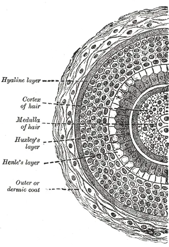
毛包は縦に3つの部分に分ける。
- 漏斗部(infundibulum)——表面の開口から脂腺の導管が開くところまで
- 峡部(isthmus)——脂腺の導管から立毛筋の付着部まで。膨隆部(幹細胞)がある
- 下部——その下。底の毛球(bulbus)に毛母細胞とメラノサイトがあり、真皮の毛乳頭(血管を含む結合組織)を包み込む
下部の横断面を中心から外へ数えると次のようになる。
| 層 | 特徴 | |
|---|---|---|
| 毛(毛幹) | 毛髄質 | 中心の、空胞の多い大きな細胞。細い毛には無い |
| 毛皮質 | 毛の大部分。硬いケラチンとメラニンを含む | |
| 毛小皮 | 扁平な細胞が毛先の方向に瓦のように重なる | |
| 内毛根鞘 | 鞘小皮 | 毛根の方向に重なり、毛小皮とかみ合う |
| Huxley 層 | 1〜3層。トリコヒアリン顆粒を持つ | |
| Henle 層 | 1層。最も早く角化する | |
| 外毛根鞘 | — | 表皮の続き。下部ではグリコーゲンを含む明るい細胞 |
| 硝子膜 | — | 厚い基底膜 |
| 結合組織性毛包 | — | 真皮由来の線維性の鞘 |
立毛筋は平滑筋で、真皮の乳頭層と、脂腺より深い位置の結合組織性毛包を結ぶ。
内毛根鞘は、毛の形を決める鋳型である。毛と一緒に上へ伸びながら角化し、鞘小皮と毛小皮が逆向きにかみ合っているので、毛は鞘の中で滑らない。内毛根鞘は峡部の高さで崩れて消えるので、皮膚の表面には毛だけが出てくる。
毛の色は、毛球のメラノサイトが毛皮質の細胞にメラノソームを渡して決まる。白髪は、膨隆部にあるメラノサイトの幹細胞が尽きて、新しい周期の毛球にメラノサイトが補充されなくなった状態である。
立毛筋が縮むと毛が立ち(鳥肌)、同時に脂腺が押されて皮脂が出る。支配は交感神経である。
毛包のどの高さから生じたかで、皮膚の嚢腫や腫瘍の名前が変わる。
| 病変 | 由来 | 組織の手がかり |
|---|---|---|
| 表皮嚢腫(粉瘤) | 漏斗部 | 表皮と同じく顆粒層を経て、層状の角質をためる |
| 外毛根鞘性嚢腫(毛根鞘嚢腫) | 峡部の外毛根鞘 | 顆粒層を経ずに角化し、密な角質をためる。頭皮に多い |
| 毛母腫(石灰化上皮腫) | 毛母 | 核が抜けた影のような細胞(陰影細胞)と石灰化。小児に多い |
瘢痕性脱毛が元に戻らないのは、炎症が峡部の膨隆部(幹細胞)まで壊すからである。毛球だけが傷む脱毛は、次の周期で生え直す。
English version
Core
The follicle has three vertical parts.
- Infundibulum — from the surface opening down to where the sebaceous duct opens.
- Isthmus — from the sebaceous duct down to the insertion of the arrector pili. It holds the bulge (stem cells).
- Inferior segment — below that. At the base, the hair bulb holds matrix cells and melanocytes and wraps around the dermal hair papilla (connective tissue with vessels).
Counting a cross-section of the lower part from the centre outward:
| Layer | Features | |
|---|---|---|
| Hair (shaft) | Medulla | large vacuolated cells in the centre; absent in fine hairs |
| Cortex | most of the hair; hard keratin and melanin | |
| Hair cuticle | flat cells overlapping like tiles, pointing to the tip | |
| Inner root sheath | Sheath cuticle | overlaps toward the root and interlocks with the hair cuticle |
| Huxley layer | 1–3 cell layers with trichohyalin granules | |
| Henle layer | one layer; keratinises first | |
| Outer root sheath | — | continuous with the epidermis; clear glycogen-rich cells in the lower part |
| Glassy membrane | — | a thick basement membrane |
| Connective tissue sheath | — | a fibrous sheath from the dermis |
The arrector pili is smooth muscle joining the papillary dermis to the connective tissue sheath below the sebaceous gland.
Going further
The inner root sheath is the mould that shapes the hair. It grows upward with the hair and keratinises, and its cuticle interlocks with the hair cuticle in the opposite direction, so the hair cannot slip. The inner root sheath breaks down at the level of the isthmus, so only the hair reaches the surface.
Hair colour comes from bulb melanocytes handing melanosomes to cortex cells. Grey hair appears when melanocyte stem cells in the bulge run out and new bulbs in the next cycle receive no melanocytes.
When the arrector pili contracts, the hair stands up (goose bumps) and the sebaceous gland is squeezed. It is supplied by sympathetic nerves.
In the clinic
The level of the follicle a cyst or tumour comes from decides its name.
| Lesion | Origin | Histological clue |
|---|---|---|
| Epidermoid cyst | infundibulum | keratinises through a granular layer like the epidermis; layered keratin |
| Trichilemmal (pilar) cyst | outer root sheath of the isthmus | keratinises without a granular layer; compact keratin; common on the scalp |
| Pilomatricoma | hair matrix | "shadow cells" that have lost their nuclei, and calcification; common in children |
Scarring alopecia is permanent because inflammation destroys the bulge (stem cells) in the isthmus. Hair loss that damages only the bulb regrows in the next cycle.
爪 — 表皮が硬く変わった付属器Unguis
爪は表皮の角化が特殊化したもので、毛と同じく表皮の付属器である。
- 爪母——爪を作っている場所。爪半月(白い半円)が見えている部分が爪母の先端である
- 爪床——爪の下の上皮。爪はここを滑って前へ出る
- 爪郭・爪上皮——側方と後方の皮膚のひだ。爪上皮(甘皮)は爪母への入口を封じている
伸びる速さは、手が 1 か月に約 3 mm、足はその半分以下。手の爪が全部生え替わるのに 4〜6 か月、足は 12〜18 か月かかる。この速さが、爪の所見を「時間の記録」にしている。
爪母で何かが起きた時期を、爪の位置から逆算できる。
| Beau 線 | 横方向の溝 | 重症疾患・化学療法・高熱で爪母が一時停止した跡。溝の位置から、いつ起きたか計算できる |
| Mees 線 | 横方向の白い帯 | ヒ素・タリウム中毒、重症疾患 |
| さじ状爪 | 中央がへこむ | 鉄欠乏性貧血 |
| ばち指 | 爪甲が丸く盛り上がり、爪郭角が消える | 慢性の低酸素・肺疾患・心疾患・炎症性腸疾患 |
| 爪甲剥離 | 先端から浮く | 甲状腺機能亢進症・乾癬・薬剤・外傷 |
| 爪の点状陥凹 | 小さなくぼみが多発 | 乾癬(爪母の不全角化)・円形脱毛症 |
⚠️ 爪の色素線条は、悪性黒色腫でありうる。幅が広い・不均一・時間とともに拡がる・色素が爪郭の皮膚まではみ出す(Hutchinson 徴候)ときは要注意である。日本人を含むアジア人では、末端黒子型黒色腫が黒色腫の中で占める割合が高いので、手足の爪と足底を見る習慣を持つ。
爪母を傷つけると永久に変形が残る。爪の外傷や手術では、爪母を温存できるかどうかが結果を分ける。
全身状態を診るとき、爪を見るのは費用のかからない診察である。貧血・低酸素・甲状腺・乾癬・中毒の手がかりが同時に得られる。
English version
Core
The nail is a specialisation of epidermal keratinisation and, like hair, an appendage of the epidermis.
- Nail matrix — where the nail is made. The visible lunula (the white crescent) is the distal end of the matrix.
- Nail bed — the epithelium beneath the nail, over which the nail slides forward.
- Nail folds and cuticle — the folds of skin at the sides and behind; the cuticle seals the entrance to the matrix.
Growth — about 3 mm a month on the hand, less than half of that on the foot. A fingernail is replaced entirely in 4–6 months, a toenail in 12–18. This speed makes the findings on a nail a record of time.
Going further
From the position of a change in the nail, the time at which something happened in the matrix can be calculated backwards.
| Sign | Appearance | Meaning |
|---|---|---|
| Beau’s lines | Transverse grooves | The mark of the matrix pausing in severe illness, chemotherapy or high fever. From the position of the groove, when it happened can be calculated |
| Mees’ lines | Transverse white bands | Arsenic or thallium poisoning, severe illness |
| Koilonychia | Spooned, hollow in the centre | Iron deficiency anaemia |
| Clubbing | The nail plate curves and the nail fold angle is lost | Chronic hypoxia, lung disease, heart disease, inflammatory bowel disease |
| Onycholysis | Lifts from the tip | Hyperthyroidism, psoriasis, drugs, trauma |
| Pitting | Multiple small depressions | Psoriasis (parakeratosis of the matrix), alopecia areata |
⚠️ A pigmented band in the nail may be a melanoma. Be careful when it is broad, uneven, widens with time, or the pigment extends onto the skin of the nail fold (Hutchinson’s sign). In Asian populations, including the Japanese, acral lentiginous melanoma makes up a high proportion of melanomas, so make a habit of looking at the nails of the hands and feet and at the soles.
Damage to the matrix leaves a permanent deformity. In injury or surgery of the nail, whether the matrix can be preserved decides the result.
In the clinic
In assessing a patient’s general condition, looking at the nails is an examination that costs nothing. Clues to anaemia, hypoxia, thyroid disease, psoriasis and poisoning are obtained at the same time.
皮膚と付属器の発生 — 外胚葉の表皮と、中胚葉の真皮が組み合わさる
皮膚は2つの胚葉の合作である。
| 部分 | 由来 |
|---|---|
| 表皮・毛・脂腺・汗腺・爪・乳腺 | 表面外胚葉 |
| 真皮・皮下組織 | 中胚葉(背中は皮筋節、体幹の前と四肢は側板中胚葉の壁側葉)。顔と頸は神経堤 |
| メラノサイト | 神経堤から胎生初期に移動してくる |
| Langerhans 細胞 | 造血系(骨髄系)の前駆細胞 |
表皮ははじめ1層の外胚葉で、まもなく表面を扁平な周皮(periderm)が覆う。その下に中間層ができ、胎生4か月ころまでに基底層・有棘層・顆粒層・角層がそろう。剥がれた周皮は皮脂と混ざって胎脂になり、羊水の中で皮膚を守る。
毛は胎生3か月の終わりころ、基底層の細胞が真皮へ落ち込む毛芽として始まる。先端が膨らんで毛球になり、その底を中胚葉の細胞が押し上げて毛乳頭になる。最初の細い毛(うぶ毛)は出生のころに抜けて生え替わる。
脂腺とアポクリン汗腺は毛包の壁から芽を出す。だから毛包へ開く。エクリン汗腺は毛包と無関係に表皮から直接真皮へ伸び、手掌・足底で最も早く現れる。乳腺は胸の乳腺堤から生じる、形を変えた汗腺である。
真皮の乳頭と表皮の突起(表皮稜)は胎生期の中ごろに作られ、指先では指紋になる。指紋の模様は生まれる前に決まり、一生変わらない。
爪は指先の背側に生じ、胎生の後半に指先まで伸びる。手の爪が指先に届くのはおよそ32週、足の爪は36週ころで、新生児の成熟度の目安になる。
毛・汗腺・歯・爪は、どれも表面外胚葉が肥厚した小さな板(プラコード)から、下の中胚葉とシグナルをやりとりして作られる。共通のシグナル(EDA と、その受容体)が壊れると、これらがまとめて作られない。
低汗性外胚葉形成不全症(多くは X 連鎖の EDA 変異)では、毛が少なく、歯が少なく尖り、汗腺がほとんど無い。汗で熱を逃がせないので、乳児が原因不明の高熱を繰り返す。同じプラコードから生まれる器官が、同時に欠ける。
副乳は、腋窩から鼠径部へ走る乳腺堤の上ならどこにでもできる。乳腺堤の残りである。
早産児の皮膚は角層がまだ薄く、バリアが未熟である。水分が皮膚から逃げやすく、皮膚に塗った薬や消毒薬が吸収されやすいので、保育器の加湿や外用薬の量に注意する。
English version
Core
The skin is a joint product of two germ layers.
| Part | Origin |
|---|---|
| Epidermis, hair, sebaceous and sweat glands, nails, mammary gland | surface ectoderm |
| Dermis and subcutis | mesoderm (dermomyotome for the back; somatic layer of lateral plate mesoderm for the front of the trunk and limbs); neural crest for the face and neck |
| Melanocytes | migrate from the neural crest early in fetal life |
| Langerhans cells | haemopoietic (myeloid) precursors |
The epidermis starts as one layer of ectoderm and is soon covered by flat periderm. An intermediate layer forms beneath, and by about the fourth month basal, spinous, granular and horny layers are present. Shed periderm mixes with sebum to form vernix caseosa, which protects the skin in amniotic fluid.
Hair starts near the end of the third month as a hair germ, basal cells growing down into the dermis. Its tip swells into the bulb, and mesoderm cells push into its base to become the hair papilla. The first fine hair (lanugo) is shed around birth.
Sebaceous and apocrine glands bud from the wall of the hair follicle, which is why they open into it. Eccrine glands grow from the epidermis directly, independent of follicles, and appear first on palms and soles. The mammary gland is a modified sweat gland arising from the mammary ridge on the chest.
Going further
Dermal papillae and epidermal ridges form in mid-fetal life; on the fingertips they become fingerprints. The pattern is fixed before birth and never changes.
Nails form on the back of the fingertips and grow toward the tip later in pregnancy. Fingernails reach the tip at about 32 weeks and toenails at about 36 weeks, a sign of a newborn’s maturity.
Hair, sweat glands, teeth and nails all form from small thickened plates of surface ectoderm (placodes) exchanging signals with mesoderm below. If a shared signal (EDA and its receptor) fails, they are all missing together.
In the clinic
In hypohidrotic ectodermal dysplasia (often X-linked EDA mutation), hair is sparse, teeth are few and pointed, and sweat glands are almost absent. Without sweating, infants have repeated unexplained high fevers. Organs from the same placodes are missing at the same time.
Accessory nipples can appear anywhere on the mammary ridge from the axilla to the groin; they are leftovers of that ridge.
Preterm infants have a thin stratum corneum and an immature barrier. Water escapes easily and drugs or antiseptics on the skin are absorbed easily, so incubator humidity and the amount of topical agents need care.
皮膚の加齢 — 年を重ねた皮膚と、日光を浴びた皮膚
皮膚の老化は2種類を分けて考える。同じ人でも、日光の当たらない臀部と、当たり続けた顔では見た目がまったく違う。違いを作っているのは紫外線である。
| 内因性の老化(年齢) | 光老化(紫外線) | |
|---|---|---|
| 表皮 | 薄くなる。入れ替わりが遅い | 厚くなったり薄くなったりする。細胞の異型が出る |
| 表皮と真皮の境 | 表皮稜と真皮乳頭が平らになる | 同じく平らになる |
| 真皮 | 線維芽細胞とコラーゲンが減り、薄く、たるむ | 異常な弾性物質が上層にたまる(日光弾性線維症) |
| 見た目 | 細かいしわ、乾燥 | 深いしわ、ごわごわした皮膚、しみ |
このほか、加齢ではメラノサイトと Langerhans 細胞が減り、汗腺と脂腺の働きが落ち、真皮の血管が減る。表皮のビタミン D 合成も下がる。
光老化の主役は、真皮まで届く長い波長の紫外線(UVA)である。UVA が線維芽細胞などにMMP(コラーゲンを分解する酵素)を作らせ、コラーゲンが壊れていく。代わりに、形の崩れた弾性物質が真皮上層に積もる。HE 染色では、この部分が青灰色の無構造な帯として見える(日光弾性線維症)。
表皮と真皮の噛み合わせが平らになると、2層をずらす力に弱くなる。表皮の再生も遅いので、高齢者の傷は治りにくい。
老人性紫斑は、高齢者の前腕の背側によく見る紫色の斑である。真皮が薄くなって血管を支えられず、軽くぶつけただけで出血する。日光の当たる部位に多いのは、光老化が加わるからである。
皮脂と汗が減るので乾皮症と、それに続く皮脂欠乏性湿疹が冬に多い。汗腺が減ることは、高齢者が熱中症になりやすい理由の1つでもある。
光老化した皮膚の表皮の異型は、日光角化症として現れ、一部が有棘細胞癌へ進む。顔や手の甲の、ざらざらした赤い斑は見逃さない。
English version
Core
Think of skin ageing as two kinds. In the same person, the buttock that never sees sunlight looks very different from a face that always has. The difference is made by ultraviolet light.
| Intrinsic ageing (age) | Photoageing (UV) | |
|---|---|---|
| Epidermis | thinner; slower turnover | thicker or thinner; cell atypia appears |
| Dermo-epidermal junction | ridges and papillae flatten | also flatten |
| Dermis | fewer fibroblasts and less collagen; thin and lax | abnormal elastic material collects in the upper dermis (solar elastosis) |
| Appearance | fine wrinkles, dryness | deep wrinkles, leathery skin, spots |
With age, melanocytes and Langerhans cells also decrease, sweat and sebaceous glands work less, and dermal vessels decrease. Vitamin D synthesis in the epidermis also falls.
Going further
The main actor in photoageing is long-wave UV (UVA), which reaches the dermis. UVA makes fibroblasts and other cells produce MMPs (collagen-degrading enzymes), and collagen breaks down. Instead, misshaped elastic material builds up in the upper dermis. In H&E this area appears as a blue-grey structureless band (solar elastosis).
When the junction flattens, the skin becomes weak against forces that slide the two layers. Epidermal renewal is also slow, so wounds in older people heal poorly.
In the clinic
Senile purpura is the purple patch often seen on the back of the forearm in older people. The thin dermis cannot support the vessels, so a light knock causes bleeding. It favours sun-exposed sites because photoageing adds to it.
Less sebum and sweat lead to xerosis and then asteatotic eczema, common in winter. Fewer sweat glands are also one reason older people get heat stroke easily.
Epidermal atypia in photoaged skin appears as actinic keratosis, some of which progress to squamous cell carcinoma. Do not miss rough red patches on the face or the back of the hand.
まとめ
| 表皮 | 重層扁平上皮(角化型)。深部から順に。 |
|---|---|
| 表皮増殖単位 — 表皮は柱ごとに入れ替わる | 表皮の入れ替わりは、基底層のどの細胞もばらばらに分裂して起きるのではない。 |
| メラノサイト(と表皮の間借り人) | 表皮には角化細胞のほかに由来の違う3種が住む。 |
| メラニンの合成と色素沈着の調節 — 紫外線とホルモンが同じ受容体を押す | メラニンはメラノサイトの中のメラノソーム(リソソームに近い小器官)で作られる。 |
| 真皮 | 表皮を支える結合組織。2層に分ける。 |
| 皮膚付属器 — 毛・脂腺・汗腺 | 毛包——表皮が真皮へ落ち込んだ管。毛周期(成長期・退行期・休止期)を繰り返す。 |
| 細胞間の接着装置 — どこが壊れるかで、水疱の深さが決まる | 表皮の細胞は2種類の接着装置で繋がれている。 |
| 角層 — レンガとモルタルで、水を逃がさない | 角層は「レンガとモルタル」の模型で理解する。 |
| 毛周期 — 抜けるかどうかは、いまどの時期にいるかで決まる | 毛は連続して伸びるのではなく、3つの時期を繰り返す。 |
| 毛包の層構造 — 毛と鞘を、内から外へ数える | 毛包は縦に3つの部分に分ける。漏斗部(infundibulum)——表面の開口から脂腺の導管が開くところまで峡部(isthmus)——脂腺の導管から立毛筋の付着部まで。 |
| 爪 — 表皮が硬く変わった付属器 | 爪は表皮の角化が特殊化したもので、毛と同じく表皮の付属器である。 |
| 皮膚と付属器の発生 — 外胚葉の表皮と、中胚葉の真皮が組み合わさる | 皮膚は2つの胚葉の合作である。部分由来表皮・毛・脂腺・汗腺・爪・乳腺表面外胚葉真皮・皮下組織中胚葉(背中は皮筋節、体幹の前と四肢は側板中胚葉の壁側葉)。 |
| 皮膚の加齢 — 年を重ねた皮膚と、日光を浴びた皮膚 | 皮膚の老化は2種類を分けて考える。同じ人でも、日光の当たらない臀部と、当たり続けた顔では見た目がまったく違う。 |
このノートは医学生が授業の内容を自分の言葉でまとめたもので、診療の判断に使うものではありません。誤りに気づいたらお知らせください。